Article

Effects of Nb Addition on Microstructures and Mechanical Properties of Nbx-CoCrFeMnNi High Entropy Alloy Films

Department of Materials Science and Engineering, National Taiwan University, Taipei 10617, Taiwan
*
Author to whom correspondence should be addressed.
Coatings 2021, 11(12), 1539; https://doi.org/10.3390/coatings11121539
Submission received: 16 November 2021 / Revised: 6 December 2021 / Accepted: 10 December 2021 / Published: 14 December 2021
(This article belongs to the Special Issue High Entropy Alloy Films)

Abstract

:
In the present work, Nbx-CoCrFeMnNi high entropy alloy films (HEAFs, 0 to 7.2 at.% Nb) were fabricated by radio frequency (RF) magnetron co-sputtering of CoCrFeMnNi alloy and Nb targets. The effects of Nb addition on the microstructures and mechanical properties of HEAFs were systematically investigated. For Nb-free film (0 at.% Nb), the face-centered cubic (FCC) peaks were identified in the X-ray diffraction (XRD) pattern. The addition of Nb resulted in a broadening of diffraction peaks, a decrease in peak intensity, and the vanishment of high-angle peaks. Transmission electron microscope (TEM) images indicated the formation of nanotwins at low Nb concentrations, and a transition from a single phase FCC solid solution to an amorphous phase was observed with the increasing Nb concentration. The films were strengthened with an increase in Nb concentration. Specifically, the hardness characterized by nanoindentation increased from 6.5 to 8.1 GPa. The compressive yield strength and fracture strength measured from micropillar compression tests were improved from 1.08 GPs and 2.56 GPa to 2.70 GPa and 5.76 GPa, respectively, whereas the fracture strain decreased from >29.4% (no fracture) to 15.8%. Additionally, shear banding was observed in the presence of amorphous phase.

Graphical Abstract

1. Introduction

Since high entropy alloys (HEAs) were first reported by Yeh et al. [1] and Cantor et al. [2] in 2004, a great deal of research has been designed to investigate their attractive and versatile properties such as excellent strength and ductility [1,3,4], superior cryogenic fracture toughness [5,6], enhanced corrosion resistance [7,8], high wear resistance [9,10], good irradiation resistance [11], and thermal stability [12], etc. Compared to the traditional designs of alloys, which contain one or, rarely, two base elements, HEAs have been defined as alloys consisting of five or more principal elements with each atomic concentration between 5 and 35 at.% with the configuration entropy ΔS higher than 1.5 R where R (=8.314 J/mol/K) is the gas constant [1]. Due to the high-entropy effect, the HEAs are favorable to form simple solid solution phases such as a face-centered cubic (FCC) [2,13,14], body-centered cubic (BCC) [4,15,16], or hexagonal close-packed (HCP) [17,18] structure, rather than the brittle intermetallic compounds. A comprehensive review of the concept, complexity, thermodynamics, stabilization, lattice strains, resulting excellent mechanical properties, and the potential for further optimization and applications was recently performed by Cantor [19]. The equiatomic CoCrFeMnNi HEA (i.e., Cantor alloy [2]), one of the most studied HEAs, crystallizes as a single phase FCC solid solution. It possesses high ductility and strength at cryogenic temperatures but shows a limited strength at room temperature [5,13]. Therefore, the enhancement in strength is of paramount importance in order to use CoCrFeMnNi HEAs in industry applications.
Doping another element in the HEAs with different concentrations has been used as an effective method for microstructure modifications and property regulations. Many HEAs based on CoCrFeMnNi alloy have been investigated in recent years. For instance, He et al. [20] found that the addition of Al into CoCrFeMnNi alloy resulted in a transition from FCC to BCC and increased strength with a sacrifice in ductility. Stepanov et al. [21] reported that the addition of V into CoCrFeMnNi alloy led to the formation of the intermetallic sigma phase, which enhanced the microhardness and yield strength. Qin et al. [22] claimed that the addition of Ti promoted the growth of the second phases (Laves and sigma phases), which played a positive role in improving the yield strength. Since Nb has a negative enthalpy of mixing with the constituent components in CoCrFeMnNi alloy (the mixing enthalpies of Nb with Co, Cr, Fe, Mn, and Ni atomic pairs are −25, −7, −16, −4, and −30 kJ/mol, respectively, [23]), it would favor the formation of second phases or intermetallic compounds in HEAs. In addition, Nb has a massive size (Nb: 0.143 nm) as compared to the five base elements (0.124 to 0.128 nm), which would result in a large atomic size mismatch and lattice distortion. As a result, the characteristics of Nb have received extensive attention, and some research has proved the improved mechanical properties with the Nb addition. For example, Qin et al. indicated that the volume fraction of second phase in (CoCrFeMnNi)100–xNbx increased with an increase in Nb content (0 to 16 at.%), and the yield strength was improved from 202 to 1010 MPa, whereas the fracture strain was reduced from 60% (no fracture) to 12% [22]. In Braeckman et al.’s work [24,25], Nbx-CoCrCuFeNi thin films deposited by magnetron sputtering displayed high hardness. As a result of the formation of nanocomposite in the presence of heavy solute elements such as Nb, the nanocomposite could give rise to an increase in hardness. Other researches, such as CoCrFeNiNbx [26,27], AlCoCrFeNbxNi [28], CoFeNi2V0.5Nb0.75 [29], and CoCrCuFeNiNbx [30], also proved that adequately doping Nb into the base alloy could show its strengthening characteristics. It is worth noting that, by adding rare earth elements (Sm) and refractive elements (Zr, Ti) in the AlCoFeNiSmTiVZr system and using the selective laser melting technique, excellent mechanical and corrosion resistant properties of HEAs could be obtained [31,32].
High entropy alloy films (HEAFs) have also attracted extensive research attention over the last decade due to their unique mechanical properties. The recent research and development of HEAFs, including basic concept, preparation technologies, microstructures, appealing properties, applications and suggested future research work, have been reviewed by Li et al. [33]. Several studies have claimed that the HEAFs have a higher yield strength and hardness compared with their bulk counterparts [33,34,35,36,37,38]. Apart from the difference in mechanical properties, distinctive phase transformations have also been observed in HEAFs. Different from second phases commonly formed in bulk HEAs, amorphous phases are usually found in HEAFs fabricated by magnetron sputtering when the solute concentration exceeds the certain threshold [37,39]. Braeckman et al. reported that the transformations of Gex-CoCrCuFeNi and Inx-CoCrCuFeNi HEAFs from FCC to the amorphous phase with an increasing additive concentration were driven by thermodynamic and topological factors, respectively [39].
In this work, a series of Nbx-CoCrFeMnNi HEAFs synthesized by radio frequency (RF) magnetron co-sputtering, a PVD process to control the accurate compositions with the homogenous distribution, was designed. In addition, the differences between Nbx-CoCrFeMnNi HEAFs and their bulk counterparts were compared. To provide an insight into the influence of Nb addition on the microstructures and mechanical properties of the HEAFs, a systematic investigation was carried out using X-ray diffraction (XRD), scanning electron microscopy (SEM), transmission electron microscope (TEM), and nanoindentation and micro-pillar compression tests.

2. Materials and Methods

2.1. Fabrication of Nbx-CoCrFeMnNi HEAFs

Nbx-CoCrFeMnNi HEAFs were deposited on 0.55 mm thick (111)-oriented silicon substrates by RF magnetron co-sputtering of CoCrFeMnNi alloy and Nb targets. Both targets, with a diameter of 76 mm and thickness of 6 mm, were prepared by vacuum arc melting with 99.99 wt.% pure elements. Before the deposition process, the silicon substrates were ultrasonically cleaned and rinsed in acetone, ethanol, and deionized water sequentially for 15 min in each step. To reduce any contamination on the surface and to equilibrate the surface composition of the two targets, argon ion bombardment was conducted with 100 W in RF mode for at least 30 min before film deposition. After the high vacuum below 5 × 10⁻⁵ Pa was maintained, the 99.9% pure argon gas was introduced at a flow rate of 20 sccm. The working distance and working pressure were fixed at 10 cm and 0.4 Pa, respectively. The rotation speed of the substrate was set at 40 rpm to ensure a homogeneous film thickness and composition. The film composition was regulated by the different powers applied on the target. The power applied on CoCrFeMnNi alloy target was fixed at 400 W, and the different powers applied on the Nb target were regulated, respectively, at 0, 20, 30, 40, and 60 W. All the films were deposited at room temperature for 1 h, and the film thicknesses ranging from 1.8 to 2.0 μm were obtained.

2.2. Characterization of Composition and Structure

The chemical composition, surface morphology, and film thickness of the films were determined by a field-emission scanning electron microscope (SEM, NOVA NANO SEM 450, FEI, Hillsboro, OR, USA) with an energy dispersive X-ray spectroscope (EDS, Oxford, UK). The average values of the chemical composition were measured in more than six regions of the films to ensure the reliability of the data. The crystalline structures of the films were examined by a glancing-incidence (1°) X-ray diffraction (XRD, Rigaku TTRAX 3, Tokyo, Japan) with Cu-Kα radiation (λ = 0.15406 nm), an acceleration voltage of 50 kV, and a working current of 250 mA. The XRD patterns were recorded at room temperature in the angular range (2θ) of 20–100° with the scanning speed of 4°/min. The microstructures of the films were analyzed by transmission electron microscope (TEM, FEI Tecnai G2 F20, Hillsboro, OR, USA) at 200 keV accelerating voltage. Specimens for TEM investigation and micropillar compression tests were prepared by a dual-beam focused ion beam (FIB, FEI Quanta 3D FEG, Hillsboro, OR, Oregon, USA). Before the film was milled, the surface was protected from ion beam damage using deposition of Pt protective layer. The EDS-mapping measurement in TEM was used to confirm the distribution of each element in the films.

2.3. Characterization of Mechanical Properties

The hardness and reduced Young’s modulus of the films were measured using nanoindentation (Hysitron TI 950 TriboIndenter, Bruker, Billerica, MA, USA) with a Berkovich diamond indenter tip (tip radius of 150 nm). All tests were performed under the displacement-controlled mode with a rate of 14 nm/s to a depth of 70 nm and a holding time of 5 s between loading and unloading. The penetration depth was less than 1/10 of the film thickness to avoid any influence from the silicon substrate. Each datum was the average from at least seven measurements. The engineering compressive yield strength, fracture strength, fracture strain, and deformation behavior of the films were characterized by the in-situ SEM micro-pillar compression tests. The tests were performed at room temperature under the displacement-rate-controlled mode at a nominal strain rate of 1 × 10⁻³/s with the picoindenter (Hysitron PI 85 SEM PicoIndenter, Bruker, Billerica, MA, USA) equipped with a diamond flat-punch tip (tip radius of 2.5 μm). Three micropillars were prepared for each composition. The micropillars with an aspect ratio (the ratio of length to diameter) of ~2.5 and a taper angle of <3° were fabricated. Firstly, a large pillar with a diameter of ~4 μm was shaped using coarse FIB milling (Ga+ ion source at 30 keV voltage and 3 nA current). Then, fine milling was performed with Ga+ ion voltage of 5 keV and a current of 16 pA to form the micropillar with a diameter ranging from 563 to 708 nm. It is worth noting that an alternative bottom-up method could be used for the pillar formation. Specifically, nano-pillars could be formed by electrochemical deposition on through-hole porous anodic aluminum oxide membranes [40,41,42].

3. Results and Discussion

3.1. Compositions and Structures

The chemical compositions (at.%) of the CoCrFeMnNi alloy target measured by EDS were 20.1% ± 1.1% of Co, 19.6% ± 0.9% of Cr, 19.9% ± 1.4% of Fe, 22.0% ± 0.7% of Mn, and 18.4% ± 0.8% of Ni. The molar ratio of each element was close to equimolar. The Nb concentrations (0 to 7.2 at.%) of the Nbx-CoCrFeMnNi HEAFs were regulated by the different powers applied on the Nb target, and their chemical compositions measured by EDS are listed in Table 1. The concentration of Nb increased with the increasing power applied on the Nb target.
The cross-sections of the films were examined using FE-SEM to observe the morphology and to measure the film thickness, as shown in Figure 1a–e. An extremely thin layer resulting from the scraping and break separation process of the specimen was observed on the top surface of each film, and it was also observed previously in the cross-sections of ZrNi films on Si substrates prepared by notching and breaking [43]. All the films showed good bonding with substrates. Figure 1a reveals that the fracture surface of the Nb-free film (0 at.% Nb) was composed of three layers. Region 1 showed dimples on the fracture surface, signifying ductile fracture, while regions 2 and 3 displayed a relatively smooth and crack-free morphology. The detailed microstructure evolution of the film during sputter deposition has been studied elsewhere [44,45]. Specifically, Wang et al. [44] also observed the three-layer structure from the TEM images and selected area diffraction patterns of the CoCrFeMnNi HEAF with a thickness of 1400 nm. In their case, region 3 consisted of an amorphous structure with a small amount of nanocrystals, region 2 had the single crystal structure with nanotwins, and region 1 consisted of nanotwinned columnar grains perpendicular to the substrate surface with a preferred (111) orientation. In the present study, as the Nb atoms were added in the film, the large atoms of Nb promoted the pinning effect [46]. The Nb atoms pinned on the substrate favored the formation of clusters with the same lattice orientation [44,46], forming only a single layer in the films (Figure 1b–e). Figure 1b revealed that the film with 0.6 at.% Nb had a nanosized columnar morphology. Figure 1c–e revealed that the films with higher Nb concentrations (1.8, 4.1 and 7.2 at.% Nb) transformed to a relatively dense and smooth morphology.
The deposition rate could be calculated from the measured thickness divided by the deposition time, and the deposition rate as a function of the sputtering power applied on the Nb target is shown in Figure 1f. Basically, a higher sputtering power would lead to a higher sputtering yield to generate more atoms from the target and give rise to an increase in film thickness. However, the highest deposition rate of 33.1 nm/min occurred in the Nb-free film, and it decreased to ~30.6 nm/min as the power applied on the Nb target increased from 0 to 60 W, which may be explained by the following. An increase in the power applied on the Nb target would raise the production of the Nb atoms in the chamber, resulting in a decrease in the mean free path of the gas molecules. Additionally, the big atomic size of Nb atoms increased the chance of atom collisions, which would hinder the arrival of atoms on the substrate and retard the deposition of the film [47,48].
Figure 2 shows the XRD patterns of Nbx-CoCrFeMnNi HEAFs (0 to 7.2 at.%). The crystal structure of the Nb-free film was a single phase FCC solid solution associated with the sharp and strong (111) peak and high-angle weak (200), (220), and (311) peaks because of the high-entropy effect. It was clear that the addition of Nb resulted in the broadening of the diffraction peak, a decrease in the (111) peak intensity, and the vanishment of the higher angle diffraction peaks. Compared with the bulk Nbx-CoCrFeMnNi HEA [22], the films in the present study were prepared by magnetron sputtering process, which is a rapid quenching method to limit the diffusion of elements and inhibit the grain growth to obtain the smaller grain sizes and produce the metastable solid solution without the presence of Laves phase or any precipitates. The full width at half maximum (FWHM) of the diffraction peak significantly increased from 0.47° to 4.72° with the initial addition of Nb (0.6 at.% Nb), and then slightly increased to 5.02° with the further increase in Nb concentration (7.2 at.% Nb) resulting from the formation of amorphous phase. Moreover, the great difference in atomic size between Nb and CoCrFeMnNi alloy tended to induce severe lattice distortion and caused the instability of crystalline structures.

3.2. Microstructures

The microstructures of the cross-sections of Nbx-CoCrFeMnNi HEAFs and the corresponding selected area electron diffraction (SAED) patterns were characterized by TEM, as shown in Figure 3. Nano-sized columnar grains with the similar orientation and the single phase FCC solid solution were observed in the Nb-free film (Figure 3a), and the columnar growth direction was perpendicular to the substrate surface during the sputtering process. In Figure 3b, the nanotwins were observed in the film with 0.6 at.% Nb. The formation of nanotwins was mainly due to the atomic peening effect [49,50]. Since Nb atom was two times heavier than Ar atom, the atomic peening effect was highly enhanced. More backscattered Ar atoms were introduced with the increasing power applied on the Nb target [25]. These energetic particles stroke the substrate and caused bombardments on the film, leading to an increase in compressive stress and the formation of defects in the film and inducing the formation of nanotwins [51,52]. The HRTEM image of the film with 0.6 at.% Nb and the corresponding fast Fourier transform (FFT) image are shown in Figure 4, confirming the thin nanotwins (~4 nm) in the FCC matrix.
With the further addition of Nb, the amount of nanotwins decreased and the amorphous phase became dominant. The nanocrystallites embedded in the amorphous matrix could be observed in the films with 1.8 and 4.1 at.% Nb (Figure 3c,d), and the corresponding SAED patterns indicated the decreasing crystalline structure (FCC phase and nanotwins) and the appearance of halo rings. No crystallites were detected in the film with 7.2 at.% Nb, and this film was fully amorphous (Figure 3e). Hence, with the increasing Nb concentration in the film, the microstructures of the films transformed from a single FCC phase (Figure 3a,b) to a mixture of FCC phase and amorphous phase (Figure 3c,d), and to a totally amorphous phase (Figure 3e).
The molar ratio of each element was close to equimolar. The Nb concentrations (0 to 7.2 at.%) of the Nbx-CoCrFeMnNi HEAFs were regulated by the different powers applied on the Nb target, and their chemical compositions measured by EDS are listed in Table 1. The concentration of Nb increased with the increasing power applied on the Nb target. The tendency of crystalline-to-amorphous configuration could be attributed to two factors. First is the atomic size mismatch (δ) of the film, defined as [53,54]:
δ = i = 1 N c i 1 r i j = 1 N c j r j 2
where N is the number of constituent elements in the HEA, c i and r i are the atomic fraction and radius of element i, respectively. Since Nb has a massive size (Nb: 0.143 nm) as compared to the five base elements (0.124 to 0.128 nm), the addition of Nb would lead to an increase in atomic size mismatch from 1.11% (for 0 at.% Nb) to 3.62% (for 7.2 at.% Nb), as listed in Table 2. The significant atomic size mismatch and the fast cooling rate during film deposition could validate the topological instability and promote the formation of the amorphous phase. Second, on the basis of Braeckman et al.’s study [39], the large negative mixing enthalpies of Nb–TM (TM = transition metals) atomic pairs, as listed in Table 3, stabilize the amorphous phase. The TEM results also agreed with the XRD experimental results, both showing a transition from a single phase FCC solid solution to an amorphous phase with an increasing Nb concentration in Nbx-CoCrFeMnNi HEAFs.
Figure 5a shows the cross-section bright field TEM image of the film with 1.8 at.% Nb, and Figure 5b shows the elemental distributions of Co, Cr, Fe, Mn, Ni, and Nb in the framed area in Figure 5a. As evidently observed from these images, the constituent elements were uniformly distributed without the presence of any second phase or precipitate.

3.3. Mechanical Properties

The hardness and reduced Young’s modulus of the Nbx-CoCrFeMnNi HEAFs were measured by nanoindentation based on the Oliver and Pharr method [55], and the results are presented in Figure 6a,b as functions of the power applied on Nb target and the Nb concentration, respectively. The hardness increased from 6.5 to 8.1 GPa when the Nb concentration increased from 0 to 7.2 at.%. Strengthening of Nbx-CoCrFeMnNi HEAFs shown in the present study could be attributed to the following:
(i)
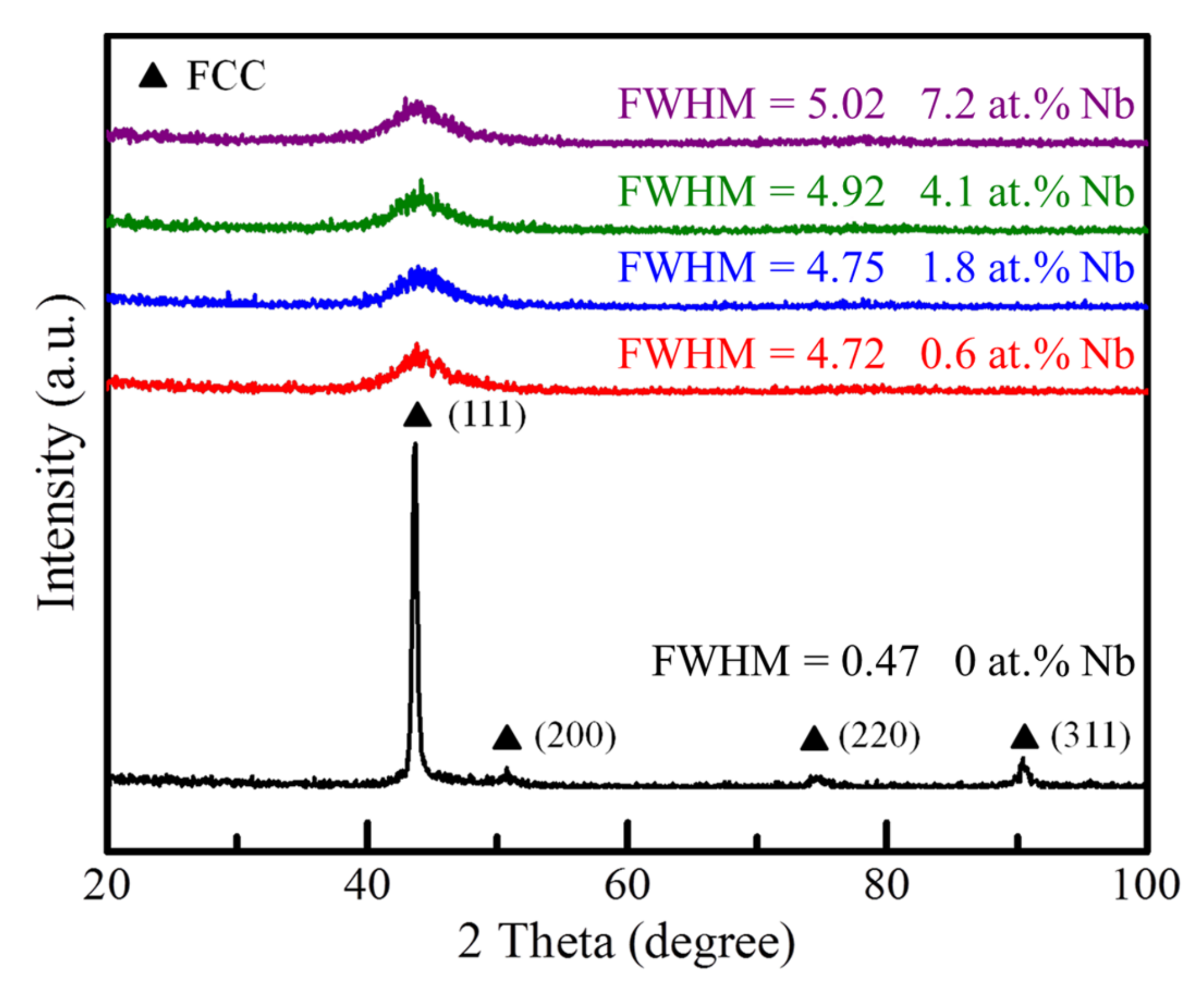
Grain boundary strengthening (Hall–Petch relation): The tendency of forming smaller grains in the films with Nb addition could be observed in the XRD results. According to the classical Hall–Petch relation, the grain boundary strengthening increased with the decreasing grain size [35];
(ii)
Solid solution strengthening: The solid solution strengthening was introduced by the lattice distortion which was relevant to the atomic size mismatch [35,56]. Since Nb had a large atomic radius, doping Nb into CoCrFeMnNi HEAFs contributed to the increase in atomic size mismatch (see Table 2) that enhanced the solid solution effect.
Figure 6. The hardness (H) and reduced Young’s modulus (Er) of Nbx-CoCrFeMnNi HEAFs as functions of (a) the power applied on Nb target and (b) Nb concentration.
Coatings 11 01539 g006
The Young’s moduli of the constituent elements in Nbx-CoCrFeMnNi are also listed in Table 3. Since Nb exhibited the lowest Young’s modulus of the six constituents [57], the reduced Young’s modulus decreased with the addition of Nb in the film.
To further characterize the mechanical properties of Nbx-CoCrFeMnNi HEAFs, in-situ micropillar compression tests were performed. Figure 7a–e shows the SEM images of as-compressed micropillars with different Nb concentrations from 0 to 7.2 at.%. As shown in Figure 7, except for the micropillars of the films with 0 and 0.6 at.% Nb, the other films (1.8, 4.1, and 7.2 at.% Nb) showed the increasing shear bands with the increasing Nb concentration after compression. This was attributed to the differences in microstructures in these micropillars. During the plastic deformation, the amorphous phase, not having well-defined defects as the crystalline structure, would lead to the formation of shear bands [58,59,60].
The engineering compressive stress–strain curves of Nbx-CoCrFeMnNi HEAFs subjected to micropillar compression tests are plotted in Figure 8. Distinct from typical curves, slight serrations were observed in micropillar with 1.8 at.% Nb and heavy serrations were observed in micropillars with 4.1 and 7.2 at.% Nb. Each strain burst in serrated curves indicated a shear band propagation, and this result agreed with the SEM images shown in Figure 7. The compressive yield strength values in these micropillars were evaluated using the 0.2% offset method. The fracture strength and the fracture strain were obtained, respectively, from the maximum load and the corresponding strain of the stress–strain curve. All the measured values are listed in Table 4. As shown in Figure 8, a tendency of the increasing yield strength and fracture strength was observed with the increasing Nb concentration in the films. The yield strength and fracture strength were improved from 1.08 GPa and 2.56 GPa for 0 at.% Nb to 2.70 GPa and 5.76 GPa for 7.2 at.% Nb, respectively. However, suffering from the strength–ductility trade-off, the fracture strain reduced from >29.4% (no fracture) for 0 at.% Nb to 15.8% for 7.2 at.% Nb. For the bulk materials in Qin et al.’s study [22], the yield strength of (CoCrFeMnNi)100-xNbx HEA was improved with the increasing Nb concentration (0.2 GPa at 0 at.% Nb to 1.01 GPa at 16 at.% Nb). It could be seen that the films possessed a much higher yield strength than their bulk counterparts.

4. Conclusions

The Nbx-CoCrFeMnNi HEAFs were successfully prepared by magnetron co-sputtering. The effects of Nb addition on the microstructures and mechanical properties of HEAFs were systematically investigated, and the following conclusions could be drawn:
(1)
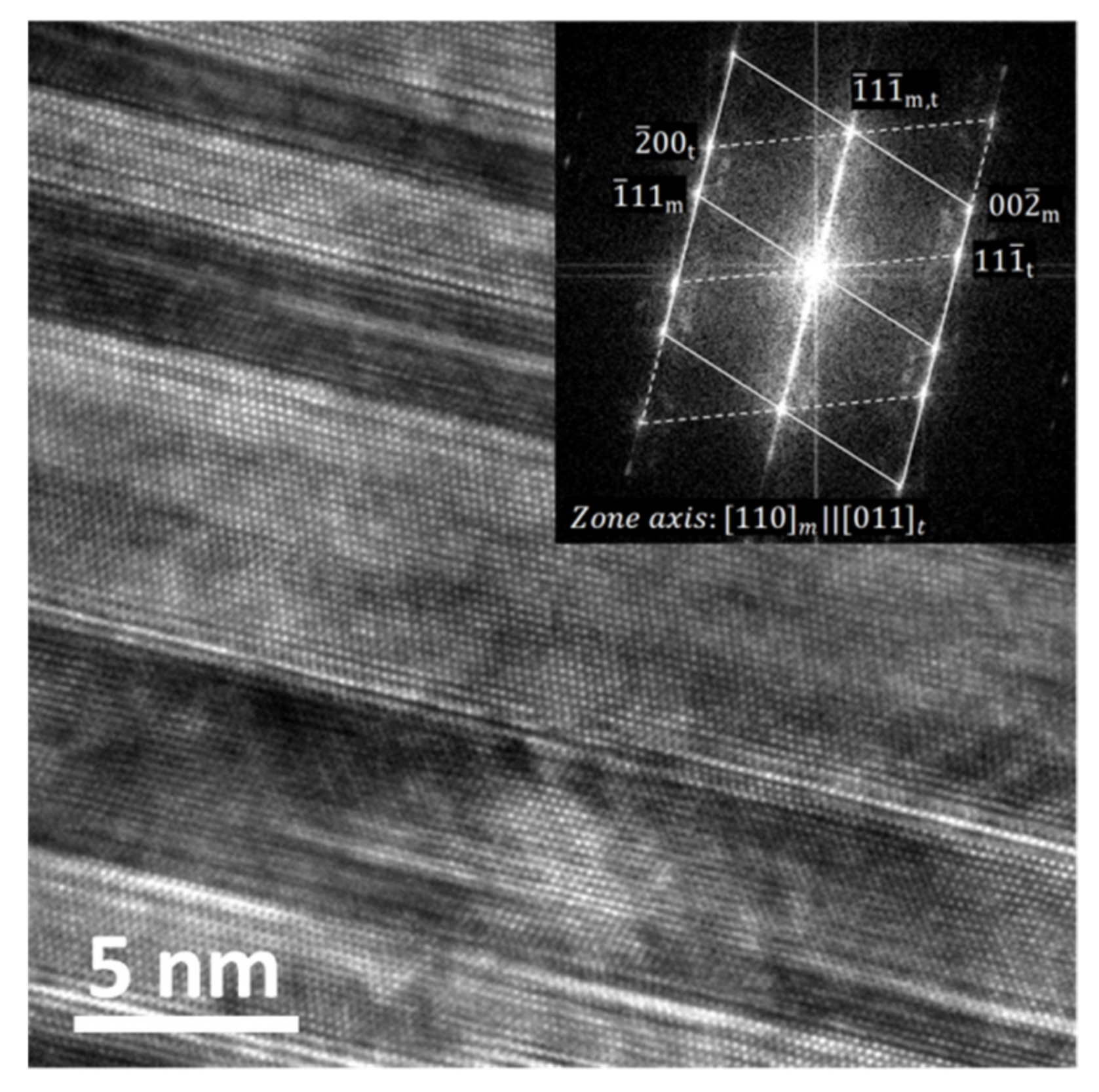
The structure transferred from a single phase FCC solid solution to an amorphous phase with an increasing Nb concentration in the films. The Nb-free film was a single phase FCC solid solution phase. The films with 1.8 and 4.1 at.% Nb showed nanocrystals embedded in amorphous matrix, while the films with 7.2 at.% Nb completely transferred to an amorphous phase;
(2)
Nanotwins were first found in the film with 0.6 at.% Nb, and the disappearance of nanotwins was observed with the further increase in Nb concentration as the amorphous phase became dominant;
(3)
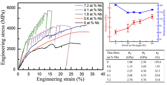
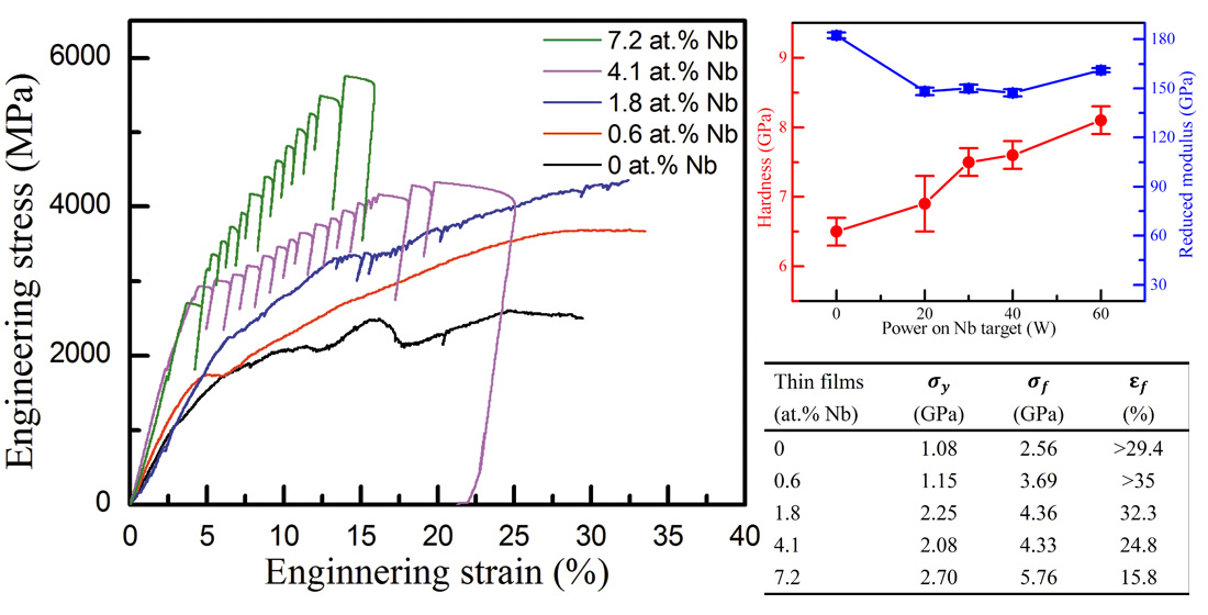
The nanoindentation results showed an increase in the hardness of the film with an increasing Nb concentration. The hardness increased from 6.5 GPa to 8.1 GPa as the Nb concentration increased from 0 to 7.2 at.%;
(4)
The micropillar compression tests indicated the strengthening of the films with the increasing Nb concentration. From 0 to 7.2 at.% Nb, the compressive yield strength and fracture strength increased from 1.08 GPa and 2.56 GPa to 2.70 GPa and 5.76 GPa, respectively. However, the fracture strain reduced from >29.4% (no fracture) to 15.8% due to the strength–ductility trade-off. Additionally, shear bands were observed in the presence of the amorphous phase.

Author Contributions

Y.-H.L.: Conceptualization, Methodology, Investigation, Writing—original draft. C.-L.L.: Investigation. C.-H.H.: Supervision, Writing—review and editing, Funding acquisition. All authors have read and agreed to the published version of the manuscript.

Funding

This work was supported by the Ministry of Science and Technology, Taiwan under Contract no. MOST 109-2221-E-002-124.

Institutional Review Board Statement

Not applicable.

Informed Consent Statement

Not applicable.

Data Availability Statement

Not applicable.

Acknowledgments

We thank Y.C. Lu for the technical assistance in TEM.

Conflicts of Interest

The authors declare no conflict of interest.

References

  1. Yeh, J.W.; Chen, S.K.; Lin, S.J.; Gan, J.Y.; Chin, T.S.; Shun, T.T.; Tsau, C.H.; Chang, S.Y. Nanostructured high-entropy alloys with multiple principal elements: Novel alloy design concepts and outcomes. Adv. Eng. Mater. 2004, 6, 299–303. [Google Scholar] [CrossRef]
  2. Cantor, B.; Chang, I.T.H.; Knight, P.; Vincent, A.J.B. Microstructural development in equiatomic multicomponent alloys. Mater. Sci. Eng. A 2004, 375, 213–218. [Google Scholar] [CrossRef]
  3. Li, Z.M.; Pradeep, K.G.; Deng, Y.; Raabe, D.; Tasan, C.C. Metastable high-entropy dual-phase alloys overcome the strength-ductility trade-off. Nature 2016, 534, 227–230. [Google Scholar] [CrossRef]
  4. Senkov, O.N.; Scott, J.M.; Senkova, S.V.; Miracle, D.B.; Woodward, C.F. Microstructure and room temperature properties of a high-entropy TaNbHfZrTi alloy. J. Alloys Compd. 2011, 509, 6043–6048. [Google Scholar] [CrossRef]
  5. Gludovatz, B.; Hohenwarter, A.; Catoor, D.; Chang, E.H.; George, E.P.; Ritchie, R.O. A fracture-resistant high-entropy alloy for cryogenic applications. Science 2014, 345, 1153–1158. [Google Scholar] [CrossRef] [PubMed] [Green Version]
  6. Ye, Y.F.; Wang, Q.; Lu, J.; Liu, C.T.; Yang, Y. High-entropy alloy: Challenges and prospects. Mater. Today 2016, 19, 349–362. [Google Scholar] [CrossRef]
  7. Ren, B.; Liu, Z.X.; Li, D.M.; Shi, L.; Cai, B.; Wang, M.X. Corrosion behavior of CuCrFeNiMn high entropy alloy system in 1M sulfuric acid solution. Mater. Corros. 2012, 63, 828–834. [Google Scholar] [CrossRef]
  8. Chen, Y.Y.; Duval, T.; Hung, U.D.; Yeh, J.W.; Shih, H.C. Microstructure and electrochemical properties of high entropy alloys—A comparison with type-304 stainless steel. Corros. Sci. 2005, 47, 2257–2279. [Google Scholar] [CrossRef]
  9. Chai, B.B.; Xiong, J.; Guo, Z.X.; Liu, J.B.; Ni, L.; Xiao, Y.; Chen, C. Structure and high temperature wear characteristics of CVD coating on HEA-bonded cermet. Ceram. Int. 2019, 45, 19077–19085. [Google Scholar] [CrossRef]
  10. Fang, Y.H.; Chen, N.; Du, G.P.; Zhang, M.X.; Zhao, X.R.; Cheng, H.; Wu, J.B. High-temperature oxidation resistance, mechanical and wear resistance properties of Ti(C,N)-based cermets with Al0.3CoCrFeNi high-entropy alloy as a metal binder. J. Alloys Compd. 2020, 815, 152486. [Google Scholar] [CrossRef]
  11. Kumar, N.; Li, C.; Leonard, K.J.; Bei, H.; Zinkle, S.J. Microstructural stability and mechanical behavior of FeNiMnCr high entropy alloy under ion irradiation. Acta Mater. 2016, 113, 230–244. [Google Scholar] [CrossRef] [Green Version]
  12. Jiang, L.; Lu, Y.P.; Dong, Y.; Wang, T.M.; Cao, Z.Q.; Li, T.J. Annealing effects on the microstructure and properties of bulk high-entropy CoCrFeNiTi0.5 alloy casting ingot. Intermetallics 2014, 44, 37–43. [Google Scholar] [CrossRef]
  13. Otto, F.; Dlouhy, A.; Somsen, C.; Bei, H.; Eggeler, G.; George, E.P. The influences of temperature and microstructure on the tensile properties of a CoCrFeMnNi high-entropy alloy. Acta Mater. 2013, 61, 5743–5755. [Google Scholar] [CrossRef] [Green Version]
  14. Dwivedi, A.; Koch, C.C.; Rajulapati, K.V. On the single phase fcc solid solution in nanocrystalline Cr-Nb-Ti-V-Zn high-entropy alloy. Mater. Lett. 2016, 183, 44–47. [Google Scholar] [CrossRef]
  15. Senkov, O.N.; Wilks, G.B.; Miracle, D.B.; Chuang, C.P.; Liaw, P.K. Refractory high-entropy alloys. Intermetallics 2010, 18, 1758–1765. [Google Scholar] [CrossRef]
  16. Yao, J.Q.; Liu, X.W.; Gao, N.; Jiang, Q.H.; Li, N.; Liu, G.; Zhang, W.B.; Fan, Z.T. Phase stability of a ductile single-phase BCC Hf0.5Nb0.5Ta0.5Ti1.5Zr refractory high-entropy alloy. Intermetallics 2018, 98, 79–88. [Google Scholar] [CrossRef]
  17. Uporov, S.; Estemirova, S.K.; Bykov, V.A.; Zamyatin, D.A.; Ryltsev, R.E. A single-phase ScTiZrHf high-entropy alloy with thermally stable hexagonal close-packed structure. Intermetallics 2020, 122, 106802. [Google Scholar] [CrossRef]
  18. Feuerbacher, M.; Heidelmann, M.; Thomas, C. Hexagonal High-entropy Alloys. Mater. Res. Lett. 2015, 3, 1–6. [Google Scholar] [CrossRef]
  19. Cantor, B. Multicomponent high-entropy Cantor alloys. Prog. Mater. Sci. 2021, 120, 100754. [Google Scholar] [CrossRef]
  20. He, J.Y.; Liu, W.H.; Wang, H.; Wu, Y.; Liu, X.J.; Nieh, T.G.; Lu, Z.P. Effects of Al addition on structural evolution and tensile properties of the FeCoNiCrMn high-entropy alloy system. Acta Mater. 2014, 62, 105–113. [Google Scholar] [CrossRef]
  21. Stepanov, N.D.; Shaysultanov, D.G.; Salishchev, G.A.; Tikhonovsky, M.A.; Oleynik, E.E.; Tortika, A.S.; Senkov, O.N. Effect of V content on microstructure and mechanical properties of the CoCrFeMnNiVx high entropy alloys. J. Alloys Compd. 2015, 628, 170–185. [Google Scholar] [CrossRef]
  22. Qin, G.; Li, Z.B.; Chen, R.R.; Zheng, H.T.; Fan, C.L.; Wang, L.; Su, Y.Q.; Ding, H.S.; Guo, J.J.; Fu, H.Z. CoCrFeMnNi high-entropy alloys reinforced with Laves phase by adding Nb and Ti elements. J. Mater. Res. 2019, 34, 1011–1020. [Google Scholar] [CrossRef]
  23. Takeuchi, A.; Inoue, A. Classification of bulk metallic glasses by atomic size difference, heat of mixing and period of constituent elements and its application to characterization of the main alloying element. Mater. Trans. 2005, 46, 2817–2829. [Google Scholar] [CrossRef] [Green Version]
  24. Braeckman, B.R.; Misjak, F.; Radnoczi, G.; Caplovicova, M.; Djemia, P.H.; Tetard, F.; Belliard, L.; Depla, D. The nanostructure and mechanical properties of nanocomposite Nbx-CoCrCuFeNi thin films. Scr. Mater. 2017, 139, 155–158. [Google Scholar] [CrossRef]
  25. Braeckman, B.R.; Depla, D. Structure formation and properties of sputter deposited Nbx-CoCrCuFeNi high entropy alloy thin films. J. Alloys Compd. 2015, 646, 810–815. [Google Scholar] [CrossRef]
  26. Liu, W.H.; He, J.Y.; Huang, H.L.; Wang, H.; Lu, Z.P.; Liu, C.T. Effects of Nb additions on the microstructure and mechanical property of CoCrFeNi high-entropy alloys. Intermetallics 2015, 60, 1–8. [Google Scholar] [CrossRef]
  27. Jiang, H.; Jiang, L.; Qiao, D.X.; Lu, Y.P.; Wang, T.M.; Cao, Z.Q.; Li, T.J. Effect of niobium on microstructure and properties of the CoCrFeNbxNi high entropy alloys. J. Mater. Sci. Technol. 2017, 33, 712–717. [Google Scholar] [CrossRef]
  28. Ma, S.G.; Zhang, Y. Effect of Nb addition on the microstructure and properties of AlCoCrFeNi high-entropy alloy. Mater. Sci. Eng. A 2012, 532, 480–486. [Google Scholar] [CrossRef]
  29. Jiang, L.; Lu, Y.P.; Wu, W.; Cao, Z.Q.; Li, T.J. Microstructure and mechanical properties of a CoFeNi2V0.5Nb0.75 eutectic high entropy alloy in as-cast and heat-treated conditions. J. Mater. Sci. Technol. 2016, 32, 245–250. [Google Scholar] [CrossRef]
  30. Cheng, J.B.; Liang, X.B.; Xu, B.S. Effect of Nb addition on the structure and mechanical behaviors of CoCrCuFeNi high-entropy alloy coatings. Surf. Coat. Technol. 2014, 240, 184–190. [Google Scholar] [CrossRef]
  31. Sarswat, P.K.; Sarkar, S.; Murali, A.; Huang, W.; Tan, W.; Free, M.L. Additive manufactured new hybrid high entropy alloys derived from the AlCoFeNiSmTiVZr system. Appl. Surf. Sci. 2019, 476, 242–258. [Google Scholar] [CrossRef]
  32. Sarkar, S.; Sarswat, P.K.; Free, M.L. Elevated temperature corrosion resistance of additive manufactured single phase AlCoFeNiTiV0.9Sm0.1 and AlCoFeNiV0.9Sm0.1 HEAs in a simulated syngas atmosphere. Addit. Manuf. 2019, 30, 100902. [Google Scholar] [CrossRef]
  33. Li, W.; Liu, P.; Liaw, P.K. Microstructures and properties of high-entropy alloy films and coatings: A review. Mater. Res. Lett. 2018, 6, 199–229. [Google Scholar] [CrossRef] [Green Version]
  34. Zou, Y.; Ma, H.; Spolenak, R. Ultrastrong ductile and stable high-entropy alloys at small scales. Nat. Commun. 2015, 6, 7748. [Google Scholar] [CrossRef] [PubMed] [Green Version]
  35. Feng, X.B.; Fu, W.; Zhang, J.Y.; Zhao, J.T.; Li, J.; Wu, K.; Liu, G.; Sun, J. Effects of nanotwins on the mechanical properties of AlxCoCrFeNi high entropy alloy thin films. Scr. Mater. 2017, 139, 71–76. [Google Scholar] [CrossRef]
  36. Liao, W.B.; Lan, S.; Gao, L.B.; Zhang, H.T.; Xu, S.; Song, J.; Wang, X.L.; Lu, Y. Nanocrystalline high-entropy alloy (CoCrFeNiAl0.3) thin-film coating by magnetron sputtering. Thin Solid Films 2017, 638, 383–388. [Google Scholar] [CrossRef]
  37. Fang, S.; Wang, C.; Li, C.L.; Luan, J.H.; Jiao, Z.B.; Liu, C.T.; Hsueh, C.H. Microstructures and mechanical properties of CoCrFeMnNiVx high entropy alloy films. J. Alloys Compd. 2020, 820, 153388. [Google Scholar] [CrossRef]
  38. Hsu, Y.C.; Li, C.L.; Hsueh, C.H. Effects of Al addition on microstructures and mechanical properties of CoCrFeMnNiAlx high entropy alloy films. Entropy 2020, 22, 2. [Google Scholar] [CrossRef] [Green Version]
  39. Braeckman, B.R.; Misjak, F.; Radnoczi, G.; Depla, D. The influence of Ge and In addition on the phase formation of CoCrCuFeNi high-entropy alloy thin films. Thin Solid Films 2016, 616, 703–710. [Google Scholar] [CrossRef]
  40. Tishkevich, D.I.; Vorobjova, A.I.; Trukhanov, A.V. Thermal Stability of Nano-Crystalline Nickel Electrodeposited into Porous Alumina. Solid State Phenom. 2020, 299, 281–286. [Google Scholar] [CrossRef]
  41. Tishkevich, D.I.; Vorobjova, A.I.; Vinnik, D.A. Formation and Corrosion Behavior of Nickel/Alumina Nanocomposites. Solid State Phenom. 2020, 299, 100–106. [Google Scholar] [CrossRef]
  42. Vorobjova, A.I.; Shimanovich, D.L.; Sycheva, O.A.; Ezovitova, T.I.; Tishkevich, D.I.; Trykhanov, A.V. Studying the Thermodynamic Properties of Composite Magnetic Material Based on Anodic Alumina. Russ. Microelectron. 2019, 48, 107–118. [Google Scholar] [CrossRef]
  43. Ghidelli, M.; Gravier, S.; Blandin, J.-J.; Raskin, J.-P.; Lani, F.; Pardoen, T. Size-dependent failure mechanisms in ZrNi thin metallic glass films. Scr. Mater. 2014, 89, 9–12. [Google Scholar] [CrossRef]
  44. Wang, Z.; Wang, C.; Zhao, Y.L.; Hsu, Y.C.; Li, C.L.; Kai, J.J.; Liu, C.T.; Hsueh, C.H. High hardness and fatigue resistance of CoCrFeMnNi high entropy alloy films with ultrahigh-density nanotwins. Int. J. Plast. 2020, 131, 102726. [Google Scholar] [CrossRef]
  45. Xie, L.; Brault, P.; Thomann, A.L.; Yang, X.; Zhang, Y.; Shang, G.Y. Molecular dynamics simulation of Al-Co-Cr-Cu-Fe-Ni high entropy alloy thin film growth. Intermetallics 2016, 68, 78–86. [Google Scholar] [CrossRef] [Green Version]
  46. Li, F.C.; Liu, T.; Zhang, J.Y.; Shuang, S.; Wang, Q.; Wang, A.D.; Wang, J.G.; Yang, Y. Amorphous–nanocrystalline alloys: Fabrication, properties, and applications. Mater. Today Adv. 2019, 4, 100027. [Google Scholar] [CrossRef]
  47. Stadler, B.J.H. Chapter 7—Vapor Processes. In Materials Processing; Francis, L.F., Ed.; Academic Press: Boston, MA, USA, 2016; pp. 513–588. [Google Scholar] [CrossRef]
  48. Chaoumead, A.; Sung, Y.M.; Kwak, D.J. The effects of RF sputtering power and gas pressure on structural and electrical properties of ITiO thin film. Adv. Condens. Matter Phys. 2012, 2012, 651587. [Google Scholar] [CrossRef] [Green Version]
  49. Huo, W.Y.; Liu, X.D.; Tan, S.Y.; Fang, F.; Xie, Z.H.; Shang, J.K.; Jiang, J.Q. Ultrahigh hardness and high electrical resistivity in nano-twinned, nanocrystalline high-entropy alloy films. Appl. Surf. Sci. 2018, 439, 222–225. [Google Scholar] [CrossRef]
  50. Dheurle, F.M. Aluminum films deposited by rf sputtering. Metall. Trans. 1970, 1, 725–732. [Google Scholar] [CrossRef]
  51. Muller, K.H. Stress and microstructure of sputter-deposited thin films: Molecular dynamics investigations. J. Appl. Phys. 1987, 62, 1796–1799. [Google Scholar] [CrossRef]
  52. Windischmann, H. An intrinsic stress scaling law for polycrystalline thin films prepared by ion beam sputtering. J. Appl. Phys. 1987, 62, 1800–1807. [Google Scholar] [CrossRef]
  53. Miracle, D.B.; Senkov, O.N. A critical review of high entropy alloys and related concepts. Acta Mater. 2017, 122, 448–511. [Google Scholar] [CrossRef] [Green Version]
  54. Zhang, Y.; Zuo, T.T.; Tang, Z.; Gao, M.C.; Dahmen, K.A.; Liaw, P.K.; Lu, Z.P. Microstructures and properties of high-entropy alloys. Prog. Mater. Sci. 2014, 61, 1–93. [Google Scholar] [CrossRef]
  55. Oliver, W.C.; Pharr, G.M. An improved technique for determining hardness and elastic modulus using load and displacement sensing indentation experiments. J. Mater. Res. 1992, 7, 1564–1583. [Google Scholar] [CrossRef]
  56. Wang, Z.; Fang, Q.H.; Li, J.; Liu, B.; Liu, Y. Effect of lattice distortion on solid solution strengthening of BCC high-entropy alloys. J. Mater. Sci. Technol. 2018, 34, 349–354. [Google Scholar] [CrossRef]
  57. Cardarelli, F. Materials Handbook: A Concise Desktop Reference; Springer: Berlin/Heidelberg, Germany, 2008. [Google Scholar]
  58. Greer, A.L.; Cheng, Y.Q.; Ma, E. Shear bands in metallic glasses. Mater. Sci. Eng. R Rep. 2013, 74, 71–132. [Google Scholar] [CrossRef]
  59. Chen, C.S.; Yiu, P.; Li, C.L.; Chu, J.P.; Shek, C.H.; Hsueh, C.H. Effects of annealing on mechanical behavior of Zr-Ti-Ni thin film metallic glasses. Mater. Sci. Eng. A 2014, 608, 258–264. [Google Scholar] [CrossRef]
  60. Wu, Y.H.; Wang, C.; Hsueh, C.H.; Li, T.H.; Chang, C.H.; Chen, H.C.; Jang, J.S.C.; Huang, J.C.; Ma, Z.H. Microstructure and mechanical properties of Zr-Ti-Cu-Nd metallic glass composites. J. Alloys Compd. 2017, 702, 318–326. [Google Scholar] [CrossRef]
Figure 1. FE-SEM cross-section micrographs of the Nbx-CoCrFeMnNi HEAFs with various Nb concentrations: (a) 0, (b) 0.6, (c) 1.8, (d) 4.1, and (e) 7.2 at.%. (f) The deposition rate as a function of the sputtering power applied on the Nb target.
Coatings 11 01539 g001
Figure 2. XRD patterns of the Nbx-CoCrFeMnNi HEAFs.
Coatings 11 01539 g002
Figure 3. Cross-section bright field TEM images and the corresponding SAED patterns of the Nbx-CoCrFeMnNi HEAFs with the Nb concentrations of (a) 0, (b) 0.6, (c) 1.8, (d) 4.1, and (e) 7.2 at.%. (m: matrix, t: twin).
Coatings 11 01539 g003
Figure 4. The HRTEM image of the film with 0.6 at.% Nb with the corresponding FFT image in the inset. (m: matrix, t: twin).
Coatings 11 01539 g004
Figure 5. (a) The cross-section bright field TEM image of the film with 1.8 at.% Nb and (b) elemental distributions of Co, Cr, Fe, Mn, Ni, and Nb in the framed area in (a).
Coatings 11 01539 g005
Figure 7. SEM images of as-compressed micropillars for (a) 0, (b) 0.6, (c) 1.8, (d) 4.1, and (e) 7.2 at.% Nb.
Coatings 11 01539 g007
Figure 8. Engineering compressive stress–strain curves of micropillar compression tests of Nbx-CoCrFeMnNi HEAFs.
Coatings 11 01539 g008
Table 1. Chemical compositions of Nbx-CoCrFeMnNi HEAFs with different powers applied on the Nb target.
Power on
CoCrFeMnNi
Target (W)
Power on
Nb
Target (W)
Chemical Composition (at.%)
NbCoCrFeMnNi
4000021.7
± 0.2
18.1
± 0.1
20.9
± 0.1
18.0
± 0.2
21.3
± 0.1
200.6
± 0.1
19.5
± 0.1
18.7
± 0.1
20.0
± 0.2
21.6
± 0.2
19.7
± 0.3
301.8
± 0.1
20.1
± 1.1
18.6
± 0.5
21.1
± 1.0
19.2
± 0.8
19.2
± 0.7
404.1
± 0.0
19.3
± 0.5
19.4
± 0.3
20.5
± 0.1
17.9
± 0.2
18.9
± 0.3
607.2
± 0.0
18.8
± 0.2
17.9
± 0.3
19.7
± 0.2
17.2
± 0.7
19.1
± 0.4
Table 2. The atomic size mismatch (δ) of Nbx-CoCrFeMnNi HEAFs with different Nb concentrations.
Nb Concentration (at.%)Atomic Size Mismatch (δ)
01.11%
0.61.52%
1.82.10%
4.12.87%
7.23.62%
Table 3. Mixing enthalpies of different atom pairs Δ H A B m i x (kJ/mol) [23].
Element
(Atomic Size, Young’s Modulus)
CoCrFeMnNiNb
Co (0.125 nm, 211 GPa)-−4−1−50−25
Cr (0.128 nm, 279 GPa) -−12−7−7
Fe (0.126 nm, 208 GPa) -0−2−16
Mn (0.127 nm, 191 GPa) -−8−4
Ni (0.124 nm, 200 GPa) -−30
Nb (0.143 nm, 105 GPa) -
Table 4. The compressive yield strength ( σ y ), fracture strength ( σ f ), and fracture strain ( ε f ) of Nbx-CoCrFeMnNi HEAFs.
Thin Films
(at.% Nb)
σ y
(GPa)
σ f
(GPa)
ε f
(%)
01.082.56>29.4
0.61.153.69>35
1.82.254.3632.3
4.12.084.3324.8
7.22.705.7615.8
Publisher’s Note: MDPI stays neutral with regard to jurisdictional claims in published maps and institutional affiliations.

