
Prevalence and Phylogenetic Analysis of Lipoprotein-Gene ragB-1 of Porphyromonas gingivalis—A Pilot Study

Abstract
:1. Introduction
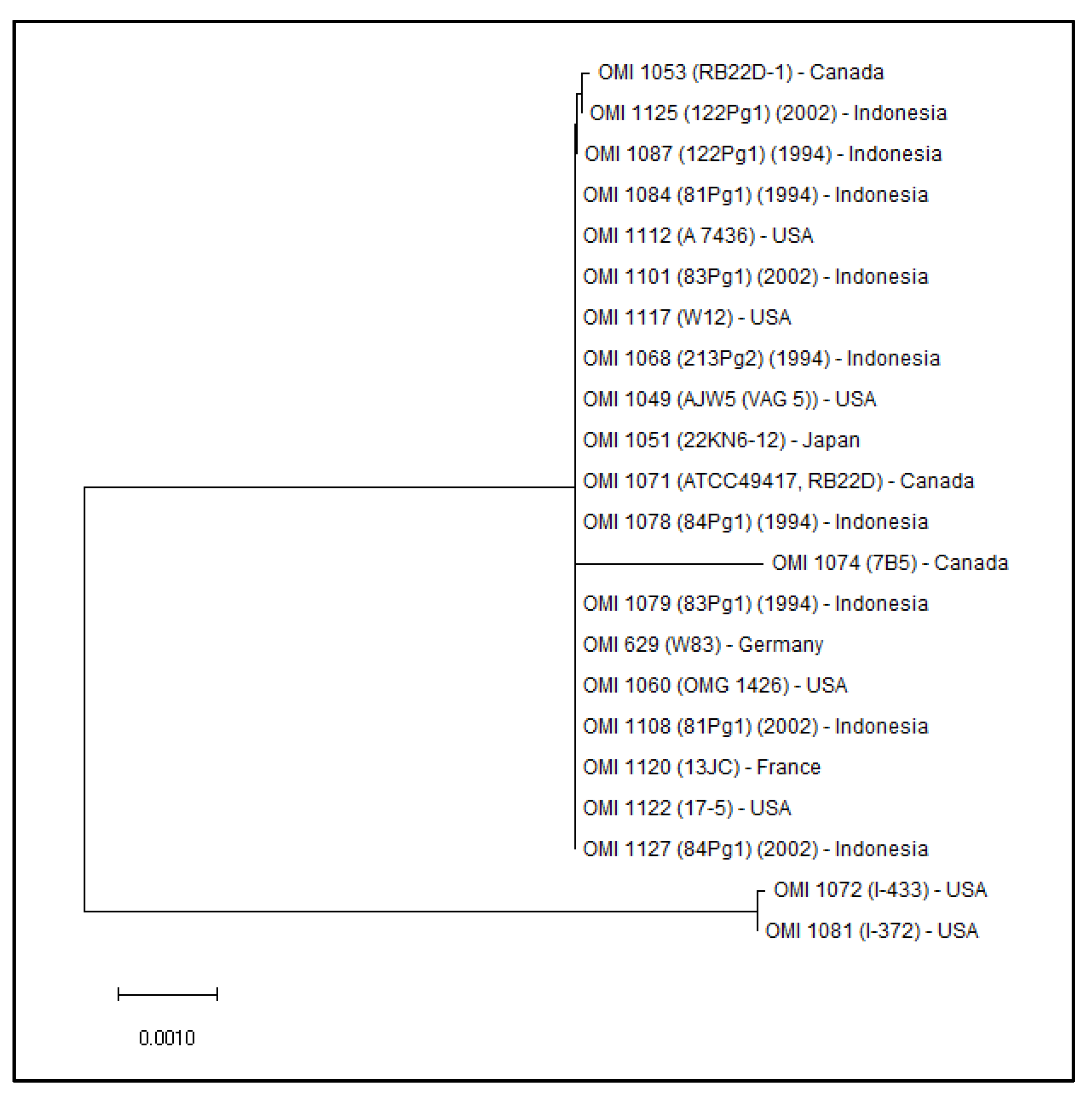
2. Results
3. Materials and Methods
4. Discussion
5. Conclusions
Author Contributions
Funding
Institutional Review Board Statement
Informed Consent Statement
Data Availability Statement
Acknowledgments
Conflicts of Interest
References
- Henry, L.G.; McKenzie, R.M.E.; Robles, A.; Fletcher, H.M. Oxidative stress resistance in Porphyromonas gingivalis. Future Microbiol. 2012, 7, 497–512. [Google Scholar] [CrossRef]
- Dahlén, G.; Gmür, R.; Yoshino, T. Phenotypes, serotypes and antibiotic susceptibility of Swedish Porphyromonas gingivalis isolates from periodontitis and periodontal abscesses. Oral Microbiol. Immunol. 2007, 22, 80–86. [Google Scholar] [CrossRef] [PubMed]
- Amano, A.; Nakagawa, I.; Kataoka, K.; Morisaki, I.; Hamada, S. Distribution of Porphyromonas gingivalis strains with fimA genotypes in periodontitis patients. J. Clin. Microbiol. 1999, 37, 1426–1430. [Google Scholar] [CrossRef]
- Griffen, A.L.; Lyons, S.R.; Becker, M.R.; Moeschberger, M.L.; Leys, E.J. Porphyromonas gingivalis strain variability and periodontitis. J. Clin. Microbiol. 1999, 37, 4028–4033. [Google Scholar] [CrossRef]
- Missailidis, C.G.; Umeda, J.E.; Ota-Tsuzuki, C.; Anzai, D.; Mayer, M.P.A. Distribution of fimA genotypes of Porphyromonas gingivalis in subjects with various periodontal conditions. Oral Microbiol. Immunol. 2004, 19, 224–229. [Google Scholar] [CrossRef] [PubMed]
- Nakagawa, I.; Amano, A.; Ohara-Nemoto, Y.; Endoh, N.; Morisaki, I.; Kimura, S.; Kawabata, S.; Hamada, S. Identification of a new variant of fimA gene of Porphyromonas gingivalis and its distribution in adults and disabled populations with periodontitis. J. Periodontal Res. 2002, 37, 425–432. [Google Scholar] [CrossRef] [PubMed]
- Hall, L.M.C.; Fawell, S.C.; Shi, X.; Faray-Kele, M.-C.; Aduse-Opoku, J.; Whiley, R.A.; Curtis, M.A. Sequence diversity and antigenic variation at the rag locus of Porphyromonas gingivalis. Infect. Immun. 2005, 73, 4253–4262. [Google Scholar] [CrossRef]
- Frandsen, E.V.; Poulsen, K.; Curtis, M.A.; Kilian, M. Evidence of recombination in Porphyromonas gingivalis and random distribution of putative virulence markers. Infect. Immun. 2001, 69, 4479–4485. [Google Scholar] [CrossRef]
- Sawada, K.; Kokeguchi, S.; Hongyo, H.; Sawada, S.; Miyamoto, M.; Maeda, H.; Nishimura, F.; Takashiba, S.; Murayama, Y. Identification by subtractive hybridization of a novel insertion sequence specific for virulent strains of Porphyromonas gingivalis. Infect. Immun. 1999, 67, 5621–5625. [Google Scholar] [CrossRef] [PubMed]
- Potempa, J.; Madej, M.; Scott, D.A. The RagA and RagB proteins of Porphyromonas gingivalis. Mol. Oral Microbiol. 2021, 36, 225–232. [Google Scholar] [CrossRef]
- Kuhn, C. Genetische Polymorphismen in Porphyromonas gingivalis und deren Auswirkung auf dessen Virulenz. Ph.D. Dissertation, Heinrich-Heine-Universität, Düsseldorf, Germany, 2018. [Google Scholar]
- Nagano, K.; Murakami, Y.; Nishikawa, K.; Sakakibara, J.; Shimozato, K.; Yoshimura, F. Characterization of RagA and RagB in Porphyromonas gingivalis: Study using gene-deletion mutants. J. Med. Microbiol. 2007, 56, 1536–1548. [Google Scholar] [CrossRef]
- Bonass, W.A.; Marsh, P.D.; Percival, R.S.; Aduse-Opoku, J.; Hanley, S.A.; Devine, D.A.; Curtis, M.A. Identification of ragAB as a temperature-regulated operon of Porphyromonas gingivalis W50 using differential display of randomly primed RNA. Infect. Immun. 2000, 68, 4012–4017. [Google Scholar] [CrossRef]
- Curtis, M.A.; Hanley, S.A.; Aduse-Opoku, J. The rag locus of Porphyromonas gingivalis: A novel pathogenicity island. J. Periodontal Res. 1999, 34, 400–405. [Google Scholar] [CrossRef] [PubMed]
- Masuda, T.; Murakami, Y.; Noguchi, T.; Yoshimura, F. Effects of various growth conditions in a chemostat on expression of virulence factors in Porphyromonas gingivalis. Appl. Environ. Microbiol. 2006, 72, 3458–3467. [Google Scholar] [CrossRef] [PubMed]
- Hanley, S.A.; Aduse-Opoku, J.; Curtis, M.A. A 55-kilodalton immunodominant antigen of Porphyromonas gingivalis W50 has arisen via horizontal gene transfer. Infect. Immun. 1999, 67, 1157–1171. [Google Scholar] [CrossRef] [PubMed]
- Shi, X.; Hanley, S.A.; Faray-Kele, M.-C.; Fawell, S.C.; Aduse-Opoku, J.; Whiley, R.A.; Curtis, M.A.; Hall, L.M.C. The rag locus of Porphyromonas gingivalis contributes to virulence in a murine model of soft tissue destruction. Infect. Immun. 2007, 75, 2071–2074. [Google Scholar] [CrossRef]
- Madej, M.; White, J.B.R.; Nowakowska, Z.; Rawson, S.; Scavenius, C.; Enghild, J.J.; Bereta, G.P.; Pothula, K.; Kleinekathoefer, U.; Baslé, A.; et al. Structural and functional insights into oligopeptide acquisition by the RagAB transporter from Porphyromonas gingivalis. Nat. Microbiol. 2020, 5, 1016–1025. [Google Scholar] [CrossRef] [PubMed]
- Glenwright, A.J.; Pothula, K.R.; Bhamidimarri, S.P.; Chorev, D.S.; Baslé, A.; Firbank, S.J.; Zheng, H.; Robinson, C.V.; Winterhalter, M.; Kleinekathöfer, U.; et al. Structural basis for nutrient acquisition by dominant members of the human gut microbiota. Nature 2017, 541, 407–411. [Google Scholar] [CrossRef]
- Goulas, T.; Garcia-Ferrer, I.; Hutcherson, J.A.; Potempa, B.A.; Potempa, J.; Scott, D.A.; Gomis-Rüth, F.X. Structure of RagB, a major immunodominant outer-membrane surface receptor antigen of Porphyromonas gingivalis. Mol. Oral Microbiol. 2016, 31, 472–485. [Google Scholar] [CrossRef]
- Curtis, M.A.; Slaney, J.M.; Carman, R.J.; Johnson, N.W. Identification of the major surface protein antigens of Porphyromonas gingivalis using IgG antibody reactivity of periodontal case-control serum. Oral Microbiol. Immunol. 1991, 6, 321–326. [Google Scholar] [CrossRef]
- Imai, M.; Murakami, Y.; Nagano, K.; Nakamura, H.; Yoshimura, F. Major outer membrane proteins from Porphyromonas gingivalis: Strain variation, distribution, and clinical significance in periradicular lesions. Eur. J. Oral Sci. 2005, 113, 391–399. [Google Scholar] [CrossRef] [PubMed]
- Zeller, I.; Hutcherson, J.A.; Lamont, R.J.; Demuth, D.R.; Gumus, P.; Nizam, N.; Buduneli, N.; Scott, D.A. Altered antigenic profiling and infectivity of Porphyromonas gingivalis in smokers and non-smokers with periodontitis. J. Periodontol. 2014, 85, 837–844. [Google Scholar] [CrossRef] [PubMed]
- Hutcherson, J.A.; Bagaitkar, J.; Nagano, K.; Yoshimura, F.; Wang, H.; Scott, D.A. Porphyromonas gingivalis RagB is a proinflammatory signal transducer and activator of transcription 4 agonist. Mol. Oral Microbiol. 2015, 30, 242–252. [Google Scholar] [CrossRef] [PubMed]
- Liu, Y.; Zhang, Y.; Wang, L.; Guo, Y.; Xiao, S. Prevalence of Porphyromonas gingivalis four rag locus genotypes in patients of orthodontic gingivitis and periodontitis. PLoS ONE 2013, 8, e61028. [Google Scholar] [CrossRef]
- Nelson, K.E.; Fleischmann, R.D.; DeBoy, R.T.; Paulsen, I.T.; Fouts, D.E.; Eisen, J.A.; Daugherty, S.C.; Dodson, R.J.; Durkin, A.S.; Gwinn, M.; et al. Complete genome sequence of the oral pathogenic bacterium Porphyromonas gingivalis strain W83. J. Bacteriol. 2003, 185, 5591–5601. [Google Scholar] [CrossRef]
- Xu, J.; Bjursell, M.K.; Himrod, J.; Deng, S.; Carmichael, L.K.; Chiang, H.C.; Hooper, L.V.; Gordon, J.I. A genomic view of the human-Bacteroides thetaiotaomicron symbiosis. Science 2003, 299, 2074–2076. [Google Scholar] [CrossRef]
- Chen, T.; Hosogi, Y.; Nishikawa, K.; Abbey, K.; Fleischmann, R.D.; Walling, J.; Duncan, M.J. Comparative whole-genome analysis of virulent and avirulent strains of Porphyromonas gingivalis. J. Bacteriol. 2004, 186, 5473–5479. [Google Scholar] [CrossRef] [PubMed]
- Roberts, A.P.; Pratten, J.; Wilson, M.; Mullany, P. Transfer of a conjugative transposon, Tn5397 in a model oral biofilm. FEMS Microbiol. Lett. 1999, 177, 63–66. [Google Scholar] [CrossRef]
- Waters, V.L. Conjugative transfer in the dissemination of beta-lactam and aminoglycoside resistance. Front. Biosci. 1999, 4, D433–D456. [Google Scholar] [CrossRef]
- Li, Y.H.; Lau, P.C.; Lee, J.H.; Ellen, R.P.; Cvitkovitch, D.G. Natural genetic transformation of Streptococcus mutans growing in biofilms. J. Bacteriol. 2001, 183, 897–908. [Google Scholar] [CrossRef]
- Li, Y.-H.; Tang, N.; Aspiras, M.B.; Lau, P.C.Y.; Lee, J.H.; Ellen, R.P.; Cvitkovitch, D.G. A quorum-sensing signaling system essential for genetic competence in Streptococcus mutans is involved in biofilm formation. J. Bacteriol. 2002, 184, 2699–2708. [Google Scholar] [CrossRef] [PubMed]
- Wang, B.Y.; Chi, B.; Kuramitsu, H.K. Genetic exchange between Treponema denticola and Streptococcus gordonii in biofilms. Oral Microbiol. Immunol. 2002, 17, 108–112. [Google Scholar] [CrossRef] [PubMed]
- Stork, M.; Bos, M.P.; Jongerius, I.; de Kok, N.; Schilders, I.; Weynants, V.E.; Poolman, J.T.; Tommassen, J. An outer membrane receptor of Neisseria meningitidis involved in zinc acquisition with vaccine potential. PLoS Pathog. 2010, 6, e1000969. [Google Scholar] [CrossRef] [PubMed]
- Kong, F.; Zheng, D.; She, P.; Ni, P.; Zhu, H.; Xu, H.; Su, Z. Porphyromonas gingivalis B cell Antigen Epitope Vaccine, pIRES-ragB’-mGITRL, Promoted RagB-Specific Antibody Production and Tfh Cells Expansion. Scand. J. Immunol. 2015, 81, 476–482. [Google Scholar] [CrossRef] [PubMed]
- Zheng, D.; Sun, Q.; Su, Z.; Kong, F.; Shi, X.; Tong, J.; Shen, P.; Peng, T.; Wang, S.; Xu, H. Enhancing specific-antibody production to the ragB vaccine with GITRL that expand Tfh, IFN-γ(+) T cells and attenuates Porphyromonas gingivalis infection in mice. PLoS ONE 2013, 8, e59604. [Google Scholar] [CrossRef]
- National Center for Biotechnology Information. [Internet]. Bethesda (MD): National Library of Medicine (US), National Center for Biotechnology Information. 1998. Available online: https://www.ncbi.nlm.nih.gov/ (accessed on 19 March 2023).
- Fournier, D.; Mouton, C.; Lapierre, P.; Kato, T.; Okuda, K.; Ménard, C. Porphyromonas gulae sp. nov., an anaerobic, gram-negative coccobacillus from the gingival sulcus of various animal hosts. Int. J. Syst. Evol. Microbiol. 2001, 51, 1179–1189. [Google Scholar] [CrossRef]
- Hall, T.; Biosciences, I.; Carlsbad, C. BioEdit: An important software for molecular biology. GERF Bull Biosci 2011, 2, 60–61. [Google Scholar]
- Hall, T.A. BioEdit: A User-Friendly Biological Sequence Alignment Editor and Analysis Program for Windows 95/98/NT; Nucleic Acids Symposium Series; Oxford University Press: Oxford, UK, 1999. [Google Scholar]
- Tamura, K.; Stecher, G.; Kumar, S. MEGA11: Molecular Evolutionary Genetics Analysis Version 11. Mol. Biol. Evol. 2021, 38, 3022–3027. [Google Scholar] [CrossRef]
- Dolgilevich, S.; Rafferty, B.; Luchinskaya, D.; Kozarov, E. Genomic comparison of invasive and rare non-invasive strains reveals Porphyromonas gingivalis genetic polymorphisms. J. Oral Microbiol. 2011, 3, 5764. [Google Scholar] [CrossRef]
- Bunte, K.; Kuhn, C.; Walther, C.; Peters, U.; Aarabi, G.; Smeets, R.; Beikler, T. Clinical significance of ragA, ragB, and PG0982 genes in Porphyromonas gingivalis isolates from periodontitis patients. Eur. J. Oral Sci. 2021, 129, e12776. [Google Scholar] [CrossRef]
- Su, Z.; Kong, F.; Wang, S.; Chen, J.; Yin, R.; Zhou, C.; Zhang, Y.; He, Z.; Shi, Y.; Xue, Y.; et al. The rag locus of Porphyromonas gingivalis might arise from Bacteroides via horizontal gene transfer. Eur. J. Clin. Microbiol. Infect. Dis. 2010, 29, 429–437. [Google Scholar] [CrossRef]
- Dashper, S.G.; Mitchell, H.L.; Seers, C.A.; Gladman, S.L.; Seemann, T.; Bulach, D.M.; Chandry, P.S.; Cross, K.J.; Cleal, S.M.; Reynolds, E.C. Porphyromonas gingivalis Uses Specific Domain Rearrangements and Allelic Exchange to Generate Diversity in Surface Virulence Factors. Front. Microbiol. 2017, 8, 48. [Google Scholar] [CrossRef]
- Laine, M.L.; van Winkelhoff, A.J. Virulence of six capsular serotypes of Porphyromonas gingivalis in a mouse model. Oral Microbiol. Immunol. 1998, 13, 322–325. [Google Scholar] [CrossRef] [PubMed]
- Anderson, K.L.; Salyers, A.A. Genetic evidence that outer membrane binding of starch is required for starch utilization by Bacteroides thetaiotaomicron. J. Bacteriol. 1989, 171, 3199–3204. [Google Scholar] [CrossRef] [PubMed]
- Koropatkin, N.M.; Martens, E.C.; Gordon, J.I.; Smith, T.J. Starch catabolism by a prominent human gut symbiont is directed by the recognition of amylose helices. Structure 2008, 16, 1105–1115. [Google Scholar] [CrossRef] [PubMed]
- Phansopa, C.; Roy, S.; Rafferty, J.B.; Douglas, C.W.I.; Pandhal, J.; Wright, P.C.; Kelly, D.J.; Stafford, G.P. Structural and functional characterization of NanU, a novel high-affinity sialic acid-inducible binding protein of oral and gut-dwelling Bacteroidetes species. Biochem. J. 2014, 458, 499–511. [Google Scholar] [CrossRef] [PubMed]
- Brunner, J.; Wittink, F.R.A.; Jonker, M.J.; de Jong, M.; Breit, T.M.; Laine, M.L.; Soet, J.J.d.; Crielaard, W. The core genome of the anaerobic oral pathogenic bacterium Porphyromonas gingivalis. BMC Microbiol. 2010, 10, 252. [Google Scholar] [CrossRef] [PubMed]
- Fujiwara-Takahashi, K.; Watanabe, T.; Shimogishi, M.; Shibasaki, M.; Umeda, M.; Izumi, Y.; Nakagawa, I. Phylogenetic diversity in fim and mfa gene clusters between Porphyromonas gingivalis and Porphyromonas gulae, as a potential cause of host specificity. J. Oral Microbiol. 2020, 12, 1775333. [Google Scholar] [CrossRef]


| Strain (OMI) | Species | Original Code | Origin Species | Isolation Year | Country |
|---|---|---|---|---|---|
| 629 | P. gingivalis | W83 | Human | 1991 | Bonn, Germany |
| 1049 | P. gingivalis | AJW5 (VAG 5) | Human | 1991 | Buffalo, NY, USA |
| 1051 | P. gingivalis | 22KN6-12 | Human | Tokushima, Japan | |
| 1060 | P. gingivalis/gulae | OMG 1426 | Monkey | 1989 | Florida, USA |
| 1053 | P. gingivalis | RB22D-1 | Human | Quebec, Canada | |
| 1068 | P. gingivalis | 213Pg2 | Human | 1994 | Indonesia |
| 1071 | P. gingivalis | ATCC49417, RB22D | Human | 1993 | Quebec, Canada |
| 1072 | P. gingivalis/gulae | I-433 | Monkey | 1989 | Florida, USA |
| 1074 | P. gingivalis | 7B5 | Human | Quebec, Canada | |
| 1078 | P. gingivalis | 84Pg1-a | Human | 1994 | Indonesia |
| 1079 | P. gingivalis | 83Pg1-a | Human | 1994 | Indonesia |
| 1081 | P. gingivalis/gulae | I-372 | Monkey | 1989 | Florida, USA |
| 1084 | P. gingivalis | 81Pg1-a | Human | 1994 | Indonesia |
| 1087 | P. gingivalis | 122Pg1-a | Human | 1994 | Indonesia |
| 1101 | P. gingivalis | 83Pg1-b | Human | 2002 | Indonesia |
| 1108 | P. gingivalis | 81Pg1-b | Human | 2002 | Indonesia |
| 1112 | P. gingivalis | A 7436 | Human | Georgia, USA | |
| 1117 | P. gingivalis | W12 | Human | Alabama, USA | |
| 1120 | P. gingivalis | 13JC | Human | Rennes, France | |
| 1122 | P. gingivalis | 17-5 | Human | Minneapolis, MN, USA | |
| 1125 | P. gingivalis | 122Pg1-b | Human | 2002 | Indonesia |
| 1127 | P. gingivalis | 84Pg1-b | Human | 2002 | Indonesia |
Disclaimer/Publisher’s Note: The statements, opinions and data contained in all publications are solely those of the individual author(s) and contributor(s) and not of MDPI and/or the editor(s). MDPI and/or the editor(s) disclaim responsibility for any injury to people or property resulting from any ideas, methods, instructions or products referred to in the content. |
© 2023 by the authors. Licensee MDPI, Basel, Switzerland. This article is an open access article distributed under the terms and conditions of the Creative Commons Attribution (CC BY) license (https://creativecommons.org/licenses/by/4.0/).
Share and Cite
Böcher, S.; Meyer, H.L.; Dafni, E.; Conrads, G. Prevalence and Phylogenetic Analysis of Lipoprotein-Gene ragB-1 of Porphyromonas gingivalis—A Pilot Study. Antibiotics 2023, 12, 1458. https://doi.org/10.3390/antibiotics12091458
Böcher S, Meyer HL, Dafni E, Conrads G. Prevalence and Phylogenetic Analysis of Lipoprotein-Gene ragB-1 of Porphyromonas gingivalis—A Pilot Study. Antibiotics. 2023; 12(9):1458. https://doi.org/10.3390/antibiotics12091458
Chicago/Turabian StyleBöcher, Sarah, Hendrik L. Meyer, Evdokia Dafni, and Georg Conrads. 2023. "Prevalence and Phylogenetic Analysis of Lipoprotein-Gene ragB-1 of Porphyromonas gingivalis—A Pilot Study" Antibiotics 12, no. 9: 1458. https://doi.org/10.3390/antibiotics12091458