Review

Enterococcal Phages: Food and Health Applications

by
Carlos Rodríguez-Lucas
1,2,* and
Victor Ladero
3,4,*
1
Microbiology Laboratory, Hospital Universitario Central de Asturias, 33011 Oviedo, Spain
2
Translational Microbiology Group, Instituto de Investigación Sanitaria del Principado de Asturias (ISPA), 33011 Oviedo, Spain
3
Department of Technology and Biotechnology of Dairy Products, Dairy Research Institute, IPLA CSIC, 33300 Villaviciosa, Spain
4
Molecular Microbiology Group, Instituto de Investigación Sanitaria del Principado de Asturias (ISPA), 33011 Oviedo, Spain
*
Authors to whom correspondence should be addressed.
Antibiotics 2023, 12(5), 842; https://doi.org/10.3390/antibiotics12050842
Submission received: 28 March 2023 / Revised: 24 April 2023 / Accepted: 30 April 2023 / Published: 2 May 2023

Abstract

:
Enterococcus is a diverse genus of Gram-positive bacteria belonging to the lactic acid bacteria (LAB) group. It is found in many environments, including the human gut and fermented foods. This microbial genus is at a crossroad between its beneficial effects and the concerns regarding its safety. It plays an important role in the production of fermented foods, and some strains have even been proposed as probiotics. However, they have been identified as responsible for the accumulation of toxic compounds—biogenic amines—in foods, and over the last 20 years, they have emerged as important hospital-acquired pathogens through the acquisition of antimicrobial resistance (AMR). In food, there is a need for targeted measures to prevent their growth without disturbing other LAB members that participate in the fermentation process. Furthermore, the increase in AMR has resulted in the need for the development of new therapeutic options to treat AMR enterococcal infections. Bacteriophages have re-emerged in recent years as a precision tool for the control of bacterial populations, including the treatment of AMR microorganism infections, being a promising weapon as new antimicrobials. In this review, we focus on the problems caused by Enterococcus faecium and Enterococcus faecalis in food and health and on the recent advances in the discovery and applications of enterococcus-infecting bacteriophages against these bacteria, with special attention paid to applications against AMR enterococci.

1. Introduction

The discovery of antibiotics in the mid-20th century is one of the scientific advances that has had the most significant impacts on increasing life expectancy. The prescription of antibiotics and other antimicrobials to treat bacterial infections, which can often cause permanent damage and even end a patient’s life, has become a routine treatment. However, in recent decades, the misuse of antimicrobials has led to a rapid increase in the isolation of antimicrobial-resistant (AMR) bacteria. Today, AMR bacteria pose a great health problem; in Europe alone, up to 133,000 deaths in 2019 were attributable to infections caused by AMR bacteria [1], with an estimated cost for health services of over EUR 1000 million every year [2]. Seven pathogenic species were responsible for most of the deaths registered, namely, Enterococcus faecium, Staphylococcus aureus, Klebsiella pneumoniae, Acinetobacter baumannii, Pseudomonas aeruginosa and Enterobacter spp., listed together as the ESKAPE group by the World Health Organization (WHO, Geneva, Switzerland) [1,3]. Because of the great impact of these pathogens in terms of nosocomial infections, deaths and the economic losses of health services, the WHO has encouraged the scientific community to search for new ways to combat them [3]. In the context of this problem, bacteriophages have re-emerged as a potential weapon to fight AMR bacteria.
In this review, we focus on the advances in the characterization and application of enterococcal phages as rediscovered weapons against AMR E. faecium and Enterococcus faecalis.

2. E. faecium and E. faecalis

Enterococci are a diverse group of Gram-positive bacteria belonging to the lactic acid bacteria (LAB) group. The members of this genus are Gram-positive coccus-shaped bacteria that possess a versatile metabolism allowing them to adapt to very diverse environments and to resist rough conditions [4,5]. E. faecium and E. faecalis are the most studied species from the genus Enterococcus due to their role in human health [6]. These species are considered commensal bacteria of the gastrointestinal tract in mammals, including that of human beings [6,7,8]. They have also been isolated from a wide variety of environments that are mostly, but not exclusively, related to animal and human facilities, for example, cattle facilities [7], farms [9], hospitals [10] and wastewater facilities [11]. In fact, due to their persistent presence in the intestinal habitat, their robustness and endurance and the ease of their cultivation in the laboratory, enterococci are used as indicators of fecal contamination [12]. In addition, due to their presence in the gut, feces and milk of animals [13], they are also commonly found in foods of animal origin, such as meat and dairy products [11,14].
Although E. faecium and E. faecalis are considered harmless commensal bacteria, some strains are used as safe and effective probiotics, and they are present in certain cheeses in which they participate in the elimination of foodborne pathogens via the production of bacteriocins [15,16]. They can behave as opportunistic pathogens, and in recent years, they have been established as one of the major nosocomial pathogens. E. faecalis is considered the most pathogenic, but E. faecium has gained more concern due to the increasing acquisition of AMR [6,16].

2.1. E. faecium and E. faecalis in Food

As previously mentioned, their presence in the gut, feces and milk of mammals results in their presence in raw materials of animal origin, such as meat and dairy products. Their resistance to adverse environmental conditions allows them to grow in a wide range of pH values, temperatures and salt concentrations and to colonize foods, including fermented foods [17]. Enterococci have an ambiguous status regarding food safety. In fact, although these microorganisms belong to the LAB group (safe bacteria involved in the production of fermented foods), they have been granted with neither the Generally Regarded as Safe (GRAS) status nor the Qualified Presumption of Safety (QPS) status. Enterococci can be considered a valuable asset in cheese making, as some strains can be used as adjunct starter cultures [18,19]. The role of enterococci during cheese making is based on the large variety of technologically interesting enzymatic activities, such as protease, peptidase and lipolytic activities, which contribute to the organoleptic properties during the maturation process [18]. In addition, some strains are able to produce enterocins—so-called bacteriocins—and these produced by different strains of enterococcus that can inhibit the growth of several foodborne pathogens, such as Staphylococcus aureus, Listeria monocytogenes and Salmonella enterica [20]. However, their presence in foods has also been associated with the production of biogenic amines (BAs), toxic compounds that can cause food poisoning [21,22]. In fact, E. faecalis has been identified as the main species responsible for the accumulation of elevated concentrations of tyramine and putrescine [23,24,25], two of the most frequent BAs in dairy products [26,27]. Moreover, due to the increase in recent years of multidrug-resistant (MDR) bacteria, including enterococci, and the coining of the One Health concept to prevent their proliferation [28], concern regarding MDR enterococci in food has emerged [29]. These AMR enterococci can reach the food chain, where they can be transmitted directly or indirectly to humans and act as reservoirs of AMR genes that can be transferred to human-adapted strains or to other pathogenic bacteria [29]. Of special concern is the increase in vancomycin-resistant enterococci (VRE) from different food sources [30,31,32,33]. Thus, although the presence of enterococci in food could be considered beneficial in some scenarios, in general, they are considered a potential health threat.

2.2. E. faecium and E. faecalis in Human Health

Enterococci are considered a commensal organism of the human gastrointestinal (GI) tract, and they can be found in the GI microbiota of more than 90% of healthy people [34]. The first description of an enterococcal human infection was in 1899, when MacCallum and Hastings reported infective endocarditis (IE) caused by a bacterium that they called Micrococcus zymogenes, later identified as a member of the Enterococcus genus [35,36]. Enterococci were subsequently shown to be the cause of several kinds of infections, both community- (e.g., including pelvic infections, urinary tract infections and IE) and healthcare-associated infections (HAIs) (e.g., including surgical site infections, and urinary and bloodstream catheter-related infections) [37]. Therefore, enterococci usually display low levels of virulence, but they can also act as an opportunistic pathogen, causing severe infections, mainly in vulnerable patients, such as those who are immunocompromised, have undergone invasive procedures (e.g., the insertion of urinary or blood catheters) and have previously received antimicrobial treatments [34].

2.2.1. Epidemiology of Enterococcal Infections

E. faecium and E. faecalis account for more than 90% of the enterococci recovered from clinical samples in humans. Among them, E. faecalis is the most frequent species (80–90%) causing human infection, followed by E. faecium (5–10%) and other species (less than 10%) [38]. In recent decades, enterococci have become a first-rate clinical problem, being one of the most common microorganisms of HAIs around the world [39,40]. Several factors related to the host and to the microorganism have contributed to this conversion of a commensal pathogen into one of the major causes of HAIs. The most relevant factors associated with enterococci are their intrinsic resistance to some antimicrobials (e.g., aminoglycosides, cephalosporins and clindamycin); their ability to acquire and disseminate AMR determinants (e.g., linezolid and vancomycin resistance); and the plasticity of their genome, which may contribute to improve their adaptation to harsh environments. Moreover, the increasing number of patients undergoing immunomodulatory therapies, undergoing invasive procedures or receiving multiple antimicrobial treatments, all of which are factors associated with the host, favors the role of enterococci to cause disease [34,41].

2.2.2. Antimicrobial Resistance in Enterococci

Resistance to ß-Lactams

Enterococci are intrinsically resistant to cephalosporins, and they present a natural reduced susceptibility to penicillin due to the expression of low-affinity penicillin binding proteins (PBPs), designated PBP4 in E. faecalis and PBP5 in E. faecium [34,42]. Moreover, many enterococci strains show tolerance to the bactericidal activity of ß-lactams, with the minimal bactericidal concentrations being higher than the minimum inhibitory concentrations (MICs) [5]. This situation can be solved with the addition of an aminoglycoside (typically streptomycin or gentamicin) to an active ß-lactam, which results in bactericidal synergism [43,44].
A higher-level resistance to penicillin or ampicillin resistance in enterococci can be due to the overexpression of chromosomal PBP4 and PBP5 in E. faecalis and E. faecium, respectively, or through acquired mechanisms [45,46,47]. The former is anecdotic, as acquired mechanisms are the most frequent cause of ampicillin resistance. Acquired mechanisms include ß-lactamase production and mutation acquisition in low-affinity PBP4 and PBP5 [48,49]. Currently, ampicillin resistance in enterococci is mainly mediated by the acquisition of mutations in PBP, and it is far more prevalent in E. faecium than in E. faecalis [49]. Ampicillin-resistant E. faecium due to acquired mutations in the PBP5-encoding gene has been linked to a hospital-associated (HA) clade, and it emerged in the late 1970s in the United States (US) [34]. Today, there is a high rate of ampicillin resistance in E. faecium strains, and it exceeds 70% in many countries [5,50].

Resistance to Aminoglycosides

Enterococci are intrinsically resistant to clinically achievable concentrations of aminoglycosides due to the poor penetration of these agents through the bacterial cell wall in E. faecalis and due to two chromosomally encoded genes, namely 6′-N-aminoglycoside acetyltransferase (aac(6′)-Ii) and rRNA methyltransferase (efmM) in E. faecium [5,34]. As previously mentioned, this type of resistance can be overcome with the addition of an agent that disrupts cell wall synthesis, such as ß-lactams. Some strains can also exhibit a high level of aminoglycoside resistance (MIC > 500 mg/L for gentamycin and MIC > 2000 mg/L for streptomycin) through the acquisition of aminoglycoside-modifying enzymes (phosphotransferases, acetyltransferases and nucleotidyltransferases), which inhibit the aforementioned synergic effect [5,51].

Resistance to Glycopeptides

Vancomycin, the main member of the glycopeptide family, was the first-line treatment of ampicillin-resistant E. faecium for decades, without reports of VRE strains until the 1980s [52,53,54]. Glycopeptide resistance in enterococci is mediated by the acquisition of eight different genes of the van operon (vanA, vanB, vanD, vanE, vanG, vanL, vanM and vanN). Moreover, E. casseliflavus and E. gallinarum exhibit intrinsic low-level resistance to glycopeptides through the presence of a vanC gene in their chromosome [55,56]. These genes code for the terminal amino acids of peptidoglycan precursors different from the original form (D-Ala-D-Ala). Thus, the modified amino acids D-Ala-D-Lactate and D-Ala-D-Serine present a lower affinity to glycopeptides, leading to high-level and low-level resistance to glycopeptides, respectively [57]. The vanA and vanB genes are the main mechanism of resistance to glycopeptides in enterococci, mainly being present in E. faecium [55,58]. The prevalence of glycopeptide-resistant E. faecium varies widely between continents and countries. Accordingly, the percentage of resistance to glycopeptides in E. faecium invasive isolates is more than 60% in the US, 37% in Australia and 16.8% in European countries (with national percentages ranging from 0.0 to 56.6%) [49,50,59,60,61]. HA ampicillin-resistant E. faecium clones often acquire resistance to glycopeptides, highlighting the importance of E. faecium as a nosocomial pathogen [62,63]. The scarce active antimicrobials available to treat infections caused by this MDR microorganism are a global cause for concern.

Resistance to Linezolid

Although linezolid resistance in enterococci remains uncommon, the number of linezolid-resistant enterococci (LRE) has increased in recent years. The main mechanism of linezolid resistance in Gram-positive bacteria is point mutations in the central loop of domain V of the 23S rRNA gene, among which the G2576T (Escherichia coli numbering) nucleotide mutation is the most described [64,65]. Other point mutations in the genes rplC, rplD and rplV, which code for the L3, L4 and L22 ribosomal proteins, respectively, are also associated with a decreased susceptibility to linezolid; however, they play a minor role [66,67]. Moreover, the acquisition and dissemination of transferable linezolid resistance genes, namely, cfr-like, optrA and poxtA genes, have been increasingly reported in linezolid-resistant Gram-positive bacteria in recent years [10,67,68,69,70]. The cfr-like genes encode a 23S rRNA methyltransferase, which confers resistance to phenicols, lincosamides, oxazolidinones, pleuromutilins and streptogramin A (PhLOPSA phenotype) [71,72]. However, the optrA and poxtA genes code for the ribosomal protection proteins of the ABC-F family, and they confer resistance to oxazolidinones and phenicols, as well as tetracyclines in the case of poxtA [73,74]. Nowadays, the cfr, cfr(B), cfr(D), optrA and poxtA genes have been described among enterococci from different sources (animal, human and environmental samples) and countries [75]. The main linezolid resistance mechanisms are mutations in the 23S rRNA in E. faecium and the optrA gene in E. faecalis [68]. The spread of these transferable linezolid resistance genes to difficult-to-treat bacteria, such as VRE, is a cause for concern. Unfortunately, outbreaks caused by E. faecium strains that are resistant to vancomycin and linezolid (optrA-positive) have already been reported [76,77].

Resistance to Daptomycin

Daptomycin-resistant enterococci (previously called daptomycin-nonsusceptible enterococci) are uncommon, and they have often been associated with prior exposure to the drug [67]. Daptomycin resistance in enterococci is mainly mediated by structural alterations of the cell envelope through a variety of mutations, mainly in the three-component regulatory system LiaFSR [5,78]. This alteration of the cell envelope produces a repulsion of daptomycin from the membrane. Moreover, daptomycin resistance in E. faecium is also associated with mutations in the cls gene [49]. Daptomycin resistance is more common in E. faecium than in E. faecalis, which is probably related to the use of this drug to treat vancomycin-resistant E. faecium infections [78].

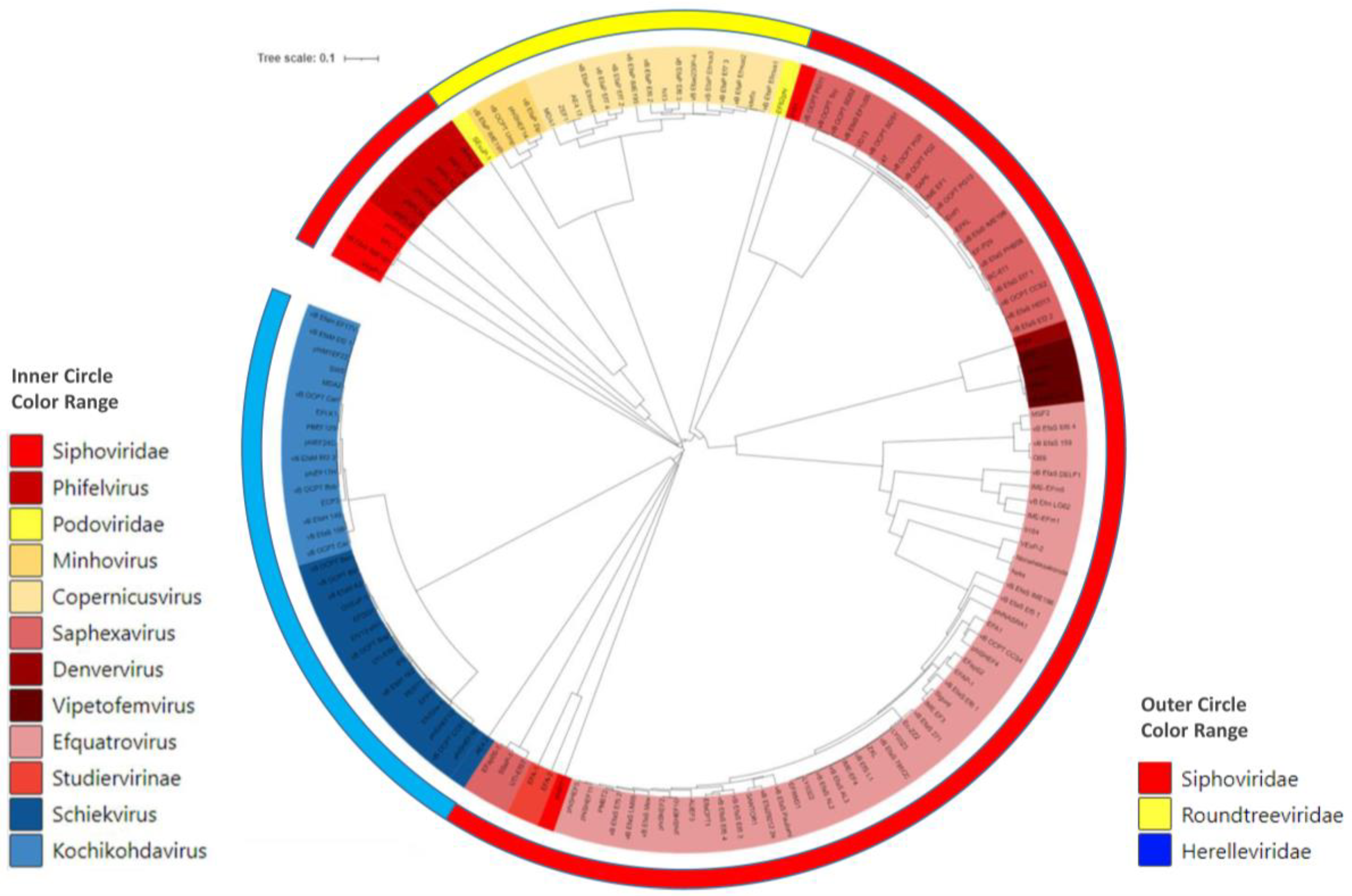
3. Bacteriophages of E. faecium and E. faecalis

Bacteriophages, or phages, have emerged in recent years as a potential bioweapon to combat MDR bacteria [79,80]. Phages are viruses that infect and kill bacteria. They are the most abundant entities on Earth and the most genetically diverse biological entities due to their mosaic genome structure and ability to mutate and recombinate [81,82]. In addition, they are ubiquitous in all types of environments, from the sea to the human gut [83]. As they are natural predators of bacteria, they have been suggested to be one of the most promising alternative therapeutic agents against MDR bacterial infections [79]. The use of phages as therapeutic agents (phage therapy) was suggested immediately after their discovery in the early XX century by Frederick Twort and Félix d’Herelle. However, the discovery of antibiotics, with a broader spectrum of action, meant that their use declined rapidly [79], except for in some countries in Eastern Europe and in the former Soviet Union where phage therapy was active, as in the Eliava Institute in Georgia [84], a reference center for phage therapy worldwide. The global problem of MDR bacteria and their consequences in terms of lives and health system costs [2] have led to a renewed interest in the study of phages and their application in phage therapy. Moreover, under the umbrella of phage therapy, the use of phages has been proposed in other fields, including food safety [85,86,87].
In this context, in recent years several enterococcal-infecting phages have been isolated and characterized—genetically and functionally (Table S1). It is remarkable that the number of E. faecalis-infecting phages that have been characterized is higher than that of E. faecium-infecting phages [88], as, in the last year, the number of E. faecium phages has increased. In Table S1, we can see that 101 genomes of E. faecalis-infecting phages are available, whereas only 16 of E. faecium can be found. Whether or not this bias is related to abundance, the ease of isolation under laboratory conditions or different searching pressures is unclear. It is astonishing that although there is a large number of molecular techniques available for the precise identification of bacterial species, there is still a large number of phage genomes (24) identified as infecting Enterococcus spp. (Table S1). However, it is remarkable that some phages are able to infect strains of both species, that is, E. faecalis and E. faecium [89,90,91]. This could be considered an advantage if a general phage cocktail designed to treat enterococcal infections is intended.
The Enterococcus-infecting phages that have been isolated to date are taxonomically widely diverse, as there are phages belonging to eleven different genera from four families (Figure 1; Table S1). Regarding the genus, the most abundant genera, accounting for almost half of the phages, are Efquatrovirus (represented by 39 phages) and Saphexavirus (represented by 25 phages), both belonging to the Siphoviridae family. Siphoviridae is the most abundant morphology, with three times more isolates than the Myoviridae and Podoviridae morphologies. The genome size distribution has a wide range, from approximately 18 kbp to 150 Kbp, but this heterogeneity is mostly related to taxonomic differences rather than genome diversity. Small genomes are typical of the Rountreeviridae family (previously known as Podoviridae), whereas large genomes are characteristic of the Herelleviridae family (previously known as Myoviridae) [92,93]. Nevertheless, within the same genera, heterogeneity in the genome size is observed, thus indicating differences among their members (Table S1).
Most of the reported enterococcal-infecting phages are virulent, at least those included in databases as single entries. In general, temperate phages are described as prophages that are identified and characterized as part of an analysis of one strain or isolate genome [94]. This is linked to the fact that temperate phages are not a good option for phage therapy due to their known involvement in the phenomenon of horizontal gene transfer, one of the main mechanisms involved in the spread of virulence and AMR genes [95]. In addition, after entering a new infecting cell, if a temperate phage undergoes the lysogenic cycle, the infected bacteria do not die; they can continue to spread and become resistant to infection by that same phage. Nevertheless, in some cases, if no alternative exists, temperate phages can be converted into virulent ones by selecting or constructing mutant phage variants [96,97], as has been achieved in the case of the E. faecalis ΦEf11 prophage, resulting in an additional increase in host range and progeny [98]. As previously mentioned, the lifestyle (virulent or temperate) of the described phages could be biased by the fact that this is an exclusion criterion for phage therapy due to their putative role in the transference of antimicrobial resistance genes [95]. In this sense, the absence of these genes is also a requirement. Most probably, this is related to the lack of such genes in the genomes of the described phages.
Although enterococci have been documented in many different ecosystems, most of the reported isolation sources are sewage and wastewater (Table S1). This could be related to the role of Enterococcus as an indicator of fecal contamination in water [12]. Nevertheless, other sources, in the search for increased phage diversity or specific target applications, have been assayed, such as human stools [89] and cheese [87,88] (Table S1).

4. Food Applications of Enterococcal Bacteriophages

As mentioned in a previous section, Enterococcus plays a yin–yang role in foods, where it can be considered a beneficial player responsible for the accumulation of toxic compounds or reservoirs of AMR genes.
E. faecalis has been identified as the main cause of the accumulation of tyramine [23,99] and putrescine [24]—together with Lactococcus lactis [100]—in dairy products, one of the food matrixes in which BAs can reach the highest concentration [27,101]. Strategies for the reduction of BAs in food have been proposed, for example, eliminating BAs after they have formed and accumulated via the addition of BA-degrading microorganisms [102,103,104] or reducing the number of BA-producing microorganisms via different treatments, such as the use of pasteurization or high-pressure technologies [105,106], with the latter being the technical process most employed during food production. However, these methods work by generally reducing the microorganisms present in the food matrix, thus affecting other bacteria that participate in the fermentation process. As enterococci belong to the LAB group, the methodologies applied to reduce their presence also act on other LAB, affecting the development of the organoleptic characteristic of the final product. Thus, tailored measures targeting only the Enterococcus population are needed. In this sense, phages infecting E. faecalis have been proposed as highly specific tools to reduce the content of BAs in dairy products [88]. The E. faecalis Q69 phage has been applied to reduce the presence of tyramine, one of the most toxic BAs found in cheese [107,108], in an experimental cheese model [87]. The phage was added directly to milk [multiplicity of infection (MOI 0.1)] used for cheese making, and after 60 days of ripening, reductions in tyramine concentrations of about 85% were achieved [87], and most importantly, the concentration of tyramine was reduced below the safety threshold level proposed [109]. In another assay, the E. faecalis-infecting phage 156 was applied (MOI 0.1) to reduce tyramine and putrescine, another toxic BA frequently found in cheese [21,26,110], taking advantage of the fact that E. faecalis is responsible for the accumulation of elevated concentrations of both BAs. In this case, significative reductions in tyramine and putrescine of 95% and 77%, respectively, were achieved [111] after 60 days of ripening. Interestingly, both phages showed the ability to control the population of E. faecalis from the early stages of cheese making, and both were partially resistant to the pasteurization process, allowing for both technologies to be applied if desired. The fact that some of the phages proposed as a tool to reduce the content of BAs in dairy products can infect MDR enterococci, including VRE [88,111], suggests that bacteriophages could also be applied to reduce the presence of MDR enterococci in food [112], thus contributing to the One Health strategy’s aim of reducing the amount of MDR bacteria in the environment.
Biofilms in the food industry also present a food safety threat, as they can act as reservoirs of foodborne spoilage or pathogenic bacteria [113,114], including BA-producing ones [115]. Different phages have been described as potential tools to eliminate biofilms formed by E. faecalis or E. faecium [91,116,117,118]. Although the great potential of enterococcal phages as biofilm elimination agents in food facilities surfaces, to the best of our knowledge, there are no reports regarding this interesting application, thus opening the opportunity for further research.

5. Human Health Applications of Enterococcal Bacteriophages

The continued increase in AMR among enterococci and their abilities to form biofilms and survive in harsh environments have been related to poor clinical outcomes in some cases [119]. Different studies have reported the use of enterococcal phages in the treatment of Enterococcus infections through in vitro and in vivo studies, including the use of biofilm, root canal and animal models. However, the use of phage therapy in human patients is limited to some case reports, and, at present, no clinical trials are being carried out.

5.1. In Vitro Models

5.1.1. Biofilm Models

The use of a single phage, a phage cocktail or their combination with antimicrobials in the treatment of bacterial biofilms has been explored in several studies [120,121]. Biofilm-associated infections are related to poor microbiological and clinical outcomes when antimicrobials are used. Phage therapy, in some cases, has been proven to be more effective against MDR biofilm infections than antimicrobials [91]. Several studies have reported the ability of different phages to infect and disrupt biofilms [120,121]. Anti-biofilm activity is mainly tested using microtiter plates with the crystal violet method and confocal laser scanning microscopy [121]. Using these methodologies, the anti-biofilm activity of several phages belonging to different families (Herelleviridae: vB_EfaH_EF1TV; Siphoviridae: Efa02, EfaS-SRH2, SHEF2 and vB_EfsS_V583; and Podoviridae: vB_ZEFP) has been demonstrated against E. faecalis [119,122,123,124,125]. Moreover, phage therapy has been shown to be a potential weapon against the biofilms formed by MDR Enterococcus. In a previous study, it was found that the EFDG1 phage was able to infect vancomycin-resistant E. faecium and E. faecalis strains. Moreover, EFDG1 significantly reduced a 2-week-old biofilm formed by a vancomycin-resistant E. faecalis strain V583 [91]. Furthermore, the vB_EfsS_V583 phage inhibited the biofilm formation by a vancomycin-resistant E. faecalis strain for 7 days. However, a poor ability to eradicate mature biofilms was revealed, with significant disruption only being observed in 1- to 2-day-old biofilms [124].
Some authors have developed models closely resembling biofilm formation during in vivo infections to avoid the possible limitations of microtiter plate studies. Thus, El-Atrees et al. studied the effects of the EPA, EPC and EPE phages on E. faecalis (EF104, EF134 and EF151) adherence to urinary catheter surfaces [126]. Their findings proved the ability to prevent biofilm formation by reducing the number of cells adhering to the catheter surface to a range of 30.8–43.8%. Moreover, they were also able to eradicate the number of cells pre-adhering to the catheter surface to a range of 48.2–71.1%. Nevertheless, when the anti-biofilm activities of the same phages were evaluated on microtiter plates, they showed more efficacy in both the prevention of biofilm formation and the eradication of the preformed biofilm, achieving ranges of 38–39.9% and 71–78.4%, respectively [126]. In a similar study, silicone Foley catheters were covered with an E. faecalis biofilm, and then they were exposed to the vB_EfaS-271 phage for 3, 6 or 24 h. A significant decrease in the number of viable E. faecalis cells was observed after three hours when a higher MOI was used. However, lower MOI ratios needed a longer time (6 h) for considerable effects to be observed. Unexpected results were observed at 24 h, with a large number of E. faecalis cells surviving in samples treated with 10 MOI compared to those treated with 0.0001 or 0.01 MOI. The authors speculated that a greater selection of phage-resistant mutants could occur under high-MOI conditions. However, such mutants seemed to be less competitive than wild-type cells [127]. In another approximation of biofilm formation during in vivo infections, Melo et al. developed an in vitro collagen wound model (CWM) of biofilm formation with two phages: vB_EfaS-Zip (Rountreeviridae) and vB_EfaP-Max (Siphoviridae). Both phages showed lytic activity against E. faecium and E. faecalis. In the CWM, vB_EfaP-Max and vB_EfaS-Zip were able to reduce the number of viable cells of E. faecalis and E. faecium, respectively, during the first eight hours. However, in both cases, the number of cells in the control and phage-treated biofilms in the CWM was similar at 24 h. In a new CWM, a cocktail comprising the two phages was used to infect a dual-species (E. faecium and E. faecalis) biofilm. In this last assay, a statistically significant reduction in the concentrations of the cells in the treated biofilms was observed at 3, 6 and 8 h compared to those in the control, and a residual reduction was also detected at 24 h. The emergence of phage resistance might be related to the loss of or a reduction in anti-biofilm activity when a phage alone or a cocktail phage is applied, respectively [90]. Although the selection of phage-resistant mutants is an issue in phage therapy, in some cases, this selection may be an opportunity. Liu et al. described the strong lytic activity of the EFap02 phage against the E. faecalis strain Efa02 and identified the glycosyltransferase gene Group 2 (gtr2) as its receptor. Unfortunately, the rapid emergence of phage-resistant mutants was observed by the authors. The phage-resistant strain EFa02R had loss-of-function mutations in the gtr2 gene, responsible for the biosynthesis of capsular polysaccharides. Not only does the loss of receptors in EFa02R prevent phage adsorption, but it also impairs the biofilm formation ability of these mutants. Therefore, capsular polysaccharide loss could revert the inactivation of some antimicrobials caused by the biofilm [125].
A phage alone or a phage cocktail combined with antimicrobials could prevent the emergence of phage resistance and enhance their activities separately [128]. Phage–antimicrobial synergy has been reported, even against bacteria resistant to the antimicrobial used in the combination [129]. Similar to this, daptomycin plus a phage cocktail (113 and 9184) showed synergic bactericidal activity against daptomycin-nonsusceptible E. faecium [130]. Likewise, phage–antimicrobial synergy against vancomycin-resistant E. faecalis V583 was also observed when a combined treatment of vancomycin and the EFLK1 phage was applied. This combination was able to reduce viable bacterial counts by nearly 8 logs in a well-established biofilm, whereas treatment with the phage alone only achieved a reduction of 4 logs, and vancomycin alone failed entirely [131].

5.1.2. Human Root Canal Model (Ex Vivo)

E. faecalis is frequently detected in asymptomatic and persistent endodontic infections, with prevalence ranging from 24 to 77% [132]. Its ability to invade the dentinal tubes of the root canal walls of human teeth, their aforementioned ability to form biofilms and their ability to survive in harsh environments allow this microorganism to cause persistent infections, and it is difficult to treat them [121,132,133]. The treatment of these infections includes mechanical debridement and chemical agents, such as chlorhexidine and sodium hypochlorite (NaOCl), which are generally effective [132,133]. However, several treatment failures have been reported, and therefore, the development of new therapeutic alternatives is necessary [132]. In this context, enterococcal phage therapy has been studied in ex vivo models of root canal infection. In a previous study, it was found that the EFDG1 phage was able to prevent E. faecalis root canal infection in an ex vivo model performed with human-extracted teeth [91]. Likewise, the vB_ZEFP phage showed a greater ability to reduce bacterial leakage from the root apex than other treatments (NaOCl and NaOCl plus EDTA) [123]. Similar studies have demonstrated the efficacy of different phages to destroy E. faecalis biofilms in root canal systems: vB_Efa29212_2e (Siphoviridae), vB_Efa29212_3e (Herelleviriae) and vB_EfaS_HEf13 (Siphoviridae) [134,135,136,137]. With regard to MDR Enterococcus, Tinoco et al. evaluated the activity of ΦEf11/ΦFL1C(∆36)PnisA, an engineered phage, against vancomycin-resistant E. faecalis V583 in an ex vivo model of root canal [138]. The treatment with the phage generated a reduction of 99% for the V583-infected models. In contrast, a scarce reduction of 18% was observed in biofilms formed by the E. faecalis JH2-2 strain (a fusidic acid- and rifampicin-resistant, vancomycin-susceptible strain) [138].

5.2. In Vivo Models

In vivo studies using animal models are essential to evaluate the safety and efficiency of new therapies, including phage therapy. Among them, the most frequently used include models performed in Galleria mellonella, zebrafish embryos and mice [139]. The G. mellonella animal model has previously been used to assess the virulence of VRE [89]. Although this model is cost-effective in the evaluation of the potential of phage therapy, to date, only two studies have assessed the efficacy of phage therapy against larvae infected by Enterococcus [89,140]. In the first study, the administration of a phage cocktail comprising the MDA1 (Rountreeviridae) and MDA2 (Herelleviridae) phages was effective against larvae infected with a vancomycin-resistant E. faecium strain (VRE004). The larvae were injected with 107 colony-forming units (CFU)/10 μL of the VRE004 strain, and two groups were employed. These groups were injected with the phage cocktail at a concentration of 2 × 10⁶ plaque-forming units (PFU)/106 μL, one of them 1 h prior to (the prophylactic group) and the other 1 h after (the treatment group) the VRE injection. After 48 h of follow-up, both groups demonstrated efficacy, being 3.7 (the treatment group) and 6.5 (the prophylactic group) times more likely to survive than the larvae injected with VRE only [140]. In the second study, the activity of the phage vB_EfaH_163 (Herelleviridae) against larvae infected with a vancomycin-resistant E. faecium (VRE-13) strain was studied. The larvae were injected with the VRE-13 strain at a concentration of 105 CFU/larva. After 1 h, the larvae were injected with PBS (the control group) or a phage suspension at an MOI of 0.1, and the number of deaths was monitored for five days. Treatment with the vB_EfaH_163 phage increased larval survival by 20% compared with the control group, although no statistically significant differences were observed [89]. In another assay, the therapeutic potential of phage SHEF2 (Siphoviridae) in treating systemic E. faecalis infections in an in vivo zebrafish embryo infection model was studied. In this model, the zebrafish embryos were infected with E. faecalis (OS16 strain), and two hours later, they were injected with SHEF2 or a heat-killed sample of SHEF2 at an MOI of 20 (with respect to the E. faecalis inoculum). The zebrafish infected with OS16 alone or with heat-killed SHEF2 showed a mortality rate of 73%, whereas those injected with SHEF2 showed a mortality rate of 16% (p < 0.0001) [119]. Nowadays, a number of studies are evaluating enterococcal phage therapy against different kinds of E. faecalis infection using mice models. Enterococcal phage therapy has been shown to be a promising candidate for the treatment of E. faecalis endophthalmitis (a rare cause of postoperative infection) in mice models. Thus, Kishimoto et al. demonstrated a decrease in the number of viable bacteria and the infiltration of neutrophils in mice eyes infected with vancomycin-susceptible and -resistant E. faecalis when they were treated with phages [ΦEF24C-P2, ΦEF7H, ΦEF14H1 and ΦEF19G (Herelleviridae)] [141,142]. Several studies have evaluated the efficacy of phage therapy in an E. faecalis sepsis mice model using intraperitoneal injections. The intraperitoneal administration of phage IME-EF1 or its endolysin, at a 10 MOI, 30 min after E. faecalis 002 inoculation resulted in survival rates of 60% and 80%, respectively [143]. A single injection of 3 × 10⁸ PFU of the phage ENB6, administered 45 min after a vancomycin-resistant E. faecium (CRMEN 44) challenge, was able to rescue 100% of the mice [144]. A single intraperitoneal administration of other phages (at different doses and times following bacterium inoculation) was also enough to protect all the mice infected with enterococcus (including VRE): ΦEF24C (MOI 0.01/20 min), EF-P29 (4 × 10⁵ PFU/1 h), SSsP-1 and GVEsP-1 (3 × 10⁹ phage stock/3 h) and a phage cocktail (comprising the phages EFDG1 and EFLK1) (2 × 10⁸ PFU/0 h and 1 h) [145,146,147,148].

5.3. Phage Therapy in Humans

To date, only a few case reports have described the use of phage therapy against enterococcus infections in humans (Table 1) [149,150,151,152,153]. Three patients suffering from chronic bacterial prostatitis caused by E. faecalis, previously unsuccessfully treated with long-term targeted antimicrobials, autovaccines and laser bio-stimulation, were selected for phage therapy. Phage treatment was rectally applied, twice daily, with 10 mL of bacterial phage lysate (with a phage titter between 107 and 109 PFU/mL) for 30 days. Encouraging results were obtained in the three patients regarding bacterial eradication, the abatement of clinical symptoms and the lack of early disease recurrence [152]. In another case of chronic bacterial prostatitis, this time polymicrobial (with different staphylococcal species, E. faecalis and Streptococcus mitis), three phage preparations from Eliava Institute (Pyo, Intesti and Staphylococcal Bacteriophage preparations), with an approximate phage titter between 105 and 107 PFU/mL, were used. These preparations were applied via three routes: the oral route (20 mL of Pyo and Intesti Bacteriophage per day for 14 days), the rectal route (Staphylococcal Bacteriophage suppositories twice a day for 10 days) and the urethral route (Intesti Bacteriophage instillations once a day for 10 days). A significant improvement in symptoms was observed after phage therapy, and the patient was considered in full remission [149]. A commercial preparation of Pyo Bacteriophage (with an unknown phage titter) (Eliava Institute, Tbilisi, Georgia) was also applied in a case of a recurrent femur osteomyelitis infection after multiple failed medical and surgical therapy regimens. The infected bone was rinsed with 40 mL of the phage solution after the debridement surgery (intraoperative), followed three times per day with 10–20 mL for seven days using a draining system (the draining system was closed to allow a contact time of 10 min). The patient was concomitantly treated with amoxicillin for three months. A follow-up of the patient after eight months showed no signs of clinical or radiological recurrence, and the patient was considered infection-free [151]. Pyo and Intesti Bacteriophages (with an unknown phage titter) were also used in a case of an E. faecalis hip prosthetic joint infection. In this case, the phages were administered orally (10 mL), with the first one administered in the morning and the second one administered in the evening, for two periods of 19 days with a pause of 2 weeks between both periods. The patient was treated concomitantly with amoxicillin in the first phage treatment period and doxycycline in the second one. Antimicrobial therapy was suspended after a final course of doxycycline for four months. Over the next two years, the patient recovered and had no hip complaints [153]. Lastly, Paul et al. reported the first case of a vancomycin-resistant E. faecium abdominal infection treated with intravenous injections of cocktail phages (comprising the EFgrKN and EFgrNG phages, with a joint titter between 107 and 108 PFU/mL). Phage therapy was intravenously administered (2 mL/Kg/12 h over 2 h) for 20 days in a one-year-old girl, critically ill, needing three successive liver transplants with a persistent vancomycin-resistant E. faecium infection. Although the disease course was complex, the authors linked the clinical improvement to the phage application [150].
As can be seen in these cases, the use of phage therapy carried out on a compassionate-use basis has provided encouraging results in the healing of patients who suffered from difficult-to-treat infections and did not have other alternative treatment options. To date, clinical trials have assessed the effect of phage therapy on a few pathogens or different types of infections (e.g., urinary tract infections) with discordant results, but no clinical trials focusing on Enterococcus have been performed [120,154]. The safety and feasibility of phage therapy, including when it is administrated intravenously, have been demonstrated in several reports [155,156]. Therefore, the development of double-blind randomized clinical trials is necessary to assess the true efficacy of phage therapy in human health. Moreover, these studies must answer some questions and assess the contribution of various factors to the outcome of phage therapy, such as the quality of phage preparation; their titter, dosage and route of administration; and the concomitant use of antimicrobials.

6. Conclusions

AMR in pathogenic microorganisms is a major global threat and a leading and increasing cause of mortality worldwide. The interest in the possibilities of phage therapy as a new weapon to fight AMR microorganisms has boosted research on bacteriophages, particularly that on Enterococcus-infecting phages. As shown in this review, there are a large number of characterized phages that fulfill the requirements for application in phage therapy and feed. However, the lack of legislation regulating their use in food limits their possible application in most countries. Similarly, the lack of procedures, as well as the lack of a definition of good production practices for phage suspensions, limits their application to compassionate use in patients for whom there is no other treatment alternative. Thus, a step forward is still needed to standardize procedures that allow for their systematic use in practical clinical applications beyond compassionate use.

Supplementary Materials

The following is available online at https://www.mdpi.com/article/10.3390/antibiotics12050842/s1, Table S1: Complete bacteriophage genomes used for the construction of the phylogenetic tree.

Author Contributions

Conceptualization, writing—original draft and writing—review and editing, C.R.-L. and V.L. All authors have read and agreed to the published version of the manuscript.

Funding

This research was funded by the Plan for Science, Technology and Innovation of the Principality of Asturias 2018–2022, co-financed by FEDER (AYUD/2021/50916).

Institutional Review Board Statement

Not applicable.

Informed Consent Statement

Not applicable.

Data Availability Statement

Data are contained within the article.

Conflicts of Interest

The authors declare no conflict of interest.

References

  1. Mestrovic, T.; Aguilar, G.R.; Swetschinski, L.R.; Ikuta, K.S.; Gray, A.P.; Weaver, N.D.; Han, C.; Wool, E.E.; Hayoon, A.G.; Hay, S.I.; et al. The burden of bacterial antimicrobial resistance in the WHO European region in 2019: A cross-country systematic analysis. Lancet Public Health 2022, 7, e897–e913. [Google Scholar] [CrossRef]
  2. O’Neill, J. Tackling Drug-Resistant Infections Globally: Final Report and Recomendations; HM Government: London, UK, 2016.
  3. Mulani, M.S.; Kamble, E.E.; Kumkar, S.N.; Tawre, M.S.; Pardesi, K.R. Emerging Strategies to Combat ESKAPE Pathogens in the Era of Antimicrobial Resistance: A Review. Front. Microbiol. 2019, 10, 539. [Google Scholar] [CrossRef] [PubMed]
  4. Gaca, A.O.; Lemos, J.A. Adaptation to Adversity: The Intermingling of Stress Tolerance and Pathogenesis in Enterococci. Microbiol. Mol. Biol. Rev. 2019, 83, e00008-19. [Google Scholar] [CrossRef] [PubMed]
  5. García-Solache, M.; Rice, L.B. The Enterococcus: A Model of Adaptability to Its Environment. Clin. Microbiol. Rev. 2019, 32, e00058-18. [Google Scholar] [CrossRef]
  6. Ramos, S.; Silva, V.; Dapkevicius, M.L.E.; Igrejas, G.; Poeta, P. Enterococci, from Harmless Bacteria to a Pathogen. Microorganisms 2020, 8, 1118. [Google Scholar] [CrossRef] [PubMed]
  7. Zaheer, R.; Cook, S.R.; Barbieri, R.; Goji, N.; Cameron, A.; Petkau, A.; Polo, R.O.; Tymensen, L.; Stamm, C.; Song, J.; et al. Surveillance of Enterococcus spp. reveals distinct species and antimicrobial resistance diversity across a One-Health continuum. Sci. Rep. 2020, 10, 3937. [Google Scholar] [CrossRef] [PubMed]
  8. Ladero, V.; Fernandez, M.; Alvarez, M.A. Isolation and identification of tyramine-producing enterococci from human fecal samples. Can. J. Microbiol. 2009, 55, 215–218. [Google Scholar] [CrossRef]
  9. Kim, S.-H.; Kim, D.-H.; Lim, H.-W.; Seo, K.-H. High prevalence of non-faecalis and non-faecium Enterococcus spp. in farmstead cheesehouse and their applicability as hygiene indicators. LWT 2020, 126, 109271. [Google Scholar] [CrossRef]
  10. Rodríguez-Lucas, C.; Fernández, J.; Vázquez, X.; de Toro, M.; Ladero, V.; Fuster, C.; Rodicio, R.; Rodicio, M.R. Detection of the optrA Gene Among Polyclonal Linezolid-Susceptible Isolates of Enterococcus faecalis Recovered from Community Patients. Microb. Drug Resist. 2022, 28, 773–779. [Google Scholar] [CrossRef]
  11. Vec, P.; Franz, C.M.A.P. The Family Enterococcaceae. In Lactic Acid Bacteria: Biodiversity and Taxonomy; Holzapfel, W.H., Wood, B.B.J., Eds.; John Wiley & Sons, Incorporated: Berlin/Heidelberg, Germany, 2014; pp. 171–244. ISBN 9781118655252. [Google Scholar]
  12. Gowda, H.N.; Kido, H.; Wu, X.; Shoval, O.; Lee, A.; Lorenzana, A.; Madou, M.; Hoffmann, M.; Jiang, S.C. Development of a proof-of-concept microfluidic portable pathogen analysis system for water quality monitoring. Sci. Total Environ. 2022, 813, 152556. [Google Scholar] [CrossRef]
  13. Jiménez, E.; Ladero, V.; Chico, I.; Maldonado-Barragán, A.; López, M.; Martín, V.; Fernández, L.; Fernández, M.; Álvarez, A.M.; Torres, C.; et al. Antibiotic resistance, virulence determinants and production of biogenic amines among enterococci from ovine, feline, canine, porcine and human milk. BMC Microbiol. 2013, 13, 288. [Google Scholar] [CrossRef] [PubMed]
  14. Martín-Platero, A.M.; Valdivia, E.; Maqueda, M.; Martínez-Bueno, M. Characterization and safety evaluation of enterococci isolated from Spanish goats’ milk cheeses. Int. J. Food Microbiol. 2009, 132, 24–32. [Google Scholar] [CrossRef] [PubMed]
  15. Khan, H.; Flint, S.; Yu, P.-L. Enterocins in food preservation. Int. J. Food Microbiol. 2010, 141, 1–10. [Google Scholar] [CrossRef]
  16. Hanchi, H.; Mottawea, W.; Sebei, K.; Hammami, R. The Genus Enterococcus: Between Probiotic Potential and Safety Concerns—An Update. Front. Microbiol. 2018, 9, 1791. [Google Scholar] [CrossRef] [PubMed]
  17. Krawczyk, B.; Wityk, P.; Gałęcka, M.; Michalik, M. The Many Faces of Enterococcus spp.—Commensal, Probiotic and Opportunistic Pathogen. Microorganisms 2021, 9, 1900. [Google Scholar] [CrossRef]
  18. Moreno, M.F.; Sarantinopoulos, P.; Tsakalidou, E.; De Vuyst, L. The role and application of enterococci in food and health. Int. J. Food Microbiol. 2006, 106, 1–24. [Google Scholar] [CrossRef] [PubMed]
  19. Giraffa, G. Functionality of enterococci in dairy products. Int. J. Food Microbiol. 2003, 88, 215–222. [Google Scholar] [CrossRef]
  20. Giraffa, G. Enterococci from foods. FEMS Microbiol. Rev. 2002, 26, 163–171. [Google Scholar] [CrossRef]
  21. Wu, Y.; Pang, X.; Wu, Y.; Liu, X.; Zhang, X. Enterocins: Classification, Synthesis, Antibacterial Mechanisms and Food Applications. Molecules 2022, 27, 2258. [Google Scholar] [CrossRef] [PubMed]
  22. Ladero, V.; Calles-Enriquez, M.; Fernandez, M.; Alvarez, M.A. Toxicological Effects of Dietary Biogenic Amines. Curr. Nutr. Food Sci. 2010, 6, 145–156. [Google Scholar] [CrossRef]
  23. Ladero, V.; Fernández, M.; Calles-Enríquez, M.; Sánchez-Llana, E.; Cañedo, E.; Martín, M.C.; Alvarez, M.A. Is the production of the biogenic amines tyramine and putrescine a species-level trait in enterococci? Food Microbiol. 2012, 30, 132–138. [Google Scholar] [CrossRef]
  24. Ladero, V.; Martínez, N.; Martín, M.C.; Fernández, M.; Alvarez, M.A. qPCR for quantitative detection of tyramine-producing bacteria in dairy products. Food Res. Int. 2010, 43, 289–295. [Google Scholar] [CrossRef]
  25. Ladero, V.; Cañedo, E.; Pérez, M.; Martin, M.C.; Fernandez, M.; Alvarez, M.A. Multiplex qPCR for the detection and quantification of putrescine-producing lactic acid bacteria in dairy products. Food Control 2012, 27, 307–313. [Google Scholar] [CrossRef]
  26. O’sullivan, D.J.; Fallico, V.; O’sullivan, O.; McSweeney, P.L.H.; Sheehan, J.J.; Cotter, P.D.; Giblin, L. High-throughput DNA sequencing to survey bacterial histidine and tyrosine decarboxylases in raw milk cheeses. BMC Microbiol. 2015, 15, 266. [Google Scholar] [CrossRef]
  27. Fernández, M.; Linares, D.M.; del Río, B.; Ladero, V.; Alvarez, M.A. HPLC quantification of biogenic amines in cheeses: Correlation with PCR-detection of tyramine-producing microorganisms. J. Dairy Res. 2007, 74, 276–282. [Google Scholar] [CrossRef]
  28. EFSA Panel on Biological Hazards (BIOHAZ). EFSA Scientific Opinion on risk based control of biogenic amine formation in fermented foods. EFSA J. 2011, 9, 2393–2486. [Google Scholar] [CrossRef]
  29. Hernando-Amado, S.; Coque, T.M.; Baquero, F.; Martínez, J.L. Defining and combating antibiotic resistance from One Health and Global Health perspectives. Nat. Microbiol. 2019, 4, 1432–1442. [Google Scholar] [CrossRef]
  30. Economou, V.; Gousia, P. Agriculture and food animals as a source of antimicrobial-resistant bacteria. Infect. Drug Resist. 2015, 8, 49–61. [Google Scholar] [CrossRef]
  31. Gaglio, R.; Couto, N.; Marques, C.; Lopes, M.D.F.S.; Moschetti, G.; Pomba, C.; Settanni, L. Evaluation of antimicrobial resistance and virulence of enterococci from equipment surfaces, raw materials, and traditional cheeses. Int. J. Food Microbiol. 2016, 236, 107–114. [Google Scholar] [CrossRef] [PubMed]
  32. Silvetti, T.; Morandi, S.; Brasca, M. Does Enterococcus faecalis from Traditional Raw Milk Cheeses Serve as a Reservoir of Antibiotic Resistance and Pathogenic Traits? Foodborne Pathog. Dis. 2019, 16, 359–367. [Google Scholar] [CrossRef]
  33. Klein, G.; Pack, A.; Reuter, G. Antibiotic Resistance Patterns of Enterococci and Occurrence of Vancomycin-Resistant Enterococci in Raw Minced Beef and Pork in Germany. Appl. Environ. Microbiol. 1998, 64, 1825–1830. [Google Scholar] [CrossRef] [PubMed]
  34. Lawpidet, P.; Tengjaroenkul, B.; Saksangawong, C.; Sukon, P. Global Prevalence of Vancomycin-Resistant Enterococci in Food of Animal Origin: A Meta-Analysis. Foodborne Pathog. Dis. 2021, 18, 405–412. [Google Scholar] [CrossRef]
  35. Arias, C.A.; Murray, B.E. The rise of the Enterococcus: Beyond vancomycin resistance. Nat. Rev. Microbiol. 2012, 10, 266–278. [Google Scholar] [CrossRef] [PubMed]
  36. Maccallum, W.G.; Hastings, T.W. A case of acute endocarditis caused by micrococcus zymogenes (Nov. spec.), with a description of the microorganism. J. Exp. Med. 1899, 4, 521–534. [Google Scholar] [CrossRef]
  37. Frobisher, M.; Denny, E.R. A study of micrococcus zymogenes. J. Bacteriol. 1928, 16, 301–314. [Google Scholar] [CrossRef]
  38. Murray, B.E. The life and times of the Enterococcus. Clin. Microbiol. Rev. 1990, 3, 46–65. [Google Scholar] [CrossRef]
  39. Růžičková, M.; Vítězová, M.; Kushkevych, I. The characterization of Enterococcus genus: Resistance mechanisms and inflammatory bowel disease. Open Med. 2020, 15, 211–224. [Google Scholar] [CrossRef] [PubMed]
  40. Ruiz-Garbajosa, P.; Regt, M.; Bonten, M.; Baquero, F.; Coque, T.M.; Cantón, R.; Harmsen, H.J.M.; Willems, R.J.L. High-density fecal Enterococcus faecium colonization in hospitalized patients is associated with the presence of the polyclonal subcluster CC17. Eur. J. Clin. Microbiol. Infect. Dis. 2012, 31, 519–522. [Google Scholar] [CrossRef]
  41. Mendes, R.E.; Castanheira, M.; Farrell, D.J.; Flamm, R.K.; Sader, H.S.; Jones, R.N. Longitudinal (2001–14) analysis of enterococci and VRE causing invasive infections in European and US hospitals, including a contemporary (2010–13) analysis of oritavancin in vitro potency. J. Antimicrob. Chemother. 2016, 71, 3453–3458. [Google Scholar] [CrossRef]
  42. Malani, P.N.; Kauffman, C.A.; Zervos, M.J. Enterococcal disease, epidemiology, and treatment. In The Enterococci; ASM Press: Washington, DC, USA, 2014; pp. 385–408. [Google Scholar]
  43. Fontana, R.; Ligozzi, M.; Pittaluga, F.; Satta, G. Intrinsic Penicillin Resistance in Enterococci. Microb. Drug Resist. 1996, 2, 209–213. [Google Scholar] [CrossRef]
  44. Bisno, A.L. Antimicrobial Treatment of Infective Endocarditis due to Viridans Streptococci, Enterococci, and Staphylococci. J. Am. Med. Assoc. 1989, 261, 1471. [Google Scholar] [CrossRef]
  45. Moellering, R.C.; Wennersten, C.; Weinberg, A.N. Synergy of Penicillin and Gentamicin against Enterococci. J. Infect. Dis. 1971, 124, S207–S213. [Google Scholar] [CrossRef]
  46. Zhang, X.; Paganelli, F.L.; Bierschenk, D.; Kuipers, A.; Bonten, M.J.M.; Willems, R.J.L.; van Schaik, W. Genome-Wide Identification of Ampicillin Resistance Determinants in Enterococcus faecium. PLoS Genet. 2012, 8, e1002804. [Google Scholar] [CrossRef]
  47. Duez, C.; Zorzi, W.; Sapunaric, F.; Amoroso, A.; Thamm, I.; Coyette, J. The penicillin resistance of Enterococcus faecalis JH2-2r results from an overproduction of the low-affinity penicillin-binding protein PBP4 and does not involve a psr-like gene. Microbiology 2001, 147, 2561–2569. [Google Scholar] [CrossRef]
  48. Infante, V.H.P.; Conceição, N.; de Oliveira, A.G.; Darini, A.L.D.C. Evaluation of polymorphisms in pbp4 gene and genetic diversity in penicillin-resistant, ampicillin-susceptible Enterococcus faecalis from hospitals in different states in Brazil. FEMS Microbiol. Lett. 2016, 363, fnw044. [Google Scholar] [CrossRef] [PubMed]
  49. Murray, B.E. Beta-lactamase-producing enterococci. Antimicrob. Agents Chemother. 1992, 36, 2355–2359. [Google Scholar] [CrossRef] [PubMed]
  50. Prieto, A.M.G.; van Schaik, W.; Rogers, M.R.C.; Coque, T.M.; Baquero, F.; Corander, J.; Willems, R.J.L. Global Emergence and Dissemination of Enterococci as Nosocomial Pathogens: Attack of the Clones? Front. Microbiol. 2016, 7, 788. [Google Scholar] [CrossRef]
  51. ECDC. Antimicrobial resistance in the EU in 2012. Vet. Rec. 2014, 174, 341. [Google Scholar] [CrossRef]
  52. Hegstad, K.; Mikalsen, T.; Coque, T.M.; Werner, G.; Sundsfjord, A. Mobile genetic elements and their contribution to the emergence of antimicrobial resistant Enterococcus faecalis and Enterococcus faecium. Clin. Microbiol. Infect. 2010, 16, 541–554. [Google Scholar] [CrossRef]
  53. Leclercq, R.; Derlot, E.; Duval, J.; Courvalin, P. Plasmid-Mediated Resistance to Vancomycin and Teicoplanin in Enterococcus faecium. N. Engl. J. Med. 1988, 319, 157–161. [Google Scholar] [CrossRef]
  54. Uttley, A.H.C.; George, R.C.; Naidoo, J.; Woodford, N.; Johnson, A.P.; Collins, C.H.; Morrison, D.; Gilfillan, A.J.; Fitch, L.E.; Heptonstall, J. High-level vancomycin-resistant enterococci causing hospital infections. Epidemiol. Infect. 1989, 103, 173–181. [Google Scholar] [CrossRef] [PubMed]
  55. Kirst, H.A.; Thompson, D.G.; Nicas, T.I. Historical Yearly Usage of Vancomycin. Antimicrob. Agents Chemother. 1998, 42, 1303–1304. [Google Scholar] [CrossRef] [PubMed]
  56. Cattoir, V.; Leclercq, R. Twenty-five years of shared life with vancomycin-resistant enterococci: Is it time to divorce? J. Antimicrob. Chemother. 2013, 68, 731–742. [Google Scholar] [CrossRef]
  57. Murray, B.E. Vancomycin-Resistant Enterococcal Infections. N. Engl. J. Med. 2000, 342, 710–721. [Google Scholar] [CrossRef]
  58. Cetinkaya, Y.; Falk, P.; Mayhall, C.G. Vancomycin-Resistant Enterococci. Clin. Microbiol. Rev. 2000, 13, 686–707. [Google Scholar] [CrossRef] [PubMed]
  59. Torres, C.; Alonso, C.A.; Ruiz-Ripa, L.; Leon-Sampedro, R.; Del Campo, R.; Coque, T.M.; León-Sampedro, R.; Del Campo, R.; Coque, T.M. Antimicrobial Resistance in Enterococcus spp. of animal origin. Microbiol. Spectr. 2018, 6, 185–227. [Google Scholar] [CrossRef]
  60. Greenstein, R.J.; Collins, M.T. Emerging pathogens: Is Mycobacterium avium subspecies paratuberculosis zoonotic? Lancet 2004, 364, 396–397. [Google Scholar] [CrossRef]
  61. Weiner-Lastinger, L.M.; Abner, S.; Benin, A.L.; Edwards, J.R.; Kallen, A.J.; Karlsson, M.; Magill, S.S.; Pollock, D.; See, I.; Soe, M.M.; et al. Antimicrobial-resistant pathogens associated with pediatric healthcare-associated infections: Summary of data reported to the National Healthcare Safety Network, 2015–2017. Infect. Control Hosp. Epidemiol. 2020, 41, 19–30. [Google Scholar] [CrossRef]
  62. Rodríguez-Lucas, C.; Fernández, J.; Raya, C.; Bahamonde, A.; Quiroga, A.; Muñoz, R.; Rodicio, M.R. Establishment and Persistence of Glycopeptide-Resistant Enterococcus faecium ST80 and ST117 Clones Within a Health Care Facility Located in a Low-Prevalence Geographical Region. Microb. Drug Resist. 2022, 28, 217–221. [Google Scholar] [CrossRef]
  63. Leavis, H.L.; Bonten, M.J.; Willems, R.J. Identification of high-risk enterococcal clonal complexes: Global dispersion and antibiotic resistance. Curr. Opin. Microbiol. 2006, 9, 454–460. [Google Scholar] [CrossRef]
  64. Tedim, A.P.; Ruíz-Garbajosa, P.; Rodríguez, M.C.; Rodríguez-Baños, M.; Lanza, V.F.; Derdoy, L.; Zurita, G.C.; Loza, E.; Cantón, R.; Baquero, F.; et al. Long-term clonal dynamics of Enterococcus faecium strains causing bloodstream infections (1995–2015) in Spain. J. Antimicrob. Chemother. 2017, 72, 48–55. [Google Scholar] [CrossRef]
  65. Mendes, R.E.; Deshpande, L.; Streit, J.M.; Sader, H.S.; Castanheira, M.; Hogan, P.A.; Flamm, R.K. ZAAPS programme results for 2016: An activity and spectrum analysis of linezolid using clinical isolates from medical centres in 42 countries. J. Antimicrob. Chemother. 2018, 73, 1880–1887. [Google Scholar] [CrossRef] [PubMed]
  66. Pfaller, M.A.; Mendes, R.E.; Streit, J.M.; Hogan, P.A.; Flamm, R.K. Five-Year Summary of In Vitro Activity and Resistance Mechanisms of Linezolid against Clinically Important Gram-Positive Cocci in the United States from the LEADER Surveillance Program (2011 to 2015). Antimicrob. Agents Chemother. 2017, 61, e00609-17. [Google Scholar] [CrossRef]
  67. Long, K.S.; Vester, B. Resistance to Linezolid Caused by Modifications at Its Binding Site on the Ribosome. Antimicrob. Agents Chemother. 2012, 56, 603–612. [Google Scholar] [CrossRef] [PubMed]
  68. Bender, J.K.; Cattoir, V.; Hegstad, K.; Sadowy, E.; Coque, T.M.; Westh, H.; Hammerum, A.M.; Schaffer, K.; Burns, K.; Murchan, S.; et al. Update on prevalence and mechanisms of resistance to linezolid, tigecycline and daptomycin in enterococci in Europe: Towards a common nomenclature. Drug Resist. Updat. 2018, 40, 25–39. [Google Scholar] [CrossRef]
  69. Deshpande, L.M.; Castanheira, M.; Flamm, R.K.; Mendes, R.E. Evolving oxazolidinone resistance mechanisms in a worldwide collection of enterococcal clinical isolates: Results from the SENTRY Antimicrobial Surveillance Program. J. Antimicrob. Chemother. 2018, 73, 2314–2322. [Google Scholar] [CrossRef] [PubMed]
  70. Elghaieb, H.; Freitas, A.R.; Abbassi, M.S.; Novais, C.; Zouari, M.; Hassen, A.; Peixe, L. Dispersal of linezolid-resistant enterococci carrying poxtA or optrA in retail meat and food-producing animals from Tunisia. J. Antimicrob. Chemother. 2019, 74, 2865–2869. [Google Scholar] [CrossRef]
  71. Moure, Z.; Lara, N.; Marín, M.; Sola-Campoy, P.J.; Bautista, V.; Gómez-Bertomeu, F.; Gómez-Dominguez, C.; Pérez-Vázquez, M.; Aracil, B.; Campos, J.; et al. Interregional spread in Spain of linezolid-resistant Enterococcus spp. isolates carrying the optrA and poxtA genes. Int. J. Antimicrob. Agents 2020, 55, 105977. [Google Scholar] [CrossRef]
  72. Schwarz, S.; Werckenthin, C.; Kehrenberg, C. Identification of a Plasmid-Borne Chloramphenicol-Florfenicol Resistance Gene in Staphylococcus sciuri. Antimicrob. Agents Chemother. 2000, 44, 2530–2533. [Google Scholar] [CrossRef]
  73. Shen, J.; Wang, Y.; Schwarz, S. Presence and dissemination of the multiresistance gene cfr in Gram-positive and Gram-negative bacteria. J. Antimicrob. Chemother. 2013, 68, 1697–1706. [Google Scholar] [CrossRef]
  74. Wang, Y.; Lv, Y.; Cai, J.; Schwarz, S.; Cui, L.; Hu, Z.; Zhang, R.; Li, J.; Zhao, Q.; He, T.; et al. A novel gene, optrA, that confers transferable resistance to oxazolidinones and phenicols and its presence in Enterococcus faecalis and Enterococcus faecium of human and animal origin. J. Antimicrob. Chemother. 2015, 70, 2182–2190. [Google Scholar] [CrossRef] [PubMed]
  75. Antonelli, A.; D’Andrea, M.M.; Brenciani, A.; Galeotti, C.L.; Morroni, G.; Pollini, S.; Varaldo, P.E.; Rossolini, G.M. Characterization of poxtA, a novel phenicol–oxazolidinone–tetracycline resistance gene from an MRSA of clinical origin. J. Antimicrob. Chemother. 2018, 73, 1763–1769. [Google Scholar] [CrossRef] [PubMed]
  76. Ruiz-Ripa, L.; Feßler, A.T.; Hanke, D.; Eichhorn, I.; Azcona-Gutiérrez, J.M.; Pérez-Moreno, M.O.; Seral, C.; Aspiroz, C.; Alonso, C.A.; Torres, L.; et al. Mechanisms of Linezolid Resistance Among Enterococci of Clinical Origin in Spain—Detection of optrA- and cfr(D)-Carrying E. faecalis. Microorganisms 2020, 8, 1155. [Google Scholar] [CrossRef] [PubMed]
  77. Egan, S.; Corcoran, S.; McDermott, H.; Fitzpatrick, M.; Hoyne, A.; McCormack, O.; Cullen, A.; Brennan, G.; O’Connell, B.; Coleman, D. Hospital outbreak of linezolid-resistant and vancomycin-resistant ST80 Enterococcus faecium harbouring an optrA-encoding conjugative plasmid investigated by whole-genome sequencing. J. Hosp. Infect. 2020, 105, 726–735. [Google Scholar] [CrossRef] [PubMed]
  78. Lazaris, A.; Coleman, D.; Kearns, A.M.; Pichon, B.; Kinnevey, P.; Earls, M.R.; Boyle, B.; O’connell, B.; Brennan, G.I.; Shore, A.C. Novel multiresistance cfr plasmids in linezolid-resistant methicillin-resistant Staphylococcus epidermidis and vancomycin-resistant Enterococcus faecium (VRE) from a hospital outbreak: Co-location of cfr and optrA in VRE. J. Antimicrob. Chemother. 2017, 72, 3252–3257. [Google Scholar] [CrossRef]
  79. Palmer, K.L.; Daniel, A.; Hardy, C.; Silverman, J.; Gilmore, M.S. Genetic Basis for Daptomycin Resistance in Enterococci. Antimicrob. Agents Chemother. 2011, 55, 3345–3356. [Google Scholar] [CrossRef]
  80. Hesse, S.; Adhya, S. Phage Therapy in the Twenty-First Century: Facing the Decline of the Antibiotic Era; Is It Finally Time for the Age of the Phage? Annu. Rev. Microbiol. 2019, 73, 155–174. [Google Scholar] [CrossRef]
  81. Maimaiti, Z.; Li, Z.; Xu, C.; Chen, J.; Chai, W. Global trends and hotspots of phage therapy for bacterial infection: A bibliometric visualized analysis from 2001 to 2021. Front. Microbiol. 2023, 13, 1067803. [Google Scholar] [CrossRef]
  82. Abedon, S.T. Chapter 1 Phage Evolution and Ecology. Adv. Appl. Microbiol. 2009, 67, 1–45. [Google Scholar] [CrossRef]
  83. Pedulla, M.L.; Ford, M.E.; Houtz, J.M.; Karthikeyan, T.; Wadsworth, C.; Lewis, J.A.; Jacobs-Sera, D.; Falbo, J.; Gross, J.; Pannunzio, N.R.; et al. Origins of Highly Mosaic Mycobacteriophage Genomes. Cell 2003, 113, 171–182. [Google Scholar] [CrossRef]
  84. Dion, M.B.; Oechslin, F.; Moineau, S. Phage diversity, genomics and phylogeny. Nat. Rev. Microbiol. 2020, 18, 125–138. [Google Scholar] [CrossRef] [PubMed]
  85. Kutateladze, M. Experience of the Eliava Institute in bacteriophage therapy. Virol. Sin. 2015, 30, 80–81. [Google Scholar] [CrossRef]
  86. Moye, Z.D.; Woolston, J.M.; Sulakvelidze, A. Bacteriophage Applications for Food Production and Processing. Viruses 2018, 10, 205. [Google Scholar] [CrossRef] [PubMed]
  87. García, P.; Martínez, B.; Obeso, J.; Rodríguez, A. Bacteriophages and their application in food safety. Lett. Appl. Microbiol. 2008, 47, 479–485. [Google Scholar] [CrossRef]
  88. Eladero, V.; Egomez-Sordo, C.; Esanchez-Llana, E.; del Rio, B.; Redruello, B.; Efernandez, M.; Martin, M.C.; Alvarez, M.A. Q69 (an E. faecalis-Infecting Bacteriophage) As a Biocontrol Agent for Reducing Tyramine in Dairy Products. Front. Microbiol. 2016, 7, 445. [Google Scholar] [CrossRef]
  89. Del Rio, B.; Sánchez-Llana, E.; Martínez, N.; Fernández, M.; Ladero, V.; Alvarez, M.A. Isolation and Characterization of Enterococcus faecalis-Infecting Bacteriophages From Different Cheese Types. Front. Microbiol. 2021, 11, 592172. [Google Scholar] [CrossRef] [PubMed]
  90. Pradal, I.; Casado, A.; del Rio, B.; Rodriguez-Lucas, C.; Fernandez, M.; Alvarez, M.A.; Ladero, V. Enterococcus faecium Bacteriophage vB_EfaH_163, a New Member of the Herelleviridae Family, Reduces the Mortality Associated with an E. faecium vanR Clinical Isolate in a Galleria mellonella Animal Model. Viruses 2023, 15, 179. [Google Scholar] [CrossRef] [PubMed]
  91. Melo, L.D.R.; Ferreira, R.; Costa, A.R.; Oliveira, H.; Azeredo, J. Efficacy and safety assessment of two enterococci phages in an in vitro biofilm wound model. Sci. Rep. 2019, 9, 6643. [Google Scholar] [CrossRef]
  92. Khalifa, L.; Brosh, Y.; Gelman, D.; Coppenhagen-Glazer, S.; Beyth, S.; Poradosu-Cohen, R.; Que, Y.-A.; Beyth, N.; Hazan, R. Targeting Enterococcus faecalis Biofilms with Phage Therapy. Appl. Environ. Microbiol. 2015, 81, 2696–2705. [Google Scholar] [CrossRef] [PubMed]
  93. Barylski, J.; Kropinski, A.M.; Alikhan, N.-F.; Adriaenssens, E.M. ICTV Report Consortium ICTV Virus Taxonomy Profile: Herelleviridae. J. Gen. Virol. 2020, 101, 362–363. [Google Scholar] [CrossRef] [PubMed]
  94. Adriaenssens, E.M.; Sullivan, M.B.; Knezevic, P.; van Zyl, L.J.; Sarkar, B.L.; Dutilh, B.E.; Alfenas-Zerbini, P.; Łobocka, M.; Tong, Y.; Brister, J.R.; et al. Taxonomy of prokaryotic viruses: 2018–2019 update from the ICTV Bacterial and Archaeal Viruses Subcommittee. Arch. Virol. 2020, 165, 1253–1260. [Google Scholar] [CrossRef] [PubMed]
  95. Matos, R.C.; Lapaque, N.; Rigottier-Gois, L.; Debarbieux, L.; Meylheuc, T.; Gonzalez-Zorn, B.; Repoila, F.; Lopes, M.D.F.; Serror, P. Enterococcus faecalis Prophage Dynamics and Contributions to Pathogenic Traits. PLoS Genet. 2013, 9, e1003539. [Google Scholar] [CrossRef]
  96. Fernández, L.; Gutiérrez, D.; García, P.; Rodríguez, A. The Perfect Bacteriophage for Therapeutic Applications—A Quick Guide. Antibiotics 2019, 8, 126. [Google Scholar] [CrossRef] [PubMed]
  97. Dedrick, R.M.; Guerrero-Bustamante, C.A.; Garlena, R.A.; Russell, D.A.; Ford, K.; Harris, K.; Gilmour, K.C.; Soothill, J.; Jacobs-Sera, D.; Schooley, R.T.; et al. Engineered bacteriophages for treatment of a patient with a disseminated drug-resistant Mycobacterium abscessus. Nat. Med. 2019, 25, 730–733. [Google Scholar] [CrossRef] [PubMed]
  98. Ladero, V.; García, P.; Bascarán, V.; Herrero, M.; Alvarez, M.A.; Suárez, J.E. Identification of the Repressor-Encoding Gene of the Lactobacillus Bacteriophage A2. J. Bacteriol. 1998, 180, 3474–3476. [Google Scholar] [CrossRef]
  99. Zhang, H.; Fouts, D.E.; DePew, J.; Stevens, R.H. Genetic modifications to temperate Enterococcus faecalis phage ϕEf11 that abolish the establishment of lysogeny and sensitivity to repressor, and increase host range and productivity of lytic infection. Microbiology 2013, 159, 1023–1035. [Google Scholar] [CrossRef]
  100. Ladero, V.; Fernández, M.; Cuesta, I.; Alvarez, M.A. Quantitative detection and identification of tyramine-producing enterococci and lactobacilli in cheese by multiplex qPCR. Food Microbiol. 2010, 27, 933–939. [Google Scholar] [CrossRef]
  101. Del Rio, B.; Linares, D.M.; Ladero, V.; Redruello, B.; Fernández, M.; Martin, M.C.; Alvarez, M.A. Putrescine production via the agmatine deiminase pathway increases the growth of Lactococcus lactis and causes the alkalinization of the culture medium. Appl. Microbiol. Biotechnol. 2015, 99, 897–905. [Google Scholar] [CrossRef]
  102. Linares, D.M.; Martín, M.; Ladero, V.; Alvarez, M.A.; Fernández, M. Biogenic Amines in Dairy Products. Crit. Rev. Food Sci. Nutr. 2011, 51, 691–703. [Google Scholar] [CrossRef]
  103. Herrero-Fresno, A.; Martínez, N.; Sánchez-Llana, E.; Díaz, M.; Fernández, M.; Martin, M.C.; Ladero, V.; Alvarez, M.A. Lactobacillus casei strains isolated from cheese reduce biogenic amine accumulation in an experimental model. Int. J. Food Microbiol. 2012, 157, 297–304. [Google Scholar] [CrossRef]
  104. Capozzi, V.; Russo, P.; Ladero, V.; Fernández, M.; Fiocco, D.; Alvarez, M.A.; Grieco, F.; Spano, G. Biogenic Amines Degradation by Lactobacillus plantarum: Toward a Potential Application in Wine. Front. Microbiol. 2012, 3, 122. [Google Scholar] [CrossRef]
  105. Alvarez, M.A.; Moreno-Arribas, M.V. The problem of biogenic amines in fermented foods and the use of potential biogenic amine-degrading microorganisms as a solution. Trends Food Sci. Technol. 2014, 39, 146–155. [Google Scholar] [CrossRef]
  106. Calzada, J.; Del Olmo, A.; Picon, A.; Gaya, P.; Nuñez, M. Proteolysis and biogenic amine buildup in high-pressure treated ovine milk blue-veined cheese. J. Dairy Sci. 2013, 96, 4816–4829. [Google Scholar] [CrossRef]
  107. Linares, D.M.; Del Río, B.; Ladero, V.; Martínez, N.; Fernández, M.; Martín, M.C.; Álvarez, M.A. Factors Influencing Biogenic Amines Accumulation in Dairy Products. Front. Microbiol. 2012, 3, 180. [Google Scholar] [CrossRef]
  108. Del Rio, B.; Redruello, B.; Ladero, V.; Cal, S.; Obaya, A.J.; Alvarez, M.A. An altered gene expression profile in tyramine-exposed intestinal cell cultures supports the genotoxicity of this biogenic amine at dietary concentrations. Sci. Rep. 2018, 8, 17038. [Google Scholar] [CrossRef]
  109. Linares, D.M.; del Rio, B.; Redruello, B.; Ladero, V.; Martin, M.C.; Fernandez, M.; Ruas-Madiedo, P.; Alvarez, M.A. Comparative analysis of the in vitro cytotoxicity of the dietary biogenic amines tyramine and histamine. Food Chem. 2016, 197, 658–663. [Google Scholar] [CrossRef]
  110. Brink, B.T.; Damink, C.; Joosten, H.; Veld, J.H.I. Occurrence and formation of biologically active amines in foods. Int. J. Food Microbiol. 1990, 11, 73–84. [Google Scholar] [CrossRef]
  111. Del Rio, B.; Redruello, B.; Linares, D.M.; Ladero, V.; Ruas-Madiedo, P.; Fernandez, M.; Martin, M.C.; Alvarez, M.A. The biogenic amines putrescine and cadaverine show in vitro cytotoxicity at concentrations that can be found in foods. Sci. Rep. 2019, 9, 120. [Google Scholar] [CrossRef]
  112. Del Rio, B.; Sánchez-Llana, E.; Redruello, B.; Magadan, A.H.; Fernández, M.; Martin, M.C.; Ladero, V.; Alvarez, M.A. Enterococcus faecalis Bacteriophage 156 Is an Effective Biotechnological Tool for Reducing the Presence of Tyramine and Putrescine in an Experimental Cheese Model. Front. Microbiol. 2019, 10, 566. [Google Scholar] [CrossRef]
  113. Gołaś-Prądzyńska, M.; Łuszczyńska, M.; Rola, J.G. Dairy Products: A Potential Source of Multidrug-Resistant Enterococcus faecalis and Enterococcus faecium Strains. Foods 2022, 11, 4116. [Google Scholar] [CrossRef]
  114. Alvarez-Ordóñez, A.; Coughlan, L.M.; Briandet, R.; Cotter, P.D. Biofilms in Food Processing Environments: Challenges and Opportunities. Annu. Rev. Food Sci. Technol. 2019, 10, 173–195. [Google Scholar] [CrossRef]
  115. Galiè, S.; García-Gutiérrez, C.; Miguélez, E.M.; Villar, C.J.; Lombó, F. Biofilms in the Food Industry: Health Aspects and Control Methods. Front. Microbiol. 2018, 9, 898. [Google Scholar] [CrossRef]
  116. Diaz, M.; Ladero, V.; Del Rio, B.; Redruello, B.; Fernández, M.; Martin, M.C.; Alvarez, M.A. Biofilm-Forming Capacity in Biogenic Amine-Producing Bacteria Isolated from Dairy Products. Front. Microbiol. 2016, 7, 591. [Google Scholar] [CrossRef]
  117. Topka-Bielecka, G.; Bloch, S.; Nejman-Faleńczyk, B.; Grabski, M.; Jurczak-Kurek, A.; Górniak, M.; Dydecka, A.; Necel, A.; Węgrzyn, G.; Węgrzyn, A. Characterization of the Bacteriophage vB_EfaS-271 Infecting Enterococcus faecalis. Int. J. Mol. Sci. 2020, 21, 6345. [Google Scholar] [CrossRef]
  118. Yang, D.; Chen, Y.; Sun, E.; Hua, L.; Peng, Z.; Wu, B. Characterization of a Lytic Bacteriophage vB_EfaS_PHB08 Harboring Endolysin Lys08 Against Enterococcus faecalis Biofilms. Microorganisms 2020, 8, 1332. [Google Scholar] [CrossRef]
  119. Coyne, A.J.K.; Stamper, K.; Kebriaei, R.; Holger, D.J.; El Ghali, A.; Morrisette, T.; Biswas, B.; Wilson, M.; Deschenes, M.V.; Canfield, G.S.; et al. Phage Cocktails with Daptomycin and Ampicillin Eradicates Biofilm-Embedded Multidrug-Resistant Enterococcus faecium with Preserved Phage Susceptibility. Antibiotics 2022, 11, 1175. [Google Scholar] [CrossRef]
  120. Al-Zubidi, M.; Widziolek, M.; Court, E.K.; Gains, A.F.; Smith, R.E.; Ansbro, K.; Alrafaie, A.; Evans, C.; Murdoch, C.; Mesnage, S.; et al. Identification of Novel Bacteriophages with Therapeutic Potential That Target Enterococcus faecalis. Infect. Immun. 2019, 87, e00512-19. [Google Scholar] [CrossRef]
  121. Canfield, G.S.; Duerkop, B.A. Molecular mechanisms of enterococcal-bacteriophage interactions and implications for human health. Curr. Opin. Microbiol. 2020, 56, 38–44. [Google Scholar] [CrossRef]
  122. Bolocan, A.S.; Upadrasta, A.; Bettio, P.H.A.; Clooney, A.G.; Draper, L.A.; Ross, R.P.; Hill, C. Evaluation of Phage Therapy in the Context of Enterococcus faecalis and Its Associated Diseases. Viruses 2019, 11, 366. [Google Scholar] [CrossRef]
  123. D’andrea, M.M.; Frezza, D.; Romano, E.; Marmo, P.; De Angelis, L.H.; Perini, N.; Thaller, M.C.; Di Lallo, G. The lytic bacteriophage vB_EfaH_EF1TV, a new member of the Herelleviridae family, disrupts biofilm produced by Enterococcus faecalis clinical strains. J. Glob. Antimicrob. Resist. 2020, 21, 68–75. [Google Scholar] [CrossRef]
  124. El-Telbany, M.; El-Didamony, G.; Askora, A.; Ariny, E.; Abdallah, D.; Connerton, I.F.; El-Shibiny, A. Bacteriophages to Control Multi-Drug Resistant Enterococcus faecalis Infection of Dental Root Canals. Microorganisms 2021, 9, 517. [Google Scholar] [CrossRef]
  125. Goodarzi, F.; Hallajzadeh, M.; Sholeh, M.; Talebi, M.; Mahabadi, V.P.; Amirmozafari, N. Anti-biofilm Activity of a Lytic Phage Against Vancomycin-Resistant Enterococcus faecalis. Iran. J. Pathol. 2022, 17, 285–293. [Google Scholar] [CrossRef]
  126. Liu, J.; Zhu, Y.; Li, Y.; Lu, Y.; Xiong, K.; Zhong, Q.; Wang, J. Bacteriophage-Resistant Mutant of Enterococcus faecalis Is Impaired in Biofilm Formation. Front. Microbiol. 2022, 13, 913023. [Google Scholar] [CrossRef]
  127. El-Atrees, D.M.; El-Kased, R.F.; Abbas, A.M.; Yassien, M.A. Characterization and anti-biofilm activity of bacteriophages against urinary tract Enterococcus faecalis isolates. Sci. Rep. 2022, 12, 13048. [Google Scholar] [CrossRef]
  128. Topka-Bielecka, G.; Nejman-Faleńczyk, B.; Bloch, S.; Dydecka, A.; Necel, A.; Węgrzyn, A.; Węgrzyn, G. Phage–Bacteria Interactions in Potential Applications of Bacteriophage vB_EfaS-271 against Enterococcus faecalis. Viruses 2021, 13, 318. [Google Scholar] [CrossRef]
  129. Song, M.; Wu, D.; Hu, Y.; Luo, H.; Li, G. Characterization of an Enterococcus faecalis Bacteriophage vB_EfaM_LG1 and Its Synergistic Effect With Antibiotic. Front. Cell. Infect. Microbiol. 2021, 11, 698807. [Google Scholar] [CrossRef]
  130. Lev, K.; Coyne, A.J.K.; Kebriaei, R.; Morrisette, T.; Stamper, K.; Holger, D.J.; Canfield, G.S.; Duerkop, B.A.; Arias, C.A.; Rybak, M.J. Evaluation of Bacteriophage-Antibiotic Combination Therapy for Biofilm-Embedded MDR Enterococcus faecium. Antibiotics 2022, 11, 392. [Google Scholar] [CrossRef]
  131. Morrisette, T.; Lev, K.L.; Canfield, G.S.; Duerkop, B.A.; Kebriaei, R.; Stamper, K.C.; Holger, D.; Lehman, S.M.; Willcox, S.; Arias, C.A.; et al. Evaluation of Bacteriophage Cocktails Alone and in Combination with Daptomycin against Daptomycin-Nonsusceptible Enterococcus faecium. Antimicrob. Agents Chemother. 2022, 66, e01623-21. [Google Scholar] [CrossRef]
  132. Shlezinger, M.; Coppenhagen-Glazer, S.; Gelman, D.; Beyth, N.; Hazan, R. Eradication of Vancomycin-Resistant Enterococci by Combining Phage and Vancomycin. Viruses 2019, 11, 954. [Google Scholar] [CrossRef]
  133. Stuart, C.H.; Schwartz, S.A.; Beeson, T.J.; Owatz, C.B. Enterococcus faecalis: Its Role in Root Canal Treatment Failure and Current Concepts in Retreatment. J. Endod. 2006, 32, 93–98. [Google Scholar] [CrossRef]
  134. Voit, M.; Trampuz, A.; Moreno, M.G. In Vitro Evaluation of Five Newly Isolated Bacteriophages against E. faecalis Biofilm for Their Potential Use against Post-Treatment Apical Periodontitis. Pharmaceutics 2022, 14, 1779. [Google Scholar] [CrossRef]
  135. Moryl, M.; Palatyńska-Ulatowska, A.; Maszewska, A.; Grzejdziak, I.; de Oliveira, S.D.; Pradebon, M.C.; Steier, L.; Różalski, A.; de Figueiredo, J.A.P. Benefits and Challenges of the Use of Two Novel vB_Efa29212_2e and vB_Efa29212_3e Bacteriophages in Biocontrol of the Root Canal Enterococcus faecalis Infections. J. Clin. Med. 2022, 11, 6494. [Google Scholar] [CrossRef] [PubMed]
  136. Lee, D.; Im, J.; Na, H.; Ryu, S.; Yun, C.-H.; Han, S.H. The Novel Enterococcus Phage vB_EfaS_HEf13 Has Broad Lytic Activity Against Clinical Isolates of Enterococcus faecalis. Front. Microbiol. 2019, 10, 2877. [Google Scholar] [CrossRef]
  137. Paisano, A.F.; Spira, B.; Cai, S.; Bombana, A.C. In vitro antimicrobial effect of bacteriophages on human dentin infected with Enterococcus faecalis ATCC 29212. Oral Microbiol. Immunol. 2004, 19, 327–330. [Google Scholar] [CrossRef] [PubMed]
  138. Tinoco, J.M.; Buttaro, B.; Zhang, H.; Liss, N.; Sassone, L.M.; Stevens, R. Effect of a genetically engineered bacteriophage on Enterococcus faecalis biofilms. Arch. Oral Biol. 2016, 71, 80–86. [Google Scholar] [CrossRef]
  139. Tinoco, J.M.; Liss, N.; Zhang, H.; Nissan, R.; Gordon, W.; Tinoco, E.; Sassone, L.; Stevens, R. Antibacterial effect of genetically-engineered bacteriophage ϕEf11/ϕFL1C(Δ36)P nisA on dentin infected with antibiotic-resistant Enterococcus faecalis. Arch. Oral Biol. 2017, 82, 166–170. [Google Scholar] [CrossRef]
  140. Cieślik, M.; Bagińska, N.; Górski, A.; Jończyk-Matysiak, E. Animal Models in the Evaluation of the Effectiveness of Phage Therapy for Infections Caused by Gram-Negative Bacteria from the ESKAPE Group and the Reliability of Its Use in Humans. Microorganisms 2021, 9, 206. [Google Scholar] [CrossRef] [PubMed]
  141. El Haddad, L.; Angelidakis, G.; Clark, J.R.; Mendoza, J.F.; Terwilliger, A.L.; Chaftari, C.P.; Duna, M.; Yusuf, S.T.; Harb, C.P.; Stibich, M.; et al. Genomic and Functional Characterization of Vancomycin-Resistant Enterococci-Specific Bacteriophages in the Galleria mellonella Wax Moth Larvae Model. Pharmaceutics 2022, 14, 1591. [Google Scholar] [CrossRef]
  142. Kishimoto, T.; Ishida, W.; Fukuda, K.; Nakajima, I.; Suzuki, T.; Uchiyama, J.; Matsuzaki, S.; Todokoro, D.; Daibata, M.; Fukushima, A. Therapeutic Effects of Intravitreously Administered Bacteriophage in a Mouse Model of Endophthalmitis Caused by Vancomycin-Sensitive or -Resistant Enterococcus faecalis. Antimicrob. Agents Chemother. 2019, 63, e01088-19. [Google Scholar] [CrossRef]
  143. Kishimoto, T.; Ishida, W.; Nasukawa, T.; Ujihara, T.; Nakajima, I.; Suzuki, T.; Uchiyama, J.; Todokoro, D.; Daibata, M.; Fukushima, A.; et al. In Vitro and In Vivo Evaluation of Three Newly Isolated Bacteriophage Candidates, phiEF7H, phiEF14H1, phiEF19G, for Treatment of Enterococcus faecalis Endophthalmitis. Microorganisms 2021, 9, 212. [Google Scholar] [CrossRef]
  144. Zhang, W.; Mi, Z.; Yin, X.; Fan, H.; An, X.; Zhang, Z.; Chen, J.; Tong, Y. Characterization of Enterococcus faecalis Phage IME-EF1 and Its Endolysin. PLoS ONE 2013, 8, e80435. [Google Scholar] [CrossRef]
  145. Biswas, B.; Adhya, S.; Washart, P.; Paul, B.; Trostel, A.N.; Powell, B.; Carlton, R.; Merril, C.R. Bacteriophage Therapy Rescues Mice Bacteremic from a Clinical Isolate of Vancomycin-Resistant Enterococcus faecium. Infect. Immun. 2002, 70, 1664. [Google Scholar] [CrossRef]
  146. Cheng, M.; Liang, J.; Zhang, Y.; Hu, L.; Gong, P.; Cai, R.; Zhang, L.; Zhang, H.; Ge, J.; Ji, Y.; et al. The Bacteriophage EF-P29 Efficiently Protects against Lethal Vancomycin-Resistant Enterococcus faecalis and Alleviates Gut Microbiota Imbalance in a Murine Bacteremia Model. Front. Microbiol. 2017, 8, 837. [Google Scholar] [CrossRef]
  147. Uchiyama, J.; Rashel, M.; Takemura, I.; Wakiguchi, H.; Matsuzaki, S. In Silico and In Vivo Evaluation of Bacteriophage φEF24C, a Candidate for Treatment of Enterococcus faecalis Infections. Appl. Environ. Microbiol. 2008, 74, 4149–4163. [Google Scholar] [CrossRef]
  148. Tkachev, P.V.; Pchelin, I.M.; Azarov, D.V.; Gorshkov, A.N.; Shamova, O.V.; Dmitriev, A.V.; Goncharov, A.E. Two Novel Lytic Bacteriophages Infecting Enterococcus spp. Are Promising Candidates for Targeted Antibacterial Therapy. Viruses 2022, 14, 831. [Google Scholar] [CrossRef]
  149. Gelman, D.; Beyth, S.; Lerer, V.; Adler, K.; Poradosu-Cohen, R.; Coppenhagen-Glazer, S.; Hazan, R. Combined bacteriophages and antibiotics as an efficient therapy against VRE Enterococcus faecalis in a mouse model. Res. Microbiol. 2018, 169, 531–539. [Google Scholar] [CrossRef]
  150. Johri, A.V.; Johri, P.; Hoyle, N.; Pipia, L.; Nadareishvili, L.; Nizharadze, D. Case Report: Chronic Bacterial Prostatitis Treated With Phage Therapy After Multiple Failed Antibiotic Treatments. Front. Pharmacol. 2021, 12, 6926148. [Google Scholar] [CrossRef]
  151. Paul, K.; Merabishvili, M.; Hazan, R.; Christner, M.; Herden, U.; Gelman, D.; Khalifa, L.; Yerushalmy, O.; Coppenhagen-Glazer, S.; Harbauer, T.; et al. Bacteriophage Rescue Therapy of a Vancomycin-Resistant Enterococcus faecium Infection in a One-Year-Old Child following a Third Liver Transplantation. Viruses 2021, 13, 1785. [Google Scholar] [CrossRef]
  152. Onsea, J.; Soentjens, P.; Djebara, S.; Merabishvili, M.; Depypere, M.; Spriet, I.; De Munter, P.; Debaveye, Y.; Nijs, S.; Vanderschot, P.; et al. Bacteriophage Application for Difficult-To-Treat Musculoskeletal Infections: Development of a Standardized Multidisciplinary Treatment Protocol. Viruses 2019, 11, 891. [Google Scholar] [CrossRef]
  153. Letkiewicz, S.; Międzybrodzki, R.; Fortuna, W.; Weber-Dabrowska, B.; Gorski, A. Eradication of Enterococcus faecalis by phage therapy in chronic bacterial prostatitis—Case report. Folia Microbiol. 2009, 54, 457–461. [Google Scholar] [CrossRef]
  154. Neuts, A.-S.; Berkhout, H.J.; Hartog, A.; Goosen, J.H.M. Bacteriophage therapy cures a recurrent Enterococcus faecalis infected total hip arthroplasty? A case report. Acta Orthop. 2021, 92, 678–680. [Google Scholar] [CrossRef]
  155. Górski, A.; Borysowski, J.; Międzybrodzki, R. Phage Therapy: Towards a Successful Clinical Trial. Antibiotics 2020, 9, 827. [Google Scholar] [CrossRef] [PubMed]
  156. Aslam, S.; Lampley, E.; Wooten, D.; Karris, M.; Benson, C.; Strathdee, S.; Schooley, R.T. Lessons Learned From the First 10 Consecutive Cases of Intravenous Bacteriophage Therapy to Treat Multidrug-Resistant Bacterial Infections at a Single Center in the United States. Open Forum Infect. Dis. 2020, 7, ofaa389. [Google Scholar] [CrossRef] [PubMed]
Figure 1. Phylogenetic tree of Enterococcus-infecting bacteriophages based on the major capsid proTable S1. The tree was generated by using the unweighted pair group method with arithmetic means (UPGMA) and by employing MAFFT v.7 software (https://mafft.cbrc.jp/alignment/server/ (accessed on 12 January 2023)). The generated phylogenetic tree was visualized using the iTOL web server (https://itol.embl.de/ (accessed on 7 March 2023)).
Antibiotics 12 00842 g001
Table 1. Characteristics of available studies of phage therapy against Enterococcus in humans.
Type of Infection and No of Subjects (n)Target StrainPhageApplication RouteConcomitant Antimicrobial UseOutcomesReference
Chronic bacterial prostatitis (n = 3)E. faecalisNo dataRectalNoBacterial eradication
Abatement of symptoms
Lack of early disease recurrence
[146]
Chronic bacterial prostatitis (n = 1)E. faecalis aPyo b, Intesti b and Staphylococcal bacteriophage bOral, rectal and urethralNoBacterial eradication
Significant improvement in symptoms
[147]
Femur osteomyelitis (n = 1)E. faecalisPyo bacteriophage bDirect rise of the infection siteYes (amoxicillin)No signs of clinical or radiological recurrence[148]
Hip prosthetic joint infection (n = 1)E. faecalisPyo b and Intesti bacteriophage bOralYes (amoxicillin and doxycycline)Not hip complaints[149]
Intrabdominal infection (n=1)VR E. faeciumEFgrKN and EFgrNGIntravenousYes (linezolid)Clinical improvement[150]
VR: vancomycin-resistant. a. Polymicrobial infection which include members of the Staphylococcal species (including S. aureus), Streptococcus mitis and E. faecalis. b. Standard phage preparations made by Eliava Institute (Tbilisi, Georgia).
Disclaimer/Publisher’s Note: The statements, opinions and data contained in all publications are solely those of the individual author(s) and contributor(s) and not of MDPI and/or the editor(s). MDPI and/or the editor(s) disclaim responsibility for any injury to people or property resulting from any ideas, methods, instructions or products referred to in the content.

