Article

Identification of Efflux Pump Mutations in Pseudomonas aeruginosa from Clinical Samples

1
Centre of Biotechnology and Microbiology, University of Peshawar, Peshawar 25120, Pakistan
2
Department of Pharmacy, Abasyn University, Peshawar 25000, Pakistan
3
Department of Computer Science and Physics, Center for Soft Matter and Biological Physics, Virginia Tech, Blacksburg, VA 24060, USA
4
Department of Health and Biological Sciences, Abasyn University, Peshawar 25000, Pakistan
5
Department of Pharmacology and Toxicology, College of Pharmacy, King Saud University, P.O. Box 2455, Riyadh 11451, Saudi Arabia
*
Authors to whom correspondence should be addressed.
Antibiotics 2023, 12(3), 486; https://doi.org/10.3390/antibiotics12030486
Submission received: 18 January 2023 / Revised: 23 February 2023 / Accepted: 23 February 2023 / Published: 1 March 2023

Abstract

:
Efflux pumps are a specialized tool of antibiotic resistance used by Pseudomonas aeruginosa to expel antibiotics. The current study was therefore conducted to examine the expression of MexAB-OprM and MexCD-OprJ efflux pump genes. In this study, 200 samples were collected from Khyber Teaching Hospital (KTH) and Hayatabad Medical Complex (HMC) in Peshawar, Pakistan. All the isolates were biochemically identified by an Analytical Profile Index kit and at the molecular level by Polymerase Chain Reaction (PCR) utilizing specific primers for the OprL gene. A total of 26 antibiotics were tested in the current study using the guidelines of the Clinical and Laboratory Standard Institute (CLSI) and high-level resistance was shown to amoxicillin-clavulanic acid (89%) and low-level to chloramphenicol (1%) by the isolates. The antibiotic-resistant efflux pump genes MexA, MexB, OprM, MexR, MexC, MexD, OprJ, and NfxB were detected in 178 amoxicillin-clavulanic acid-resistant isolates. Mutations were detected in MexA, MexB, and OprM genes but no mutation was found in the MexR gene as analyzed by I-Mutant software. Statistical analysis determined the association of antibiotics susceptibility patterns by ANOVA: Single Factor p = 0.05. The in silico mutation impact on the protein structure stability was determined via the Dynamut server, which revealed the mutations might increase the structural stability of the mutants. The docking analysis reported that MexA wild protein showed a binding energy value of −6.1 kcal/mol with meropenem and the mexA mutant (E178K) value is −6.5 kcal/mol. The mexB wild and mutant binding energy value was −5.7 kcal/mol and −8.0 kcal/mol, respectively. Efflux pumps provide resistance against a wide range of antibiotics. Determining the molecular mechanisms of resistance in P. aeruginosa regularly will contribute to the efforts against the spread of antibiotic resistance globally.

1. Introduction

Pseudomonas aeruginosa is a predominant Gram-negative, aerobic, motile rod belonging to the family Pseudomonadaceae [1]. P. aeruginosa is present in soil and water and is a well-known pathogen causing diseases in humans, animals, and plants. Due to pigment production, pyoverdine, pyocyanin, and pyorubin by P. aeruginosa are easily detected on agar plates [2]. In comparison to other bacteria, the genome size of P. aeruginosa is very large (5.5–7 Mbp) and encodes many regulatory proteins/enzymes important for metabolism, development, and efflux system (hence for antibiotic resistance). Due to this huge encoding ability, P. aeruginosa becomes more stable and adapts to a variety of harsh environments [3]. P. aeruginosa is ubiquitous and causes severe infections in immunocompromised individuals. It causes healthcare-associated infections including sepsis, respiratory tract infections, hospital-acquired pneumonia, urinary tract infections, skin infections, bacterial keratitis, bacterial colitis, and otitis externa [4]. The treatment for the infections caused by P. aeruginosa includes mono and combination therapy [5]. The combination therapy may reduce the mortality rate in patients infected with P. aeruginosa. However, the well-documented antibiotic-resistant mechanisms of P. aeruginosa to a wide range of antibiotics are the main hurdle in treatment. Moreover, the over and misuse of antibiotics are responsible for antibiotic resistance in P. aeruginosa which is often multidrug resistant. P. aeruginosa has developed resistance against major antibiotic families including β-lactams, aminoglycosides, quinolones, and carbapenem [6]. The resistance mechanisms include adaptive resistance, acquired resistance, and intrinsic resistance [7]. The formation of biofilm protects against many antibiotics and contributes to the adaptive resistance of P. aeruginosa [8]. The antibiotic resistance genes can be acquired from the environment by P. aeruginosa via horizontal gene transfer and mutations are further adding to the phenomenon of acquired resistance [9]. The overexpression of efflux pumps diminished outer membrane permeability, and the production of enzymes for inactivating antibiotics are the main contributors to the intrinsic resistance of P. aeruginosa [10]. The efflux pumps of the Resistant Nodulation Division (RND) family are among the main efflux pumps of P. aeruginosa which contribute to resistance to many antibiotics. The MexAB-OprM is the first efflux pump detected in P. aeruginosa, regulated by the mexR gene, and is able to expel a wide range of antibiotics such as β-lactams, fluoroquinolones, tetracycline, macrolides, β-lactamase inhibitors, chloramphenicol, and sulfonamides. The efflux pump MexCD-OprJ, regulated by the nfxB gene is similar to the MexAB-OprM efflux pump [11]. Other efflux pumps such as MexEF-OprN and MexXY-OprM show resistance to a narrower spectrum of antibiotics [12]. There is a need to investigate the role of efflux pumps in clinical isolates of P. aeruginosa so that appropriate strategies and antibiotics can be used to manage the respective diseases. The current study focused on the expression and mutations of MexAB-OprM and MexCD-OprJ efflux pumps in clinical isolates of P. aeruginosa and correlated the expression of genes with antibiotic susceptibility profiles of P. aeruginosa.

2. Materials and Methods

2.1. Isolation and Identification of Bacterial Isolates

The current research was carried out at the Molecular Microbiology laboratory of the Centre of Biotechnology and Microbiology (COBAM), University of Peshawar.
A total of 200 clinical samples of P. aeruginosa were collected, of which 52 were from the Pathology and Microbiology laboratory of Khyber Teaching Hospital (KTH) Peshawar and 148 from the Hayatabad Medical Complex (HMC) Peshawar. All the samples were inoculated on nutrient agar and MacConkey agar plates and were incubated at 37 °C for 24 h for bacterial growth. After incubation, bacterial colonies were subjected to phenotypic and genotypic identification. The phenotypic identification was carried out by Gram staining to determine the Gram-negative status of the bacteria [13]. For biochemical identification, Analytical Profile Index (API 20E) strips were used [14].

2.2. Extraction of Genomic DNA

After the identification of isolates, 24 h old bacterial cultures were used for the extraction of bacterial DNA via a GJC®DNA purification kit. After DNA extraction, DNA samples were run on 1.5% agarose gel and visualized under Bio-Rad Molecular Imager® Gel Doc™.

2.3. Molecular Identification of Bacterial Isolates

For confirmation of isolates, genotypic identification was performed via the oprL gene by using a specific primer under optimized PCR conditions (Table 1) After PCR, the PCR product was run on 1.5% agarose gel and visualized under Bio-Rad Molecular Imager® Gel Doc™.

2.4. Antibiotic Susceptibility Testing

The antibiotic susceptibility pattern of the identified isolates was performed by the Kirby Bauer disc diffusion method against selected antibiotics (Table 2) as prescribed by the Clinical and Laboratory Standards Institute (CLSI) 2019. Sterile plates of Muller Hinton Agar (MHA) were prepared, and selected antibiotic discs were placed and incubated for 24 h at 37 °C. The zones of inhibition were measured and interpreted as susceptible, intermediate, and resistant according to the CLSI guidelines [15].

2.5. Molecular Detection of Efflux Pump Resistance Genes

The efflux pump-resistant genes MexA-MexB-OprM and MexC-MexD-OprJ, with regulators mexR and nfxB, respectively, were investigated in all isolates by PCR. The PCR mixture was prepared by adding 12.5 µL GoTaq® Green Master Mix 2X, 1 µL upstream primer, 1 µL downstream primer, 25 µL PCR grade water, and 1 µL DNA template and run under optimized conditions (Table 1). After that, samples were run on 1.5% agarose gel and visualized under the gel documentation system.

2.6. Mutational Analysis of PCR Products

After the amplification of efflux pump-resistant genes, PCR products were sent to Macrogen for sequencing using the next-generation sequencing (NGS) method. The sequences of genes were analyzed through the BioEdit Sequence Alignment Editor Software (Borland, Vista, CA, USA). The consensus sequence of each gene was checked through the Basic Local Alignment Search Tool (BLAST) which checked the local similarity between the sequences. Interpretation of I-mutant results was used to predict either an increase or decrease in the function of the respective proteins.

2.7. Computational Studies

By using the Expasy translater tool (https://web.expasy.org/translate/ accessed on 8 September 2022), the nucleotide sequences of the genes were converted into amino acid sequences to be used for structure modeling and docking studies. The SWISS-MODEL server (https://swissmodel.expasy.org/) was used for the structural modeling of wild and mutant proteins. SWISS-MODEL accepts the protein sequence in FASTA format. The protein structure visualization was performed through UCSF Chimera v1.16 (http://www.cgl.ucsf.edu/chimera/ accessed on 15 September 2022). The mutation effect on the protein structure and overall conformational stability was determined by the Dynamut server available at https://biosig.lab.uq.edu.au/dynamut/prediction accessed on 20 September 2022. The PyRx 0.8 virtual screening software (https://pyrx.sourceforge.io/ accessed on 25 September 2022) was used for molecular docking studies to determine the intermolecular binding conformation of wild and mutant proteins with meropenem. The docking was performed on Intel® Core(TM) i5-3230M CPU @ 2.60 GHz with 64-bit Windows 8.1. The grid box dimensions were set manually to cover the whole protein. For mexA wild-type protein, the dimensions were x = 346.21 Å, y = 317.80 Å, and z = 333.04 Å. The docking dimensions for the mexA mutant were set to 74.19 Å on x = 342.03 Å, 282.35 Å on the y-axis, and 329.09 Å on the z-axis. The box dimensions for mexB wild were set to 79.64 Å on the x-axis, −45.72 Å on the y-axis, and −17.71 Å on the z-axis. For the mexB mutant, the dimensions used were x = −34.72 Å, y-axis = −22.56 Å, and z-axis = 20.64 Å. The docking complexes were analyzed by UCSF Chimera v1.16 and Discovery Studio (DS) Visualizer v2021.

2.8. Statistical Analysis

A chi-square analysis was conducted using SPSS version 20 to find the association between the expected value of E. coli with the observed p ≤ 0.05. For that, the number of samples was (n) set at 150 and the degree of freedom was taken at n-1. For comparative analysis, one-way analysis of variance (ANOVA) among the continuous values of antibiotics with P. aeruginosa was performed and p ≤ 0.05 values were considered statistically significant.

3. Results

The clinical isolates of P. aeruginosa were collected from the KTH and the HMC, Peshawar, from different sources: wound swab, urine, pus, blood, ear pus, and cerebrospinal fluid (Table 3). One hundred and eight patients (54%) were male and 92 (46%) were female and of different age groups. Among 200 isolates of P. aeruginosa, a high rate of prevalence was recorded in the age group of 21 to 30 (21.5%) followed by the age group of 31 to 40 (18.5%) (Table 4).

3.1. Antibiotics Susceptibility Testing

The antibiotic sensitivity pattern of the isolates revealed sensitivity to AK, SCF, and TZP and high resistance to AMC, CTX, CFM, and SXT (Table 5)

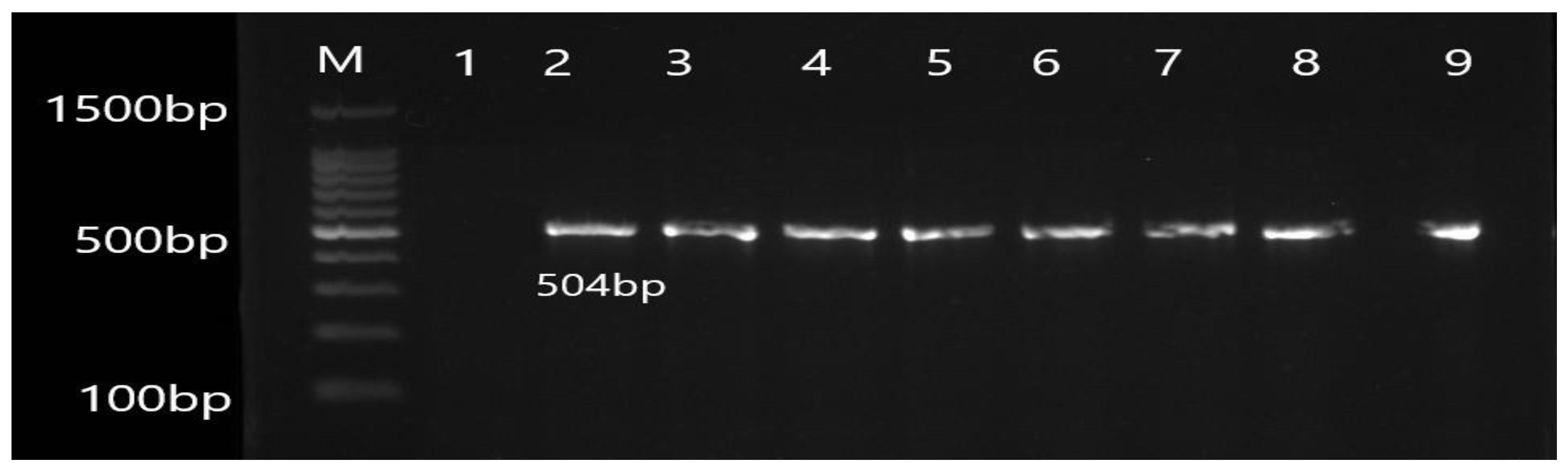
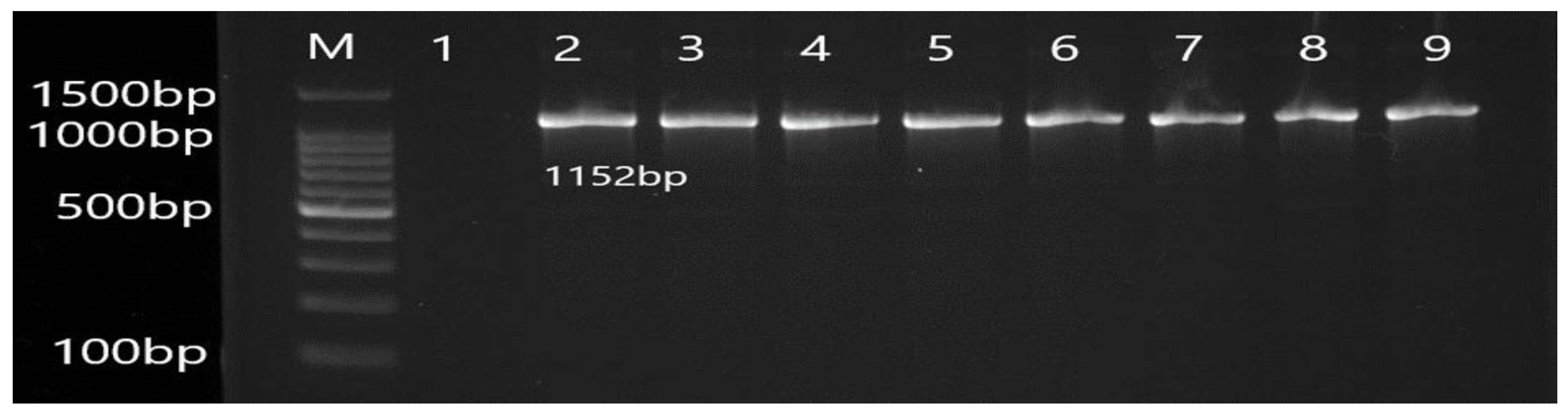
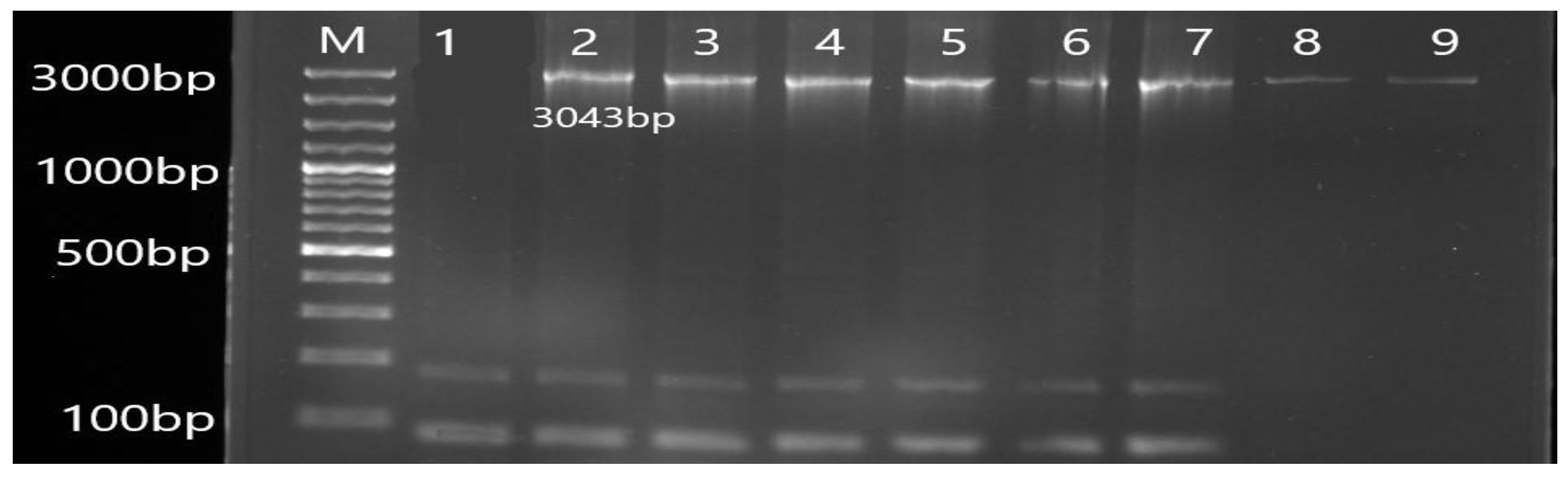
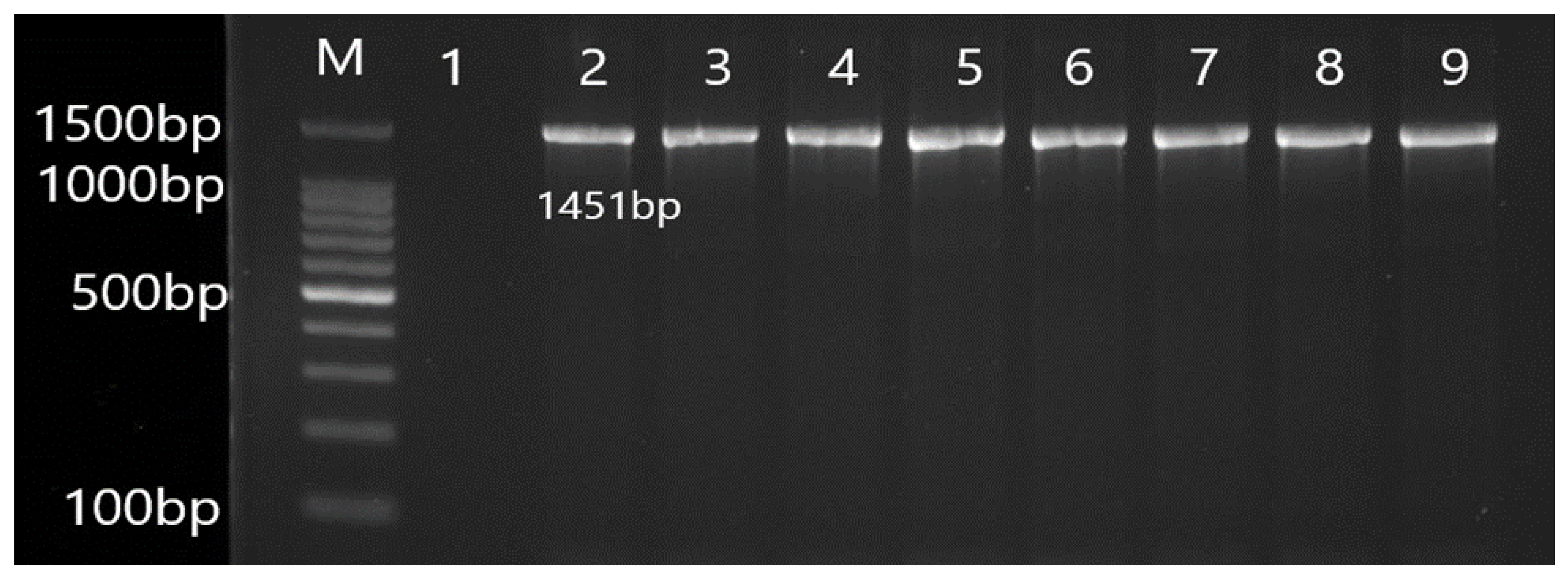
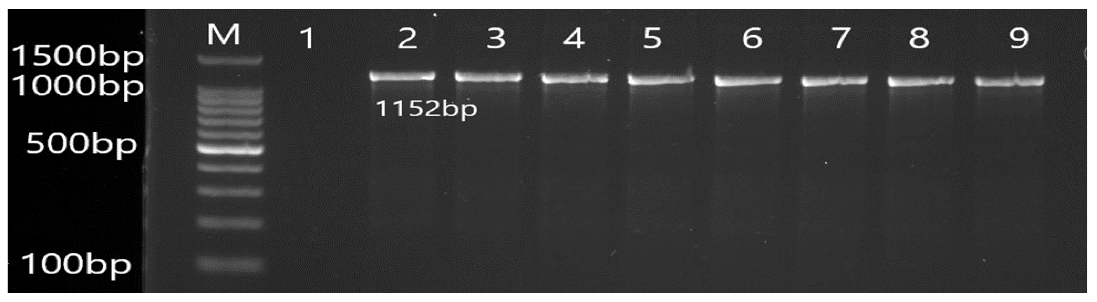
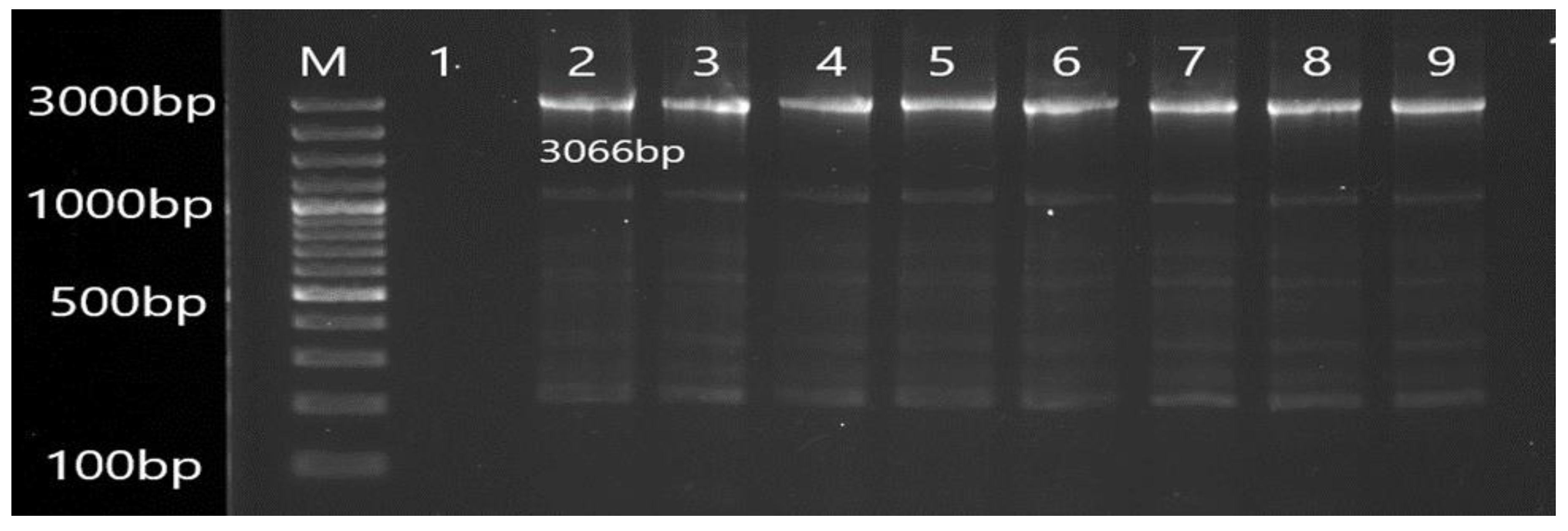
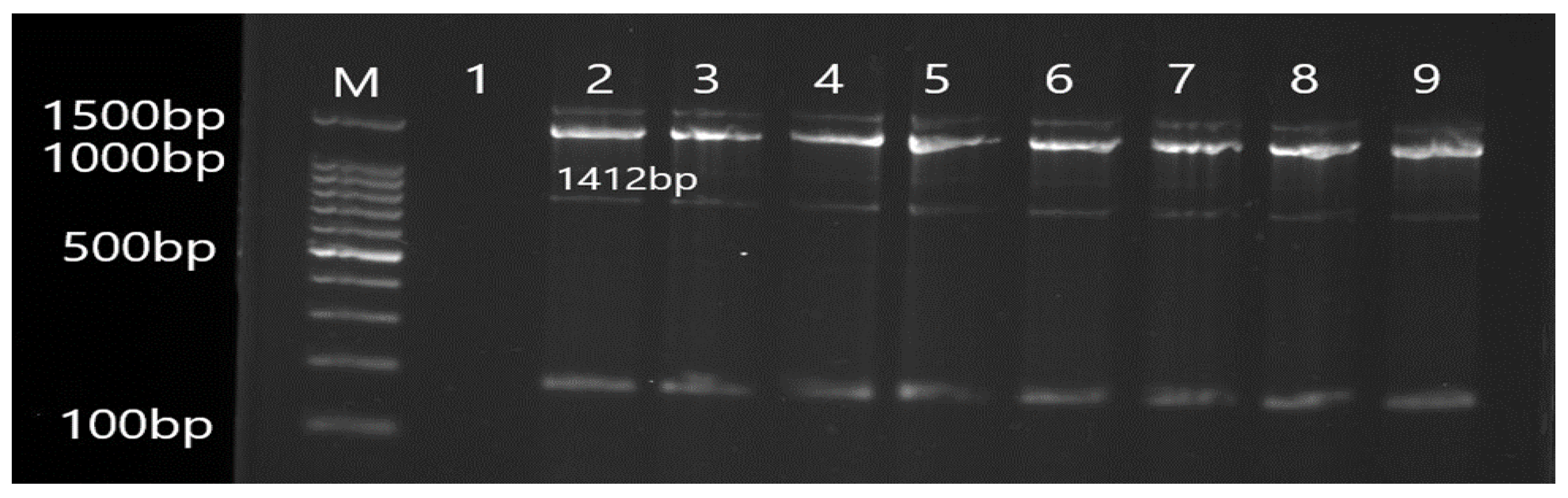
3.2. Molecular Detection of Efflux Pump Resistance Genes in Isolates of P. aeruginosa

The PCR results revealed the presence of efflux pump genes in P. aeruginosa isolates (Figure 1, Figure 2, Figure 3, Figure 4, Figure 5, Figure 6, Figure 7 and Figure 8). By comparing the results of PCR with the antibiotic susceptibility pattern of isolates, it was concluded that efflux pump resistance genes were detected mostly among amoxicillin/clavulanic acid-resistant isolates (n = 178; 89%) (Table 6).

3.3. Mutational Analysis of Antibiotic-Resistant Efflux Pump Genes

The mutational analysis was performed for the mexA, mexB, oprM, and mexR genes. In the sequences of mexA (Table 7 and Table 8), mexB (Table 9 and Table 10), and oprM gene (Table 11 and Table 12) mutations were detected while no mutation was detected in the mexR gene.

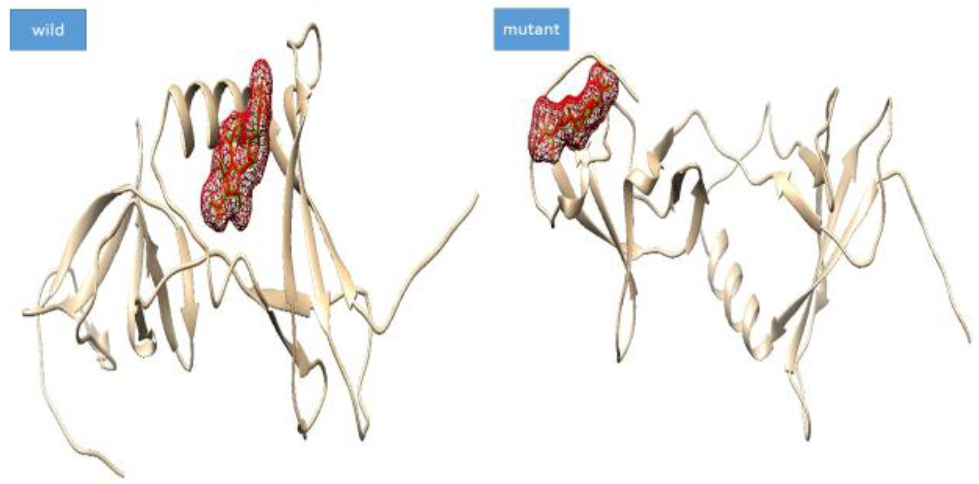
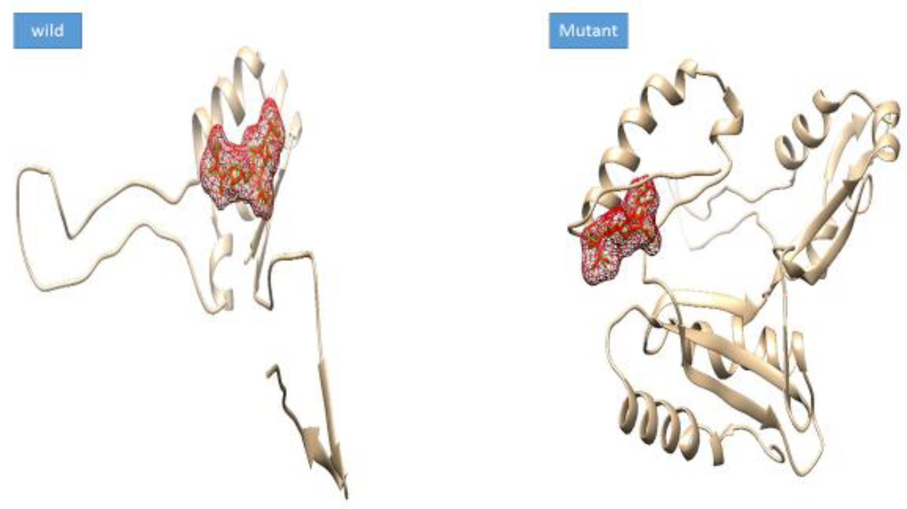
3.4. Mutation Impact on Structure Stability

The impact of mutations on the thermodynamic characteristics of wild-type and mutant proteins was revealed through the Dynamut server. The Dynamut predicts each mutation’s impact on protein conformational energy. As given in Table S1, the mutation effect determines the increased stability of mutant proteins compared to wild proteins. The E178K of mexA showed a destabilizing effect. In case of mexB, mutations such as R2T, W4T, L5V, D6T, P7F, A8E, N9Q, L10G, N11T, S12D, Y13P, Q14D, L15I, T16A, P17Q, G18V, D19Q, S21Q, S22N, A23K, I24L, H25Q, A26L, Q27A, N28T, V29P, Q30L, I31L, S32P, S33Q, G34E, Q35V, L36Q, G37R, G38Q, L39G, P40I, N43T, G44K, Q45A, H46V, L47K, A49F, T50L, I51M, I52V, G53V, K54G, T55V, R56V, L57S, Q58T, T59D, A60G, E61S, Q62M, F63T, E64K, N65E, I66D, L68S, K69N, V70Y, N71I, P72V, D73S, G74N, S75I, V77D, R78P, K80S, D81R, V82T, A83K, D84G, L87D, G88F, G89Q, H90V, D91F, Y92G, I94Q, N95Y, A96R, Q97S, F98M, N99R, G100I, S101W, P102L, G103D, V104P, R105A, Y106K, R107L, D108N, Q109S, and A110Y reported a destabilizing effect on the wild mexB protein. The vibrational entropy energy between the wild and mutant types was recorded in kcal/mol.

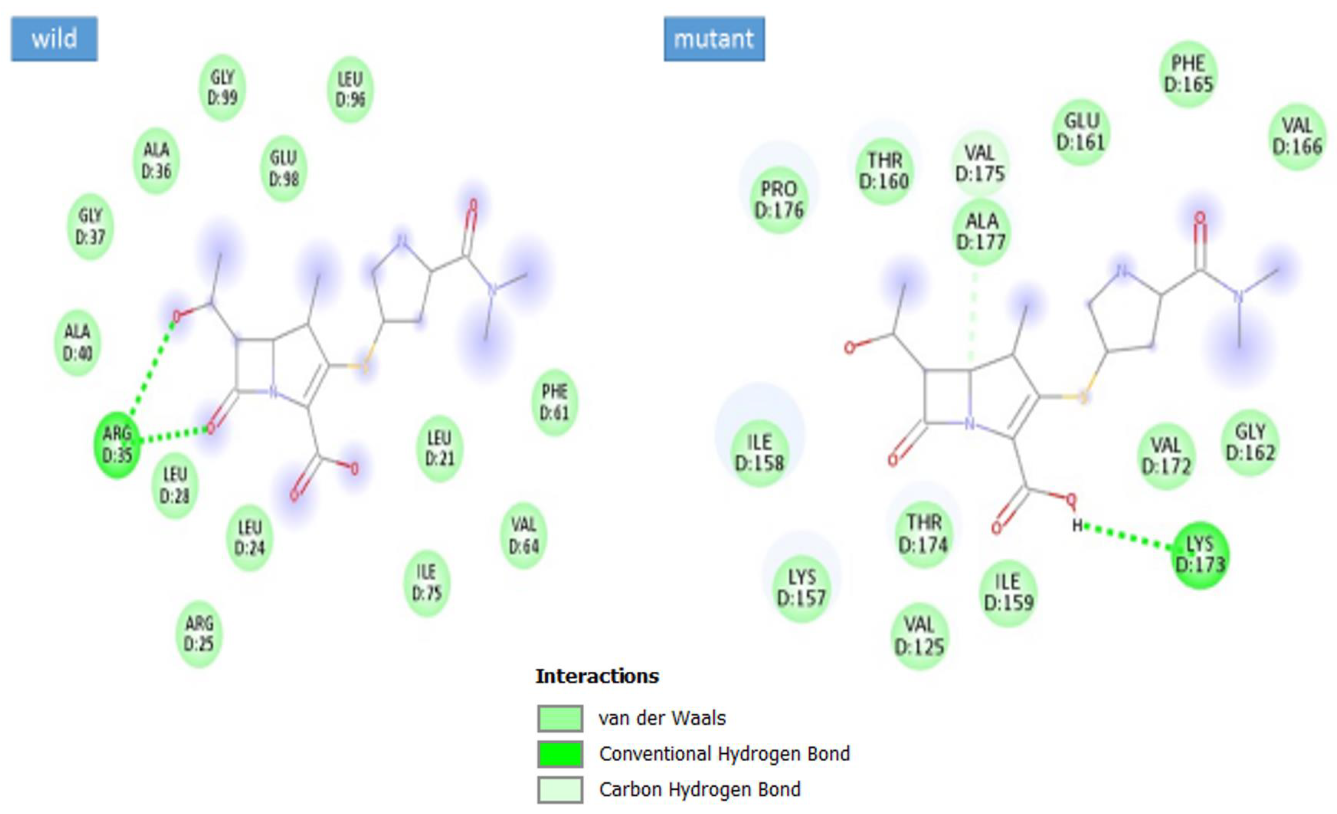
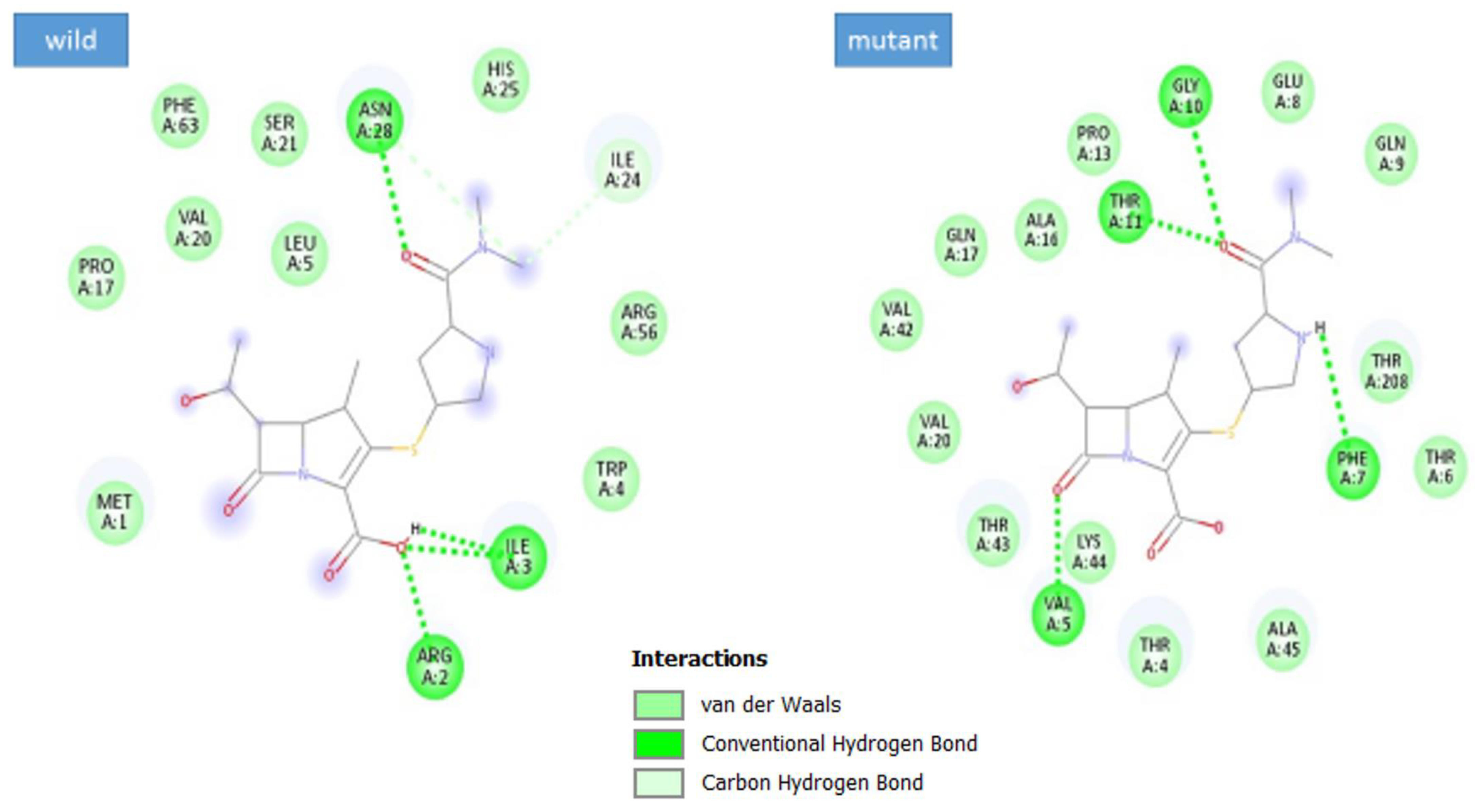
3.5. Docking Analysis

Molecular docking is a computational-based technique for intermolecular binding conformation. Here, the objective was to determine the mutation impact on meropenem drug binding with wild and mutant phenotypes of the genes. The docking results are provided in Table 13. The mexA wild protein complex binding energy value was −6.1 kcal/mol and the mexA mutant (E178K) value was −6.5 kcal/mol. The mexB wild protein complex binding energy value was −5.7 kcal/mol and the mexB mutant protein complex binding energy value was −8.0 kcal/mol. The binding conformation of meropenem with the mexA and mexB is shown in Figure 9 and Figure 10.
Through discovery studio visualizer v2021 software, the binding interactions between protein and drug were determined. The wild-type MexA is involved in van der Waals and conventional hydrogen bonds with the drug, while the mutant formed van der Waals conventional hydrogen, and carbon-hydrogen bonds. The wild MexA active residue such as Arg35 is attached to the hydroxybutanal with the help of a conventional hydrogen bond while 1-azabicyclo[3.2.0]hept-2-ene of the drug produced chemical bonding with Ala40, Gly37, Ala 36, Gly99, Glu58, Lue96, Leu28, Leu24, Arg25, leu21, Phe61, Val64, and Ile75. In mutant MexA, Lys173 is attached to the 1-azabicyclo[3.2.0]hept-2-ene through a conventional hydrogen bond. The val175 is attached to the 1-azabicyclo[3.2.0]hept-2-ene with the help of a carbon-hydrogen bond. The active residues such as Pro176,Thr160, Ala177, Glu161, Phe165,Val 166,Ile158, Lys157, The174, Val125, Ile159, Val172, and Gly162 were engaged with 1-azabicyclo [3.2.0]hept-2-ene by Van der Waals bonding (Figure 11). In mexB wild, binding interactions involve Arg2 and Ile3 attached to the 1-azabicyclo[3.2.0]hept-2-ene-2-carboxylic acid by a conventional hydrogen bond. The Asn28 is attached to the pyrrolidine-2-carboxamide chemical moiety via a conventional hydrogen bond. The active residues such as Pro17, Val20, Phe63, Ser21, Leu5, His25, Ile24, Arg56, Trp4, and Met1 interact with the drug by van der Waals interactions (Figure 12). The mexB mutant binding interactions involve Thr11, Gly10, and Phe7 with the pyrrolidine-2-carboxamide through conventional hydrogen bonding while Val5 is seen with 1-azabicyclo[3.2.0]hept-2-en-7-one. The active site residues Val42, Gln17, Ala16, Pro13, Glu8, Gln9, Thr208, Thr6, Ala45, Thr4, Lys44, Lys44, Thr43, and Val20 formed bonding to the protein via van der Waals interactions.

4. Discussion

A recent study investigated the expression of the MexA (88.2%) and MexB genes (70.5%) in 136 MDR and PDR isolates of P. aeruginosa. The study reported 69% MexB gene expression followed by 28.7% MexC expression, 43.4% MexE expression, and 74.6% MexY expression among isolates from the ICU. They were highly resistant to ticarcillin (80%), ciprofloxacin (74%), and meropenem (71%) [13].
In another study, antibiotic resistance-conferring efflux pumps were investigated in the isolates that were carbapenem-resistant (63.15%). The PCR results revealed overexpression in 19 (79.1%) isolates [14]. In the present study, MexAB-OprM and MexCD-OprJ efflux pumps were expressed in all the amoxicillin/clavulanic acid-resistant isolates. Mohseni et al., [15] investigated the efflux pumps conferring resistance among isolates collected from both human and animal sources. The PCR results showed an increased expression of the MexA gene as compared to the MexB gene. The isolates were 100% resistant to trimethoprim/sulfamethoxazole, cefazolin, ampicillin, kanamycin, and amoxicillin/clavulanic acid.
Efflux pump systems also mediate fluoroquinolone resistance in P. aeruginosa. In another study, out of 36 isolates, 88% were resistant to ofloxacin while 85% of them were resistant to sparfloxacin. Thus, the resistance mediated by efflux pump systems must be considered when introducing novel fluoroquinolones [16]. A study by Rudy et al. detected the expression of MexA-MexB-OprM efflux pump in 80% of isolates that were all ciprofloxacin resistant [17]. In the current study, 79 (39.5%) isolates were resistant to ciprofloxacin. The MexA, MexB, OprM, and MexR genes were detected in these ciprofloxacin-resistant isolates in accordance with the reported literature [18,19].

5. Conclusions

P. aeruginosa is known to adapt efficiently in harsh environments. All isolates in the present study were highly resistant to various families of antibiotics including beta-lactams, aminoglycosides, tetracycline, and carbapenems. Among 200 isolates, 178 were highly resistant and expressed all the selected efflux pump-resistant genes. For the better treatment of infections by P. aeruginosa, combination therapies may be a good choice to overcome the multidrug-resistant mechanisms of P. aeruginosa.

6. Future Recommendations

All isolates in the present study were highly resistant showing expression of efflux pumps. To overcome this hurdle, the implementation of efflux pump inhibitors with antibiotics would be helpful. Research for novel antibiotics and efflux pump inhibitors could be an interesting strategy for the better management of infections caused by P. aeruginosa.

Supplementary Materials

The following supporting information can be downloaded at: https://www.mdpi.com/article/10.3390/antibiotics12030486/s1, Table S1: Dynamut result of mexA and mexB.

Author Contributions

Conceptualization, I.K. and S.A. (Sajjad Ahmad); methodology, S.Q.; software, M.U.H., S.A. (Sadiq Azam); validation, I.K., S.A. (Sadiq Azam) and S.A. (Sajjad Ahmad); formal analysis, Z.L.; investigation, S.Q.; resources, M.A.; data curation, S.Q.; writing—original draft preparation, S.Q.; writing—review and editing, I.K., S.A. (Sadiq Azam), S.A. (Sajjad Ahmad); visualization, M.U.H.; supervision, I.K.; project administration, S.A. (Sajjad Ahmad); funding acquisition, M.A. All authors have read and agreed to the published version of the manuscript.

Funding

The authors express their gratitude to the Researchers Supporting Project number (RSP2023R462), King Saud University, Riyadh, Saudi Arabia.

Institutional Review Board Statement

Not required for our study.

Informed Consent Statement

Informed consent has been obtained from all the patients.

Data Availability Statement

The data generated in the research study is presented in the manuscript.

Conflicts of Interest

The authors declare no conflict of interest.

References

  1. Peix, A.; Martha-Helena, B.; Encarna, V. Historical evolution and current status of the taxonomy of genus Pseudomonas. Infect. Genet. Evol. 2009, 9, 1132–1147. [Google Scholar] [CrossRef] [PubMed]
  2. Meyer, J.M. Pyoverdines: Pigments, siderophores and potential taxonomic markers of fluorescent Pseudomonas species. Arch. Microbiol. 2000, 174, 135–142. [Google Scholar] [CrossRef] [PubMed]
  3. Meliani, A. The multifactorial resistance of Pseudomonas aeruginosa. EXCLI J. 2020, 19, 813–816. [Google Scholar] [PubMed]
  4. Streeter, K.; Katouli, M. Pseudomonas aeruginosa: A review of their Pathogenesis and Prevalence in Clinical Settings and the Environment. Epidemiol. Infect. 2016, 2, 25–32. [Google Scholar] [CrossRef]
  5. El Solh, A.A.; Alhajhusain, A. Update on the treatment of Pseudomonas aeruginosa pneumonia. J. Antimicrob. Chemother. 2009, 64, 229–238. [Google Scholar] [CrossRef] [PubMed] [Green Version]
  6. Hirsch, E.B.; Vincent, T.H. Impact of multidrug-resistant Pseudomonas aeruginosa infection on patient outcomes. Expert Rev. Pharmacoecon. Outcomes Res. 2010, 10, 441–451. [Google Scholar] [CrossRef] [PubMed] [Green Version]
  7. Pang, Z.; Raudonis, R.; Glick, B.R.; Lina, T.J.; Cheng, Z. Antibiotic resistance in Pseudomonas aeruginosa: Mechanisms and alternative therapeutic strategies. Biotechnol. Adv. 2019, 37, 177–192. [Google Scholar] [CrossRef] [PubMed]
  8. Mulcahy, L.R.; Jane, L.B.; Stephen, L.; Kim, L. Emergence of Pseudomonas aeruginosa strains producing high levels of persister cells in patients with cystic fibrosis. J. Bacteriol. Res. 2010, 192, 6191–6199. [Google Scholar] [CrossRef] [PubMed] [Green Version]
  9. Breidenstein, E.B.; Elena, B.M.; de la Fuente-Núñez, C.; Robert, E.H. Pseudomonas aeruginosa: All roads lead to resistance. Trends Microbiol. 2011, 19, 419–426. [Google Scholar] [CrossRef] [PubMed]
  10. Blair, J.M.; Vassiliy, N.B.; Vito, R.; Niraj, M.; Pierpaolo, C.; Ulrich, K.; Paolo, R.; Vargiu, A.; Baylay, A.; Smith, H.; et al. AcrB drug-binding pocket substitution confers clinically relevant resistance and altered substrate specificity. Proc. Natl. Acad. Sci. USA 2015, 112, 3511–3516. [Google Scholar] [CrossRef] [PubMed] [Green Version]
  11. Suresh, M.; Narayanan, N.; Vimal, K.; Jayasree, P.; Kumar, P. Mutational and Phylogenetic Analysis of nfxB Gene in Multidrug-Resistant Clinical Isolates of Pseudomonas aeruginosa Hyperexpressing MexCD-OprJ Efflux Pump. Adv. Microbiol. 2019, 9, 993. [Google Scholar] [CrossRef] [Green Version]
  12. Kohler, T.; Michéa-Hamzehpour, M.; Henze, U.; Gotoh, N.; Kocjancic, C.L.; Pechère, J. Characterization of MexE–MexF–OprN, a positively regulated multidrug efflux system of Pseudomonas aeruginosa. Mol. Microbiol. 1997, 23, 345–354. [Google Scholar] [CrossRef] [PubMed]
  13. Bialvaei, A.Z.; Rahbar, M.; Hamidi-Farahani, R.; Asgari, A.; Esmailkhani, A.; Soleiman-Meigooni, S. Expression of RND efflux pumps mediated antibiotic resistance in Pseudomonas aeruginosa clinical strains. Microb. Pathog. 2021, 153, 104789. [Google Scholar] [CrossRef] [PubMed]
  14. Younes, K.; Memar, M.Y.; Farajnia, S.; Adibkia, K.; Kafil, H.S.; Ghotaslou, R. Molecular epidemiology and carbapenem resistance of Pseudomonas aeruginosa isolated from patients with burns. J. Wound Care 2021, 30, 2. [Google Scholar]
  15. Mohseni, N.; Rad, M.; Khorramian Toosi, B.; Mokhtari, A.R.; Yahyaraeyat, R.; Salehi, T.Z. Evaluation of MexAB-OprM efflux pump and determination of antimicrobial susceptibility in Pseudomonas aeruginosa human and veterinary isolates. Bulg. J. Vet. Med. 2019, 24, 200–207. [Google Scholar] [CrossRef]
  16. Ehiagbe, I.; Ehiagbe, F.; Akinshipe, B.; Ilobanafor, R. Fluoroquinolone efflux pump of Pseudomonas aeruginosa. NISEB J. 2019, 11, 4. [Google Scholar]
  17. Rudy, M.A.; Dolatabadi, S.; Zare, H.; Ghazvini, K. Characterization of efflux pump-mediated resistance against fluoro-quinolones among clinical isolates of Pseudomonas aeruginosa in the northeast of Iran and its association with mortality of infected patients. Cell 2019, 98, 1248938. [Google Scholar]
  18. Ali, Z.; Mumtaz, N.; Naz, S.A.; Jabeen, N.; Shafique, M. Multi-Drug Resistant Pseudomonas Aeruginosa: A threat of nosocomial infections in tertiary care hospitals. J. Pak. Med. Assoc. 2015, 65, 12. [Google Scholar] [PubMed]
  19. Kishk, R.; Abdalla, M.; Hashish, A.; Nemr, N.; El Nahhas, N.; Alkahtani, S.; Abdel-Daim, M.; Kishk, S. Efflux MexAB-mediated resistance in P. aeruginosa isolated from patients with healthcare associated infections. Pathogens 2020, 9, 471. [Google Scholar] [CrossRef] [PubMed]
Figure 1. Electrophoresis showing amplicons of P. aeruginosa mexB gene. Lane M: 100 bp plus molecular marker, Lane 1: Negative control, Lane 2–9: Positive isolates of mexB gene.
Antibiotics 12 00486 g001
Figure 2. Electrophoresis showing amplicons of P. aeruginosa mexA gene. Lane M: 100 bp molecular marker, Lane 1: Negative control, Lane 2–9: Positive isolates of mexA gene.
Antibiotics 12 00486 g002
Figure 3. Electrophoresis showing amplicons of P. aeruginosa oprL gene. Lane M: 100 bp molecular marker, Lane 1: Negative control, Lane 2: Positive control, Lane 3–9: Positive isolates of oprL.
Antibiotics 12 00486 g003
Figure 4. Electrophoresis showing amplicons of P. aeruginosa mexC gene. Lane M: 100 bp molecular marker, Lane 1: Negative control, Lane 2–9: Positive isolates of mexC gene.
Antibiotics 12 00486 g004
Figure 5. Electrophoresis showing amplicons of P. aeruginosa mexR gene. Lane M: 100 bp molecular marker, Lane 1: Negative control, Lane 2–9: Positive isolates of mexR gene.
Antibiotics 12 00486 g005
Figure 6. Electrophoresis showing amplicons of P. aeruginosa oprM gene. Lane M: 100 bp molecular marker, Lane 1: Negative control, Lane 2–9: Positive isolates of oprM gene.
Antibiotics 12 00486 g006
Figure 7. Electrophoresis showing amplicons of P. aeruginosa mexD gene. Lane M: 100 bp plus molecular marker, Lane 1: Negative control, Lane 2–9: Positive isolates of mexD gene.
Antibiotics 12 00486 g007
Figure 8. Electrophoresis showing amplicons of P. aeruginosa oprJ gene. Lane M: 100 bp molecular marker, Lane 1: Negative control, Lane 2–9: Positive isolates of oprJ gene.
Antibiotics 12 00486 g008
Figure 9. MexA wild and mutant intermolecular-docked complex with meropenem. The proteins are shown in tan cartoon style while the ligands are given in mesh.
Antibiotics 12 00486 g009
Figure 10. Binding conformation of meropenem with the mexB wild and mutant proteins. The proteins are shown in tan cartoon style while the ligands are given in mesh.
Antibiotics 12 00486 g010
Figure 11. MexA wild and mutant binding interactions with meropenem. The compound is presented in a 2D line.
Antibiotics 12 00486 g011
Figure 12. MexB wild and mutant binding interactions with meropenem. The compound is presented in a 2D line.
Antibiotics 12 00486 g012
Table 1. Primer sequences with optimized PCR conditions.
GenePrimerProduct Size (bp)Annealing Temperature (°C)
OprLF ATGGAAATGCTGAAATTCGGC
R CTTCTTCAGCTCGACGCGACG
50455
MexAF CTATGCAACGAACGCCAGC
R AGCCCTTGCTGTCGGTTTTC
115256
MexBF TAGGCCCATTTTCGCGTGG
R CGGTACCCAGAAGATCGCC
304356
OprMF CGGTCCTTCCTTTCCCTGG
R CAAGCCTGGGGATCTTCCTT
145155
MexRF CAAGCGGTTGCGCGG
R CCCCGTGAATCCCGACCTG
42556
MexCF TTACTGTTGCGGCGCAGG
R CGTGCAATAGGAAGGATCGG
115255
MexDF CAGCAGCCAGACGAAACAGA
R TTCTTCATCAAGCGGCCGAA
306656
OprJF CTGCCGCCTCGATGTACC
R GTATCGGCGCTGCTGATCG
141255
NfxBF GACCCTGATTTCCCATGACG
R GGAACATCTGCTCCAGGGTAT
53056
Table 2. List of antibiotics.
S. NoAntibiotics (µg)Family (Symbol)
1Amikacin (20)Aminoglycoside (AK)
2Gentamicin (10)Aminoglycoside (CN)
3Azithromycin (30)Macrolide (AZM)
4Tigecycline (15)Tetracycline (TGC)
5Chloramphenicol (30)Chloramphenicol (C)
6Ciprofloxacin (5)Fluoroquinolone (CIP)
7Levofloxacin (5)Fluoroquinolone (LEV)
8Moxifloxacin (5)Fluoroquinolone (MXF)
9Amoxicillin (25)β-lactam (penicillin) (AML)
10Amoxicillin-clavulanic acid (30)β-lactam (penicillin) (AMC)
11Piperacillin-tazobactam (110)β-lactam (penicillin) (TZP)
12Aztreonam (30)β-lactam (monobactams) (ATM)
13Cefotaxime (30)β-lactam (cephalosporin) (CTX)
14Cefepime (30)β-lactam (cephalosporin) (FEP)
15Ceftazidime (30)β-lactam (cephalosporin) (CAZ)
16Cefoperazone (75)β-lactam (cephalosporin) (CFP)
17Cefoperazone-sulbactam (105)β-lactam (cephalosporin) (SCF)
18Ceftriaxone (30)β-lactam (cephalosporin) (CRO)
19Cefixime (5)β-lactam (cephalosporin) (CFM)
20Meropenem (10)β-lactam (carbapenem) (MEM)
21Imipenem (10)β-lactam (carbapenem) (IMP)
22Fosfomycin (50)Fosfomycin (FOS)
23Colistin (10)Polymyxin (CT)
24Polymyxin B (300)Polymyxin (PB)
25Trimethoprim-sulfamethoxazole (25)Sulfonamide (SXT)
26Nitrofurantoin (300)Nitrofurantoin (F)
Table 3. Collection of clinical samples of P. aeruginosa from various sources.
SourceNumber (Percentage)
Urine catheter1 (0.5)
Stone analysis1 (0.5)
Urine28 (14)
Pus57 (28.5)
Wound swab94 (47)
Blood7 (3.5)
Sputum9 (4.5)
CSF1 (0.5)
Ear swab2 (1.0)
Total200
Table 4. Frequency of patients’ gender and age.
ParameterFrequencyPercentage
GenderMale10854.0
Female9246.0
Age Group (Years)1–10126
11–203015
21–304321.5
31–403718.5
41–502311.5
51–602512.5
61–702110.5
71–8084
81–9010.5
Table 5. Antibiotic susceptibility pattern of P. aeruginosa.
AntibioticsResistant (n)Percentage (%)Intermediate (n)Percentage (%)Susceptible (n)Percentage (%)
AK40204215678
CN884410510251
CIP7939.594.511258
LEV7135.52311.510653
MXF8040115.510954.5
AML63--10.5
AMC1788910.52110.5
TZP4924.552.514673
ATM7135.5168.011356.5
CTX1286452.56733.5
FEP723673.512160.5
CAZ7336.5115.511658
CEP7236157.511356.5
SCF4924.5105.014170.5
CRO9648115.59346.5
CFM1587973.53517.5
MEM6331.584.012964.5
IMP6331.5115.512663
AZM----73.5
TGC100501268844
CT6231178.512160.5
PB6331.52110.511658
FOS63212211
C21--52.5
SXT12562.552.57035
F157.5--157.5
Table 6. Polymerase chain reactions of Antibiotic resistance efflux pump genes.
Positive Isolates of Efflux Pump GenesGenesPositive Result
AMC-resistant isolatesMexA178 (89%)
MexB178 (89%)
OprM178 (89%)
MexR178 (89%)
MexC178 (89%)
MexD178 (89%)
OprJ178 (89%)
NfxB178 (89%)
Table 7. Non-synonymous mutation of the mexA gene.
Codon
Position
Reference Amino AcidAltered Amino AcidAmino Acid Position
389GGT (Glycine)AGT (Serine)368
Table 8. mexA Prediction result of I-Mutant software.
Wild TypeNewI-Mutant Prediction EffectDDG ValueReliability
Index (RI)
Temperature pH
G (Glycine)S (Serine)Decrease−18257
Table 9. Synonymous and non-synonymous mutations of the mexB gene.
Codon PositionReference Amino Acid PositionAltered Amino Acid PositionAmino Acid Position
Synonymous mutation of mexB gene
148TCC-TCGSerine129
154AGC-AGTSerine130
184GTC-GTGValine142
256CCT-CCGProline166
259CTC-CTALeucine167
302AAA-AAGLysine290
308GTA-GTCValine291
635CAA-CAGGlutamine673
Non-synonymous mutation of the mexB gene
126Asparagine (AAC)Aspartate (GAC)123
129Tyrosine (TAT)Asparagine (AAT)124
136Leucine (CTC)Arginine (CGC)126
138Phenylalanine (TTC)Tyrosine (TAC)127
140Phenylalanine (TTC)Isoleucine (ATC)128
151Aspartate (GAC)Glutamate (GAG)131
165Alanine (GCC)Glycine (GGC)136
167Cysteine (TGC)Serine (AGC)137
170Proline (CCG)Methionine (ATG)138
191Glutamine (CAA)Glutamate (GAA)145
197Leucine (CTC)Glycine (GGC)147
200Proline (CCC)Threonine (ACC)148
203Asparagine (AAC)Aspartate (GAC)149
215Proline (CCC)Alanine (GCC)143
219Leucine (CTG)Glutamine (CAG)154
228Alanine (GCC)Valine (GTG)157
231Leucine (CTC)Glutamine (CAG)158
244Histidine (CAC)Glutamine (CAA)162
269Glutamine (CAA)Glutamate (GAA)171
283Histidine (CAT)Glutamine (CAG)175
292Histidine (CAC)Arginine (CGG)287
303Serine (TCG)Alanine (GCG)291
321Leucine (CTG)Methionine (ATG)296
324Leucine (CTG)Valine (GTG)298
327Leucine (CTG)Valine (GTG)299
330Arginine (CGT)Glycine (GGT)300
340Proline (CCT)Valine (GTT)302
365Asparagine (AAC)Lysine (AAG)311
378Histidine (CAC)Asparagine (AAC)316
388Alanine (GCT)Valine (GTT)319
424Alanine (GCC)Glycine (GGC)331
429Cysteine (TGC)Glycine (GGT)333
439Proline (CCG)Glutamine (CAG)336
441Leucine (CTG)Valine (GTG)337
456Histidine (CAC)Tyrosine (TAC)342
488Asparagine (AAT)Lysine (AAG)472
536Histidine (CAT)Glutamine (CAG)488
590Asparagine (AAC)Lysine (AAG)506
599Histidine (CAT)Tyrosine (CAG)509
732Histidine (CAT)Tyrosine (CAG)673
Table 10. MexB gene Prediction results of I-Mutant software.
Wild TypeNew TypeI-Mutant Prediction EffectDDG ValueReliability
Index (RI)
TemperaturepH
NDDecrease−0.957257
YNIncrease−0.240257
LRDecrease−0.957257
FYDecrease−0.857257
FIDecrease−1.999257
DEDecrease−0.597257
AGDecrease−1.037257
CSDecrease−0.531257
PMDecrease−0.961257
QEDecrease−0.294257
LGIncrease0.222257
PTDecrease−0.021257
NDIncrease0.115257
PADecrease−1.024257
LQDecrease0.141257
AVDecrease−0.936257
LQDecrease0.003257
HQDecrease−0.617257
QEDecrease−0.111257
HQDecrease−0.617257
HRDecrease−1.379257
SADecrease−0.908257
LMDecrease−0.805257
LVDecrease−1.306257
LVDecrease−1.326257
RGDecrease−0.481257
PVDecrease−1.574257
NKIncrease−0.483257
HNDecrease−0.669257
AVDecrease−1.377257
AGIncrease −0.511257
CGDecrease−0.760257
PQDecrease−0.416257
LVDecrease−0.744257
HYDecrease0.041257
NKIncrease0.044257
HQDecrease−0.536257
NKDecrease−0.552257
HQDecrease−0.978257
HQDecrease−0.916257
Table 11. Synonymous and non-synonymous mutations of the oprM gene.
Codon PositionReference Amino Acid PositionAltered Amino Acid PositionAmino Acid Position
Non-synonymous mutation of the OprM gene
11Glutamine (CAA)Arginine (CGC)7
50Valine (GTG)Alanine (GCG)20
Synonymous mutation of the OprM gene
43ACT-ACCT17
Table 12. OprM gene Prediction results of I-Mutant software.
Wild TypeNew TypeI-Mutant Prediction EffectDDG ValueReliability
Index (RI)
TemperaturePH
Q (Glutamine)R (Arginine)Increase−0.111257
V (Valine)A (Alanine)Decrease−1.668257
Table 13. Docking energy score in kcal/mol.
ComplexDocking Score
max-A wild_meropenem−6.1
max-A mutant (E178K) meropenem−6.5
max-B wild_meropenem−5.7
max-B mutant_meropenem−8
Disclaimer/Publisher’s Note: The statements, opinions and data contained in all publications are solely those of the individual author(s) and contributor(s) and not of MDPI and/or the editor(s). MDPI and/or the editor(s) disclaim responsibility for any injury to people or property resulting from any ideas, methods, instructions or products referred to in the content.

