Article

Virulence Characteristics and Molecular Typing of Carbapenem-Resistant ST15 Klebsiella pneumoniae Clinical Isolates, Possessing the K24 Capsular Type

1
Department of Medical Biology and Central Electron Microscope Laboratory, Medical School, University of Pécs, 7624 Pécs, Hungary
2
Department of Biotechnology, Nanophagetherapy Center, Enviroinvest Corporation, 7632 Pécs, Hungary
3
Bioinformatics Research Group, Genomics and Bioinformatics Core Facility, Szentágothai Research Centre, University of Pécs, 7624 Pécs, Hungary
4
Molecular Pharmacology Group, Department of Pharmacology and Pharmacotherapy, Neuroscience Centre, Medical School, University of Pécs, 7624 Pécs, Hungary
5
Division of Microbiological Reference Laboratories, National Public Health Center, 1097 Budapest, Hungary
6
Department of Medical Microbiology and Immunology, Medical School, University of Pécs, 7624 Pécs, Hungary
*
Author to whom correspondence should be addressed.
Antibiotics 2023, 12(3), 479; https://doi.org/10.3390/antibiotics12030479
Submission received: 1 February 2023 / Revised: 21 February 2023 / Accepted: 23 February 2023 / Published: 28 February 2023

Abstract

:
Klebsiella pneumoniae is an opportunistic pathogen that frequently causes nosocomial and community-acquired (CA) infections. Until now, a limited number of studies has been focused on the analyses of changes affecting the virulence attributes. Genotypic and phenotypic methods were used to characterise the 39 clinical K. pneumoniae isolates; all belonged to the pan-drug resistant, widespread clone ST 15 and expressed the K24 capsule. PFGE has revealed that the isolates could be divided into three distinct genomic clusters. All isolates possessed allS and uge genes, known to contribute to the virulence of K. pneumoniae and 10.25% of the isolates showed hypermucoviscosity, 94.87% produced type 1 fimbriae, 92.3% produced type 3 fimbriae, and 92.3% were able to produce biofilm. In vivo persistence could be supported by serum resistance 46.15%, enterobactin (94.87%) and aerobactin (5.12%) production and invasion of the INT407 and T24 cell lines. Sequence analysis of the whole genomes of the four representative strains 11/3, 50/1, 53/2 and 53/3 has revealed high sequence homology to the reference K. pneumoniae strain HS11286. Our results represent the divergence of virulence attributes among the isolates derived from a common ancestor clone ST 15, in an evolutionary process that occurred both in the hospital and in the community.

1. Introduction

Klebsiella pneumoniae is one of the most important Gram-negative bacteria causing nosocomial and community-acquired (CA) infections, resulting in pneumonia, urinary tract infections (UTI), septicaemia, bronchitis and intra-abdominal infections [1]. K. pneumoniae is one of the most prevalent causes of catheter-associated urinary tract infections (CAUTIs), next to Escherichia coli. A high incidence of CAUTIs has substantial costs related to extended periods of hospital access. Furthermore, CAUTIs can affect the kidneys and enter the bloodstream causing systemic disease such as septicaemia [2]. Carbapenem-resistant Klebsiella pneumoniae (CR-Kp) has become a significant global public health challenge [3].
Severeness of the infection is basically determined by (i) physiological state of the host and (ii) virulence potential of the pathogenic agent. Hospitalization, antimicrobial therapy, prolonged use of invasive medical devices, and major surgeries are considered as the most important environmental factors that contribute to infection [4]. CR-Kp is a major concern for nosocomial infections worldwide since emerging antibiotic resistance makes treatments difficult and are more frequently associated with high morbidity and mortality rates [5]. The therapeutic challenge is large, because CR-Kp isolates are resistant against not only a wide variety of antibiotics, but also against last resorts of clinically used drugs, like carbapenems and colistin [6,7].
Today, the factors considered to be important in the survival and virulence of K. pneumoniae are the capsule, lipopolysaccharide (LPS), fimbriae, and siderophores (iron chelators). Certain capsule types together with the LPS layer, protect the K. pneumoniae against phagocytosis and the bactericidal activity of serum and enable survival and endurance in and out of the host. The phase variable (fim switch) type 1 fimbriae, are found to be frequently present in strains of K. pneumoniae [8] and mediate adhesion to mannose-containing structures. Type 3 fimbriae are present in almost all K. pneumoniae isolates and mediate binding in vitro to human-derived extracellular matrix (ECM) proteins [2] prior to biofilm formation on these surfaces. Its role is pivotal in urinary tract infections (UTI) [9]. The MrkD protein mediates binding to this substrate, whereas the MrkA peptide constitutes the major fimbrial subunit that is polymerized to form the fimbrial shaft [10]. Bacterial biofilms are frequently observed on the surfaces of tissues [11] and biomaterials [12] at the site of persistent infections. Once in the biofilm, extracellular polymeric substance shield bacteria from opsonisation and phagocytosis [13]. In addition, in vitro experiments have demonstrated that the bacteria in biofilms are considerably less susceptible to antibiotics than their planktonic counterparts [14] and can also ensure survival on abiotic surfaces [15].
Klebsiella isolates have been shown to produce high-affinity iron-chelating siderophores, that were shown to contribute to the virulence of K. pneumoniae [8]. The role of the catechol-type siderophore, enterobactin, in virulence is still uncertain, in contrast to the widely studied hydroxamate-type siderophore, aerobactin [16].
Although dissemination is associated with highly diverse isolates of K. pneumoniae, certain clonal groups (CG) are dominant in this process. One group is CG15 in which the pan-drug resistant ST 15 clone belongs [17]. This clone was recently identified worldwide from Brazil [18] to China [19] and Europe [20] and was frequently associated with carbapenemase production [18] and nosocomial infections. Recent epidemiologic surveys have revealed that K24 is the most frequent capsule type linked to ST 15 [18], this capsule type does not belong to the classical virulent capsule types [21], but could contribute to survival and therefore persistence of the bacterium either in the hospital environment or in the community [22,23].
Emerging antibiotic resistance including carbapenem resistant Klebsiella pneumoniae (CR-Kp) is one of the most important rising public health threats since the early 2000s [24]. One major concern is the predominance of TEM, SHV and CTX-M type extended-spectrum β-lactamases that mainly mediate resistance against β-lactams. Recently the appearance and emergence of β-lactamases with carbapenem-hydrolysing activity (carbapenemases), like the serine carbapenemases KPC and OXA-48, and the metallo-β-lactamases VIM, IMP and NDM are the most concerning from therapeutic point of view [25,26]. Furthermore CR-Kp isolates have undergone extensive disseminations affecting well characterized regions and countries and certain types of carbapenemases show geographical associations. In CR-Kp isolates the VIM type is the most frequently detected metallo-β-lactamase (MBL) in the Mediterranean region, including Spain and Italy [3,24] and has become endemic in certain countries of this region such as Greece [27]. Molecular analyses of emerged VIM-producing K. pneumoniae in the northern European countries revealed genetic relatedness to those of southern and other international complexes [28].
Recently, it became evident that not only the activity of carbapenemase can have an effect on carbapenem resistance, but also the presence of certain mutations in outer membrane proteins. These mutations cause alterations in the amino acid sequences that can lead to protein modification, alter pore size and hydrophobicity and can impede uptake of an antibiotic. Mutations in ompK35 and ompK36 genes are the most known that are related to carbapenem resistance [29].
According to a recent study, infections caused by KPC-producing strains is more frequently associated with increased mortality compared with other mechanisms of resistance [30].
Recently, several studies have focused on the spread of antibiotic resistance and its evolutionary aspects in and outside of the hospital environment. However, knowledge of K. pneumoniae ecology, population structure or pathogenicity is relatively limited [17]. In this study appearance and emergence of CR-KP isolates with the same clonal origins and antibiograms in an English Teaching Hospital offered the possibility to analyse the evolutionary aspects affecting virulence attributes.

2. Results

2.1. Bacterial Identification and Antimicrobial Susceptibility Testing

Isolates were analysed by MALDI-TOF MS and the results were compared with standard conventional identification. All the isolates (n = 39) were identified at the species level (log (score value) ≥ 2.0). The result of standard biochemical tests showed that, all isolates were negative for indole probe, methyl red test, ornithine- and arginine- decarboxylase test, motility test; and all isolates were positive for the adonite test, Voges–Proskauer test, citrate test, malonate test, urease test, lysine-decarboxylase test, saccharose and lactose test.
All isolates were resistant to penicillins, cephalosporins, carbapenems and fluoroquinolones. Antibiotic resistance profile and MIC values of the K. pneumoniae isolates is provided in Supplementary Table S2a and S2b, respectively. The minority of the isolates were susceptible to amikacin (48.71%) and gentamicin (30.76%). All isolates, except one (categorized as intermediate susceptible) were resistant to tobramycin (Supplementary Table S2a,b).
Of the 39 K. pneumoniae isolates included in the study, 100% (39/39) were shown to produce carbapenemase by the Carbapenem Inactivation Method (CIM).

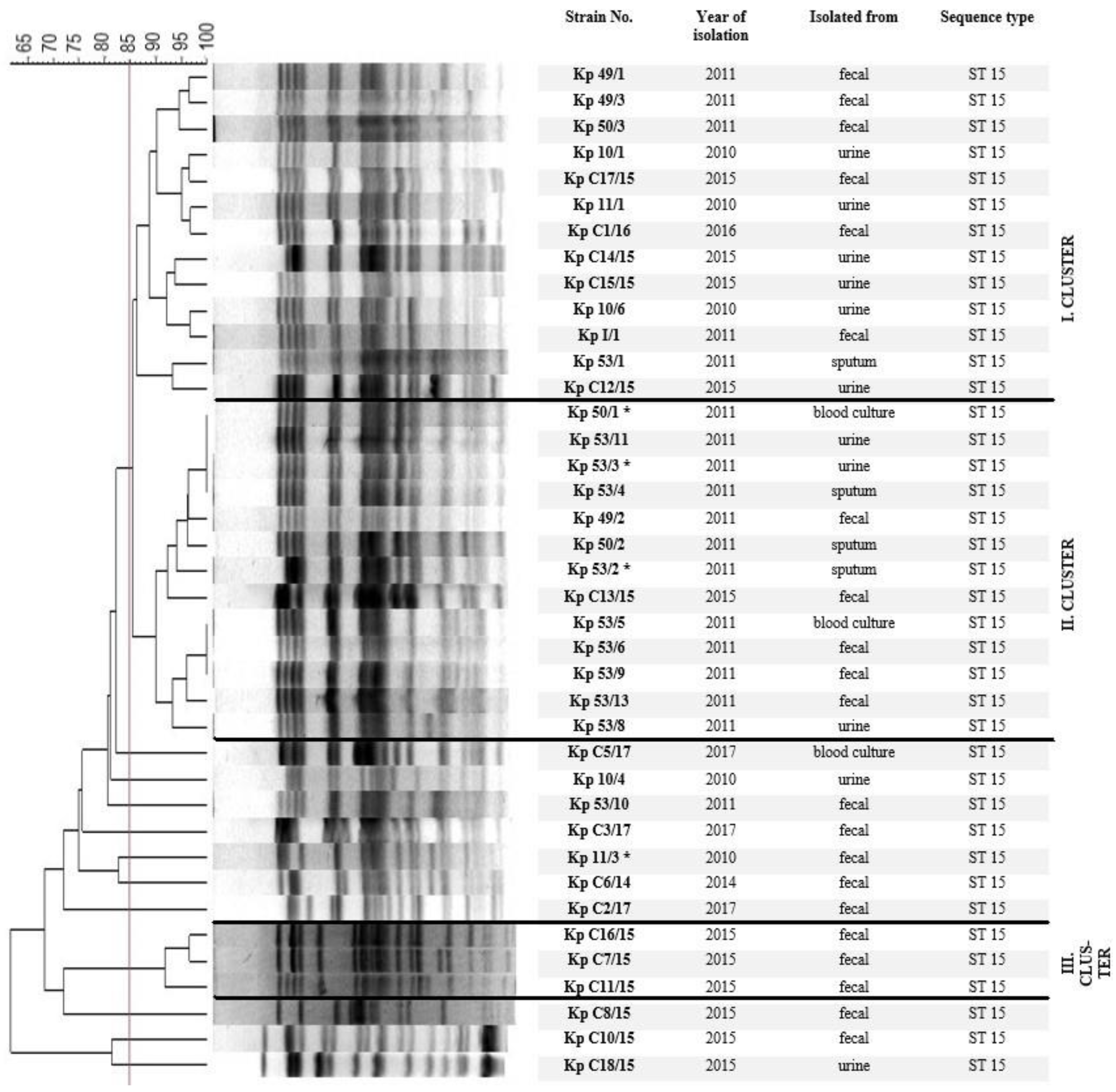
2.2. Chromosomal Macro-Restriction Fragment Polymorphism Analysis with PFGE

Isolates were typed by PFGE of XbaI-digested total genomic DNA. Strains were considered to be the same clone if they showed ≥85% pattern similarity, or fewer than six fragment differences in the PFGE profile. Based on macrorestriction profile analysis by PFGE the isolates were grouped into three major clusters (Figure 1).

2.3. Presence of Virulence-Associated Genes

The occurrence of virulence-associated genes was determined with PCR and results are summarized in Table 1. fimH-1 and mrkD genes encoding the type 1 and type 3 fimbrial adhesins, were present in 94.87% and 92.3% of the isolates, respectively. Phenotypic test (Table 2) results were in agreement with the PCR data (Supplementary Table S3). The adhesion associated genes, mrkA, mrkJ and cf29a were detected at a prevalence of 92.3%, 92.3% and 10.25%, respectively. The activator of the allantoin regulator gene, allS was detected in 100% of isolates. Siderophore genes entB (enterobactin) and iutA (aerobactin) were detected at a prevalence of 94.87% and 5.13%, respectively. Siderophore phenotypic test results were in agreement with the PCR data. Serum resistance was observed in 46.15% of isolates at 3 h. traT gene, involved in resistance to serum, was detected in 46.15% of isolates. rmpA, the genetic determinant associated with the HMV phenotype was found in 10.25% of isolates. An enhancer of the colony mucoidity, was detected in 10.25%. uge and wziK24 genes encoding the uridine diphosphate galacturonate 4-epidermase gene and K24 capsular type gene, respectively, were detected in 100% of isolates. magA gene encoding mucoviscosity-associated gene A, causes hypermucoviscosity, as defined by positive results of the string test, were detected in 10.25% of isolates.

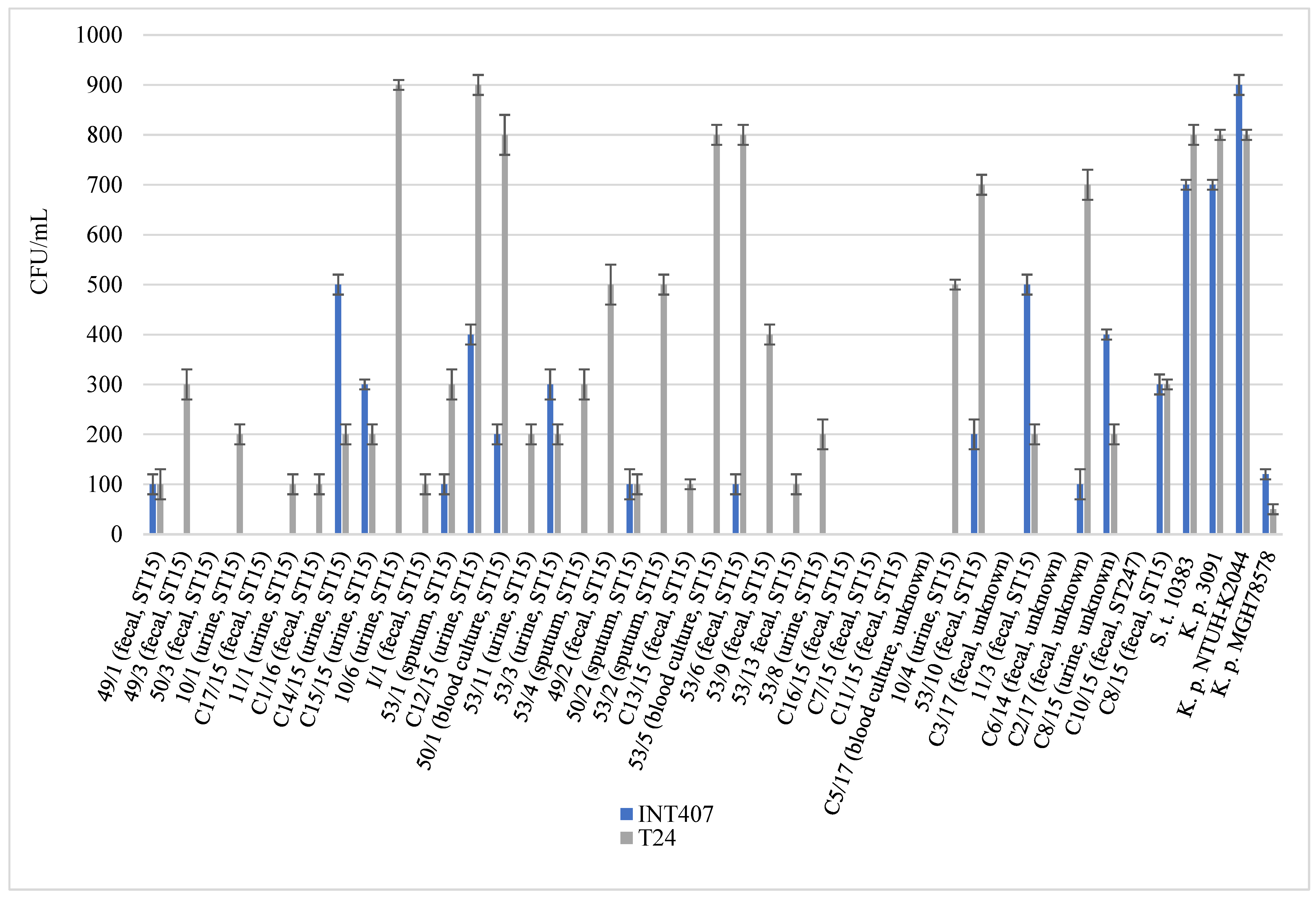
2.4. Virulence Associated Phenotypic Assays

Presence of certain virulence attributes were revealed with phenotypic tests and results are summarized in Table 2. MSHA specific to type 1 fimbriae was detected in 94.87% of the isolates, while incidence of MRHA specific to type 3 fimbriae was 92.3%.
The ability to form biofilm was detected in 92.3% of the isolates, of which, 34 isolates were high producers, 2 isolates were medium producers, and 3 isolates were poor biofilm producers (Table 2). All isolates that were able to form biofilm also produced type 3 fimbriae. No relationship was revealed between biofilm formation and isolate origin.
Phenotypic tests aiming to reveal the capacities of the CR-Kp isolates to produce siderophores showed that 94.87% of them produced enterobactin, while only 5.12% produced aerobactin (Table 2). In two cases (53/1 and C8/15) the presence of both iron scavenging systems were revealed, while in one case (C1/16) none of the tested siderophore systems could be detected.
These isolates generally exhibited poor survival in human serum, with only 46.15% of isolates proving resistant to serum bactericidal activity after 3 h (Table 2). Similarly, HMV was found in only 10.25% of isolates.
Cell internalisation assays performed on two human epithelial cell lines (INT 407 and T24) have revealed that most of the isolates (31/39; 79.48%) were able to invade one or both of the cell lines to some extent during a 3 h co-incubation (Figure 2). Fifteen isolates were internalized by both cell lines, while 16 isolates could only invade T24, but not INT407 to a detectable extent. Nine isolates were not able to invade any of the applied cell lines. No general relationship between the origin of isolation and the ability to interact with epithelial cells was established.

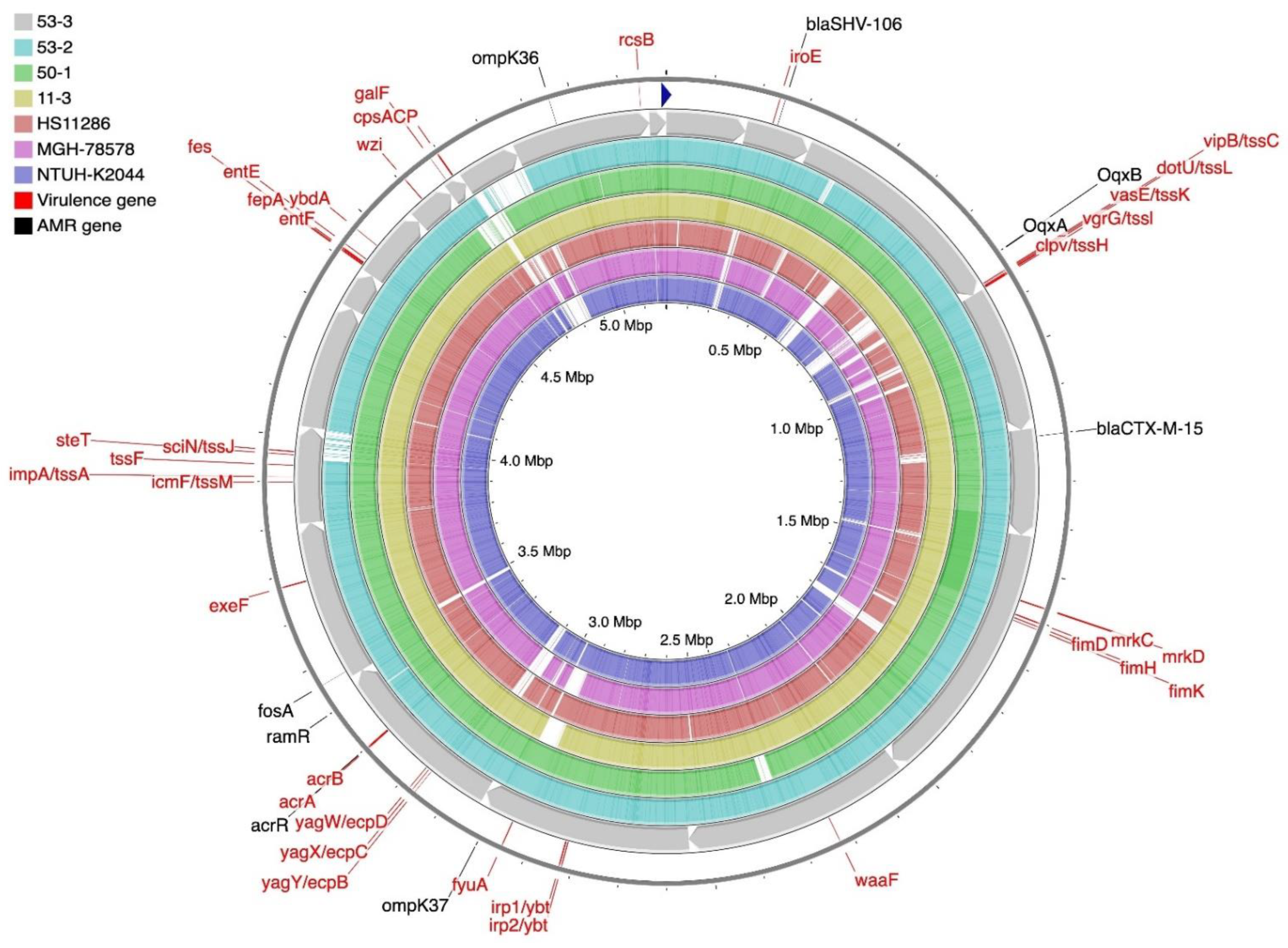
2.5. Genome Sequencing and Bioinformatic Analysis of K. pneumoniae Isolates

A representative member of clinical K. pneumoniae strains, from four different isolation sites, were selected for whole-genome sequencing.
K. pneumoniae strain 11/3 was isolated from a faecal sample. De novo assembly generated 5,517,254 bp. The average contig length was 134,567 and the average GC content of the chromosome was 57.3%. The genome contained 5180 genes and 81 tRNA genes. Analysis of the strain 11/3 sequence data revealed the presence of three types of plasmids, IncFIB (K), IncFII(K) and ColpVC. Based on ResFinder results, the presence of 21 genes related to antibiotics, including the previously characterized blaCTX-M-15, blaSHV-106 and blaVIM-4 genes, were identified. Several genes and mutations associated with resistance to antimicrobials were detected. Mutations were identified in the ompK35 (p.A217S) and ompK36 (p.I128M, p.I70M) genes that are associated with carbapenem resistance. Seven fluoroquinolone resistance mutations (acrR p.F197I, p.K201M, p.L195V, p.G164A, p.R173G, p.F172S and p.P161R) and one tigecycline resistance mutation (ramR p.A19V) were also found in the genome. Based on VFDB results, 27 virulence genes were analysed (Supplementary Table S4), of which 3 virulence genes (manB, manC and wbbM) were only present in strain 11/3. The nucleotide sequence of strain 11/3 was deposited in the GenBank database under the accession number JAJTNS000000000. The general features of genomic analysis of K. pneumoniae strain 11/3 are summarized in Table 3 and are shown in Figure 3.
K. pneumoniae strain 50/1 was isolated from blood culture. Shotgun sequences were assembled into one circular replicon measuring 5,379,211 bp. The average contig length was 298,845 and the average GC content of the chromosome was 57.4%. The genome contained 5022 genes and 73 tRNA genes. Analysis of the strain 50/1 sequence data has revealed the presence of one type of plasmid, IncFII(K). Based on ResFinder results, the presence of 14 genes related to antibiotics, including the previously characterized blaCTX-M-15 and blaSHV-106 genes were identified. Mutations were identified in the ompK35 (p.A217S) and ompK36 (p.I128M, p.I70M) genes that are associated with carbapenem resistance. Seven fluoroquinolone resistance mutations (acrR p.K201M, p.F172S, p.F197I, p.G164A, p.L195V, p.R173G and p.P161R) and one tigecycline resistance mutation (ramR p.A19V) were also found in the genome. Based on VFDB results, we analysed 29 virulence genes (Supplementary Table S4), of which 3 virulence genes (mrkB, mrkH and mrkJ) were only present in strain 50/1. The nucleotide sequence of strain 50/1 was deposited in the GenBank database under the accession number JAJTNT000000000. The general features of genomic analysis of K. pneumoniae strain 50/1 are summarized in Table 3 and are shown in Figure 3.
K. pneumoniae strain 53/2 was isolated from sputum. De novo assembly of the genome generated 5,269,114 bp. The average contig length was 292,728 and the average GC content of the chromosome was 57.4%. The genome contained 4908 genes and 81 tRNA genes. Analysis of the strain 53/2 sequence data revealed the presence of one type of plasmid, ColpVC. Based on ResFinder results, 20 genes related to antibiotics, including the previously characterized blaCTX-M-15, blaSHV-106 and blaVIM-4 genes were identified. Several genes and mutations associated with resistance to antimicrobials were detected. Mutations were identified in the ompK35 (p.A217S) and ompK36 (p.I128M, p.I70M) genes that are associated with carbapenem resistance. Seven fluoroquinolone resistance mutations (acrR p.F172S, p.P161R, p.K201M, p.L195V, p.F197I, p.G164A and p.R173G) were found in the genome. Based on VFDB results, we analysed 18 virulence genes (Supplementary Table S4). The nucleotide sequence of strain 53/2 was deposited in the GenBank database under the accession number JAJTNR000000000. The general features of genomic analysis of K. pneumoniae strain 53/2 are summarized in Table 3 and are shown in Figure 3.
K. pneumoniae strain 53/3 was isolated from urine. De novo assembly generated 5,201,283 bp. The average contig length was 273,751 and the average GC content of the chromosome was 57.4%. The genome contained 4844 genes and 63 tRNA genes. Analysis of the strain 53/3 sequence data revealed the presence of one type of plasmid, IncFII(K). Based on ResFinder results, 21 genes were related to antibiotics, including the previously characterized blaCTX-M-15 and blaSHV-106 genes. Several genes and mutations associated with resistance to antimicrobials were detected. Mutations were identified in the ompK35 (p.A217S) and ompK36 (p.I128M, p.I70M) genes that are associated with carbapenem resistance. Seven fluoroquinolone resistance mutations (acrR p.R173G, p.F197I, p.G164A, p.P161R, p.K201M, p.F172S and p.L195V) and one tigecycline resistance mutation (ramR p.A19V) were also found in the genome. Based on VFDB results, we analysed 37 virulence genes (Supplementary Table S4), of which 5 virulence genes (cpsAPC, galF, irp1/ybt, irp2/ybt and wzi) were only in strain 53/3. The nucleotide sequence of strain 53/3 was deposited in the GenBank database under the accession number JACTNU010000000. The general features of genomic analysis of K. pneumoniae strain 53/3 are summarized in Table 3 and are shown in Figure 3.

3. Discussion

Evolutionary aspects of antibiotic resistance spread and persistence and its impact on the clinical outcome in and out of the hospital environment has been the focus of recent studies [31,32,33,34,35,36]. It has been concluded that the CR-Kp infection-related mortality rate was higher than those of extended-spectrum β-lactamases (ESBL)-producing, and wild-type susceptible K. pneumoniae strains [37,38]. Moreover, infection with carbapenem-resistant strains is a risk factor for infection-related mortality [39,40,41,42,43]. Clones with common clonal origins, first identified in an English teaching hospital in 2010, provided a good opportunity to perform a comparative analysis focusing on virulence attributes.
In this study, the clones used were the first CR-Kps isolated in this hospital and originated from the first wave of the emergence (2010–2017). A common origin of the clones was confirmed as they all belonged to ST 15, possessed the moderately virulent capsule type K24, had the same antibiogram (Supplementary Table S2a,b), and showed high sequence homology based on PFGE and partially by whole genome sequence analysis. Well documented controls were used, including the hypervirulent K. pneumoniae strain NTUH K-2044 [44] and the moderately virulent reference strain MGH78578 [45]. Many attributes were common, but the slight differences among them indicated a divergence or evolutionary separation via an evolutionary process.
The common clonal origins of the K. pneumoniae isolates were supported by the fact that all isolates (n = 39) belonged to ST15 and possessed the K24 capsular type and their antibiograms showed high similarities to each other (Supplementary Table S2a,b). Furthermore, identified point mutations (location inside the gene is labelled with: p.XY) in the resistance genes of the four sequenced isolates 11/3, 50/1, 53/2 and 53/3 were localized at the same positions affecting the carbapenem resistance in the ompK35 (p.A217S) and ompK36 (p.I128M, p.I70M) [46]. This was also the case in other two inactive resistance genes, namely the acrR and ramR. The acrR gene (1492–4449 bp) confers resistance to fluoroquinolones [47,48], is located on a plasmid, and was affected with the same seven point mutations (p.F197I, p.K201M, p.L195V, p.G164A, p.R173G, p.F172S and p.P161R). Only one mutation (p.A19V) was identified in all sequenced isolates in the ramR gene, that confers resistance to tigecycline [47,48,49].
Expression the type 1 (92.3%) and type 3 (94.87%) fimbriae (Table 2) showed the capacity of the clones to colonize abiotic and biotic surfaces, and this contributed not only to survival but also to pathogenesis. These findings are in agreement with earlier studies which show that the adhesive subunit FimH in particular, of type 1 fimbriae, plays an important role in UTIs caused by K. pneumoniae [50]. Type 3 fimbriae assist adhesion to human tissue structures (e.g., lung, kidney) and promote biofilm formation on abiotic surfaces. As such, they may play a role in biofilm-associated infections in catheterized patients [2,51,52,53], but are also present in UTI E.coli isolates [54]. Correlation between the presence of mrkD, the expression of type 3 fimbriae, and strong biofilm formation was also confirmed by our study (Table 1 and Table 2), indicating its pivotal role in survival and persistence of the isolated clones. Incidence of mrkD genes (92.3%) among our CR-Kp isolates correlate with recent findings (94%) [55]. Results of our and this latter recent study are also comparable based on the incidence of magG as being 10.25% and 11%, respectively.
The high rate of enterobactin expression (94.87%, Table 2) was consistent with previous studies [56,57,58,59] and further supported the pathogenic potential of the isolates. Furthermore, aerobactin was also reported to be an important determinant of virulence of K. pneumoniae [16] if present, although it was more rarely found in Klebsiella. Its reported rates range from 3–6% [21], similar to our findings (5.12%). Although in our study no serious outcomes were registered among the patients infected by the isolates, evidence in the literature is mounting that K. pneumoniae strains carrying acquired siderophores have enhanced capacities to cause invasive diseases [17,59,60,61,62].
A high rate of siderophore production could also support cell internalization and invasion. The heterogeneous picture of the invasion capacities of the isolates with common clonal origin testify to unknown molecular biological changes that could affect the invasion capacities of these bacteria. Cell internalization in most bacteria is a still an unsolved multifactorial process, that in case of K. pneumoniae was first reported more than two decades ago [63]. Because of its shielding effect, internalization is a bacterial survival strategy that could also spoil the efficacy of targeted therapies presenting a challenge to the clinician. Despite these therapeutic consequences, cell internalization is still not in the focus of K. pneumoniae research [64,65,66]. In our study, internalization experiments (Figure 2) not only revealed the differences among the isolates, but also revealed that most isolates showed preference for the T24 bladder carcinoma cells, instead of INT 407.
In contrast, past studies have focused on the HMV phenotype of K. pneumoniae and its contribution to hypervirulence [61,67,68]. Our data confirmed the position that HMV and hypervirulence are different phenotypes that should not be described synonymously. This finding is consistent with recent opinions [69,70]. Although HMV positive clones (53/3, 53/11, 50/3, 11/1) can form biofilm, produce enterobactin and resist degradation in human serum, they were isolated either from mild UTIs or from faecal samples. Furthermore, our results confirmed earlier findings that the presence of the rmpA gene, encoding a positive regulator of mucopolysaccharides expression, is necessary for the HMV phenotype, since we detected this regulator only in the 4 HMV positive clones. To best of our knowledge this is the first study where the rmpA gene has been detected in a CR-Kp strain belonging to the ST 15 clone.
The described differences among the isolates with common clonal origins could be a result of an evolutionary process happening in the community and subsequently brought into the hospital. Based on the cluster analysis, we hypothesize that members of cluster I may originate from these two epidemiological processes. However, based on the isolation years and genomic similarities between members of cluster II and cluster III, revealed with PFGE, we postulate that two internal hospital epidemics could have occurred in 2011 and 2015, respectively. We have no available community data from the Chester region from this period, but based on the hospital data, the dominance of the ST 15 clone could be speculated to have emerged from 2010–2017.
Interestingly, sequence analysis revealed that four sequenced clones (11/3, 50/1, 53/2, 53/3) showed the highest genomic similarity to a K. pneumoniae strain HS11268 (Figure 3) isolated in China in 2011 [38]. The genomic similarity between K. pneumoniae strain 53/3 and HS11286 is 96%, strain 53/2 and HS11286 is 97%, strain 50/1 and HS11286 is 90%, strain 11/3 and HS11286 is 96%, based on NCBI Nucleotide BLAST.
Despite these similarities, it is interesting to note that no serious infections were detected among the patients. One explanation for this is the genetic background of the clones. Most clones (53.85%) were not resistant to the bactericidal effect of human serum. On the other hand, almost all clones possessed the K24 capsule, which in contrast to K1, K2, and K5 [21,71,72] is not considered as an important capsule type in the pathogenic process. Nevertheless, the ability of all clones to form a firm biofilm with the help of the K24 capsule maintains that this capsule type, together with type 3 fimbria, supports the survival of bacteria on abiotic and potentially on biotic surfaces. This survival potential of the K24 capsule type might explain why this is the most frequent capsule type linked to ST 15 [18], an otherwise pan-drug resistant widespread clone recently identified worldwide [18,19,60,72,73,74].
Although we do not know what attributes the original ST 15 clone had, we hypothesize that after its appearance this clone underwent, and is still currently undergoing a homing process, when the bacterium adapts to survival in the hospital environment by losing some of its virulence attributes, such as the ability to survive in serum and to invade eukaryotic cells. Such a process in the hospital, community, or in the environment is an important aspect of bacterial evolution with relevance to human pathogenesis and was also formerly outlined in cases of S. aureus and L. pneumophilia [75,76]. Till now no, relevant data were found in case of Klebsiella pneumoniae. This was the reason that our study mainly focused on the divergence of the virulence attributes among isolates with common clonal origin. In addition, sequence analysis could also provide an insight into the contributing mechanisms of the carbapenem resistance in the isolates i.e., mutations in ompK35 and ompK36. Isolates were resistant to cefoxitin (Supplementary Table S2a,b), indicating that the mutations detected by WGS analysis in the ompK35 and ompK36 truly manifested.

4. Materials and Methods

4.1. Bacterial Isolates, Growth Conditions

Thirty-nine K. pneumoniae isolates (one isolate per patient) collected from the Microbiology Laboratories of University Hospital in Chester, England were studied (Table 4). They were recovered from different specimens: faecal (n = 21), urine (n = 11), sputum (n = 4), and blood cultures (n = 3). Identification of the isolates was performed by Matrix-Assisted Laser Desorption/Ionization Time-of-Flight Mass Spectrometry (MALDI-TOF MS, Microflex, Bruker Daltonics, Billerica, MA, USA) and was confirmed with the standard biochemical tests (indole, adonite, Voges-Proskauer, methyl red, citrate, malonate, urease, lysine-, ornithine-, arginine- decarboxylase, saccharose, lactose and motility tests) prior to the study.
Bacteria were routinely grown on 37 °C in Luria–Bertani (LB) broth or on eosin–methylene blue (EMB) agar (Difco, Fisher Scientific, Leicestershire, UK) plates. Special media and control strains for fimbria, biofilm, and siderophore production tests, are indicated at relevant sections. K. pneumoniae strains NTUH-K2044 [44] and MGH 78578 [45] were used as controls for each experiment. Other control strains are designated in the relevant assays.

4.2. Antimicrobial Susceptibility Testing

Antimicrobial susceptibility was determined with the disc diffusion method on Mueller–Hinton (MH) agar (Oxoid, Basingstoke, UK). The inoculum was adjusted to an optical density of 0.5 McFarland, and the plates were incubated for 18 h at 35 °C. Resistance against the following antibiotics were tested: AMP: Ampicillin 10 µg, AMC: Amoxicillin/Clavulanic acid 20/10 µg, PTZ: Piperacillin/Tazobactam 30/6 µg, SAM: Ampicillin/Sulbactam 10/10 µg, FEP: Cefepime 30 µg, CTX: Cefotaxime 5 µg, CAZ: Ceftazidime 10 µg, CRO: Ceftriaxone 30 µg, CXM: Cefuroxime 30 µg, FOX: Cefoxitin 30 μg, ETP: Ertapenem 10 µg, IMP: Imipenem 10 µg, MEM: Meropenem 10 µg, CIP: Ciprofloxacin 5 µg, LEV: Levofloxacin 5 µg, AN: Amikacin 30 µg, GM: Gentamicin 10 µg and TM: Tobramycin 10 µg. Antimicrobial disks were purchased from Oxoid, Hungary. E. coli ATCC 25922 was used as a control. The results were interpreted according to the guidelines of the European Committee on Antimicrobial Susceptibility Testing (EUCAST) [77].
A new phenotypic test, called the Carbapenem Inactivation Method (CIM), was used to detect carbapenemase activity [78]. To perform the CIM, a suspension was made by suspending a full 10 μL inoculation loop of culture, taken from a Muller–Hinton (MH) agar (Oxoid, UK) plate in 400 μL Tryptic Soy Broth (TSB). Subsequently, a susceptibility-testing disk containing 10 μg meropenem (Oxoid, UK) was immersed in the suspension and incubated for 4 h at 35 °C. After incubation, the disk was removed from the suspension using tweezers, placed on a MH agar plate inoculated with a susceptible E. coli ATCC 29522 indicator strain and incubated for 18 h at 35 °C. Inoculation of the MH agar plate with the indicator strain was done with suspension of OD595 1.25, streaked in three directions using a sterile cotton swab. If the bacterial isolate produced carbapenemase, the meropenem in the susceptibility disk was inactivated allowing uninhibited growth of the susceptible indicator strain. Disks incubated in suspensions that do not contain carbapenemases yielded a clear inhibition zone. Each isolate was tested two times.

4.3. Chromosomal Macro-Restriction Fragment Polymorphism Analysis with Pulsed-Field Gel Electrophoresis (PFGE)

Pulsed-field gel electrophoresis (PFGE) was used to reveal the clonal relationship among the K. pneumoniae isolates. Strains were grown overnight (10 h) on LB agar at 37 °C. The ODs were adjusted to 1.3 to 1.4 (~108 CFU/mL) at 540 nm. Genomic DNA was prepared in low-gelling point agarose (BioRad, USA) by a procedure developed at the Centre for Disease Control (CDC) [79]. DNAs were in cube digested with XbaI (New England Biolabs, San Diego, CA, USA) restriction endonuclease for 12 h, with 10 units/mL. Separation of the fragments was performed by using the CHEF-DR II system (BioRad, Hercules, CA, USA). DNA was electrophoresed for 24 h at 14 °C in a 1.2% agarose gel (Sigma-Aldrich, St. Louis, MO, USA) at 6 V/cm with a linear gradient pulse time of 54 s. Interpretation of PFGE patterns was based on the criteria of Tenover et al. [80]. After photographing, gels were analysed and interpreted with Fingerprinting II Informatix Software (BioRad, USA). Levels of similarities were calculated with the Dice coefficient, and unweighted pair group method with arithmetic averages (UPGMA) was used for the cluster analysis of the PFGE patterns. Pulsotypes (PTs) were defined at 85% similarity between macrorestriction patterns [20].

4.4. Multilocus Sequence Typing (MLST) Analysis

Genomic DNA was isolated from bacteria using a Qiagen DNeasy blood and tissue kit (Qiagen, Valencia, CA, USA). Seven housekeeping genes (rpoB F: Vic3: GGCGAAATGGCWGAGAACCA, R: Vic2: GAGTCTTCGAAGTTGTAACC; gapA F: gapA173: TGAAATATGACTCCACTCACGG, R: gapA181: CTTCAGAAGCGGCTTTGATGGCTT; mdh F: mdh130: CCCAACTCGCTTCAGGTTCAG, R: mdh867: CCGTTTTTCCCCAGCAGCAG; pgi F: pgi1F: GAGAAAAACCTGCCTGTACTGCTGGC, R: pgi1R: CGCGCCACGCTTTATAGCGGTTAAT; phoE F: phoE604.1: ACCTACCGCAACACCGACTTCTTCGG, R: phoE604.2 TGATCAGAACTGGTAGGTGAT; infB F: infB1F: CTCGCTGCTGGACTATATTCG, R: infB1R: CGCTTTCAGCTCAAGAACTTC and tonB F: tonB1F: CTTTATACCTCGGTACATCAGGTT, R: tonB2R: ATTCGCCGGCTGRGCRGAGAG) were amplified by polymerase chain reaction (PCR), using conditions and primers designated by the PubMLST Klebsiella pneumoniae MLST Database [81], with 50 ng of genomic DNA as a template. The PCR products were purified using a QIAquick PCR purification kit (Qiagen, Valencia, CA, USA). Sequencing was performed at the Countess of Chester Hospital, Institute of Microbiology, Chester, Cheshire, UK. Sequence alignment was performed using MacVector editing software, and the resulting contig was compared against the PubMLST database to determine the designated allele. To identify the sequence type, allelic profiles were generated for each clinical isolate and compared to the MLST database.

4.5. PCR Detection of Virulence-Associated Genes

The presence of virulence-associated genes, including the activator of the allantoin regulator gene (allS), regulator of mucoid phenotype A (rmpA), siderophore genes: enterobactin (entB) and aerobactin (iutA), adhesion associated genes (cf29a, mrkA, mrkJ, mrkD, fimH-1), uridine diphosphate galacturonate 4-epimerase gene (uge), serum resistance associated gene (traT) and mucoviscosity-associated gene A (magA) was studied by PCR using primers and conditions described elsewhere (Supplementary Table S1).
Genetic determinants of the K24 capsule type were revealed after whole-genome sequencing of four representative strains and analysis of these sequences. Based on the wzi cluster analysis, the K24 specific primer pair (Fw: 5′-AGATAATAGG CAACAGCGTTCT-3′ and Rev: 5′-GATACGTTAAA CGCCTCAAGTA-3′) was designed by Primer-BLAST software [82].
The sequenced K. pneumoniae strains NTUH-K2044 [44] and MGH 78578 [45] were used as controls.
For DNA extraction, 1.5 mL of overnight (10 h) broth culture was centrifuged at 12,000 rpm for 2 min. The pellet was resuspended in 100 µL sterile deionized water and boiled for 10 min. After a final centrifugation at 12,000 rpm for 10 min, the supernatant containing template DNA was recovered, and used for the analysis. Conditions of the amplification reactions were as follows: 95 °C for 2 min for initial denaturation, which was followed by 34 cycles consisting of 95 °C denaturation for 30 s, annealing temperatures for 30 s, and a 72 °C elongation for 1 min. A one step termination was carried out at 72 °C for 10 min. Samples were electrophoresed in 1% agarose (Invitrogen, Waltham, MA, USA) gels, stained with ethidium bromide (Acros Organics, Antwerp, Belgium), and visualized under UV light.

4.6. Phenotypic Tests

4.6.1. Type 1 Fimbriae Assay (Mannose Sensitive Haemagglutination, MSHA)

The traditional S. cerevisiae agglutination test was used to detect the expression of the type 1 fimbriae—Mannose Sensitive Haemagglutinins (MSHA)—on the surface of the K. pneumoniae isolates. A cell suspension (37 µL; 1 × 10⁹ CFU/mL) of the standard Sacharomyces cerevisiae W303 was mixed with the bacterial dilution (1 × 10⁸ CFU/mL) on a glass slide, and gently rotated. To confirm the specificity, tests were also performed in the presence of α-methyl-d-mannoside (5%) (Fluka, Buchs, Switzerland) as it inhibited the specific agglutination [83]. All agglutination assays were carried out three times. K. pneumoniae strain 71 and K. pneumoniae strain 39 were used as positive controls, and K. pneumoniae strain 130 and K. pneumoniae strain 131 were used as negative controls.

4.6.2. Type 3 Fimbriae Assay (Mannose-Resistant Haemagglutination, MRHA)

Presence of the type 3 fimbriae mediated mannose-resistant haemagglutination on K. pneumoniae isolates was revealed by using the classical method of Podschun and Sahly [84]. Tannic acid (Fluka, Hungary) treated bovine erythrocyte suspension (Culex, Budapest, Hungary) was used. Bacteria were grown at 37 °C on brain heart infusion (BHI) agar plates (Oxoid, UK) for 24 h. Cells were collected, resuspended in physiological saline and their cell counts were adjusted to ~108 cells/mL in physiological saline. Thirty-two µL of bacterial suspensions and 38 µL of erythrocytes were mixed on glass slide, manually rotated, and observed for 10 min at room temperature (22 °C). Agglutination was finally read after further incubation for 5 min at 4 °C [39]. All agglutination assays were carried out three times. K. pneumoniae strain 130 and K. pneumoniae strain 131 were used as positive controls, and K. pneumoniae strain 71 and K. pneumoniae strain 79 were used as negative controls.

4.6.3. Biofilm Assay

The biofilm forming capacities of the K. pneumoniae isolates were tested by the slightly modified crystal violet binding plate assay [85]. Bacteria were subcultured in LB broth for 12 h at 37 °C, in a shaking incubator (120 RPM). The ODs were adjusted to 0.9 to 1.0 (~108 CFU/Ml) at 540 nm. Twenty µL of the adjusted bacterial cultures and 180 µL LB broth were transferred to 96-well polystyrene microtiter plates (Sarstedt, Nümbrecht, Germany) and incubated for 24 h at 37 °C. Planktonic bacteria were removed with gentle washing and fixed with 2% formalin-phosphate buffered saline (PBS; Sigma-Aldrich, USA). Intensities of biofilm formations were revealed by 1% crystal violet (Sigma-Aldrich, USA) staining for 20 min at room temperature (22 °C), and by solubilizing the layer with 1% SDS (Sigma-Aldrich, USA). Extinction of the solubilized crystal violet in each well was measured at 595 nm with a FLUOstar Optima Microplate Reader (BMG Labtech, Ortenberg, Germany). Controls were performed with crystal violet binding to the wells exposed only to the culture medium without bacteria. Biofilm assays were repeated three times in three independent experiments and in each assay, quantification was performed in four separate wells. K. pneumoniae strain 703+ was used as a positive control and K. pneumoniae strain 446- was used as a negative control. Isolates were classified as high biofilm-producers (OD ˃ 3.0), medium producers (OD 1.0–3.0) or poor producers (OD < 1.0).

4.6.4. Siderophores Production Assay

Production of enterobactin and aerobactin were detected by the cross-feeding bioassay described by Hantke [86]. Briefly, nutrient agar supplemented with 2,2′-dipyridyl (Sigma-Aldrich, USA) (final concentration 275 µM) was used as iron-restricted agar medium. Growth capacity of the CR-Kp isolates was tested in parallel in the presence of two different indicator strains. E. coli H1887 (ColV, Aer, Iut+, FepA, Fiu, Cir, aroB) was used as the indicator strain for aerobactin production and strain H1939 (FepA+, Fiu, Cir, FhuA, FhuB, aroB) for enterobactin. Aerobactin production was counterchecked with E. coli strain H1886, which is the Iut parent strain of H1887. Strain K311 (pColV-K311) served as a positive control in the aerobactin test [56]. Each isolate was tested three times.

4.6.5. Serum Bactericidal Assay

Normal human serum (NHS), pooled from healthy volunteers, was divided into equal volumes and stored at −20 °C before use. The serum bactericidal activity was measured using the method described by Podschun et al. [56], with slight modification. Bacteria were grown at 37 °C in LB broth for 24 h, with shaking at 100 rpm in an incubator. After washing with physiological saline solution, OD600 was adjusted to 0.4. Bacteria were diluted to 2–3 × 10⁶ cell/mL in physiological saline. Then, 25 µL from the bacterial suspensions and 75 µL from the undiluted NHS were mixed in the 96 well polystyrene microtiter plates (Sarstedt, Germany), and incubated at 37 °C. Samples were taken immediately after mixing and after incubation for 1 and 3 h, and serial dilutions were plated on LB agar for colony forming unit (CFU) determination. Resistance was graded by the mean 3 h survival ratio (ratio of colony count after serum treatment for 3 h compared with baseline). The highly serum resistant K. pneumoniae strain NTUH-K2044 was used as a positive control [44]. Each strain was tested three times.

4.6.6. Hypermucoviscosity (HMV) Testing

Single colonies obtained after overnight culture on blood agar plates were tested for their ability to form viscous strings when a standard inoculation loop was touched onto their surface and slowly raised. The formation of string greater than 5 mm in length is indicative of the hypermucoviscosity (HMV) positive phenotype [87]. K. pneumoniae strain NTUH-K2044 [44] was used as a positive control. Each isolate was tested twice.

4.7. Whole-Genome Sequencing

In order to get a detailed view of the genome organization of the ST 15 K. pneumoniae lineage, sequencing of four representative members from different specimens was carried out: K. pneumoniae strain 11/3 (isolated from faeces), strain 50/1 (isolated from blood culture), strain 53/2 (isolated from sputum) and strain 53/3 (isolated from urine).
DNA was extracted from K. pneumoniae strains 11/3, 50/1, 53/2 and 53/3. DNA for whole-genome sequencing were extracted from cultures grown overnight (10 h) in LB agar, using DNA extraction kit (PureLink Genomic DNA Mini Kit, Thermo Fischer Scientific, Waltham, MA, USA) following the manufacturer’s protocol. At the end of the extraction process, DNA samples were dissolved in 100 μL of sterile nuclease free water. Genomic DNA sequencing libraries were prepared using the Nextera XT Library Preparation kit (Illumina, San Diego, CA, USA). Sequencing was performed using MiSeq Reagent Kit v3 (600 cycles) on an Illumina MiSeq (Illumina, San Diego, CA, USA). Assembly of the pure sequence was performed with the MyPro pipeline [88]. Open reding frames were predicted and annotated with the Rapid Annotation using Subsystem Technology [89]. Homology searches were conducted with the BLAST tools available at the NCBI website [90].
Analysis of antibiotic resistance was performed with ResFinder 4.1 [91]. Additional software, including Pathogen Wach [92], was used for analysis of specific plasmid genetic features. Nucleotide sequences for validated Klebsiella genus virulence genes were downloaded from the Virulence Factor Database (VFDB) [93]. Nucleotide–nucleotide alignment was run using BLASTN v2.11.0 on macOS 11.6 for our Klebsiella sp. contig sequences against the downloaded virulence factor database. Our annotated sequences along with three Klebsiella pneumoniae subsp. pneumoniae reference genomes (strain HS11286, SAMN02602959; MGH78578, SAMN02603941; NTUH-K2044, SAMD00060934) were visualized using the CGView Server Beta web tool [94]. BLAST alignments for all sequences were run against the nucleotide sequence of K. pneumoniae strain 53/3 and visualized. Virulence genes for strain 11/3, 50/1, 53/2 and 53/3 identified previously using the VFDB were marked on the CGView map.

4.8. Cell Internalization Assay

Two human cell lines, the human intestinal cell line INT 407 and the bladder carcinoma cell line T24 were used to reveal the abilities of the investigated CR-Kp isolates to invade cultured cells. Invasion assays were performed essentially as described previously [95]. Briefly, semiconfluent cell monolayers were prepared (3 × 10⁵ cells/well) in RPMI 1640 medium (Lonza, Verviers, Belgium) supplemented with 10% heat-inactivated (30 min for 56 °C) calf bovine serum (Sigma-Aldrich, USA), 10,000 U/mL of penicillin, 10 μg/mL of streptomycin and 0.5 mg/mL of neomycin and incubated overnight (10 h) at 37 °C in a humidified 5% CO2 incubator. On the following day, cells were washed with PBS (pH 7) and to each well 1 mL RPMI 1640 medium (Lonza, Belgium) and bacterial suspensions (OD600 = 1, ~1 × 10⁸ CFU/mL) were added to reach 10× dilution [96]. Plates were incubated at 37 °C in a humidified, 5% CO2 incubator for 3 h. The plates were then washed three times with PBS (pH 7) to remove unbound bacteria. Fresh cell culture medium containing 100 µg/mL Polymixin B was then added to kill all extracellular bacteria and incubated for 1 h at 37 °C. Then wells were washed three times with PBS (pH 7) and lysed with 0.05% Triton X-100 (Sigma-Aldrich, Budapest, Hungary). The intracellularly surviving K. pneumoniae cell counts were determined by out-plating. All assays were performed in triplicate and were repeated independently twice. Salmonella enterica serotype Typhimurium strain ATCC14028, K. pneumoniae strain 3091 (accession number: SAMEA8948279) [97] and K. pneumoniae strain NTUH-K2044 (accession number: SAMD00060934) were used as positive controls and K. pneumoniae strain MGH78578 (accession number: SAMN02603941) was used as a negative control.

5. Conclusions

In this study an evolutionary process could be outlined for a CR-Kp ST 15 clone that first appeared in an English Teaching Hospital in 2010. We conclude that during this process, which likely occurred in parallel both in the hospital and the community, divergence of virulence attributes could be observed that support persistence of the original clone rather than virulence. Long term studies in hospital environments, supplemented with community data, could reveal, in the future, the changes in the virulence potentials of emerging clones, similar to changes that are studied for antibiotic resistance. To the best of our knowledge this is the first study where the rmpA gene has been detected in a CR-Kp strain belonging to the ST 15 clone.

Supplementary Materials

The following supporting information can be downloaded at: https://www.mdpi.com/article/10.3390/antibiotics12030479/s1, Table S1: PCR detection of virulence-associated genes. All the primers used in this study. Table S2a: Antimicrobial susceptibility pattern (EUCAST) of the carbapenem-resistant K. pneumoniae isolates of this study, Table S2b: Minimum Inhibitory Concentration (MIC) for 17 antibiotics (mg/L) against the clinical K. pneumoniae isolates (n = 39). Table S3: PFGE based virulence analysis of K. pneumoniae isolates. Table S4: Comparative pathogenomics of K. pneumoniae strain 11/3, strain 50/1, strain 53/2 and strain 53/2 with VFDB database. References [10,21,98,99,100,101] are cited in supplemantary materials.

Author Contributions

M.H. and G.S. were involved in the experimental design. M.H. performed basic virulence analysis and molecular typing, summarized the data and wrote the manuscript. T.K., J.K. and A.G. performed the sequencing and the bioinformatics analysis. I.D. performed the PFGE analysis. G.S. and Z.T. conceived and coordinated the whole work. All authors have read and agreed to the published version of the manuscript.

Funding

A.G. and J.K. were supported by the grants GINOP-2.3.4-15-2020-00010, GINOP-2.3.1-20-2020-00001 and Educating Experts of the Future: Developing Bioinformatics and Biostatistics competencies of European Biomedical Students (BECOMING, 2019-1-HU01-KA203-061251).

Institutional Review Board Statement

Not applicable.

Informed Consent Statement

Not applicable.

Data Availability Statement

All data associated with this study are presented in the main text or Supporting information.

Acknowledgments

The authors wish to thank Ildikó Kustos for the hospital-derived K. pneumoniae isolates. We also thank the Medical School of the University of Pécs for supporting our work. The research was performed in collaboration with the Genomics and Bioinformatics Core Facility at the Szentágothai Research Centre of the University of Pécs. Bioinformatics infrastructure was supported by ELIXIR Hungary (http://elixir-hungary.org/, accessed on 1 February 2023 ).

Conflicts of Interest

The authors declare no conflict of interest.

References

  1. Decré, D.; Verdet, C.; Emirian, A.; Le Gourrierec, T.; Petit, J.-C.; Offenstadt, G.; Maury, E.; Brisse, S.; Arlet, G. Emerging severe and fatal infections due to Klebsiella pneumoniae in two University Hospitals in France. J. Clin. Microbiol. 2011, 49, 3012–3014. [Google Scholar] [CrossRef] [Green Version]
  2. Schroll, C.; Barken, K.B.; Krogfelt, K.A.; Struve, C. Role of type 1 and type 3 fimbriae in Klebsiella pneumoniae biofilm formation. BMC Microbiol. 2010, 10, 179. [Google Scholar] [CrossRef] [PubMed] [Green Version]
  3. Albiger, B.; Glasner, C.; Struelens, M.J.; Grundmann, H.; Monnet, D.L. Carbapenemase-producing Enterobacteriaceae in Europe: Assessment by national experts from 38 countries. Eurosurveillance 2015, 20, 45. [Google Scholar] [CrossRef] [Green Version]
  4. Kwak, Y.G.; Choi, S.-H.; Choo, E.J.; Chung, J.-W.; Jeong, J.-Y.; Kim, N.J.; Woo, J.-H.; Ryu, J.; Kim, Y.S. Risk factors for the acquisition of carbapenem-resistant Klebsiella pneumoniae among hospitalized patients. Microb. Drug Resist. 2005, 11, 165–169. [Google Scholar] [CrossRef] [PubMed]
  5. Gomez-Simmonds, A.; Uhlemann, A.-C. Clinical implications of genomic adaptation and evolution of carbapenem-resistant Klebsiella pneumoniae. J. Infect. Dis. 2017, 215, S18–S27. J. Infect. Dis. 2017, 215, S18–S27. [Google Scholar] [CrossRef]
  6. Munoz-Price, L.S.; Poirel, L.; Bonomo, R.A.; Schwaber, M.J.; Daikos, G.L.; Cormican, M.; Cornaglia, G.; Garau, J.; Gniadkowski, M.; Hayden, M.K.; et al. Clinical epidemiology of the global expansion of Klebsiella pneumoniae carbapenemases. Lancet Infect. Dis. 2013, 13, 785–796. [Google Scholar] [CrossRef] [PubMed] [Green Version]
  7. Liu, Y.-Y.; Wang, Y.; Walsh, T.R.; Yi, L.-X.; Zhang, R.; Spencer, J.; Doi, Y.; Tian, G.; Dong, B.; Huang, X.; et al. Emergence of plasmid-mediated colistin resistance mechanism MCR-1 in animals and human beings in China: A microbiological and molecular biological study. Lancet Infect. Dis. 2016, 16, 161–168. [Google Scholar] [CrossRef] [PubMed]
  8. Struve, C.; Krogfelt, K.A. Pathogenic potential of environmental Klebsiella pneumoniae isolates. Environ. Microbiol. 2004, 6, 584–590. [Google Scholar] [CrossRef]
  9. Selden, R.S.; Lee, S.; Wang, W.L.L.; Bennett, J.V.; Eickhoff, T.C. Nosocomial Klebsiella infections: Intestinal colonization as a reservoir. Ann. Intern. Med. 1971, 74, 657–664. [Google Scholar] [CrossRef]
  10. Johnson, J.G.; Clegg, S. Role of MrkJ, a phosphodiesterase, in type 3 fimbrial expression and biofilm formation in Klebsiella pneumoniae. J. Bacteriol. 2010, 192, 3944–3950. [Google Scholar] [CrossRef] [Green Version]
  11. Potera, C. Forging a link between biofilms and disease. Science 1999, 283, 1837–1839. [Google Scholar] [CrossRef]
  12. Stickler, D.J.; Morris, N.S.; McLean, R.J.C.; Fuqua, C. Biofilms on indwelling urethral catheters produce quorum-sensing signal molecules in situ and in vitro. Appl. Environ. Microbiol. 1998, 64, 3486–3490. [Google Scholar] [CrossRef] [Green Version]
  13. Costerton, J.W.; Stewart, P.S.; Greenberg, E.P. Bacterial biofilms: A common cause of persistent infections. Science 1999, 284, 1318–1322. [Google Scholar] [CrossRef] [PubMed] [Green Version]
  14. Darouiche, R.O.; Dhir, A.; Miller, G.C.; Raad, G.C.; Musher, D.M. Vancomycin penetration into biofilm covering infected prostheses and effect on bacteria. J. Infect. Dis. 1994, 170, 720–723. [Google Scholar] [CrossRef] [PubMed]
  15. Di Martino, P.; Cafferini, N.; Joly, B.; Darfeuille-Michaud, A. Klebsiella pneumoniae type 3 pili facilitate adherence and biofilm formation on abiotic surfaces. Res. Microb. 2003, 154, 9–16. [Google Scholar] [CrossRef]
  16. Nassif, X.; Sansonetti, P.J. Correlation of the virulence of Klebsiella pneumoniae K1 and K2 with the presence of a plasmid encoding aerobactin. Infect. Immun. 1986, 54, 603–608. [Google Scholar] [CrossRef] [PubMed] [Green Version]
  17. Wyres, K.L.; Nguyen, T.N.T.; Lam, M.M.C.; Judd, L.M.; Chau, N.; Van, V.; Dance, D.A.B.; Ip, M.; Karkey, A.; Ling, C.L.; et al. Genomic surveillance for hypervirulence and multi-drug resistance in invasive Klebsiella pneumoniae from South and Southeast Asia. Genome Med. 2020, 12, 11. [Google Scholar] [CrossRef] [Green Version]
  18. Andrade, L.N.; Novais, Â.; Stegani, L.M.M.; Ferreira, J.C.; Rodrigues, C.; Darini, A.L.C.; Peixe, L. Virulence genes, capsular and plasmid types of multidrug-resistant CTX-M (-2, -8, -15) and KPC-2-producing Klebsiella pneumoniae isolates from four major hospitals in Brazil. Diagn. Microbiol. Infect. Dis. 2018, 91, 164–168. [Google Scholar] [CrossRef]
  19. Ma, Y.; Bao, C.; Liu, J.; Hao, X.; Cao, J.; Ye, L. Microbiological characterization of Klebsiella pneumoniae isolates causing bloodstream infections from five tertiary hospitals in Bejing, China. J. Glob. Antimicrob. Resist. 2018, 12, 162–166. [Google Scholar] [CrossRef]
  20. Damjanova, I.; Tóth, Á.; Pászti, J.; Hajbel-Vékony, G.; Jakab, M.; Berta, J.; Milch, H.; Füzi, M. Expansion and countrywide dissemination of ST11, ST15 and ST147 ciprofloxacin-resistant CTX-M-15-type β-lactamase-producing Klebsiella pneumoniae epidemic clones in Hungary in 2005—The new ‘MRSAs’? J. Antimicrob. Chemother. 2008, 62, 978–985. [Google Scholar] [CrossRef] [Green Version]
  21. El Fertas-Assiani, R.; Messai, Y.; Alouache, S.; Bakour, R. Virulence profiles and antibiotic susceptibility petterns of Klebsiella pneumoniae strains isolated from different clinical specimens. Pathol. Biol. 2013, 61, 209–216. [Google Scholar] [CrossRef]
  22. Effah, C.Y.; Sun, T.; Liu, S.; Wu, Y. Klebsiella pneumoniae: An increasing threat to public health. Ann. Clin. Microbiol. Antimicrob. 2020, 19, 1. [Google Scholar] [CrossRef]
  23. Caneiras, C.; Lito, L.; Melo-Cristino, J.; Duarte, A. Community- and hospital-acquired Klebsiella pneumoniae urinary tract infections in Portugal: Virulence and antibiotic resistance. Microorganisms 2019, 7, 138. [Google Scholar] [CrossRef] [Green Version]
  24. Gupta, N.; Limbago, B.M.; Patel, J.B.; Kallen, A.J. Carbapenem-resistant Enterobacteriaceae: Epidemiology and prevention. Clin. Infect. Dis. 2011, 53, 60–67. [Google Scholar] [CrossRef] [PubMed] [Green Version]
  25. Bush, K. Alarming β-lactamase-mediated resistance in multidrug-resistant Enterobacteriaceae. Curr. Opin. Microbiol. 2010, 13, 558–564. [Google Scholar] [CrossRef]
  26. Quenan, E.M.; Bush, K. Carbapenemases: The versatile beta-lactamases. Clin. Microbiol. Rev. 2007, 20, 440–458. [Google Scholar] [CrossRef] [Green Version]
  27. Papagiannitsis, C.C.; Malli, E.; Florou, Z.; Sarrou, S.; Hrabak, J.; Mantzarlis, K.; Zakynthinos, E.; Petinaki, E. Emergence of sequence type 11 Klebsiella pneumoniae coproducing NDM-1 and VIM-1 metallo-beta-lactamases in Greek hospital. Diagn. Microbiol. Infect. Dis. 2017, 87, 295–297. [Google Scholar] [CrossRef] [PubMed]
  28. Samuelsen, Ø.; Toleman, M.A.; Hasseltvedt, V.; Fuursted, K.; Leegaard, T.M.; Walsh, A.; Sundsfjord, A.; Giske, C.G. Molecular characterization of VIM-producing Klebsiella pneumoniae from Scandinavia reveals genetic relatedness with international clonal complexes encoding transferable multidrug resistance. Clin. Microbiol. Infect. 2011, 17, 1811–1816. [Google Scholar] [CrossRef] [Green Version]
  29. Shi, W.; Li, K.; Ji, Y.; Jiang, Q.; Wang, Y.; Shi, M.; Mi, Z. Carbapenem and cefoxitin resistance of Klebsiella pneumoniae strains associated with porin OmpK36 loss and DHA-1 β-lactamase production. Braz. J. Microbiol. 2013, 44, 435–442. [Google Scholar] [CrossRef] [Green Version]
  30. Mouloudi, E.; Protonariou, E.; Zagorianou, A.; Iosifidis, E.; Karapanagiotou, A.; Giasnetsova, T.; Tsioka, A.; Roilides, E.; Sofianou, D.; Gritsi-Gerogianni, N. Bloodstream infections caused by metallo-β-lactamase/Klebsiella pneumoniae carbapenemase-producing K. pneumoniae among intensive care unit patient in Greece: Risk factors for infection and impact of type of resistance on outcomes. Infect. Control. Hosp. Epidemiol. 2010, 31, 1250–1256. [Google Scholar] [CrossRef]
  31. Chong, Y.; Ito, Y.; Kamimura, T. Genetic evolution and clinical impact in extended-spectrum β-lactamase-producing Escherichia coli and Klebsiella pneumoniae. Infect. Gnetics Evol. 2011, 11, 7. [Google Scholar] [CrossRef] [PubMed]
  32. Chong, Y.; Shimoda, S.; Shimono, N. Current epidemiology, genetic evolution, and clinical impact of extended-spectrum B-lactamase-producing Escherichia coli and Klebsiella pneumoniae. Infect. Gnetics. Evol. 2018, 61, 185–188. [Google Scholar] [CrossRef] [PubMed]
  33. Lai, Y.-C.; Lu, M.-C.; Hsueh, P.-R. Hypervirulence and carbapenem resistance: Two distinct evolutionary directions that led high-risk K. pneumoniae clones to epidemic success. Taylor Fr. 2019, 19, 825–837. [Google Scholar] [CrossRef]
  34. Li, X.; Ma, W.; Quin, Q.; Liu, S.; Ye, L.; Yang, J.; Li, B. Nosocomial spread of OXA-232-producing Klebsiella pneumoniae ST15 in a teaching hospital, Shanghai, China. MBC Microbiol. 2019, 19, 235. [Google Scholar] [CrossRef] [PubMed] [Green Version]
  35. Rodrigues, C.; Sousa, C.; Lopes, J.A.; Novais, Â.; Peixea, L. A front line on Klebsiella pneumoniae capsular polysaccharide knowledge: Fourier transform infrared spectroscopy as an accurate and fast typing tool. Am. Soc. Microbiol. 2020, 5, e00386-19. [Google Scholar] [CrossRef] [Green Version]
  36. Du, Y.; Mu, S.; Liu, Y.; Yuan, Y.; Zhu, Y.; Ma, M.; Wang, Q.; Zhu, Z.; Liu, Y.; Wang, S. The genomic characterization of KPC-producing Klebsiella pneumoniae from the ICU of a Teaching Hospital in Shanghai, China. Infect. Drug Resist. 2022, 15, 69–81. [Google Scholar] [CrossRef]
  37. MacKenzie, F.M.; Forbes, K.J.; Dorai-John, T.; Amyes, S.G.; Gould, I.M. Emergence of a carbapenem-resistant Klebsiella pneumoniae. Lancet 1997, 350, 783. [Google Scholar] [CrossRef]
  38. Liu, S.W.; Chang, H.J.; Chia, J.H.; Kuo, A.J.; Wu, T.L.; Lee, M.H. Outcomes and characteristics of ertapenem-non-susceptible Klebsiella pneumoniae bacteremia at a university hospital in Northern Taiwan: A matched case-control study. J. Microbiol. Immunol. Infect. 2012, 45, 113–119. [Google Scholar] [CrossRef] [Green Version]
  39. Gasink, L.B.; Edelstein, P.H.; Lautenbach, E.; Synnestvedt, M.; Fishman, N.O. Risk factors and clinical impact of Klebsiella pneumoniae carbapenemase-producing K. pneumoniae. Infect. Control. Hosp. Epidemiol. 2009, 30, 1180–1185. [Google Scholar] [CrossRef] [Green Version]
  40. Ben-David, D.; Kordevani, R.; Keller, N.; Tal, I.; Marzel, A.; Gal-Mor, O.; Maor, Y.; Rahav, G. Outcomes of carbapenem resistant Klebsiella pneumoniae bloodstream infections. Clin. Microbiol. Infect. 2012, 18, 54–60. [Google Scholar] [CrossRef] [Green Version]
  41. Dai, G.; Xu, Y.; Kong, H.; Xie, W.; Wang, H. Risk factors for carbapenem-resistant Klebsiella pneumoniae infection and associated clinical outcomes. Am. J. Transl. Res. 2021, 13, 7276–7281. [Google Scholar] [PubMed]
  42. Chen, J.; Ma, H.; Huang, X.; Cui, Y.; Peng, W.; Zhu, F.; Ma, S.; Rao, M.; Zhang, P.; Yang, H.; et al. Risk factors and mortality of carbapenem-resistant Klebsiella pneumoniae bloodstream infection in a tertiary-care hospital in China: An eight-year retrospetcive study. Antimicrob. Resist. Infect. Control. 2022, 11, 161. [Google Scholar] [CrossRef] [PubMed]
  43. Lou, T.; Du, X.; Zhang, P.; Shi, Q.; Han, X.; Lan, P.; Yan, R.; Hu, H.; Wang, Y.; Wu, X.; et al. Risk factors for infection and mortality caused by carbapenem-resistant Klebsiella pneumoniae: A large multicentre case–control and cohort study. J. Infect. 2022, 84, 5. [Google Scholar] [CrossRef] [PubMed]
  44. Wu, K.M.; Li, L.H.; Yan, J.J.; Tsao, N.; Liao, T.L.; Tsai, H.-C.; Fung, C.-P.; Chen, H.-J.; Liu, Y.-M.; Wang, J.-T.; et al. Genome sequencing and comparative analysis of Klebsiella pneumoniae NTUH-K2044, a strain causing liver abscess and meningitis. J. Bacteriol. 2009, 191, 4492–4501. [Google Scholar] [CrossRef] [Green Version]
  45. Fang, C.T.; Chuang, Y.P.; Shun, C.T.; Chang, S.C.; Wang, J.T. A novel virulence gene in Klebsiella pneumoniae strains causing primary liver abscess and septic metastatic complications. J. Exp. Med. 2004, 5, 697–705. [Google Scholar] [CrossRef] [Green Version]
  46. Tsai, Y.-K.; Fung, C.-P.; Lin, J.-C.; Chen, J.-H.; Chang, F.-Y.; Chen, T.-L.; Siu, L.K. Klebsiella pneumoniae outer membrane porins ompK35 and ompK36 play roles in both antimicrobial resistance and virulence. Antimicrob. Agents Chemothe. 2011, 55, 1485–1493. [Google Scholar] [CrossRef] [Green Version]
  47. He, F.; Fu, Y.; Chen, Q.; Ruan, Z.; Hua, X.; Zhou, H.; Yu, Y. Tigecyclne suscebility and the role of efflux pumps in tigecycline resisnatnce in KPC-producing Klebsiella pneumoniae. PLoS ONE 2015, 10, e0119064. [Google Scholar] [CrossRef]
  48. Schneiders, T.; Amyes, S.G.B.; Levy, S.B. Role of AcrR and RamA in fluoroquinolone rsistance in clinical Klebsiella pneumoniae isolates from Singapore. Antimicrob. Agents Chemother. 2013, 47, 9. [Google Scholar] [CrossRef] [Green Version]
  49. Rosenblum, R.; Khan, E.; Gonzalez, G.; Hasan, R.; Schneiders, A. Genetic regulation of the ramA locus and its expression in clinical isolates of Klebsiella pneumoniae. Int. J. Antimicrob. Agents 2011, 38, 39–45. [Google Scholar] [CrossRef]
  50. Struve, C.; Bojer, M.; Krogfelt, K.A. Identification of a conserved chromosomal region encoding Klebsiella pneumoniae Type 1 and Type 3 fimbriae and assessment of the role of fimbriae in pathogenicity. Infect. Immun. 2009, 77, 5016–5024. [Google Scholar] [CrossRef] [Green Version]
  51. Eghbalpoor, F.; Habibi, M.; Azizi, O.; Karam, M.R.A.; Bouzar, S. Antibiotic resistance, virulence and genetic diversity of Klebsiella pneumoniae in community- and hospital-acquired urinary tract infections in Iran. Acta Microbiol. Et. Immunol. Hung. 2019, 66, 3. [Google Scholar] [CrossRef] [Green Version]
  52. Maharjan, G.; Khadka, P.; Shilpakar, G.S.; Chapagain, G.; Dhungana, G.R. Catheter-associated urinary tract infection and obstinate biofilm producers. Can. J. Infect. Dis. 2018, 2018, 7624857. [Google Scholar] [CrossRef] [Green Version]
  53. Zadeh, F.M.; Zarei, H.; Jahromy, S.H. Type 1 and 3 fimbriae phenotype and genotype as suitable markers for uropathogenic bacterial pathogenesis via attachment, cell surface hydrophobicity, and biofilm formation in catheter-associated urinary tract infections (CAUTIs). Iran. J. Basic Med. Sci. 2021, 24, 1098–1106. [Google Scholar] [CrossRef]
  54. Khonsari, M.S.; Behzadi, P.; Foroohi, F. The prevalence of type 3 fimbriae in uropathogenic Escherichia coli isolated from clinical urine samples. Meta Gene 2021, 28, 100881. [Google Scholar] [CrossRef]
  55. Ahmadi, M.; Ranjbar, R.; Behzadi, P.; Mohammadian, T. Virulence factors, antibiotic resistance patterns, and molecular types of clinical isolates of Klebsiella pneumoniae. Expert Rev. Anti-Infect. Ther. 2022, 20, 463–472. [Google Scholar] [CrossRef] [PubMed]
  56. Podschun, R.; Sievers, D.; Fischer, A.; Ullmann, U. Serotypes, hemagglutinins, siderophore synthesis, and serum resistance of Klebsiella isolates causing human urinary tract infections. J. Infect. Dis. 1993, 168, 1415–1421. [Google Scholar] [CrossRef]
  57. Koczura, R.; Kaznowski, A. Occurrence of the Yersinia high-pathogenicity island and iron uptake systems in clinical isolates of Klebsiella pneumoniae. Microb. Pathog. 2003, 35, 197–202. [Google Scholar] [CrossRef] [PubMed]
  58. Remya, P.A.; Shanthi, M.; Sekar, U. Characterisation of virulence genes associated with pathogenicity in Klebsiella pneumoniae. Indian J. Med. Microbiol. 2020, 37, 210–218. [Google Scholar] [CrossRef]
  59. Daoud, L.; Al-Marzooq, F.; Moubareck, C.A.; Ghazawi, A.; Collyns, T. Elucidating the effect of iron acquisition systems in Klebsiella pneumoniae on susceptibility to the novel siderophore-cephalosporin cefiderocol. PLoS ONE 2022, 17, e0277946. [Google Scholar] [CrossRef]
  60. Lam, M.M.C.; Wick, R.R.; Wyres, K.L.; Gorrie, C.; Judd, L.M.; Brisse, S.; Jenney, A.; Holt, K.E. Frequent emergence of pathogenic lineages of Klebsiella pneumoniae via mobilisation of yersiniabactin and colibactin. Microb. Genom. 2018, 4, 1–14. [Google Scholar] [CrossRef] [Green Version]
  61. Choby, J.E.; Howard-Anderson, J.; Weiss, D.S. Hypervirulent Klebsiella pneumoniae—Clinical and molecular perspectives. J. Intern. Med. 2020, 287, 283–300. [Google Scholar] [CrossRef] [Green Version]
  62. Zhu, J.; Wang, T.; Chen, L.; Du, H. Virulence factors in hypervirulent Klebsiella pneumoniae. Front. Microbiol. 2021, 12, 642484. [Google Scholar] [CrossRef]
  63. Sahly, H.; Podschun, R.; Oelschlaeger, T.A.; Greiwe, M.; Parolis, H.; Hasty, D.; Kekow, J.; Ullmann, U.; Ofek, I.; Sela, S. Capsule impedes adhesion to and invasion of epithelial cells by Klebsiella pneumoniae. Infect. Immun. 2000, 68, 6744–6749. [Google Scholar] [CrossRef] [Green Version]
  64. Hsieh, P.-F.; Lu, Y.-R.; Lin, T.-Z.; Lai, L.-Y.; Wang, J.-T. Klebsiella pneumoniae type VI secretion system contributes to bacterial competition, cell invasion, type-1 fimbriae expression, and in vivo colonization. J. Infect. Dis. 2019, 219, 637–647. [Google Scholar] [CrossRef] [Green Version]
  65. Kim, J.H.; Cho, Y.Y.; Choi, J.Y.; Wi, Y.M.; Ko, K.S. Two distinct genotypes of KPC-2-producing Klebsiella pneumoniae isolates from South Korea. Antibiotics 2021, 10, 911. [Google Scholar] [CrossRef] [PubMed]
  66. Cho, Y.Y.; Kim, J.H.; Kim, H.; Lee, J.; Im, S.J.; Ko, K.S. Comparison of virulence between two main clones (ST11 and ST307) of Klebsiella pneumoniae isolates from South Korea. Microorganisms 2022, 10, 1827. [Google Scholar] [CrossRef] [PubMed]
  67. Catalán-Nájera, J.C.; Garza-Ramos, U.; Barrios-Camacho, H. Hypervirulence and hypermucoviscosity: Two different but complementary Klebsiella spp. phenotypes? Virulence 2017, 8, 1111–1123. [Google Scholar] [CrossRef] [Green Version]
  68. Xu, Q.; Yang, X.; Chan, E.W.C.; Chen, S. The hypermucoviscosity of hypervirulent K. pneumoniae confers the ability to evade neutrophil-mediated phagocytosis. Virulence 2021, 12, 2050–2059. [Google Scholar] [CrossRef] [PubMed]
  69. Walker, K.A.; Miller, V.L. The intersection of capsule gene expression, hypermucoviscosity and hypervirulence in Klebsiella pneumoniae. Curr. Opin. Microbiol. 2020, 54, 95–102. [Google Scholar] [CrossRef] [PubMed]
  70. Dey, T.; Chakrabortty, A.; Kapoor, A.; Warrier, A.; Nag, V.L.; Sivashanmugam, K.; Shankar, M. Unusual hypermucoviscous clinical isolate of Klebsiella pneumoniae with no known determinants of hypermucoviscosity. Microbiol. Spectr. 2022, 10, 3. [Google Scholar] [CrossRef]
  71. Short, F.L.; Di Sario, G.; Reichmann, N.T.; Kleanthous, C.; Parkhill, J.; Taylor, P.W. Genomic profiling reveals distinct routes to complement resistance in Klebsiella pneumoniae. Am. Soc. Microbiol. 2020, 88, e00043-20. [Google Scholar] [CrossRef] [PubMed]
  72. Rodrigues, C.; Machado, E.; Ramos, H.; Peixe, L.; Novais, Â. Expansion of ESBL-producing Klebsiella pneumoniae in hospitalized patients: A successful story of international clones (ST15, ST147, ST336) and epidemic plasmids (IncR, IncFIIκ). Int. J. Med. Microbiol. 2014, 304, 1100–1108. [Google Scholar] [CrossRef]
  73. Tang, M.; Kong, X.; Hao, J.; Liu, J. Epidemiological characteristics and formation mechanisms of multidrug-resistant hypervirulent Klebsiella pneumoniae. Front. Microbiol. 2020, 11, 581543. [Google Scholar] [CrossRef] [PubMed]
  74. Dong, N.; Yang, X.; Chan, E.W.-C.; Zhang, R.; Chen, S. Klebsiella species: Taxonomy, hypervirulence and multidrug resistance. eBioMedicine 2022, 79, 103998. [Google Scholar] [CrossRef]
  75. Diep, B.A.; Carleton, H.A.; Chang, R.F.; Sensabaugh, G.F.; Perdreau-Remington, F. Roles of 34 virulence genes in the evolution of hospital- and community-associated strains of methicillin-resistant Staphylococcus aureus. J. Inf. Dis. 2006, 193, 1495–1503. [Google Scholar] [CrossRef] [Green Version]
  76. Zhan, X.-Y.; Zhu, Q.-Y. Molecular evolution of virulence genes and non-virulence genes in clinical, natural and artificial environmental Legionella pneumophila isolates. PeerJ 2017, 5, e4114. [Google Scholar] [CrossRef] [Green Version]
  77. European Society of Clinical Microbiology and Infectious Diseases, version 12.0. In The European Committee on Antimicrobial Susceptibility Testing: Breakpoint Tables for Interpretation of MICs and Zone Diameters; EUCAST: Växjö, Sweden, 2022; Available online: https://www.eucast.org/fileadmin/src/media/PDFs/EUCAST_files/Breakpoint_tables/v_12.0_Breakpoint_Tables.pdf (accessed on 26 January 2022).
  78. Van der Zwaluw, K.; de Haan, A.; Pluister, G.N.; Bootsma, H.J.; de Neeling, A.J.; Schouls, L.M. The Carbapenem Inactivation Method (CIM), a simple and low-cost alternative for the Carba NP test to assess phenotypic carbapenemase activity in gram-negative rods. PLoS ONE 2015, 10, e0123690. [Google Scholar] [CrossRef] [PubMed] [Green Version]
  79. Hunter, S.B.; Vauterin, P.; Lambert-Fair, M.A.; Van Duyne, M.S.; Kubota, K.; Graves, L.; Wrigley, D. Establishment of a universal size standard strain for use with the PulseNet standardized pulsed-field gel electrophoresis protocols: Converting the national databases to the new size standard. J. Clin. Microb. 2005, 43, 3. [Google Scholar] [CrossRef] [Green Version]
  80. Tenover, F.C.; Arbeit, R.G.; Goering, R.V.; Mickelsen, P.A.; Murray, B.E.; Persing, D.H.; Swaminathan, B. Interpreting chromosomal DNA restriction patterns produced by pulse-field gel electrophoresis: Criteria for bacterial strain typing. J. Clin. Microbiol. 1995, 33, 2233–2239. [Google Scholar] [CrossRef] [PubMed] [Green Version]
  81. PubMLST. Available online: https://pubmlst.org/bigsdb?db=pubmlst_mlst_seqdef&page=schemeInfo&scheme_id=13 (accessed on 25 January 2022).
  82. Primer-BLAST. Available online: https://www.ncbi.nlm.nih.gov/tools/primer-blast/ (accessed on 10 June 2021).
  83. Schembri, M.A.; Blom, J.; Krogfelt, K.A.; Klemm, P. Capsule and fimbria interaction in Klebsiella pneumoniae. Am. Soc. Microbiol. 2005, 73, 4626–4633. [Google Scholar] [CrossRef] [Green Version]
  84. Podschun, R.; Sahly, H. Hemagglutinins of Klebsiella pneumoniae and K. oxytoca isolated from different sources. Zenralbl. Hyg. Umweltmed. 1991, 191, 46–52. [Google Scholar]
  85. O’Toole, G.A.; Kolter, R. Initiation of biofilm formation in Pseudomonas fluorescens WCS365 proceeds via multiple, convergent signaling pathways: A genetic analysis. Mol. Microbiol. 1998, 28, 449–461. [Google Scholar] [CrossRef]
  86. Hantke, K. Dihydroxybenzoylserine—A siderophore for E. coli. FEMS Microbiol. Lett. 1990, 67, 5–8. [Google Scholar] [CrossRef] [Green Version]
  87. Wiskur, B.J.; Hunt, J.J.; Callegan, M.C. Hypermucoviscosity as a virulence factor in experimental Klebsiella pneumoniae endophthalmitis. Investig. Ophthalmol. Vis. Sci. 2008, 49, 4931–4938. [Google Scholar] [CrossRef] [PubMed] [Green Version]
  88. Liao, Y.C.; Lin, H.H.; Sabharwal, A.; Haase, E.M.; Scannapieco, F.A. MyPro: A seamless pipeline for automated prokaryotic genome assembly and annotation. J. Microbiol. Methods 2015, 113, 72–74. [Google Scholar] [CrossRef] [PubMed] [Green Version]
  89. Overbeek, R.; Olson, R.; Pusch, G.D.; Olsen, G.J.; Davis, J.J.; Disz, T.; Edwards, R.A.; Gerdes, S.; Parrello, B.; Shukla, M.; et al. The SEED and the Rapid Annotation of microbial genomes using Subsystems Technology (RAST). Nucleic Acids Res. 2014, 42, D206–D214. [Google Scholar] [CrossRef]
  90. NCBI Nucleotide BLAST. Available online: https://blast.ncbi.nlm.nih.gov/Blast.cgi (accessed on 25 January 2019).
  91. ResFinder 4.1. Available online: https://cge.cbs.dtu.dk/services/ResFinder/ (accessed on 19 November 2021).
  92. Pathogen Wach. Available online: https://pathogen.watch/ (accessed on 29 November 2021).
  93. Virulence Factor Database (VFDB). Available online: http://www.mgc.ac.cn/VFs/ (accessed on 15 November 2021).
  94. CGView Server Beta. Available online: http://cgview.ca/ (accessed on 13 December 2021).
  95. Elsinghorst, E.A.; Baron, L.S.; Kopecko, D.J. Penetration of human intestinal epihtelial cells by Salmonella: Molecular cloning and expression of Salmonella typhi invasion determinants in Escherichia coli. Proc. Natl. Acad. Sci. USA 1989, 86, 5173–5177. [Google Scholar] [CrossRef] [Green Version]
  96. Backert, S.; Hofreuter, D. Molecular methods to investigate adhesion, transmigration, invasion and intracellular survival of the foodborne pathogen Campylobacter jejuni. J. Microbiol. Methods 2013, 95, 8–23. [Google Scholar] [CrossRef] [Green Version]
  97. Oelschlaeger, T.A.; Tall, B.D. Invasion of cultured human epithelial cells by Klebsiella pneumoniae isolated from the urinary tract. Infect. Immun. 1997, 65, 2950–2958. [Google Scholar] [CrossRef] [Green Version]
  98. Abdulhasan, G.A.; Alzubaidy, S.K.; Abed, I.J. Effect of sub-inhibitory and inhibitory concentrations of some antibiotics and resemary essential oil (Rosemarinus officinalis L.) on biofilm formation of Klebsiella pneumoniae. World J. Exp. Biosci. 2016, 4, 130–135, ISSN 2313-3937. [Google Scholar]
  99. Chiang, T.-T.; Yang, Y.-S.; Yeh, K.-M.; Chiu, S.-K.; Wang, N.-C.; Lin, T.-Y.; Huang, L.-Y.; Chang, F.-Y.; Siu, L.K.; Lin, J.-C.; et al. Quantification and comparision of virulence and characteristics of differnt variants of carbapenemase-producing Klebsiella pneumoniae clinical isolates from Taiwan and the United States. J. Microbiol. Immunol. Infect. 2016, 49, 83–90. [Google Scholar] [CrossRef] [Green Version]
  100. Regué, M.; Hita, B.; Piqué, N.; Izquierdo, L.; Merino, S.; Fresno, S.; Benedí, V.J.; Tomás, J.M. A gene, uge, is essential for Klebsiella pneumoniae virulence. Infect. Immun. 2004, 72, 54–61. [Google Scholar] [CrossRef] [Green Version]
  101. Wilksch, J.J.; Yang, J.; Clements, A.; Gabbe, J.L.; Short, K.; Cao, H.; Cavaliere, R.; James, C.E.; Whitchurch, C.; Schembri, M.; et al. MrkH, a Novel c-di-GMP-Dependent Transcriptional Activator, Controls Klebsiella pneumoniae Biofilm Formation by Regulating Type 3 Fimbriae Expression. PLoS Pathog. 2011, 7, e1002204. [Google Scholar] [CrossRef] [Green Version]
Figure 1. Relationships based on the PFGE profiles of K. pneumoniae isolates have revealed that apart from the minor clones they can divided into three major groups. In gel digestion of the chromosomal DNA was performed with XbaI. Cluster analysis was performed with Fingerprinting II Informatix (BioRad, Hercules, CA, USA). Sequenced isolates are indicated with asterisks (*).
Antibiotics 12 00479 g001
Figure 2. The extent of cell internalization of 39 carbapenem-resistant K. pneumoniae isolates in the INT407 epithelial and in the T24 bladder carcinoma cell lines, after a 3 h co-incubation. S. typhimurium 10383 (S. t. 10383), K. pneumoniae 3091 (K. p. 3091) and K. pneumoniae strain NTUH-K2044 (K. p. NTUH-K2044) were used as a positive control and K. pneumoniae strain MGH78578 (K. p. MGH78578) was used as a negative control.
Antibiotics 12 00479 g002
Figure 3. Genomic comparison map of the K. pneumoniae strain 53/3, strain 53/2, strain 50/1 and strain 11/3, with relative localization of the predicted virulence determinant genes. From the outside, the dark grey shows the predicted antimicrobial resistance (AMR) genes (black label), based on ResFinder results. Red labels show the predicted virulence genes, based on VFDB analysis. The second circle (grey) shows the genomic map of the K. pneumoniae strain 53/3 chromosome. The third circle (light blue) represents the genomic map of the K. pneumoniae strain 53/2 chromosome. The fourth circle (light green) shows the genomic map of the K. pneumoniae strain 50/1 chromosome, while the fifth circle (yellow) shows the genomic map of the K. pneumoniae strain 11/3 chromosome. The three innermost circles represent the chromosomes of the reference strains, like K. pneumoniae strain SH11286 (red), K. pneumoniae strain MGH78578 (pink) and K. pneumoniae strain NTUH-K2044 (purple). Circular genomic maps of the investigated K. pneumoniae isolates were obtained using the CGView Server.
Antibiotics 12 00479 g003
Table 1. Occurrence of 13 virulence-associated genes in the genomes of the investigated Klebsiella pneumoniae clinical isolates (n = 39).
K. pneumoniae Isolates PCR Amplification
fimH-1mrkDmrkAmrk Jcf29aallSentBiutAtraTrmpAugemagAwziK24
53/1++++-++++-+-+
53/2++++-++---+-+
53/3++++-++-+++++
53/4+----++-+-+-+
53/5++++-++-+-+-+
53/6++++-++---+-+
53/8++++-++---+-+
53/9++++-++---+-+
53/10++++-++---+-+
53/11++++-++-+++++
53/13++++-++---+-+
50/1++++-++---+-+
50/2++++-++---+-+
50/3++++-++-+++++
I/1++++-++---+-+
49/1++++-++---+-+
49/2+----+--+-+-+
49/3++++-++---+-+
10/1++++-++---+-+
10/4-----++---+-+
10/6++++-++---+-+
11/1++++-++-+++++
11/3++++-++---+-+
C6/14++++-++-+-+-+
C7/15+++++++-+-+-+
C8/15-+++-+++--+-+
C10/15++++-++---+-+
C11/15+++++++-+-+-+
C12/15++++-++-+-+-+
C13/15+++++++-+-+-+
C14/15++++-++---+-+
C15/15++++-++-+-+-+
C16/15+++++++-+-+-+
C17/15++++-++-+-+-+
C18/15++++-++---+-+
C1/16++++-+----+-+
C2/17++++-++-+-+-+
C3/17++++-++-+-+-+
C5/17++++-++---+-+
NTUH-K2044++++++++++++-
MGH 78578++++++----+--
Table 2. Prevalence of virulence factors according to clinical K. pneumoniae isolates (n = 39).
K. pneumoniae Isolates Type 1
Fimbriae
Type 3
Fimbriae
Biofilm
Production
Siderophores ProductionSerum
Resistance
HMV
EnterobactinAerobactin
53/1++high+++-
53/2++high+---
53/3++high+-++
53/4+-poor+-+-
53/5++high+-+-
53/6++high+---
53/8++high+---
53/9++high+---
53/10++high+---
53/11++high+-++
53/13++high+---
50/1++high+---
50/2++high+---
50/3++high+-++
I/1++high+---
49/1++high+---
49/2+-poor--+-
49/3++high+---
10/1++high+---
10/4--poor+---
10/6++high+---
11/1++high+-++
11/3++high+---
C6/14++high+-+-
C7/15++high+-+-
C8/15-+medium++--
C10/15++high+---
C11/15++high+-+-
C12/15++high+-+-
C13/15++high+-+-
C14/15++high+---
C15/15++high+-+-
C16/15++high+-+-
C17/15++high+-+-
C18/15++high+---
C1/16++high----
C2/17++high+-+-
C3/17++medium+-+-
C5/17++high+---
NTUH-K2044++high++++
MGH 78578++high----
Table 3. Comparative genomic analysis of K. pneumoniae strain 11/3, strain 50/1, strain 53/2 and strain 53/2.
K. pneumoniae Strain 11/3 (Faecal)K. pneumoniae Strain 50/1 (Blood Culture)K. pneumoniae Strain 53/2 (Sputum)K. pneumoniae Strain 53/3 (Urine)
Genome length (bp)5,517,2545,379,2115,269,1145,201,283
No. Contigs41181819
Average contig length (bp)134,567298,845292,728273,751
Average GC content (%)57.357.457.457.4
Genes5180502249084844
tRNA81738163
CDS5098494848264780
Plasmid typesIncFIB(K), IncFII(K), ColpVCIncFII(K)ColpVCIncFII(K)
Mutation foundacrR, ramR, ompK35, ompK36acrR, ramR, ompK35, ompK36acrR, ompK35, ompK36acrR, ramR, ompK35, ompK36
Antimicrobial resistance genotypesompK35, ompK36, blaCTX-M-15, blaSHV-106, blaVIM-4, ramR, acrRompK35, ompK36, blaCTX-M-15, blaSHV-106, ramR, acrRompK35, ompK36, blaCTX-M-15, blaSHV-106, blaVIM-4, acrRompK35, ompK36, blaCTX-M-15, blaSHV-106, fosA, oqxA, oqxB, ramR, acrR
Special virulence genesmanB, manC, wbbMmrkB, mrkH, mrkJcpsAPC, galF, irp1/ybt, irp2/ybt, wzi
Accession numberJAJTNS000000000JAJTNT000000000JAJTNR000000000JACTNU010000000
Table 4. Origin and Sequence Type (ST) of the 39 clinical K. pneumoniae isolates involved in this study.
Strain No.Year of IsolationIsolated fromST
10/12010urineST 15
10/42010urineST 15
10/62010urineST 15
11/12010urineST 15
11/32010faecalST 15
53/12011sputumST 15
53/22011sputumST 15
53/32011urineST 15
53/42011sputumST 15
53/52011blood cultureST 15
53/62011faecalST 15
53/82011urineST 15
53/92011faecalST 15
53/102011faecalST 15
53/112011urineST 15
53/132011faecalST 15
50/12011blood cultureST 15
50/22011sputumST 15
50/32011faecalST 15
I/12011faecalST 15
49/12011faecalST 15
49/22011faecalST 15
49/32011faecalST 15
C6/142014faecalST 15
C7/152015faecalST 15
C8/152015faecalST 15
C10/152015faecalST 15
C11/152015faecalST 15
C12/152015urineST 15
C13/152015faecalST 15
C14/152015urineST 15
C15/152015urineST 15
C16/152015faecalST 15
C17/152015faecalST 15
C18/152015urineST 15
C1/162016faecalST 15
C2/172017faecalST 15
C3/172017faecalST 15
C5/172017blood cultureST 15
Disclaimer/Publisher’s Note: The statements, opinions and data contained in all publications are solely those of the individual author(s) and contributor(s) and not of MDPI and/or the editor(s). MDPI and/or the editor(s) disclaim responsibility for any injury to people or property resulting from any ideas, methods, instructions or products referred to in the content.

