Article

Chemical and Biological Studies of Achillea setacea Herba Essential Oil—First Report on Some Antimicrobial and Antipathogenic Features

by
Ioana Cristina Marinas
1,2,
Eliza Oprea
3,*,
Diana Madalina Gaboreanu
3,
Gratiela Gradisteanu Pircalabioru
1,4,
Mihaela Buleandra
5,
Eugenia Nagoda
6,
Irinel Adriana Badea
5 and
Mariana Carmen Chifiriuc
1,2,7
1
Research Institute of the University of Bucharest-ICUB, 91-95 Spl. Independentei, 050095 Bucharest, Romania
2
Research and Development Department of S.C. Sanimed International Impex SRL, Șos. București-Giurgiu (DN5), No. 6, 087040 Călugăreni, Romania
3
Faculty of Biology, Department of Botany and Microbiology, University of Bucharest, 1-3 Portocalilor Way, 060101 Bucharest, Romania
4
Academy of Romanian Scientists, 3rd Ilfov Street, 051157 Bucharest, Romania
5
Faculty of Chemistry, Department of Analytical Chemistry, University of Bucharest, 90-92 Panduri Street, 050663 Bucharest, Romania
6
Garden “D. Brandza”, University of Bucharest, 32 Sos. Cotroceni, 060114 Bucharest, Romania
7
The Romanian Academy, Biological Sciences Division, Calea Victoriei 125, 010071 Bucharest, Romania
*
Author to whom correspondence should be addressed.
Antibiotics 2023, 12(2), 371; https://doi.org/10.3390/antibiotics12020371
Submission received: 30 December 2022 / Revised: 4 February 2023 / Accepted: 8 February 2023 / Published: 10 February 2023
(This article belongs to the Special Issue Antimicrobial Activity of Plant Extracts)

Abstract

:
The essential oil of Achillea setacea was isolated by hydrodistillation and characterized by GC-MS. The antioxidant and antimicrobial activity of Achillea setacea essential oil was evaluated, as well as its biocompatibility (LDH and MTT methods). DPPH, FRAP, and CUPRAC methods were applied for antioxidant activity evaluation, while qualitative and quantitative assays (inhibition zone diameter, minimum inhibitory concentration, and minimum fungicidal concentration), NO release (by nitrite concentration determination), and microbial adhesion capacity to the inert substrate (the biofilm microtiter method) were used to investigate the antimicrobial potential. A total of 52 compounds were identified by GC-MS in A. setacea essential oil, representing 97.43% of the total area. The major constituents were borneol (32.97%), 1,8-cineole (14.94%), camphor (10.13%), artemisia ketone (4.70%), α-terpineol (3.23%), and γ-eudesmol (3.23%). With MICs ranging from 0.78 to 30 μg/mL, the A. setacea essential oil proved to inhibit the microbial adhesion and induce the NO release. To the best of our knowledge, the present study reports for the first time the antimicrobial activity of A. setacea EO against clinically and biotechnologically important microbial strains, such as Shigella flexneri, Listeria ivanovii, L. innocua, Saccharomyces cerevisiae, Candida glabrata, Aspergillus niger, Rhizopus nigricans, Cladosporium cladosporioides, and Alternaria alternata, demonstrating its antimicrobial applications beyond the clinical field.

1. Introduction

The genus Achillea comprises over 100 species worldwide, which are used as medicinal plants against fever, common cold, digestive complaints, slow-healing wounds and skin inflammations [1]. Achillea setacea Waldst. & Kit. is an herbaceous perennial plant, common from the steppe to the beech floor, growing in meadows, rare forests, rocky coasts, and ruderal places. A. setacea contains essential oils, flavonoids, alkaloids, glycosides, tannins, resins, organic acids, and vitamins C and K [2,3,4,5].
The chemical composition of the A. setacea essential oil (EO) depends on the period and the harvesting area, the analyzed taxa, and the studied vegetative organs. However, Karamenderes [6] has shown that 1,8-cineole (34.30–48.50%) was the major component in the majority of A. setacea samples harvested from seven different areas of Turkey. Important differences can also be observed regarding the significant compounds in the EO of A. setacea reported in different studies. Thus, eucalyptol (18.5%) and sabinene (10.8%) were reported as major compounds by Unlu [7], α-bisabolol oxide by Turkmenoglu [8], nerolidol (20%) and α-cubebene (14%) by Rezaei [9], and β-eudesmol (29.17%) and debromoallolaurinterol (14.90%) by Yener [10].
To our knowledge, there are no data available on the antioxidant activity of A. setacea essential oil. However, it has been reported that Achillea millefolium contains in very different percentages terpenoids known for their antioxidant properties (i.e., α- and β-pinene, camphene, limonene, 1,8-cineole, borneol, sabinene, α-phellandrene, α-terpinene, α-terpinolene, p-cymene, α-thujone, camphor, terpinen-4-ol, humulene, α-terpineol, β-caryophyllene, and chamazulene) [11].
There are also very few studies published so far that have focused on the antimicrobial activity of A. setacea EO, but the reported results seem very promising. The minimum inhibitory concentration (MIC) of EO varied between 0.28 and 2.25 mg/mL for Clostridium perfringens, Acinetobacter lwoffii, and Candida albicans, and camphor, borneol, terpinene- 4-ol, and eucalyptol have been considered responsible for this antimicrobial activity [7]. The EO extracted from A. setacea diluted in culture medium (5% concentration) and containing 1,8-cineole (34.30–48.50%) exhibited antimicrobial activity on Proteus vulgaris A, Salmonella thyphimurium, and C. albicans. In comparison, the EO sample in which camphor was predominant (30.2%) was active only on C. albicans [6].
The present study reports for the first time the antimicrobial activity of A. setacea EO against a wide variety of bacterial and fungal strains, such as Shigella flexneri, Listeria ivanovii, L. innocua, Saccharomyces cerevisiae, Candida glabrata, Aspergillus niger, Rhizopus nigricans, Cladosporium cladosporioides, and Alternaria alternata. Worldwide, it is considered that Alternaria alternata is a fungal allergenic source frequently responsible for the positive results of prick tests (SPT), being often associated with severe forms of bronchial asthma [12], the degree of severity of this pathology being linked to the presence of Rhizopus nigricans strains [12]. In addition, it is known that there are respiratory diseases with pulmonary localization produced by Aspergillus through direct infection (invasive pulmonary aspergillosis) or hypersensitivity reactions (allergic bronchopulmonary aspergillosis) [13].
Small conidia of Cladosporium cladosporioides are an essential cause of respiratory arrest in asthmatic patients and allergic rhinitis due to their easy dispersion in the air [14].
Listeria ivanovii and Listeria innocua are pathogens reported in ruminants and rarely in humans, especially in immunosuppressed individuals. The first was associated with spontaneous abortion in sheep, goats, and cattle, being identified in food products derived from them, while the second produced infections in various experimental models in mice and zebrafish [15,16].
Another pathogen with food relevance is Shigella flexneri, responsible for bacillary dysentery in various regions of the globe, especially in countries with a low socio-economic level. Resistant strains to azithromycin, fluoroquinolones, and generation cephalosporins have been recently reported [17].
Although S. cerevisiae was considered safe for humans, in recent years its involvement in a wide range of infections (skin, systemic, and significant organ infections, especially in immunocompromised and catheterized patients) has been established, and it is currently considered an opportunistic fungal pathogen [18]. In addition, Candida glabrata, an opportunistic fungal pathogen, can cause urinary tract, vaginal tract, and bloodstream infections, especially in transplant and diabetics patients [19].
Therefore, the aim of this study was to perform a physico-chemical characterization and to determine the antimicrobial and antioxidant activity of A. setacea essential oil against bacteria, yeast, and mold strains. A secondary aim was to calculate the selectivity index of this EO, as a key step for selecting the optimal EO concentrations with minimal cytotoxicity. The taxon analyzed in this study is A. setacea Waldst. & Kit. (bristly yarrow) from the Asteraceae family, with an area that includes continental Eurasia. A. setacea was determined based on morphological features (hairy stem, lacinia of the leaves ending with a cartilaginous mucron, involucral leaflets ovate, hairy, fimbriated at the lacerated tip, careened on the back) according to the specialized literature [20,21,22].
This study reports for the first time the inhibitory activity on microbial adhesion capacity and the correlation of this antipathogenic feature with the extracellular NO release induced by A. setacea EO, as well as the evaluation of its biocompatibility, by 3-(4,5-dimethylthiazol-2-yl)-2,5-diphenyl-2H-tetrazolium bromide (MTT) and lactate dehydrogenase (LDH) tests.

2. Results and Discussion

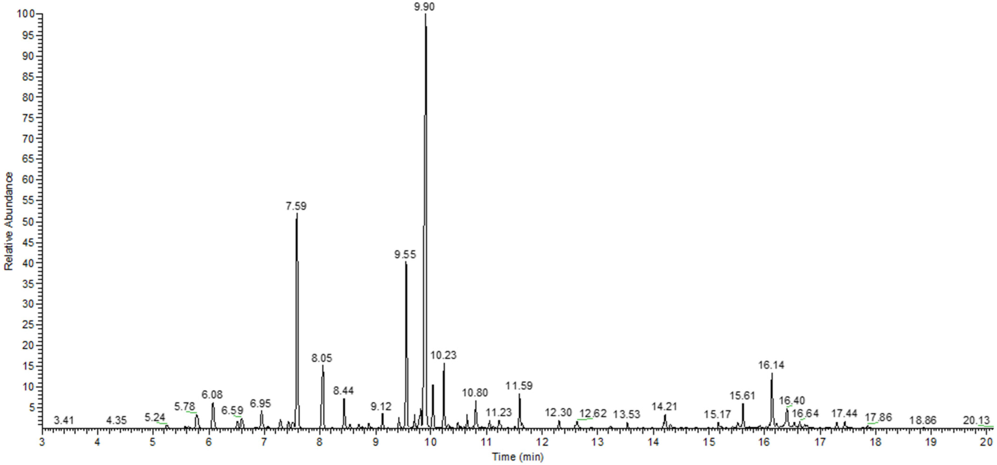
2.1. Physico-Chemical Characterization

The average content of the extracted A. setacea EO (10 determinations) was 0.41 ± 0.14%, expressed as mL EO/100 g plant. EOs had a blue color, with a density of 0.8576 ± 0.0609 g/mL.
A total of 52 compounds were identified through GC-MS in A. setacea EO, representing 97.43% of the total area. The main identified compounds were borneol (a bicycle monoterpene alcohol), eucalyptol (a bicycle monoterpene ether), and camphor (a bicycle monoterpene ketone), which have the largest relative areas 32.97%, 14.94%, and 10.13%, respectively. In addition, the EO is also rich in artemisia ketone (4.70%), an acyclic monoterpene ketone, α-terpineol (3.23%), a monoterpene alcohol, and γ-eudesmol (3.23%), a sesquiterpene alcohol.
The EO contains monoterpenes hydrocarbons (7.58%), sesquiterpenes hydrocarbons (1.19%), monoterpenes and sesquiterpenes alcohols (44.24% and 5.42%, respectively), and monoterpenes ketones (15.97%). Table 1 shows the relative content of volatile compounds from the A. setacea herba EO growing in Romania, expressed as a percentage of the total area. The gas chromatogram of A. setacea EO is shown in Figure 1.
The presence of chamazulene responsible for the blue color of A. setacea EO is confirmed only by some of the authors [6,7,23], a possible explanation being the low amounts of this compound produced only during the flowering period, and only by certain taxa, as reported by Karamenderes and Hethelyi [7,23]. In addition, the aerial parts of A. setacea could contain some (colorless) compounds that can be considered as proazulenes, such as 11,13-dehydrodeacetylmatricarin (14-deoxylactucin), rupicolin A, and rupicolin B [24], along with 13 other lactones sesquiterpenes identified by Todorova [25].
It is known that azulenogenic sesquiterpene lactones (naturally present in various species of Achillea) transform into azulenes during the EO extraction by hydrodistillation but not in case of supercritical extraction. Therefore, the extraction method applied to obtain the EOs could be another source of the compositional differences reported in the literature [26].
The presence of chamazulene in the composition of the EO influences not only the color but also the therapeutic effects, such as photodynamic antimicrobial activity, antiviral, anti-inflammatory, anti-acid effects, etc. [27].

2.2. Antioxidant Activity

The radical scavenger activity of the A. setacea EO was expressed as the amount of the antioxidants necessary to decrease the initial 2,2-diphenylpicrylhydrazyl (DPPH) absorbance by 50% (median effective concentration value, IC50), and results were compared with IC50 for butylated hydroxytoluene (BHT) (Table 2). The scavenging activity toward the DPPH radical of the EO of A. setacea (IC50 = 12.38 ± 0.63 mg/mL) was significantly higher (p < 0.0001) than the control, BHT (IC50 = 0.64 ± 0.07 mg/mL). The results are substantially different from those published by Rezaei et al. [9], probably due to the differences in the chemical composition, influenced by the pedoclimatic conditions. For this species, the antioxidant activity was determined through ferric reducing antioxidant power (FRAP) and cupric ion reducing antioxidant capacity (CUPRAC) assays for the first time and was significantly lower than that given by BHT (p < 0.0001). These methods aim to evaluate the capacity of the sample to reduce ferric or cupric ions in aqueous media. It can be observed that the antioxidant activity for essential oil in an acidic environment (FRAP) is higher than that obtained by CUPRAC, where the environment is neutral. In the case of BHT, the antioxidant activity is higher in neutral pH, suggesting that the antioxidant mechanism is different from that of A. setacea EO, probably due to the hydrophobic character [28].
Among the major components of the tested EO, borneol, camphor, and eucalyptol are known for their strong antioxidant activity [29,30,31,32]. Other compounds present in this EO that could contribute to the antioxidant effect are chamazulene [33], eudesmol [34], caryophyllene oxide [35], nerolidol [36], bornyl acetate [37], α-pinene [38], camphene [39], terpinen-4-ol [40], etc. According to Karakaya et al., caryophyllene oxide is correlated with a substantial cholinesterase inhibitory activity [35].

2.3. Antimicrobial Activity

The qualitative testing of microbial susceptibility to the A. setacea herba EO highlighted growth inhibition zones for the following microbial strains: Escherichia coli, Staphylococcus aureus, Salmonella typhimurium vs. enterica, Shigella flexneri, Bacillus cereus, Listeria ivanovii, Listeria innocua, Saccharomyces cerevisiae (SMR4), Candida glabrata, and strains isolated from seeds, Aspergillus niger, Rhizopus nigricans, Cladosporium cladosporioides, and Alternaria alternata (Table 3). In addition, the solvent (DMSO) in which the essential oil was solubilized showed no antimicrobial activity for any tested strains.
The qualitative results of the antimicrobial activity correlate with those obtained by Unlu et al. [7], the A. setacea EO being most active on B. cereus, S. aureus, and E. coli, with higher growth inhibition zones obtained in this study. According to Karamenderes et al. (2003) [6], the A. setacea EO also shows activity on S. thyphimurium, P. vulgaris, and C. albicans strains, but it is inactive on the S. aureus and E. coli strains. These significant differences could be explained by the differences in the composition of the A. setacea EO. To our knowledge, the antimicrobial activity of A. setacea EO against Shigella flexneri, Listeria ivanovii, L. innocua, Saccharomyces cerevisiae, C. glabrata, Aspergillus niger, Rhizopus nigricans, Cladosporium cladosporioides, and Alternaria alternata are reported for the first time in the literature.
The comparison between the MIC values of the stock solution and the solvent, respectively, revealed statistically significant results for the following strains: B cereus, L. ivanovii, E. coli, and C. glabrata with p < 0.001; S. aureus, L. inoccua, A. niger, A. alternata and C. cladosporioides (p < 0.01); and S. enterica and R. nigricans (p < 0.05). However, in the case of the S. cerevisiae strain, the difference is not statistically significant compared to the solvent control.
In Table 3, the results obtained for MMC are reported, highlighting that the MIC values are clearly lower than the MMCs. In the case of S. aureus and Listeria sp. the microbial growth was observed at concentrations of 3.75 μL/mL (S. aureus), 7.5 μL/mL (L. inoccua), and 0.94 μL/mL (L. ivanovii). The positive control specific to each strain and the solvent (DMSO) showed confluent colonies after 24 h of incubation in the case of all studied strains, as expected. The obtained MMCs for A. setacea EO indicate better activity on Gram-positive bacterial strains and yeasts.
Of particular importance among the components of essential oils with antimicrobial activity are the phenolic derivatives, represented by eugenol in the case of A. setacea EO, followed by aldehydes (not present in the studied extract), followed by terpene alcohols (present in large quantities, for example, borneol, terpinen-4-ol, α-terpineol, cis-piperitol), esters, and ketones, while monoterpene hydrocarbons used alone present a modest antimicrobial activity [41].
Although a mathematical prediction for complex matrices such as essential oils is difficult to achieve, the correlation data for EO of Thymus sp. showed that the synergism generated by the association of p-cymene with cis-geraniol (both present in the A. setacea EO) could be responsible for the antibacterial activity on Salmonella typhimurium and Shigella flexneri strains (thymol recorded negative correlations with bacterial strains) [42].
Among the compounds identified in the A. setacea EO, terpinen-4-ol and eucalyptol show remarkable antifungal activity against Fusarium subglutinans, F. cerealis, F. verticillioides, F. proliferatum, F. oxysporum, F. sporotrichioides, Aspergillus tubingensis, A. carbonarius, Alternaria alternata, and Penicillium sp. [43].
According to Kotan et al. [44], bornyl acetate and eucalyptol do not show antimicrobial activity, while terpinen-4-ol had a broad-spectrum antimicrobial activity against 35 strains including S. aureus, P. aeruginosa, E. coli, and S. typhimurium.
Borneol has been shown to exhibit antiadhesion effects by reducing bacterial attachment and biofilm formation [45]. Despite its low antimicrobial activity, borneol, in combination with conventional antibiotics or other antimicrobial compounds, can penetrate the bacterial membrane and impair the efflux pump activity [46], leading to a synergistic interaction and improving the antibacterial activity of other compounds found in low concentrations but with a strong antimicrobial effect, such as terpinen-4-ol [44], limonene [47], pinene [48], carvone [49], and eugenol [50].
Eugenol and its derivatives from the essential oil of Piper divaricatum are probably responsible for its antifungal action (obtained by a direct bioautography test after nebulization of fungal spores) on C. cladosporioides [51], similar to that of miconazole (MIC 0.5 μg/mL). The essential oils of Eugenia caryophyllus and Cinnamomum zeylanicum inhibited the A. alternata isolate, this activity correlating with the high percentages of eugenol, 90.5% and 80.7%, respectively [51,52]. Although present in a much lower percentage in the case of the essential oil studied in this article, eugenol certainly influences its antimicrobial activity.
Jadhav (2013) [53] showed that the A. millefolium essential oil has a bactericidal effect on L. monocytogenes and L. innocua planktonic cells. α-pinene, 1,8-cineole, terpinene-4-ol, and caryophyllene were the main components of this essential oil, the first two being attributed to the respective effect.
A hydroalcoholic extract (ethanol: water = 1:1, V:V) of A. millefolium L. inhibited the growth of A. niger and Penicillium hirsutum by 70.19 and 47.40%, respectively, while for the EO of the same plant, the inhibition ratio was greater than 85% for both strains (for 20 μL/mL of oil) [54], maybe due to the morphological changes induced by EO.
A recent study on EO isolated from two species of Artemisia (Asteraceae family) shows that sesquiterpenoids, such as bisabolol, are responsible for inhibiting the growth of A. niger, A. flavus, and Candida sp. [55].
Tanacetum chiliophyllum var. chiliophyllum, which belongs to one of the largest genera in the Asteraceae family, contains 1,8-cineole (16.1%), camphor (36.2%), borneol (2.8%), chamazulene (2.9%), and terpinene-4-ol (2.2%) in the essential oil extracted from stems. The antibacterial activity of this essential oil (with a composition close to A. setacea EO, at least in terms of the majority of compounds) isolated from plants collected from Van-Muradiye in Istanbul was remarkable because it has had the same MIC as chloramphenicol (62.5 μg/mL) for E. coli NRRL B-3008 [56].
Another species from the Asteraceae family, Dittrichia viscosa, contains an essential oil in which borneol and bornyl acetate represent over 50% and which had antimicrobial activity on E. coli, S. aureus, C. albicans, and S. cerevisiae (with MIC between 0.1 and 3.3 mg/mL) [57].

2.4. Correlation between Inhibition of Microbial Adherence and the Release of Extracellular NO

Regarding the influence on the adhesion capacity, a significant decrease in the absorbances measured at the wavelength of 490 nm was observed, the graphic representations being expressed as a function of the absorbance for each well specific to the concentrations used from the A. setacea essential oil stock solution in DMSO (1:1) in comparison with the used solvent, DMSO, and the microbial growth control.
The ability to adhere to the inert substrate was inhibited for all tested microbial strains at concentrations between MIC/2 and MIC/4 (μL/mL), the tested EO being more active on Listeria sp. (Table 4).
NO is used as a dispersal signal in many microbial species, modulating the tolerance to antimicrobial substances. An additional study demonstrated that nitrite-induced stress in S. aureus resulted in the impairment of both polysaccharide intracellular adhesion (PIA) synthesis and biofilm formation. Nitrite-induced stress led to the upregulation of genes associated with oxidative and nitrosative stress (including genes for DNA repair, iron homeostasis, and ROS detoxification) [58].
Oxidative and nitrosative stress is produced inside biofilms according to Miranda et al. [59], thus affecting their growth under different conditions and producing ROS and RNI, with a decrease in the extracellular matrix. These radicals could accumulate in the extracellular environment and thus affect the matrix.
Nitrosative stress involves the production and subsequent damage induced by RNIs, which include nitric oxide (NO), peroxynitrite (ONOO), nitric acid (HNO2), dinitrogen trioxide (N2O3), and others. RNIs are small, potentially highly reactive molecules that can be continuously produced in organisms as by-products of anaerobic respiratory metabolism. When the production of ROIs and/or RNIs overwhelms the cell’s ability to eliminate such molecules, they can generate damage to DNA, lipids, and proteins [60]. Due to their hydrophobic nature and lower density compared to water, the EO accumulates on the surface, leading to the occurrence of an anaerobic environment. The lack of oxygen induces cellular stress, and the amount of nitric oxide is increased. Although the NO synthesized endogenously by bacterial NOS inhibits aerobic respiration, the tricarboxylic acid cycle is still active, generating the NADH that could reduce the alternative electron acceptor nitrate, thus maintaining the membrane potential during microaerobiosis [61].
Table 5 shows a significant increase in the extracellular concentration of NO in most tested strains, except S. cerevisiae and A. alternata (p > 0.05) induced by the tested EO at MIC/2 concentration. The MIC/4 concentration does not significantly influence the NO release in the case of L. ivanovii, S. flexneri, E. coli, S. cerevisiae, and A. alternata strains. The highest extracellular concentration of NO was observed in the case of the L. inoccua strain (MIC/2: 4.63 ± 0.43 µM/mL, MIC/4: 5.49 ± 0.43 µM/mL), followed by C. glabrata (MIC/2: 3.47 ± 0.14 µM/mL). In the case of fungi, much higher concentrations of NO were observed compared to bacteria, probably due to the hypoxia induced by A. setacea EO. The denitrifying system coupled to the mitochondrial electron transport chain facilitates anaerobic respiration associated with ATP synthesis under hypoxic conditions. Nitrite reductases located in the intermembrane space of fungal mitochondria have been shown to reduce NO2 to NO in an NADP-dependent manner according to Arasimowicz-Jelonek and Floryszak-Wieczorek [62].
NO release has been significantly correlated with microbial adherence inhibition (p-value < 0.05), but the relationship depends on the microbial strains used, being much stronger for the C. cladosporioides and R. nigricans fungal strains (Table 6).

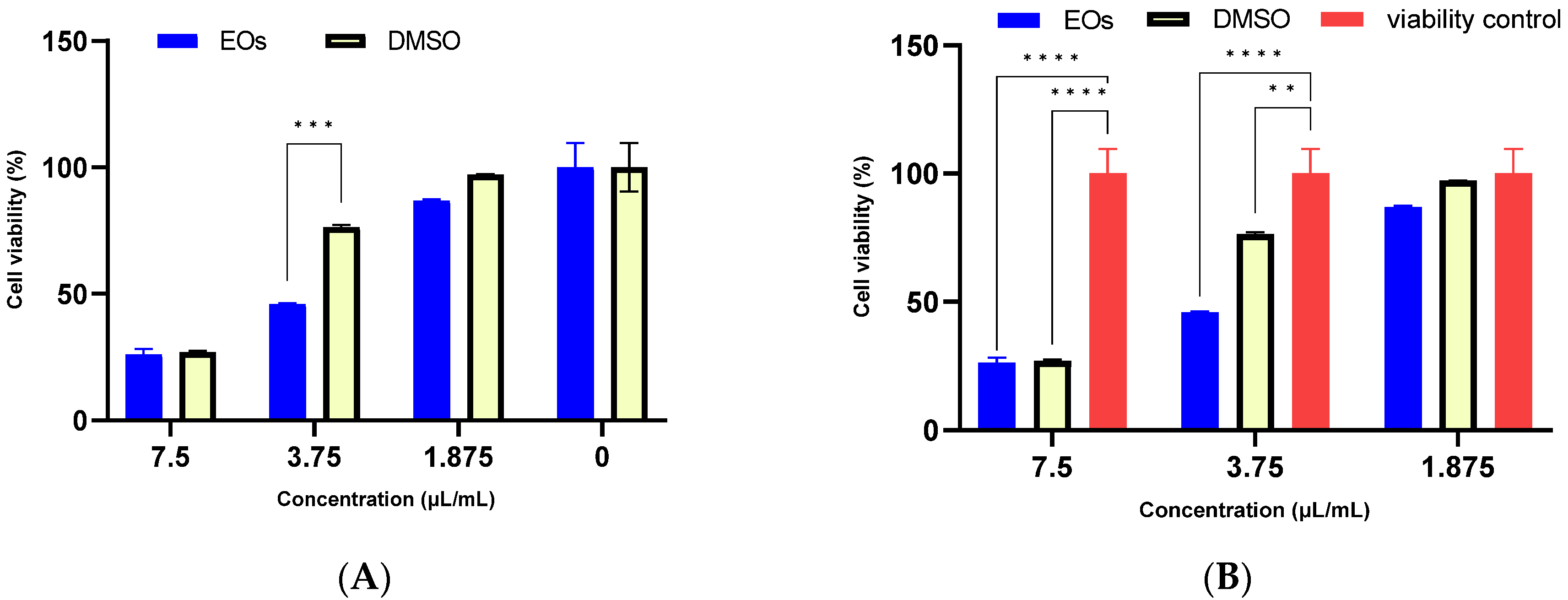
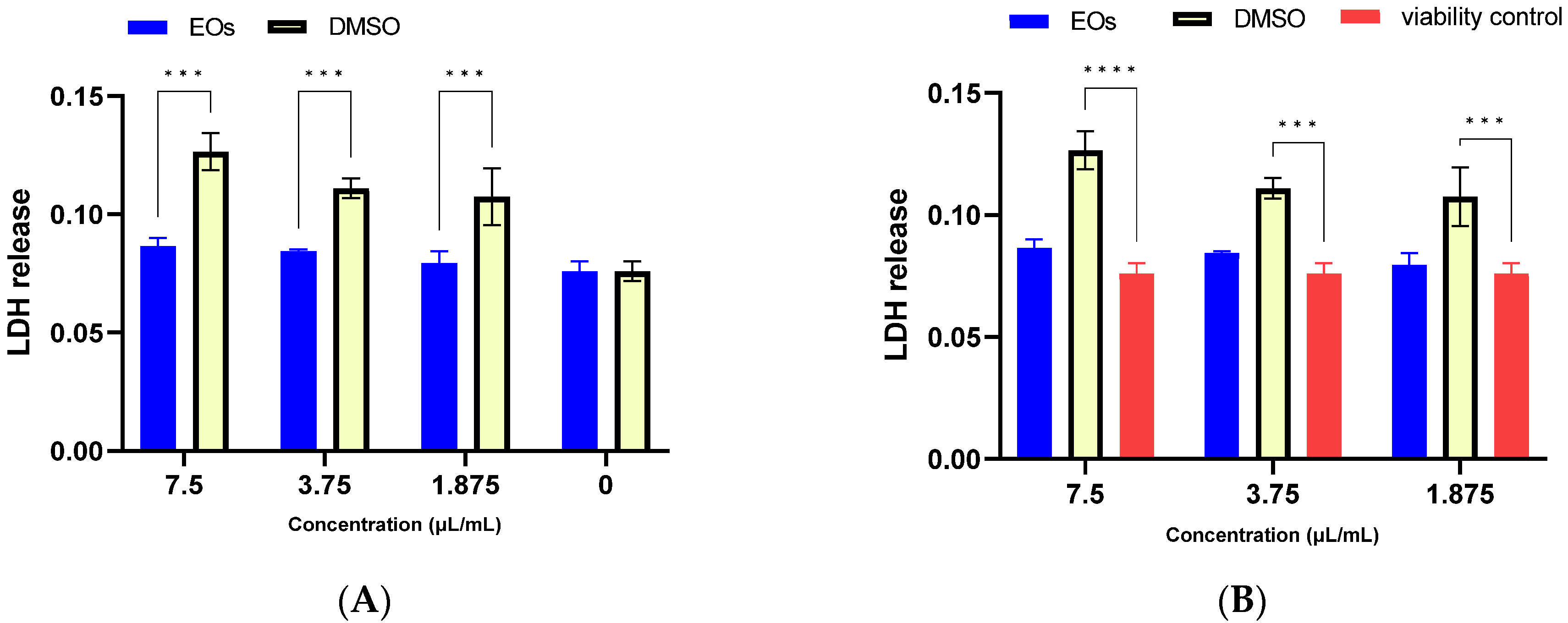
2.5. Biocompatibility of A. setacea Essential Oil

The obtained results of the MTT and LDH release assays in cultured mouse fibroblast L929 cells showed that the tested concentrations of A. setacea EO stock solution have a cytotoxic effect similar to the solvent used (DMSO) depending on the concentration used. Figure 2 shows the comparison between the solvent and the sample and the control of viable cells. Practically, the cytotoxicity of the EO stock solution is given by an additive effect of the mixture because the used solvent is highly cytotoxic. However, at the concentration of 1.875 µL/mL of essential oil, the cytotoxicity was considerably reduced. The IC50 for the essential oil stock solution was 4.68 ± 0.08 µL/mL, while for DMSO it was 5.72 ± 0.04 µL/mL.
From Figure 3, the solvent used increases the release of LDH, while the essential oil solubilized in a similar solvent concentration significantly reduced the release of LDH, which leads to the conclusion that the oil protects the cell from the aggressive action of the solvent. The differences between the essential oil stock solution and the cell viability control are not significant (p > 0.05).
Therefore, it can be concluded that the cytotoxic effect is given by the used solvent and not by the A. setacea essential oil. Moreover, due to its density being lower than water, the essential oil tends to remain on the surface and thus changes the microenvironment of eukaryotic cells.
By calculating the selectivity index (SI), the MIC obtained values indicate higher toxicity on microorganisms compared to mammalian cells (Table 7). The antifungal effect is much more pronounced than the antimicrobial one in the case of B. cereus, L. inoccua, L. ivanovii, C. glabrata, S. cerevisiae, R. nigricans, A. alternata, and C. cladosporioides strains, while the used solvent is more toxic to mammalian cells.

3. Materials and Methods

3.1. Plant Material

The plants were collected from Bucharest, Dămăroia area, near Poligrafiei Boulevard, from a field in the vicinity of the railway in July 2020 (in the blossom period). An herbarium voucher with the number [BUC 410047] was deposited in the herbarium of the “Dimitrie Brandza” Botanical Garden of the University of Bucharest (BUC).

3.2. Essential Oil Isolation

Ninety grams of A. setacea leaves were hydrodistilled in a Clevenger-type apparatus for 4 h. The essential oil was dried over anhydrous Na2SO4, stored in a dark glass bottle, and kept at 4 °C until analysis [63].

3.3. Gas Chromatography-Mass Spectrometry Analysis

The GC-MS analysis was carried out with a Thermo Electron system (Focus GC chromatograph coupled with a Polaris Q ion trap mass detector) controlled with Xcalibur® software. DB-5MS column (25 m × 0.25 mm; 0.25 μm film thickness) was used with helium as the carrier gas (1 mL/min). Both headspace (500 μL headspace gas) and liquid (1 μL injection from 1:10 hexane dilution) samples were analyzed under the same chromatographic conditions. The GC oven temperature program was kept at 60 °C for 3 min and programmed to 200 °C at a rate of 10 °C/min and after that at 12 °C/min to the final temperature of 240 °C (2 min). The injector temperature was set at 250 °C. Mass spectra were recorded at 70 eV. Mass range was from m/z 35 to 450.
Identification of the volatile components was carried out by comparison of their relative retention index to series of n-alkanes (C8-C20 in hexane, Sigma Aldrich Co., St. Louis, MO, USA) and the literature [64].

3.4. Antioxidant Activity

3.4.1. DPPH

The method of Ahmed et al. [65], with slight modification, was used to assay the DPPH radical scavenging activity. The stable free radical DPPH was dissolved in methanol to a final concentration of 300 μmol/L solution; 25 μL of the essential oil in methanol (or methanol itself as blank control) was added to 175 μL of the methanol DPPH solution. Different concentrations were tested (100, 50, 25, 12.5, and 6.25 mg/mL for oils in methanol). After further mixing, the decrease in absorbance was measured at 515 nm after 20 min. The antioxidant activity of each test sample was expressed as an IC50 value, i.e., the concentration in mg/mL that inhibits DPPH absorption by 50%, and it was calculated from the concentration–effect linear regression curve. 2,6-Ditert-butyl-4-methylphenol (IUPAC) or butylated hydroxytoluene (BHT) was used as a positive control. The DPPH radical scavenging activity was calculated as the percentage inhibition.
% Inhibition of DPPH radical activity = [(A0 − A1)/A0] × 100%
where A0 is the absorbance of the DPPH with the solvent used; A1 is the absorbance of the sample or the positive control (BHT).

3.4.2. FRAP

The determination of the antioxidant capacity of iron reduction was performed by the FRAP assay method [66]. The stock solutions included 300 mM acetate buffer, pH 3.6, 10 mM 2,4,6-tri(2-pyridinyl(-1,3,5-triazine (TPTZ) solution in 40 mM HCl, and 20 mM FeCl3 6H2O solution in a volume ratio of 10:1:1, which were warmed to 37 °C before use. A 150 µL aliquot of FRAP reagent was mixed with 25 µL of methanolic essential oil solution (100 mg/mL). After incubation, the absorbance was read at 593 nm. A 1 mM Trolox stock solution was used to plot the calibration curve, the concentration ranging between 30 and 250 µM Trolox/mL (R2 = 0.9989). The results were expressed in mM Trolox equivalent/mL (TE) of extract. BHT was used as a positive control.

3.4.3. CUPRAC

Copper ion reduction was performed as previously described [67]: 120 µL of the sample (1 mg/mL in methanol)/standard solutions of different concentrations were mixed with 100 µL CuCl2 (10 mM), 100 µL neocuproine (7.5 mM), and 100 µL ammonium acetate buffer 1 M, pH = 7.00. After 30 min, the absorbance was measured at 450 nm. The stock Trolox solutions required for the calibration curve were 2 mM, and the working concentrations were between 0.24 and 2.0 mM Trolox/mL (R2 = 0.9980). The results were expressed in mM Trolox equivalent/mL (TE) extract. BHT was used as the positive control.

3.5. Antimicrobial Activity

3.5.1. Microbial Strain

The antimicrobial activity was tested on Gram-positive (Staphylococcus aureus ATCC 25923, Bacillus cereus ATCC 11778, Listeria inoccua ATCC 33096, Listeria ivanovii ATCC 19110), and Gram-negative (Escherichia coli ATCC 25922, Salmonella typhimurium vs. enterica ATCC 14028, Shigella flexneri ATCC 12022) bacterial as well as yeast (Candida glabrata ATCC MYA-2950, Saccharomyces cerevisiae SMR4) reference strains.
Four species of fungi, Aspergillus niger, Alternaria alternata, Rhizopus nigricans, and Cladosporium cladosporioides strains, were obtained from the Microbial Strain Collection of Faculty of Biology, University of Bucharest, Romania, and confirmed by MALDI-TOF.

3.5.2. Qualitative Assay of Antimicrobial Activity

Qualitative assay was performed using microbial suspensions adjusted to 1.5 × 10⁸ CFU/mL according to 0.5 McFarland from 18–24 h cultures. The inoculation in the cloth was carried out on solid media (ALOA for Listeria sp and Muller Hilton for the rest of the bacteria, Sabouraud for yeasts, and Dichloran Glycerol Agar (DG18) for fungi). Sterile discs were distributed, on which 5 µL of stock solution was impregnated. The plates were incubated for 24 h at a temperature of 37 °C for bacteria and 72 h at 28 °C for fungi. The zone of inhibition (mm) at the spot level was measured.

3.5.3. Quantitative Assay of Antibacterial Activity

Quantitative antimicrobial activity was carried out by the method of serial binary microdilutions in liquid medium (Demi-Fraser for Listeria sp., and TSB (Trypton Soy Broth) for the rest of the bacteria, Sabouraud for yeast) in 96-well plates using a negative control (DMSO). The dilutions of the samples belong to the range of 0.94–30 µL/mL for each strain tested. Simultaneously, serial dilutions were performed with the samples without stems in order to observe the absorbance specific to the sample. Each well was inoculated with 15 µL of 0.5 McFarland microbial suspension. After 20–24 h of incubation at 37 °C, the MIC value was established both macroscopically, as the last concentration at which microbial growth was not observed with respect to the appearance of turbidity of the environment, and spectrophotometrically. The absorbance of the microbial cultures was measured at 620 nm using a FlexStation 3 UV-Vis spectrophotometer (Molecular Devices, GA, USA). The interpretation of the results was achieved by subtracting the absorbances specific to the concentration of each non-inoculated dilution from the inoculated sample.
To determine the minimum microbicidal concentrations (MMC), 10 µL was spotted from the wells where no microbial growth was observed in plates with specific solid media, Aloa for Listeria sp., Muller Hilton for the rest of the bacterial strains, and Sabouraud for yeasts.

3.5.4. Quantitative Assay of Antifungal Activity

The fungicidal activity was achieved by the method of binary serial microdilutions in a liquid medium, RPMI 1640 (5.2 g of powder in 500 mL of distilled water, at a pH greater than 7) and inoculated with 0.1 mL of 1 McFarland fungal suspension (made in a solution of 0.01% Tween 80). Essential oil stock solution concentrations ranged from 0.01 to 12.5 µL/mL. Positive control (represented by the untreated strain), solvent control (DMSO), and negative control (sterility control) were used. The tubes were incubated for 3 days at a temperature of 28 °C. On the third day after incubation, the absorbance was read at 550 nm, and they were sown in the spot on the DG18 medium (medium specific for products with a water activity below 0.95) by pipetting 10 μL of the suspension. Finally, the plates were incubated for 3–4 days at a temperature of 28 °C. The minimum fungicidal concentration was represented by the last concentration at which no fungal growth was observed.

3.5.5. Evaluation of NO Release

Nitric oxide (NO) is rapidly converted to nitrite in aqueous solutions, and therefore total nitrite can be used as an indicator of NO concentration. This was measured using a spectrophotometric analysis of total nitrite performed using the Griess reagent according to the methodology described by Quinteros et al. (2016) with some modifications. To the microbial supernatant (MIC/2 and MIC/4) obtained after 24 h incubation for bacteria and 72 h for fungi (50 μL), 50 μL of 2% sulfanilamide in 5% (v/v) H3PO4 and 50 μL of 0.1% N-(1-naphthyl)-ethylenediamine aqueous solution were added. The formation of the azo dye was measured after 30 min at λ = 540 nm. For the quantification of nitric oxide, a calibration curve was produced with NaNO2 in the range of 0–100 μM (R2 = 0.9993).

3.5.6. Microbial Adhesion Capacity to the Inert Substrate

The influence on the adhesion capacity to the inert substrate was quantified after performing the protocol for the quantitative analysis of the antimicrobial effect and establishing the MIC. Using the microtitration method, the biomass adhered to the wells of the microplates was evaluated after washing with AFS, fixation with cold methanol (10 min), and staining with crystal violet (concentration 0.1%, staining time 15 min). The absorbance of the biological material adhered and resuspended in acetic acid (CH3COOH 33%, 15 min) was determined by reading the absorbance at 490 nm.

3.6. Biocompatibility of Essential Oil

Mouse fibroblast L929 cells were cultured in Gibco Dulbecco’s Modified Eagle Medium (DMEM) supplemented with 10% fetal bovine serum and Penicillin-Streptomycin (100 U/mL) and Amphotericin B (25 µg/mL). A. setacea essential oil and DMSO were serially diluted in liquid media. The concentrations tested were 7.5, 3.75, and 1.875 µL/mL.

3.6.1. LDH

Cytotoxicity was assessed using the LDH Released Detection Kit (Roche). LDH activity was measured spectrophotometrically at λ = 490 nm with a wavelength reference λ = 600 nm.

3.6.2. MTT

The MTT assay was used to assess cell viability and proliferation in the presence of A. setacea essential oil stock solution and DMSO. The Vybrant® MTT Cell Proliferation Assay Kit (cat no. V-13154) was used for the determination according to the manufacturer’s instructions.

3.6.3. Selectivity Index

The IC50 (cytotoxicity) values were calculated as the concentration of essential oil, resulting in a 50% reduction in absorbance compared to untreated cells [68]. The SI was defined as IC50 of essential oil against eukaryotic cell line/MIC of essential oil for every microbial strain. SI > 1 means that the extract is more toxic to bacteria/fungi than to mammalian cells [69]. Selective activities of the essential oil were calculated as follows:
SI   % = IC 50   µ L / mL MIC   µ L / mL

3.7. Statistical Analysis

Data obtained in triplicate/duplicate were expressed as means ± SD. Statistical analysis was performed using GraphPad Prism v9. Data were analyzed using the unpaired t-test for antioxidant activity. The correction of multiple comparisons was carried out by the Holm–Sidak method, the comparison being sample vs. solvent control or sample vs. cell viability control. The correlation between microbial adherence and the ability of microorganisms to release extracellular NO was achieved through the Pearson correlation. The cytotoxicity assays were analyzed by ordinary two-way Anova using a two-stage linear step-up procedure of Benjamini, Krieger, and Yekutieli, with a single pooled variance method. The level of statistical significance was set at p < 0.05.

4. Conclusions

The Achillea setacea essential oil proved to be rich in borneol, eucalyptol, and camphor compounds, well known for their antimicrobial and antioxidant activity. The obtained EO has shown a strong antifungal effect and inhibited microbial adhesion, especially in the case of Gram-positive bacteria (with the S. aureus strain exception) and C. glabrata strains. The NO release favors the inhibition of microbial adhesion to the inert substrate in a strain-dependent manner. The selectivity index highlighted that the maximum concentration of EO that does not influence the mammalian cells is 3.75 µL/mL. Therefore, this EO can be used as an antibacterial and antifungal agent for food product decontamination, and in topical formulations for fungal skin infections.

Author Contributions

Conceptualization, I.C.M., E.O. and M.C.C.; methodology, I.C.M., E.O., G.G.P., M.B., I.A.B. and M.C.C.; validation, M.C.C.; formal analysis, I.C.M.; investigation, I.C.M., E.O., D.M.G., G.G.P., M.B., I.A.B. and E.N.; resources, M.C.C.; data curation, E.N.; writing—original draft preparation, I.C.M. and E.O.; writing—review and editing, I.C.M., E.O. and M.C.C.; visualization, M.C.C.; supervision, M.C.C. All authors have read and agreed to the published version of the manuscript.

Funding

This research received no external funding.

Institutional Review Board Statement

Not applicable.

Informed Consent Statement

Not applicable.

Data Availability Statement

Not applicable.

Acknowledgments

The authors thank O.I.M., research project ICUB 2150/01.02.2022. I.C.M., G.G.P. and M.C.C. acknowledge the support of the grant of the Romanian National Authority for Scientific Research, CNCS—UEFISCDI, project PN-III-P2-2.1-PED-2021-2193 (594PED/2022).

Conflicts of Interest

The authors declare no conflict of interest.

References

  1. Si, X.-T.; Zhang, M.-L.; Shi, Q.-W.; Kiyota, H. Chemical Constituents of the Plants in the GenusAchillea. Chem. Biodivers. 2006, 3, 1163–1180. [Google Scholar] [CrossRef] [PubMed]
  2. Marchart, E.; Kopp, B. Capillary Electrophoretic Separation and Quantification of Flavone-O- and C-Glycosides in Achillea setacea W. et K. J. Chromatogr. B 2003, 792, 363–368. [Google Scholar] [CrossRef] [PubMed]
  3. Eisenman, S.W.; Zaurov, D.E.; Struwe, L. Medicinal Plants of Central Asia: Uzbekistan and Kyrgyzstan; Eisenman, S.W., Zaurov, D.E., Struwe, L., Eds.; Springer: New York, NY, USA, 2013; ISBN 978-1-4614-3911-0. [Google Scholar]
  4. Mohammadhosseini, M.; Sarker, S.D.; Akbarzadeh, A. Chemical Composition of the Essential Oils and Extracts of Achillea Species and Their Biological Activities: A Review. J. Ethnopharmacol. 2017, 199, 257–315. [Google Scholar] [CrossRef]
  5. Başer, K.H.C.; Hüsnü, K.; Başer, C. Essential Oils of Achillea Species of Turkey. Volatiles Essent. Oils 2016, 3, 1–14. [Google Scholar]
  6. Karamenderes, C.; Karabay, N.Ü.; Zeybek, U. Türkiye’nin Farkli Lokalitelerinden Toplanan Achillea setacea Waldst. Kit. Uçucu Yağinin Bileşimi ve Antimikrobiyal Aktivitesi. Ankara Univ. Eczac. Fak. Derg. 2003, 32, 113–120. [Google Scholar] [CrossRef]
  7. Ünlü, M.; Daferera, D.; Dönmez, E.; Polissiou, M.; Tepe, B.; Sökmen, A. Compositions and the in Vitro Antimicrobial Activities of the Essential Oils of Achillea setacea and Achillea Teretifolia (Compositae). J. Ethnopharmacol. 2002, 83, 117–121. [Google Scholar] [CrossRef] [PubMed]
  8. Turkmenoglu, F.P.; Agar, O.T.; Akaydin, G.; Hayran, M.; Demirci, B. Characterization of Volatile Compounds of Eleven Achillea Species from Turkey and Biological Activities of Essential Oil and Methanol Extract of A. Hamzaoglui Arabaci & Budak. Molecules 2015, 20, 11432–11458. [Google Scholar] [CrossRef]
  9. Rezaei, F.; Jamei, R.; Heidari, R.; Maleki, R. Chemical Composition and Antioxidant Activity of Oil from Wild Achillea setacea and A. Vermicularis. Int. J. Food Prop. 2017, 20, 1522–1531. [Google Scholar] [CrossRef]
  10. Yener, I.; Yilmaz, M.A.; Olmez, O.T.; Akdeniz, M.; Tekin, F.; Hasimi, N.; Alkan, M.H.; Ozturk, M.; Ertas, A. A Detailed Biological and Chemical Investigation of Sixteen Achillea Species’ Essential Oils via Chemometric Approach. Chem. Biodivers. 2020, 17, e1900484. [Google Scholar] [CrossRef]
  11. Buleandra, M.; Moldovan, Z.; Badea, I.A.; David, I.G.; Popa, D.E.; Oprea, E.; Caglar, T.A.T.; Basaga, S.H. Comparative Assessment of the Volatile Profile, Antioxidant Capacity and Cytotoxic Potential of Different Preparation of Millefolli Herba Samples. Rev. Chim. 2020, 71, 69–78. [Google Scholar] [CrossRef]
  12. Sánchez, P.; Vélez-del-Burgo, A.; Suñén, E.; Martínez, J.; Postigo, I. Fungal Allergen and Mold Allergy Diagnosis: Role and Relevance of Alternaria Alternata Alt a 1 Protein Family. J. Fungi 2022, 8, 277. [Google Scholar] [CrossRef] [PubMed]
  13. Alen Coutinho, I.; Lopes, M.; Lima, F.; Ventura, C.; Rabadão, E.; Alfaro, T.; da Cunha, J.S.; Regateiro, F.S. Concomitant Allergic Bronchopulmonary Aspergillosis and Eosinophilic Granulomatosis with Polyangiitis after Aspergillus Niger Infection. Pulmonology 2022, 28, 231–234. [Google Scholar] [CrossRef]
  14. Sandoval-Denis, M.; Gené, J.; Sutton, D.A.; Wiederhold, N.P.; Cano-Lira, J.F.; Guarro, J. New Species of Cladosporium Associated with Human and Animal Infections. Persoonia-Mol. Phylogeny Evol. Fungi 2016, 36, 281–298. [Google Scholar] [CrossRef]
  15. Lianou, D.T.; Skoulakis, A.; Michael, C.K.; Katsarou, E.I.; Chatzopoulos, D.C.; Solomakos, N.; Tsilipounidaki, K.; Florou, Z.; Cripps, P.J.; Katsafadou, A.I.; et al. Isolation of Listeria Ivanovii from Bulk-Tank Milk of Sheep and Goat Farms—From Clinical Work to Bioinformatics Studies: Prevalence, Association with Milk Quality, Antibiotic Susceptibility, Predictors, Whole Genome Sequence and Phylogenetic Relationships. Biology 2022, 11, 871. [Google Scholar] [CrossRef] [PubMed]
  16. Matto, C.; D’Alessandro, B.; Mota, M.I.; Braga, V.; Buschiazzo, A.; Gianneechini, E.; Varela, G.; Rivero, R. Listeria Innocua Isolated from Diseased Ruminants Harbour Minor Virulence Genes of L. Monocytogenes. Vet. Med. Sci. 2022, 8, 735–740. [Google Scholar] [CrossRef]
  17. Nisa, I.; Qasim, M.; Yasin, N.; Ullah, R.; Ali, A. Shigella Flexneri: An Emerging Pathogen. Folia Microbiol. 2020, 65, 275–291. [Google Scholar] [CrossRef] [PubMed]
  18. Pérez-Torrado, R.; Querol, A. Opportunistic Strains of Saccharomyces Cerevisiae: A Potential Risk Sold in Food Products. Front. Microbiol. 2016, 6, 01522. [Google Scholar] [CrossRef]
  19. Kumar, K.; Askari, F.; Sahu, M.; Kaur, R. Candida Glabrata: A Lot More Than Meets the Eye. Microorganisms 2019, 7, 39. [Google Scholar] [CrossRef]
  20. Prodan, I. Achillea. In Flora României; Săvulescu, T., Ed.; Editura Academiei Române: Bucureşti, Romania, 1964; Volume 9, p. 390. [Google Scholar]
  21. Ciocârlan, V. Flora Ilustrată a României: Pteridophyta et Spermatophyta; Ceres: Bucureşti, Romania, 2009; p. 798. ISBN 978-973-40-0817-9. [Google Scholar]
  22. Sârbu, I.; Ștefan, N.; Oprea, A. Plante Vasculare Din România–Determinator Ilustrat de Teren; Victor B Victor: Bucuresti, Romania, 2013; p. 802. ISBN 9786068149080. [Google Scholar]
  23. Héthelyi, É.; Dános, B.; Tétényi, P.; Héthelyi; Dános, B.; Tétényi, P. Phytochemical Studies on the Essential Oils of Species Belonging to TheAchillea Genus by Gas Chromatography/Mass Spectrometry. Biol. Mass Spectrom. 1989, 18, 629–636. [Google Scholar] [CrossRef]
  24. Zitterl-Eglseer, K.; Jurenitsch, J.; Korhammer, S.; Haslinger, E.; Sosa, S.; Loggia, R.; Kubelka, W.; Franz, C. Entzündungshemmende Sesquiterpenlactone von Achillea setacea. Planta Med. 1991, 57, 444–446. [Google Scholar] [CrossRef]
  25. Todorova, M.; Vogler, B.; Tsankova, E. Terpenoids from Achillea setacea. Z. Naturforsch. C 2000, 55, 840–842. [Google Scholar] [CrossRef]
  26. Lemberkovics, É.; Kéry, Á.; Kakasy, A.; Szoke, É.; Simándi, B. Effect of Extraction Methods on the Composition of Essential Oils. Acta Hortic. 2004, 597, 49–56. [Google Scholar] [CrossRef]
  27. Muhammad, S.; Abdul Khalil, H.P.S.; Abd Hamid, S.; Danish, M.; Marwan, M.; Yunardi, Y.; Abdullah, C.K.; Faisal, M.; Yahya, E.B. Characterization of Bioactive Compounds from Patchouli Extracted via Supercritical Carbon Dioxide (SC-CO2) Extraction. Molecules 2022, 27, 6025. [Google Scholar] [CrossRef]
  28. Apak, R.; Özyürek, M.; Güçlü, K.; Çapanoğlu, E. Antioxidant Activity/Capacity Measurement. 1. Classification, Physicochemical Principles, Mechanisms, and Electron Transfer (ET)-Based Assays. J. Agric. Food Chem. 2016, 64, 997–1027. [Google Scholar] [CrossRef] [PubMed]
  29. Kordali, S.; Cakir, A.; Mavi, A.; Kilic, H.; Yildirim, A. Screening of Chemical Composition and Antifungal and Antioxidant Activities of the Essential Oils from Three Turkish Artemisia Species. J. Agric. Food Chem. 2005, 53, 1408–1416. [Google Scholar] [CrossRef] [PubMed]
  30. Chrysargyris, A.; Mikallou, M.; Petropoulos, S.; Tzortzakis, N. Profiling of Essential Oils Components and Polyphenols for Their Antioxidant Activity of Medicinal and Aromatic Plants Grown in Different Environmental Conditions. Agronomy 2020, 10, 727. [Google Scholar] [CrossRef]
  31. Xu, G.; Guo, J.; Sun, C. Eucalyptol Ameliorates Early Brain Injury after Subarachnoid Haemorrhage via Antioxidant and Anti-Inflammatory Effects in a Rat Model. Pharm. Biol. 2021, 59, 112–118. [Google Scholar] [CrossRef]
  32. Drikvandi, P.; Bahramikia, S.; Alirezaei, M. Modulation of the Antioxidant Defense System in Liver, Kidney, and Pancreas Tissues of Alloxan-induced Diabetic Rats by Camphor. J. Food Biochem. 2020, 44, e13527. [Google Scholar] [CrossRef] [PubMed]
  33. Capuzzo, A.; Occhipinti, A.; Maffei, M.E. Antioxidant and Radical Scavenging Activities of Chamazulene. Nat. Prod. Res. 2014, 28, 2321–2323. [Google Scholar] [CrossRef]
  34. Aati, H.; El-Gamal, A.; Kayser, O. Chemical Composition and Biological Activity of the Essential Oil from the Root of Jatropha Pelargoniifolia Courb. Native to Saudi Arabia. Saudi Pharm. J. 2019, 27, 88–95. [Google Scholar] [CrossRef]
  35. Karakaya, S.; Yilmaz, S.V.; Özdemir, Ö.; Koca, M.; Pınar, N.M.; Demirci, B.; Yıldırım, K.; Sytar, O.; Turkez, H.; Baser, K.H.C. A Caryophyllene Oxide and Other Potential Anticholinesterase and Anticancer Agent in Salvia verticillata Subsp. Amasiaca (Freyn & Bornm.) Bornm. (Lamiaceae). J. Essent. Oil Res. 2020, 32, 512–525. [Google Scholar] [CrossRef]
  36. Nogueira Neto, J.D.; de Almeida, A.A.C.; da Silva Oliveira, J.; dos Santos, P.S.; de Sousa, D.P.; de Freitas, R.M. Antioxidant Effects of Nerolidol in Mice Hippocampus After Open Field Test. Neurochem. Res. 2013, 38, 1861–1870. [Google Scholar] [CrossRef] [PubMed]
  37. Kim, S.H.; Lee, S.Y.; Hong, C.Y.; Gwak, K.S.; Park, M.J.; Smith, D.; Choi, I.G. Whitening and Antioxidant Activities of Bornyl Acetate and Nezukol Fractionated from Cryptomeria Japonica Essential Oil. Int. J. Cosmet. Sci. 2013, 35, 484–490. [Google Scholar] [CrossRef] [PubMed]
  38. Aydin, E.; Türkez, H.; Geyikoğlu, F. Antioxidative, Anticancer and Genotoxic Properties of α-Pinene on N2a Neuroblastoma Cells. Biologia 2013, 68, 1004–1009. [Google Scholar] [CrossRef]
  39. Quintans-Júnior, L.; Moreira, J.C.F.; Pasquali, M.A.B.; Rabie, S.M.S.; Pires, A.S.; Schröder, R.; Rabelo, T.K.; Santos, J.P.A.; Lima, P.S.S.; Cavalcanti, S.C.H.; et al. Antinociceptive Activity and Redox Profile of the Monoterpenes (+)-Camphene, p -Cymene, and Geranyl Acetate in Experimental Models. ISRN Toxicol. 2013, 2013, 459530. [Google Scholar] [CrossRef] [PubMed]
  40. Lee, K.-G.; Shibamoto, T. Antioxidant Activities of Volatile Components Isolated FromEucalyptus Species. J. Sci. Food Agric. 2001, 81, 1573–1579. [Google Scholar] [CrossRef]
  41. Yammine, J.; Chihib, N.-E.; Gharsallaoui, A.; Dumas, E.; Ismail, A.; Karam, L. Essential Oils and Their Active Components Applied as: Free, Encapsulated and in Hurdle Technology to Fight Microbial Contaminations. A Review. Heliyon 2022, 8, e12472. [Google Scholar] [CrossRef]
  42. Beicu, R.; Alexa, E.; Obiștioiu, D.; Cocan, I.; Imbrea, F.; Pop, G.; Circioban, D.; Moisa, C.; Lupitu, A.; Copolovici, L.; et al. Antimicrobial Potential and Phytochemical Profile of Wild and Cultivated Populations of Thyme (Thymus Sp.) Growing in Western Romania. Plants 2021, 10, 1833. [Google Scholar] [CrossRef]
  43. Morcia, C.; Malnati, M.; Terzi, V. In Vitro Antifungal Activity of Terpinen-4-Ol, Eugenol, Carvone, 1,8-Cineole (Eucalyptol) and Thymol against Mycotoxigenic Plant Pathogens. Food Addit. Contam. Part A 2011, 29, 415–422. [Google Scholar] [CrossRef]
  44. Kotan, R.; Kordali, S.; Cakir, A. Screening of Antibacterial Activities of Twenty-One Oxygenated Monoterpenes. Z. Naturforsch. C 2007, 62, 507–513. [Google Scholar] [CrossRef]
  45. Luo, L.; Li, G.; Luan, D.; Yuan, Q.; Wei, Y.; Wang, X. Antibacterial Adhesion of Borneol-Based Polymer via Surface Chiral Stereochemistry. ACS Appl. Mater. Interfaces 2014, 6, 19371–19377. [Google Scholar] [CrossRef] [PubMed]
  46. Fadli, M.; Bolla, J.-M.; Mezrioui, N.-E.; Pagès, J.-M.; Hassani, L. First Evidence of Antibacterial and Synergistic Effects of Thymus Riatarum Essential Oil with Conventional Antibiotics. Ind. Crop. Prod. 2014, 61, 370–376. [Google Scholar] [CrossRef]
  47. Haiyan, L.; Chongxin, X.; Xiao, Z.; Ying, L.; Xianjin, L. Antibacterial Effect of Limonene on Food-Borne Pathogens. J. Zhejiang Univ. (Agric. Life Sci.) 2016, 42, 306–312. [Google Scholar]
  48. Leite, A.M.; de Lima, E.O.; de Souza, E.L.; de Diniz, M.F.F.M.; Trajano, V.N.; deMedeiros, I.A. Inhibitory Effect of Beta-Pinene, Alpha-Pinene and Eugenol on the Growth of Potential Infectious Endocarditis Causing Gram-Positive Bacteria. Rev. Bras. Ciênc. Farm. 2007, 43, 121–126. [Google Scholar] [CrossRef]
  49. Moro, I.J.; Gondo, G.D.G.A.; Pierri, E.G.; Pietro, R.C.L.R.; Soares, C.P.; de Sousa, D.P.; dos Santos, A.G. Evaluation of Antimicrobial, Cytotoxic and Chemopreventive Activities of Carvone and Its Derivatives. Braz. J. Pharm. Sci. 2018, 53, e00076. [Google Scholar] [CrossRef]
  50. Catherine, A.A.; Deepika, H.; Negi, P.S. Antibacterial Activity of Eugenol and Peppermint Oil in Model Food Systems. J. Essent. Oil Res. 2012, 24, 481–486. [Google Scholar] [CrossRef]
  51. da Silva, J.K.R.; Andrade, E.H.A.; Guimarães, E.F.; Maia, J.G.S. Essential Oil Composition, Antioxidant Capacity and Antifungal Activity of Piper Divaricatum. Nat. Prod. Commun. 2010, 5, 477–480. [Google Scholar] [CrossRef] [PubMed]
  52. Castro, J.C.; Endo, E.H.; de Souza, M.R.; Zanqueta, E.B.; Polonio, J.C.; Pamphile, J.A.; Ueda-Nakamura, T.; Nakamura, C.V.; Dias Filho, B.P.; de Abreu Filho, B.A. Bioactivity of Essential Oils in the Control of Alternaria Alternata in Dragon Fruit (Hylocereus Undatus Haw.). Ind. Crop. Prod. 2017, 97, 101–109. [Google Scholar] [CrossRef]
  53. Jadhav, S.; Shah, R.; Bhave, M.; Palombo, E.A. Inhibitory Activity of Yarrow Essential Oil on Listeria Planktonic Cells and Biofilms. Food Control. 2013, 29, 125–130. [Google Scholar] [CrossRef]
  54. Fierascu, I.; Ungureanu, C.; Avramescu, S.M.; Fierascu, R.C.; Ortan, A.; Soare, L.C.; Paunescu, A. In Vitro Antioxidant and Antifungal Properties of Achillea Millefolium L. Rom. Biotechnol. Lett. 2015, 20, 10626–10636. [Google Scholar]
  55. Khan, F.A.; Khan, N.M.; Ahmad, S.; Nasruddin; Aziz, R.; Ullah, I.; Almehmadi, M.; Allahyani, M.; Alsaiari, A.A.; Aljuaid, A. Phytochemical Profiling, Antioxidant, Antimicrobial and Cholinesterase Inhibitory Effects of Essential Oils Isolated from the Leaves of Artemisia Scoparia and Artemisia Absinthium. Pharmaceuticals 2022, 15, 1221. [Google Scholar] [CrossRef]
  56. Polatoğlu, K.; Demirci, B.; Demirci, F.; Gören, N.; Başer, K.H.C. Biological Activity and Essential Oil Composition of Two New Tanacetum Chiliophyllum (Fisch. & Mey.) Schultz Bip. Var. Chiliophyllum Chemotypes from Turkey. Ind. Crop. Prod. 2012, 39, 97–105. [Google Scholar] [CrossRef]
  57. Mssillou, I.; Agour, A.; Allali, A.; Saghrouchni, H.; Bourhia, M.; El Moussaoui, A.; Salamatullah, A.M.; Alzahrani, A.; Aboul-Soud, M.A.M.; Giesy, J.P.; et al. Antioxidant, Antimicrobial, and Insecticidal Properties of a Chemically Characterized Essential Oil from the Leaves of Dittrichia Viscosa L. Molecules 2022, 27, 2282. [Google Scholar] [CrossRef]
  58. Gilmore, B.F.; Flynn, P.B.; O’Brien, S.; Hickok, N.; Freeman, T.; Bourke, P. Cold Plasmas for Biofilm Control: Opportunities and Challenges. Trends Biotechnol. 2018, 36, 627–638. [Google Scholar] [CrossRef] [PubMed]
  59. Arce Miranda, J.E.; Sotomayor, C.E.; Albesa, I.; Paraje, M.G. Oxidative and Nitrosative Stress in Staphylococcus Aureus Biofilm. FEMS Microbiol. Lett. 2011, 315, 23–29. [Google Scholar] [CrossRef]
  60. Barraud, N.; Hassett, D.J.; Hwang, S.-H.; Rice, S.A.; Kjelleberg, S.; Webb, J.S. Involvement of Nitric Oxide in Biofilm Dispersal of Pseudomonas Aeruginosa. J. Bacteriol. 2006, 188, 7344–7353. [Google Scholar] [CrossRef] [PubMed]
  61. Fang, F.C.; Vázquez-Torres, A. Reactive Nitrogen Species in Host–Bacterial Interactions. Curr. Opin. Immunol. 2019, 60, 96–102. [Google Scholar] [CrossRef] [PubMed]
  62. Arasimowicz-Jelonek, M.; Floryszak-Wieczorek, J. Nitric Oxide in the Offensive Strategy of Fungal and Oomycete Plant Pathogens. Front. Plant Sci. 2016, 7, 252. [Google Scholar] [CrossRef]
  63. Mayrs, E.B.C. British Pharmacopoeia Appendix 9; Medicines & Healthcare products Regulatory Agency: London, UK, 2017; Volume IV. [Google Scholar]
  64. Adams, R.P. Identification of Essential Oil Components by Gas Chromatography/Mass Spectrometry; Allured Publishing Corporation: Carol Stream, IL, USA, 2007; ISBN 9781932633214. [Google Scholar]
  65. Ahmed, A.F.; Attia, F.A.K.; Liu, Z.; Li, C.; Wei, J.; Kang, W. Antioxidant Activity and Total Phenolic Content of Essential Oils and Extracts of Sweet Basil (Ocimum basilicum L.) Plants. Food Sci. Hum. Wellness 2019, 8, 299–305. [Google Scholar] [CrossRef]
  66. Thaipong, K.; Boonprakob, U.; Crosby, K.; Cisneros-Zevallos, L.; Hawkins Byrne, D. Comparison of ABTS, DPPH, FRAP, and ORAC Assays for Estimating Antioxidant Activity from Guava Fruit Extracts. J. Food Compos. Anal. 2006, 19, 669–675. [Google Scholar] [CrossRef]
  67. Olszowy, M.; Dawidowicz, A.L. Essential Oils as Antioxidants: Their Evaluation by DPPH, ABTS, FRAP, CUPRAC, and β-Carotene Bleaching Methods. Mon. Für Chem.-Chem. Mon. 2016, 147, 2083–2091. [Google Scholar] [CrossRef]
  68. Bagla, V.P.; McGaw, L.J.; Elgorashi, E.E.; Eloff, J.N. Antimicrobial Activity, Toxicity and Selectivity Index of Two Biflavonoids and a Flavone Isolated from Podocarpus Henkelii (Podocarpaceae) Leaves. BMC Complement. Altern. Med. 2014, 14, 383. [Google Scholar] [CrossRef] [PubMed]
  69. Asong, J.A.; Amoo, S.O.; McGaw, L.J.; Nkadimeng, S.M.; Aremu, A.O.; Otang-Mbeng, W. Antimicrobial Activity, Antioxidant Potential, Cytotoxicity and Phytochemical Profiling of Four Plants Locally Used against Skin Diseases. Plants 2019, 8, 350. [Google Scholar] [CrossRef] [PubMed] [Green Version]
Figure 1. The gas chromatogram of A. setacea essential oil.
Antibiotics 12 00371 g001
Figure 2. Cytotoxicity assessment of A. setacea essential oil by MTT test under standard cultivation conditions. Comparison between the stock solution of essential oil: DMSO (1:1) and DMSO (A), and the cell viability control (B). The tests were performed in duplicate, and the differences were considered significant for p < 0.05 (** p < 0.01, *** p < 0.001, **** p < 0.0001).
Antibiotics 12 00371 g002
Figure 3. Cytotoxicity assessment of A. setacea essential oil by LDH release test under standard cultivation conditions. Comparison between the stock solution of essential oil: DMSO (1:1) and DMSO (A), and the cell viability control (B). The tests were performed in duplicate, and the differences were considered significant for p < 0.05 (*** p < 0.001, **** p < 0.0001).
Antibiotics 12 00371 g003
Table 1. The chemical composition of A. setacea EO resulted from gas chromatography coupled with mass spectrometry.
No.Compound NameCompound ClassesRI [l]Relative Area (%)
1Santolina trieneMH [a]9150.35
2TricycleneMH [a]9280.15
3α-ThujeneMH [a]9320.17
4α-PineneMH [a]9401.40
5CampheneMH [a]9562.45
6Sabinene (β-Thujene)MH [a]9830.58
7β-PineneMH [a]9850.89
82,3-Dehydro-1,8-cineoleME [c]9980.19
9Yomogi alcoholMA [b]10061.31
10α-PhellandreneMH [a]10130.17
11α -TerpineneMH [a]10250.69
12p-CymeneMH [a]10330.49
13LimoneneMH [a]10380.46
14Eucalyptol (1,8-Cineole)ME [c]104214.94
15Artemisia ketoneMK [e]10674.70
16cis-Sabinene hydrateMA [b]10780.07
17Artemisia alcoholMA [b]10941.73
18α-TerpinoleneMH [b]10970.28
19trans-Sabinene hydrateMA [b]11110.09
20α-ThujoneMK [e]11170.30
21cis-p-Menth-2-en-1-olMA [b]11320.85
22trans-p-Menth-2-en-1-olMA [b]11510.71
23CamphorMK [e]116010.13
24cis-ChrysanthenolMA [c]11690.76
25LavandulolMA [b]11770.45
26BorneolMA [b]118132.97
27Terpinen-4-olMA [b]11912.36
28α-TerpineolMA [b]12043.23
29cis-PiperitolMA [b]12200.15
30cis-CarveolMA [b]12320.64
31trans-Crysanthenyl acetateMAE [d]12421.62
32CarvoneMK [e]12590.38
33GeraniolMA [b]12640.14
34PiperitoneMK [e]12710.46
35cis-Chrysanthenyl acetateMAE [d]12730.24
36Bornyl acetateMAE [d]12961.49
37trans-Carvyl acetateMAE [d]13480.43
38EugenolPM [f]13720.29
39cis-JasmoneCK [g]14170.16
40β-caryophylleneSH [h]14410.36
41α-HumuleneSH [h]14760.10
42γ-MuuroleneSH [h]14940.25
43Germacrene DSH [h]15030.26
44Viridiflorene (Ledene)SH [h]15090.11
45δ-CadineneSH [h]15420.12
46trans-β-NerolidolSA [i]15750.28
47SpathulenolSA [i]16040.41
48Caryophyllene oxideSE [j]16121.33
49γ-EudesmolSA [i]16593.23
50α-EudesmolSA [i]16821.46
51α-BisabololSA [i]17040.31
52ChamazuleneAH [k]17560.35
Total 97.43
Monoterpenes hydrocarbons 8.07
Monoterpenes alcohols and esters 49.23
Monoterpenes ketones 15.97
Sesquiterpenes hydrocarbons 1.19
Sesquiterpenes alcohols and ethers 6.75
[a] MH, monoterpene hydrocarbon; [b] MA, monoterpene alcohol; [c] ME, monoterpene ether; [d] MAE, monoterpene alcohol ester; [e] MK, monoterpene ketone; [f] PH, phenolic monoterpene; [g] CK, cyclic ketone; [h] SH, sesquiterpene hydrocarbon; [i] SA, sesquiterpene alcohol; [j] SE, sesquiterpene ether; [k] AH, aromatic hydrocarbon; [l] RI, Kovats Index, the retention index relative to C8-C20 n-alkanes (DB-5MS column).
Table 2. The antioxidant properties of the A. setacea essential oil.
DPPH (IC50, mg/mL)FRAP (µM TE/mg)CUPRAC (µM TE/mg)
A. setacea essential oil12.38 ± 0.6314.70 ± 0.996.17 ± 0.68
BHT0.64 ± 0.07823.46 ± 14.69988.03 ± 11.35
p-value<0.0001<0.0001<0.0001
TE, Trolox equivalent; DPPH, 2,2-diphenylpicrylhydrazyl; FRAP, ferric reducing antioxidant power; CUPRAC, cupric ion reducing antioxidant capacity; BHT, butylated hydroxytoluene.
Table 3. The minimum inhibitory (MIC) and microbicidal (MMC) concentrations for the essential oils stock solution and DMSO on the microbial strains involved in the contamination of food products and the etiology of food poisoning.
IDZ *
(mm)
MIC (µL/mL)MMC (µL/mL)
A. setacea EODMSOA. setacea EODMSOp-ValueA. setacea EODMSO
MIC **MV% #MV% #
S. aureus13.00 ± 1.00-7.501.62 ± 0.2855.45 ± 2.530.0011167.50>30.00
B. cereus20.00 ± 2.00-3.750.71 ± 0.2982.57 ± 2.760.0005743.75>30.00
L. inoccuaNA ##-3.753.57 ± 0.3380.46 ± 3.950.00132615.00>30.00
L. ivanoviiNA-1.880.59 ± 0.42102.04 ± 2.280.0002611.88>30.00
E. coli8.33 ± 0.58-7.5023.97 ± 0.7688.59 ± 2.530.0008357.50>30.00
S. flexneris8.00 ± 2.00-30.00>3012.55 ± 8.46-30.00>30.00
S. enterica6.33 ± 0.58-15.0036.75 ± 2.0349.23 ± 1.450.01939930.00>30.00
C. glabrata10.00 ± 1.00-3.754.40 ± 0.9380.57 ± 1.870.0003763.75>30.00
S. cerevisiae13.33 ± 0.58-1.886.19 ± 0.3446.00 ± 7.740.3508353.75>30.00
A. niger8.00 ± 1.00-0.787.32 ± 3.4582.93 ± 0.000.00103912.50>12.50
R. nigricans6.33 ± 0.58-1.5632.89 ± 1.8668.42 ± 7.440.022511>12.50>12.50
A. alternata7.66 ± 0.58-1.560.00 ± 0.40114.61 ± 7.150.001946>12.50>12.50
C. cladosporioides9.33 ± 0.16-3.1257.69 ± 4.3570.77 ± 1.090.002517>12.50>12.50
* IDZ—inhibition diameter zone; ** MIC—minimum inhibition concentration; # MV—microbial viability; ## NA—Not Active.
Table 4. Microbial adherence (%) in the case of MIC/2 and MIC/4 for the essential oil stock solution and DMSO on the microbial strains involved in the contamination of food products.
EODMSOp-ValueEODMSOp-Value
MIC/2MA (%) *MA (%) *MIC/4MA (%) *MA (%) *
S. aureus3.7534.38 ± 12.6460.84 ± 2.300.1004321.8878.57 ± 11.7865.06 ± 2.270.252256
B. cereus1.8814.19 ± 1.7668.73 ± 3.710.0028220.9412.60 ± 2.4339.93 ± 3.140.010389
L. inoccua1.881.87 ± 0.6454.57 ± 0.830.0001980.941.92 ± 0.7058.70 ± 2.330.000917
L. ivanovii0.947.08 ± 0.2478.80 ± 2.160.000459<0.94---
E. coli3.75101.76 ± 2.15134.88 ± 14.750.0880981.88104.18 ± 5.69150.39 ± 21.550.099298
S. enterica7.5013.02 ± 3.6625.99 ± 2.020.0482183.7511.52 ± 0.2727.20 ± 3.730.027284
C. glabrata1.880.18 ± 0.2570.22 ± 2.720.0007600.9422.64 ± 0.97127.84 ± 2.020.000227
C. cladosporioides1.5632.81 ± 4.42105.88 ± 13.870.6872800.7840.63 ± 2.2186.27 ± 11.090.029350
R. nigricans0.7823.64 ± 1.2958.18 ± 1.290.0013920.3925.45 ± 0.0049.09 ± 2.570.005858
A. niger0.3987.5 ± 13.26229.69 ± 15.470.0101110.20120.21 ± 2.21223.42 ± 12.310.007262
A. alternata0.78118.39 ± 13.00133.33 ± 17.880.4400600.39145.98 ± 16.26112.64 ± 1.630.102062
* MA—microbial adherence.
Table 5. The concentrations of NO released in the extracellular environment for the MIC/2 and MIC/4 values in the presence of EO stock solution and DMSO on the microbial strains involved in the contamination of food products.
EODMSOp-ValueEODMSOp-Value
MIC/2ENOC *ENOC *MIC/4ENOC *ENOC *
S. aureus3.751.82 ± 0.140.39 ± 0.14<0.0000011.880.18 ± 0.141.41 ± 0.14<0.00001
L. inoccua1.884.63 ± 0.433.55 ± 0.58<0.00010.945.49 ± 0.433.25 ± 0.14<0.000001
L. ivanovii0.942.14 ± 0.14NA **<0.0000010.470.92 ± 0.140.51 ± 0.14>0.05
S. flexneri15.00NA0.82 ± 0.14<0.0017.500.82 ± 0.140.41 ± 0.14>0.05
E. coli3.752.43 ± 0.141.20 ± 0.43<0.000011.881.10 ± 0.291.00 ± 0.14>0.05
S. enterica7.501.10 ± 0.000.59 ± 0.14<0.053.751.41 ± 0.140.69 ± 0.00<0.01
C. glabrata1.883.47 ± 0.140.41 ± 0.14<0.0000010.940.41 ± 0.140.10 ± 0.00<0.001
S. cerevisiae0.940.41 ± 0.00NA>0.050.470.20 ± 0.000.31 ± 0.14>0.05
C. cladosporioides1.562.68 ± 0.071.92 ± 0.14<0.010.782.68 ± 0.072.07 ± 0.21<0.01
R. nigricans0.782.73 ± 0.071.97 ± 0.14<0.010.392.78 ± 0.142.07 ± 0.29<0.05
A. niger0.393.64 ± 0.142.32 ± 0.14<0.0010.202.07 ± 0.070.61 ± 0.14<0.0001
A. alternata0.782.89 ± 0.642.02 ± 0.64>0.050.391.87 ± 0.362.12 ± 0.07>0.05
* ENOC—extracellular nitric oxide concentration (µM/mL); ** NA—Not Active.
Table 6. The correlation coefficients (Pearson correlation) between NO release and inhibition of microbial adherence capacity.
StrainsR2p-ValueSignificance
(α = 0.05)
S. aureus0.62460.0228Yes
L. inoccua0.87900.0437Yes
L. ivanovii0.60360.0578No
E. coli0.38250.2079No
S. enterica0.90220.0152Yes
C. glabrata0.49700.1183No
C. cladosporioides0.98610.0041Yes
R. nigricans0.97720.0060Yes
A. niger0.50440.0677No
A. alternata0.35580.0561No
Table 7. Selectivity index values of A. setacea essential oil and DMSO against microbial strains.
SI (IC50/MIC)
A. setacea EODMSO
MICSIInterpretationMICSIInterpretation
S. aureus7.500.624*MTEC>60.00-*MTEC
B. cereus3.751.248**MTMC30.000.191*MTEC
L. inoccua3.751.248**MTMC>60.00-*MTEC
L. ivanovii1.882.489**MTMC60.000.095*MTEC
E. coli7.500.624*MTEC>60.00-*MTEC
S. flexneris30.000.156*MTEC30.000.191*MTEC
S. enterica15.000.312*MTEC60.000.095*MTEC
C. glabrata3.751.248**MTMC30.000.191*MTEC
S. cerevisiae1.882.489**MTMC15.000.381*MTEC
A. niger0.786.000**MTMC6.250.915*MTEC
R. nigricans1.563.000**MTMC12.500.458*MTEC
A. alternata1.563.000**MTMC>25.00-*MTEC
C. cladosporioides3.1251.498**MTMC6.250.915*MTEC
*MTEC—more toxic to eukaryotic cell; **MTMC—more toxic to microbial cell.
Disclaimer/Publisher’s Note: The statements, opinions and data contained in all publications are solely those of the individual author(s) and contributor(s) and not of MDPI and/or the editor(s). MDPI and/or the editor(s) disclaim responsibility for any injury to people or property resulting from any ideas, methods, instructions or products referred to in the content.

