Review

Nanomaterials and Coatings for Managing Antibiotic-Resistant Biofilms

Grup de Biotecnologia Molecular i Industrial, Department of Chemical Engineering, Universitat Politècnica de Catalunya, Rambla Sant Nebridi 22, 08222 Terrassa, Spain
*
Author to whom correspondence should be addressed.
Antibiotics 2023, 12(2), 310; https://doi.org/10.3390/antibiotics12020310
Submission received: 15 January 2023 / Revised: 30 January 2023 / Accepted: 31 January 2023 / Published: 2 February 2023
(This article belongs to the Special Issue Feature Reviews on Antibiofilm Strategies)

Abstract

:
Biofilms are a global health concern responsible for 65 to 80% of the total number of acute and persistent nosocomial infections, which lead to prolonged hospitalization and a huge economic burden to the healthcare systems. Biofilms are organized assemblages of surface-bound cells, which are enclosed in a self-produced extracellular polymer matrix (EPM) of polysaccharides, nucleic acids, lipids, and proteins. The EPM holds the pathogens together and provides a functional environment, enabling adhesion to living and non-living surfaces, mechanical stability, next to enhanced tolerance to host immune responses and conventional antibiotics compared to free-floating cells. Furthermore, the close proximity of cells in biofilms facilitates the horizontal transfer of genes, which is responsible for the development of antibiotic resistance. Given the growing number and impact of resistant bacteria, there is an urgent need to design novel strategies in order to outsmart bacterial evolutionary mechanisms. Antibiotic-free approaches that attenuate virulence through interruption of quorum sensing, prevent adhesion via EPM degradation, or kill pathogens by novel mechanisms that are less likely to cause resistance have gained considerable attention in the war against biofilm infections. Thereby, nanoformulation offers significant advantages due to the enhanced antibacterial efficacy and better penetration into the biofilm compared to bulk therapeutics of the same composition. This review highlights the latest developments in the field of nanoformulated quorum-quenching actives, antiadhesives, and bactericides, and their use as colloid suspensions and coatings on medical devices to reduce the incidence of biofilm-related infections.

1. Introduction

Biofilms are structured and coordinated communities of microbial cells on a surface. They are formed by initial attachment of bacterial cells, followed by proliferation and enclosure in a self-produced extracellular polymeric matrix (EPM), which is composed mainly of water (97%), exopolysaccharides, proteins, and nucleic acids [1]. The EPM formation is triggered by an intercellular communication process, known as quorum sensing (QS) [2]. During this process, bacteria secrete signaling molecules, acyl homoserine lactones (AHLs), or autoinducer peptides (AIPs) in the case of Gram-negative and Gram-positive bacteria, respectively, generally termed autoinducers (AIs) [3]. When the bacterial population reaches a certain density, which in turn leads to a certain threshold level of AIs, genes for biofilm formation and production of virulence factors are expressed [4].
Bacterial biofilms are closely related with antimicrobial resistance (AMR), which is a global concern for increased morbidity and mortality, prolonged hospitalization, and additional financial costs. A high number of bacterial species are already resistant to most of the existing antibiotics due to acquired molecular mechanisms allowing the expulsion, inactivation, or destruction of antimicrobials [5,6]. Thereby, the biofilm mode of growth is even more challenging. The formation of a densely packed EPM makes the cells 100 to 1000 times less susceptible to antibacterials than their free-floating counterparts and bacteria encased in biofilms exhibit higher tolerance to the host immune response [7,8,9]. The EPM holds the pathogens together, protects them, and reduces the diffusion of drug molecules, while the intimate biofilm environment offers excellent conditions for horizontal gene transfer and further resistance development [10,11]. Consequently, several mechanisms, such as limited drug penetration, decelerated bacterial growth, and expression of specific protective factors are recognized for the pronounced resistance of biofilms [12]. The formation of biofilms is responsible for numerous acute and chronic diseases such as cystic fibrosis, otitis, endocarditis, chronic wounds, periodontitis, and dental caries and their treatment frequently involves intensive antibiotic treatment and a combination of various antibiotics at high dosages, further aggravating the AMR issue [13,14,15]. The Gram-negative Escherichia coli, Klebsiella pneumoniae, Pseudomonas aeruginosa, and Proteus mirabilis, and the Gram-positive Enterococcus faecalis, Staphylococcus aureus, Staphylococcus epidermidis, as well as the yeast Candida albicans, are among the most common biofilm-forming species, found in up to 60% of severe and persistent hospital-acquired infections [16]. In addition, the frequent usage of indwelling devices leads to even more nosocomial infections [17]. Furthermore, biofilms have a detrimental impact on a wide range of industrial sectors such as food packaging, water filtration systems, marine equipment, and industrial bioreactors [18,19,20,21,22].
Given the growing impact of AMR, there is an urgent need to develop novel antibacterial strategies in order to outsmart the bacterial evolutionary mechanisms. Effective strategies for prevention and treatment of bacterial infections with a lower risk of AMR should integrate: (i) bacterial eradication without creating selective pressure [23], (ii) prevention and elimination of biofilm formation [24,25], (iii) biocompatibility, protection of the beneficial strains and host environment [26], (iv) long-term stability [27].
Recently, nanotechnology-based approaches have gained immense attention because the nano form endows the actives with stability and enhanced antimicrobial efficacy compared to the same material in bulk form [28,29]. Nanomaterials and nanoformulation have been employed to give a new life to obsolete antibiotics, specifically, to deliver different actives to the site of infection or act as a structural and/or functional element in medically relevant materials and coatings. Nanoparticles (NPs) have been deposited on medically relevant surfaces, medical devices, and implants by techniques like layer-by-layer, sonochemistry, and spin coating to engineer durable nanostructured coatings against biofilm formation [30,31,32]. Apart from eradicating planktonic cells and preventing biofilm formation, nano-sized actives are also highly potent in the elimination of already established drug-resistant biofilms. Thus, NPs and small vesicles have been used as delivery vehicles of biofilm dispersants (e.g., matrix-degrading enzymes, NO-donors) or direct bactericidals (e.g., antibiotics, antimicrobial peptides) [33,34,35,36]. In this review, we attempt to highlight the latest advances in the engineering of functional nano-enabled materials and coatings to combat bacterial biofilms and the spread of resistance. The use of nano-formulated enzymes, natural compounds, and their synthetic mimics able to scramble the wires of bacterial communication before it starts and consequently, to attenuate bacterial virulence and biofilm formation is discussed. The design of innovative nano-actives targeting inhibition of the initial bacterial settlement and destabilization of the protective EPM is also outlined. Finally, recent attempts in the nanoformulation of other non-antibiotic actives with broad-spectrum antibacterial activity, increased safety, and low-resistance-inducing mechanisms of action are described as prominent alternatives for managing biofilm-related infections (Table 1).

2. Anti-Virulence Approaches for Managing Biofilm Infections

Targeted interruption of the cell-to-cell communication with quorum-quenching enzymes (QQE), which degrade bacterial messengers, or by chemical QS inhibitors (QSIs), inactivating specific receptors, is an innovative and effective way to tackle the challenges of bacterial infections. Disturbing the QS lowers the expression of genes for synthesis of virulence factors and adhesive components of the EPM without affecting the bacterial viability, thus exerting low selective pressure for mutation (Figure 1). However, the practical use of the anti-QS compounds is frequently limited due to their potential toxicity, low stability and reduced therapeutic efficacy compared to antibiotics. In order to address these limitations, significant efforts have been devoted to nanoformulation.

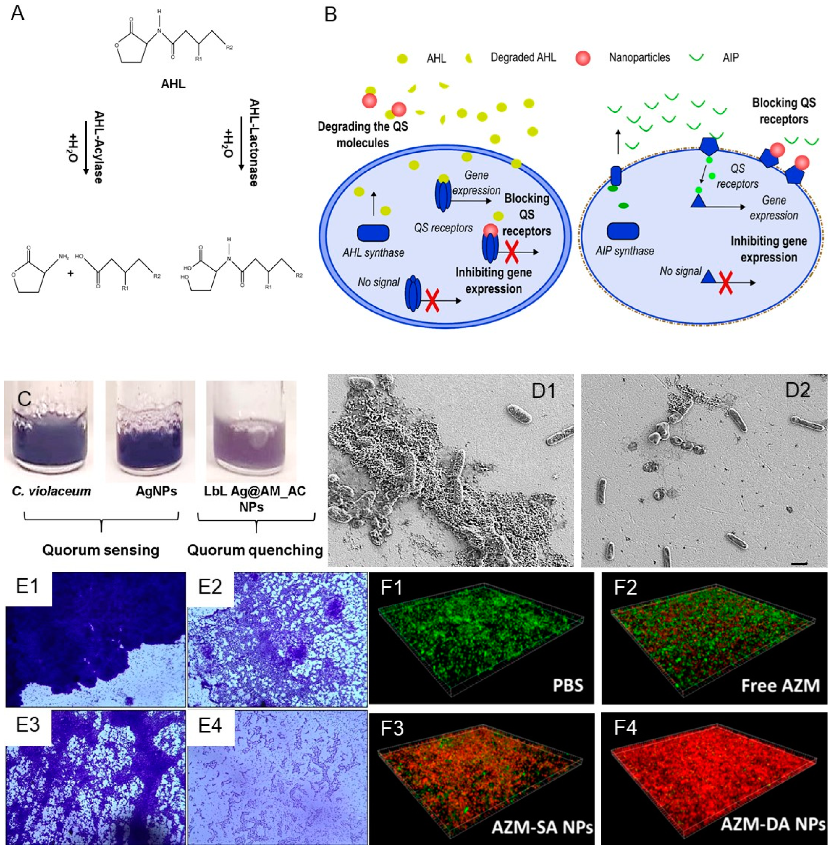
2.1. Nano-Formulated Quorum-Sensing Inhibitors

Some of the most widely used molecules that inhibit or obstruct the QS machinery are cinnamaldehyde, quercetin, azithromycin, or eugenol (Table 1). Cinnamaldehyde, an aromatic compound present in cinnamon, is able to disrupt the autoinducer-2 system of S. aureus and the LuxR-mediated transcription from the PluxI promoter in Gram-negative bacteria [37]. This compound has been incorporated in gold NPs using a silica coating and encapsulated inside chitosan NPs, which improved its effectivity compared to the bulk form [38,39]. The plant flavonoid quercetin has also been encapsulated in chitosan NPs and combined with silver NPs [40,41,42]. Azithromycin is an antibiotic that has been reported to inhibit the synthesis of AHLs at sub-inhibitory concentrations and it has been introduced in hyaluronic acid-poly(lactic-co-glycolic acid) nanovesicles in order to deliver the drug inside the biofilm [43,44]. Similar results were obtained by Gao et al. after combining this active with cationic polymers—the resulting NPs were able to penetrate the biofilm and eradicate mature P. aeruginosa biofilm in vitro and in vivo in a lung infection model (Figure 1F) [45]. Finally, eugenol is a catechol, obtained from clove, cinnamon leaf, pimento, bay, sassafras, massot bark oils, that has been reported to reduce the biofilm metabolic activity due to QS disruption in Gram-negative strains, inducing biofilm detachment [46]. Different types of eugenol nanoemulsions have been successfully generated and showed a capacity to inhibit the production of AHLs and biofilm formation in P. aeruginosa, protect surfaces when applied as hydrogel coatings, and improve the treatment outcomes of wound infection when loaded in a dressing with silver [47,48,49,50,51].

2.2. Quorum-Quenching Enzyme NPs

Two hydrolytic enzymes, which degrade AHLs—acylase and lactonase—have been widely exploited to inhibit QS-regulated virulence and biofilm growth in Gram-negative bacteria [52] (Figure 1A and Table 1). In our group, we employed acylase in combination with silver NPs, which were coated with aminocellulose and enzyme in a layer-by-layer fashion, adding a membrane-disturbing functionality. The obtained hybrid NPs reduced the QS indicator violacein in Cromobacterium violaceum and inhibited the planktonic growth and biofilm formation of P. aeruginosa at lower NP concentrations, non-toxic to human cells (Figure 1C) [33]. In another work, we nanoformulated acylase with the conventional antibiotic gentamicin in order to boost the bactericidal activity of the latter, while conferring biofilm inhibition activity on P. aeruginosa (Figure 1D) [28]. Other researchers have combined acylase with graphene oxide and mesoporous silica NPs for antifouling coatings of membranes [53,54]. In parallel, lactonase that catalyzes the breaking of ester bonds in the lactone ring has been nanohybridized with gold and silver to inhibit the biofilm production of Proteus species and K. pneumonia, respectively [55,56,57]. The extracellular disturbing of QS is among the most promising anti-QS approaches because it avoids the need to penetrate the cells or reach their receptors. However, despite the great potential of enzymes, their activity in vivo might be compromised; hence, further studies are needed to validate their antibiofilm activity in relevant environments.

2.3. Metal NPs as QS Inhibitors

Metal (e.g., silver, copper) and metal oxide (e.g., zinc oxide and copper oxide) NPs are gaining attention due to their strong antibacterial efficacy and lower probability for inducing AMR. Recent evidence has demonstrated that apart from direct killing, metal NPs may also interfere with bacterial community behavior and act as QS inhibitors (Table 1). For instance, silver NPs with an average size of 20–40 nm at concentrations of 10–25 µg/mL reduced the biofilm formation and inhibited the virulence of P. aeruginosa [58]. Gold NPs, produced using Capsicum annuum as reducing and active agent, impeded the biofilm development of P. aeruginosa and Serratia marcescens. The authors speculated that inhibition of the synthesis of QS signals and blocking of regulatory proteins is a possible QQ mechanism (Figure 1E) [59]. Nickel oxide NPs have presented hydrolase-like activity by degrading AHLs, which inhibited the violacein production in C. violaceum and biofilm formation in P. aeruginosa [60]. Copper NPs, coated with polyacrylic acid, were able to downregulate the expression of ppyR, which is related with the regulation of Psl operon that promotes bacterial adhesion of P. aeruginosa [61]. In another work, the anti-QS potential of selenium and tellurium NPs was also demonstrated. An up to 80% decrease in the QS-regulated violacein expression and a significant reduction of P. aeruginosa biofilm were obtained for both NPs types [62]. Zinc oxide NPs and, to a lesser degree, titanium oxide NPs, also displayed an ability to interfere with the AHL system of C. violaceum [63].

2.4. Mimicks of QS Signals and Inhibitors

Apart of the aforementioned direct anti-QS strategies, it is worth mentioning the design of more advanced approaches, exploiting the sequestration, camouflaging, and mimicking of QS machinery (Table 1). For example, Lu et al. downregulated the production of virulence factors and impeded biofilm production of Vibrio cholera with NPs, delivering the bacterium autoinducer CAI-1 at high concentrations [64]. In another approach, computationally designed polymers were used to sequester QS molecules of Gram-negative bacteria and consequently alter the QS-controlled phenotype [65]. However, the efficacy of the latter approach is restricted by the adsorption capacity of the polymer that, upon saturation, may instead act as a source for potentiating the QS. To address this issue, Garcia Lopez et al. generated innovative molecularly imprinted NPs able to mimic the catalytic activity of lactonases [66]. In another work, micellar imprinting of AHL-like templates yielded NPs, in which acidic zinc hydrolyzed the acyl chains of C8-AHLs [67].
Figure 1. (A) Degradation of AHLs signals by quorum-quenching enzymes lactonase and acylase. (B) Schematic representation of QS process in Gram-positive and Gram-negative bacteria and different mechanisms for its inhibition. Gram-negative bacteria produce acyl-homoserine lactones (AHLs, yellow circles), while Gram-positive secrete autoinducer peptides (AIPs, green lines). When the concentration of these molecules reaches a certain threshold, the genes related with virulence and biofilm formation are expressed. Nanoformulated QQE, QSI, or metals are employed to degrade the QS signals outside the cells or block the cognate QS receptors in bacteria. (C) Inhibition of bacterial virulence by Ag NPs coated with aminocellulose and acylase I assessed through the decrease in the QS-regulated violacein production by C. violaceum (reproduced from [33] under the terms of the Creative Commons Attribution International License (CC BY 4.0)). (D) SEM images of untreated P. aeruginosa biofilm (D1) and treated with acylase loaded NPs (D2) (reproduced from [28] under the terms of the Creative Commons Attribution International License (CC BY 4.0)). (E) Light microscopic images of the untreated P. aeruginosa (E1) and S. marcescens MTCC 97 biofilms (E3), and in the presence of AuNPs synthesized using C. annuum extract (E2 and E4, respectively) (reproduced from [59] under the terms of the Creative Commons Attribution International License (CC BY 4.0)). (F) Live/dead staining assay of P. aeruginosa biofilms (F1) treated with free azithromycin (F2), and in its nanoform (F3 and F4) (Reprinted with permission from Gao et al., ‘Size and Charge Adaptive Clustered Nanoparticles Targeting the Biofilm Microenvironment for Chronic Lung Infection Management.’ ACS Nano 2020, 14, 6588–5699. Copyright 2020 American Chemical Society [45]).
Antibiotics 12 00310 g001

3. Antifouling, Antiadhesive, and Biofilm-Dispersing Nanoactives

The anti-adhesion strategy comprises prevention of the initial bacterial adherence to the surface or detachment of already established biofilms. Inhibiting the initial bacterial adhesion by preventing the contact of surface with the bacterial cells or their anchoring components, impeding the transition from motile to sessile cells via alteration of the bacterial mechanism, or targeting the adhesive and anchoring components of the EPS are also among the most promising strategies to maintain bacteria in planktonic form (Figure 2 and Table 1).

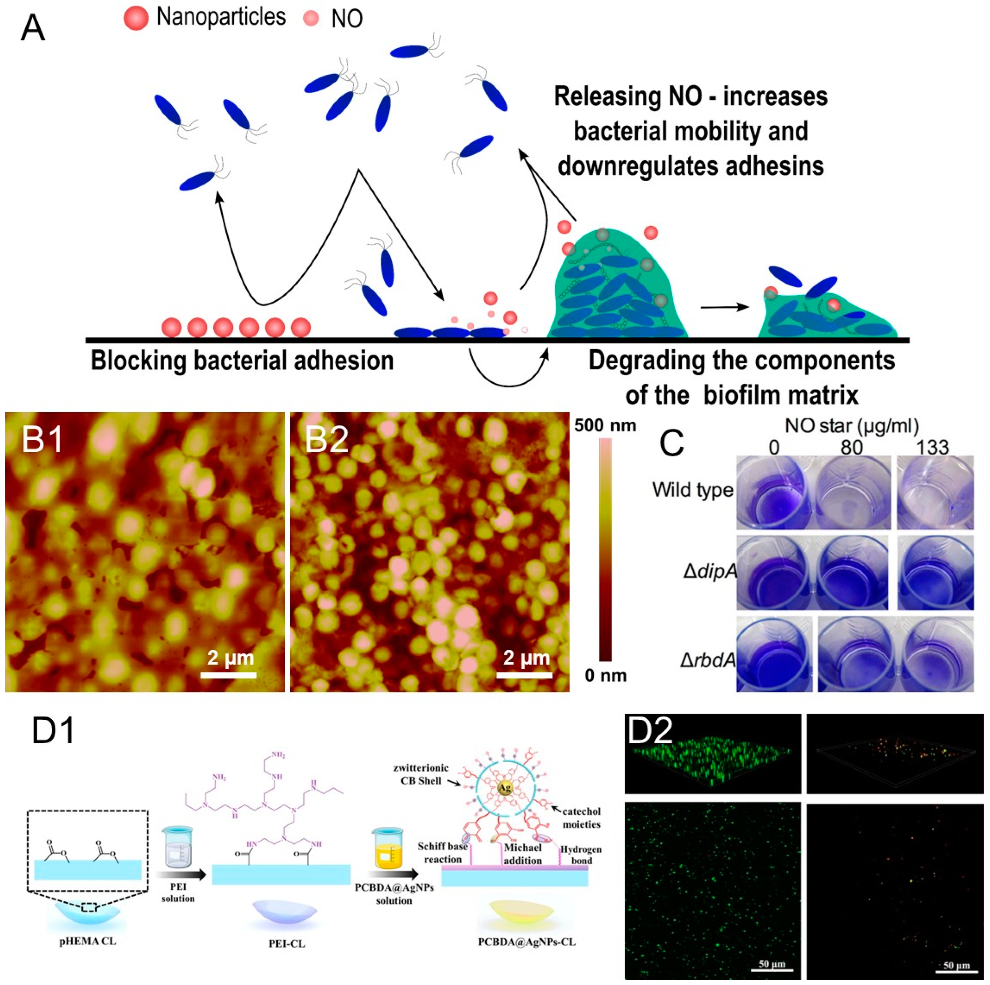
3.1. Nitric Oxide Donors Loaded in Nanocarriers

Nitric oxide (NO) is an endogenous molecule that has gained increasing attention due to its potential to impede bacterial adhesion and biofilm occurrence. NO is able to reduce the levels of the intracellular cyclic-di-GMP messenger, promoting the dispersion of biofilms [68]. Although its mechanism of action is still under investigation, there is evidence that NO enhances the activity of phosphodiesterases, upregulates genes for motility, and downregulates the expression of virulence factors and adhesins. However, due to its high reactivity, its direct application as gas is not feasible. Several NO delivery nanosystems have been developed in order to drive NO in a safe and effective way to the specific site of infection because NO has a short half-life and rapidly diffuses from the release site [69]. NO-releasing silica NPs demonstrated the capacity to kill P. aeruginosa, E. coli, S. aureus, and S. epidermidis encased in biofilms [70]. Duong et al. described how polymeric-star NPs, conjugated with spermine and NO, hindered the transition from swimming bacteria to sessile and dispersed P. aeruginosa biofilm, subsequently increasing the number of released planktonic cells in suspension (Figure 2C) (Table 1) [71]. In addition, Slomberg et al. demonstrated that the size and shape of the NO carrier play an important role in its release, small rod-shaped delivery vehicles being the most effective [72]. NO donors have been loaded into poly(vinyl alcohol) and poly(ethylene glycol) films and alginate hydrogels together with silver NPs for antibacterial topical applications (Table 1) [73,74]. Silica NPs loaded with nitroprusside acted synergistically with ampicillin and tetracycline, improving their bactericidal effect towards S. aureus and S. epidermidis [36].

3.2. Zwitterionic Materials

Zwitterions hold an immense potential for antifouling due to the formation of a hydration layer and steric hindrance effect that impedes the contact and adherence of the fouling biomolecules (e.g., proteins, polysaccharides) [75]. An essential step in the biofilm formation is the ability of bacteria to colonize surfaces via unspecific interaction of their surface proteins that serve as primary anchoring points. The equimolar number of homogenously distributed positively and negatively charged moieties along the zwitterion chain allows the binding of water molecules, creating a barrier towards protein attachment and bacterial colonization [76]. Poly(sulfobetaine methacrylate) (PSBMA) has been combined with silver NPs, which reduced the adhesion of proteins and inhibited P. aeruginosa biofilm formation on thin-film composite membranes under dynamic flow (Table 1) [77]. Xin et al. conjugated silver NPs with modified sulfobetaine in polyester membranes against E. coli and S. aureus, while Ma et al. combined NPs with poly(carboxybetaine-co-dopamine methacrylamide) (PCBDA) to coat contact lenses that reduced corneal infection in a rabbit model (Figure 2D) [78,79]. Xiang et al. immobilized PCBDA-Ag NPs on cotton gauzes, which not only inhibited bacterial adhesion and biofilm formation, but also promoted wound healing (Table 1) [80].
Antifouling surfaces and materials aim to prevent the first stage of the biofilm growth, but they do not kill the bacteria and the latter are able to colonize other surfaces and tissues. Hence, this anti-biofilm strategy may be feasible for material applications of short duration such as sutures, meshes, and drainage tubes, but not in, e.g., long-term implants.

3.3. Nanoformulated Matrix-Degrading Enzymes

Matrix-degrading enzymes target the extracellular components secreted by bacteria during biofilm formation. The polysaccharide, protein, and nucleic acid components of the EPS provide diverse benefits for bacteria. Carbohydrates play an important role in the anchoring of sessile cells, while proteins provide structural stability of the three-dimensional biofilm structure. Some of the proteins are catalytically active and possess the ability to digest large biomolecules (e.g., glycoside hydrolases and lipases) to supply nutrients, or take part in redox reactions (e.g., catalase). Nucleic acids also participate in the adhesion, aggregation, and cohesion of the biofilm, but their characteristic role is focused on the transfer of genetic information [81].
Degrading the biofilm adhesive structure by different enzymes such as α-amylase, alginate lyase, proteases, and deoxyribonucleases (DNases) is a feasible strategy to weaken biofilms and increase the bacterial susceptibility to antibacterial agents. α-amylase is a glycoside hydrolase that catalyzes the breaking of α-1,4-glycosidic bonds. Its main substrate is starch; however, it also acts on several carbohydrates present in the biofilm matrix [82]. We have developed NPs containing α-amylase and silver via gallic acid/laccase-mediated crosslinking, which were able to eradicate already established biofilms of P. aeruginosa and S. aureus (Figure 2B and Table 1) [34]. In another study, we hybridized α-amylase and zinc oxide in an NP form to coat urinary catheters in a single-step sonochemical approach. Thus, the undesirable phenomenon of unspecific protein adsorption was turned into an advantage by using the enzyme as adhesive. The resulting nanostructured coating hindered the formation of S. aureus and E. coli biofilms in a model of a catheterized bladder and significantly reduced the incidence of bacteriuria in rabbit models [31]. Abeleda et al. employed amylase to design hybrid silver/amylase NPs that inhibited and eradicated K. pneumonia and resistant S. aureus biofilms [83]. Alginate lyase is another biofilm-dispersing enzyme that breaks down polysaccharides, which are responsible for the strong adhesion and resistance of P. aeruginosa biofilms [84]. The enzyme was immobilized on chitosan and silver NPs, enhancing the effectivity of the conventional antibiotics ciprofloxacin and ceftazidime [85,86,87]. Proteases also have been nanoformulated using different approaches. For example, antibiotic-loaded shellac NPs, coated with the biofilm-degrading serine endo-peptidase alcalase, reduced the protein concentration in S. aureus EPM and effectively eradicated bacterial cells in the biofilm (Table 1) [88]. Antibiotic-carrying nanogels functionalized with antibiofilm protease were effective towards Staphylococcus aureus, Pseudomonas aeruginosa, S. epidermidis, K. pneumoniae, E. coli, and E. faecalis, while conjugating the enzyme with gold nano-rods completely eradicated S. aureus and E. coli biofilms under photothermal treatment [89,90].
Deoxyribonuclease I (DNAse I), which can degrade DNA in the biofilm matrix, has also shown a negative impact on the biofilm structure of medically relevant pathogens such as P. aeruginosa, E. coli, S. aureus, and K. pneumonia. DNAse I was integrated with ciprofloxacin to disperse already established P. aeruginosa biofilms and increase the antibiotic susceptibility. Its combination with silver NPs boosted the bactericidal effect and acted effectively against P. aeruginosa, Streptococcus mutans, and E. coli biofilms [91,92]. Combinations of several matrix-degrading enzymes have been shown to achieve better results in compromising the ability to establish or maintain stable biofilm structure. Protease and DNase have been loaded in liposomes to disperse Cutibacterium acnes biofilms in vitro and maintained their inhibition capacities in skin and catheter models without affecting cell viability in mice [93]. Although these enzyme-based approaches show very promising results, the major limitation is related to catalyst inactivation. Furthermore, the biofilm dispersion capacity is influenced by the nature of the species, which produce different matrix components, so a one-size-fits-all solution may not be feasible [94].
Figure 2. (A) Schematic representation of the anti-adhesive and biofilm-dispersing strategies covered in this review. (B) Nanoaggregates of Ag-amylase degraded EPM of S. aureus biofilm on gold disks. AFM images of the disk showing EPM matrix (B1) and its disintegration by the hybrid Ag-amylase NPs (B2) (reproduced from [34] under the terms of the Creative Commons Attribution International License (CC BY 4.0)). (C) Cristal violet staining of P. aeruginosa biofilms treated with different concentrations of NO-core cross-linked star (Adapted with permission from Duong et al., ‘Nanoparticle (Star Polymer) Delivery of Nitric Oxide Effectively Negates Pseudomonas aeruginosa Biofilm Formation’. Biomacromolecules 2014, 15, 2583–2589. Copyright 2014 American Chemical Society [71]). (D) Scheme of PCBDA@Ag NPs deposition on contact lenses (D1), and confocal microscopy of P. aeruginosa grown on pristine and coated contact lenses (D2) (Reprinted from J. Colloid Interface Sci, 610, Ma et al., Commercial Soft Contact Lenses Engineered with Zwitterionic Silver Nanoparticles for Effectively Treating Microbial Keratitis 923–933, Copyright 2022, with permission from Elsevier [79]).
Antibiotics 12 00310 g002

4. Antimicrobial Nanoactives

Biofilms reduce the susceptibility of bacteria to conventional antibiotics due to restricted diffusion in the EPS; however, nanoformulation may enhance the penetration. In addition, other compounds with wide spectrum and low-resistance-inducing mechanisms of action overcome the main limitations of conventional drugs. In this section, we focus on actives that disrupt the bacterial membrane, such as peptides and lipids, enzymes, which damage the cell wall and produce reactive oxygen species (ROS), and metal NPs with several antibacterial mechanisms, including ROS generation and release of toxic ions (Figure 3).

4.1. Antimicrobial Peptides

Antimicrobial peptides (AMP) are small oligopeptides of five to a hundred amino acids, found in bacteria, protozoa, fungi, plants, insects, and animals. AMPs form pores in the microbial membranes, which enables eradication of a broad spectrum of microorganisms and is less likely to lead to AMR. Despite the pronounced antimicrobial activity, even against multidrug-resistant strains, the low stability of AMP in biological fluids limits their practical application. To overcome this issue, different nanoformulation and hybridization strategies have been proposed. RBRBR was loaded into chitosan NPs and inhibited the formation of biofilm by reducing the number of viable motile bacteria (Table 1) [95]. Seferji et al. photoionized IVFK in order to reduce silver and form nanocomposites, which acted against S. aureus and E. coli biofilms [96]. Zhang et al. produced NPs by self-assembly of pHly-1 at acidic conditions. These peptide NPs were able to kill S. mutans in acidic cariogenic biofilm microenvironment displaying interesting properties for odontology applications [97]. The simultaneous nanoformulation of AMP and antibiotics leads to synergistic activity for eradication of P. aeruginosa. Yu et al. engineered stimuli-responsive NPs loaded with melittin and ofloxacin [98]. Gupta et al. combined AMP with quaternary ammonium polymers and the resulting NPs were able to penetrate biofilm and kill sessile S. aureus, P. aeruginosa, and Enterococcus cloacae [99]. In our group, we synthesized and deposited on silicone catheters peptide-zwitterion NPs via a one-step sonochemical approach. Thereby, catheters were incubated with polymyxin B and PSBMA under ultrasound and the formed NPs were coated through a “throwing stones” process. The coated silicones presented contact killing and antibiofilm properties without affecting mammal cells and were able to eliminate 80% of P. aeruginosa biofilms (Figure 3D and Table 1) [100]. Although the potent wide-spectrum antimicrobial activity of AMPs predicates them as a promising tool, their accumulation can lead to unspecific toxicity and they also suffer from stability issues in biological fluids once released from the nanocarrier [101].

4.2. Non-Peptide Mimics of AMPs

Non-peptide cationic steroids with enhanced stability, termed ceragenins, have been developed to overcome some of these drawbacks. Their molecular structure is based on cholic acid, appended by amine groups, which are arranged to reproduce the amphiphilic morphology of AMPs. These AMP mimics act on a variety of Gram-positive and Gram-negative pathogens, including multi-drug resistant strains in planktonic and sessile forms, but unlike AMPs, they demonstrate enhanced stability at physiological conditions and lower toxicity [102,103,104]. Due to the high positive charge and amphipathic nature, ceragenins interact with negatively charged cell membranes causing changes in the organization of the membrane lipid bilayer, change of permeability, and cell death. They have also the ability to bind bacterial endotoxins (e.g., lipopolysaccharides and lipoteichoic acid), which is further translated into anti-inflammatory effects [105]. Recently, Paprocka et al. demonstrated the benefit of using synergistic combinations of ceragenins with commercial antibiotics including ceftazidime, levofloxacin, co-trimoxazole, and colistin for managing of Stenotrophomonas maltophilia infections. Ceragenins applied simultaneously with β-lactam antibiotics (e.g., ceftolozane/tazobactam, ceftazidime/avibactam, meropenem/vaborbactam) at low concentrations also showed efficacy against clinical strains of P. aeruginosa regardless of their resistance mechanisms [106]. Rod, peanut, and star-shaped gold NPs conjugated with ceragenins exerted potent bactericidal activity against multi-drug resistant strains due to the generation of ROS, followed by membrane damage and the leakage of intracellular content [107]. Silver NPs coated with ceragenins, or other cationic antimicrobials, were found to be eight times more effective against bacteria than silver NP alone [108], while ceragenin-functionalized magnetic NPs were shown to impede not only bacterial biofilm formation but also of fungal ones [109,110]. Despite the great promise of these AMP mimics in terms of long-term stability, low toxicity, and antibiofilm activity towards a great number of pathogens, their use for coatings is scarcely reported. Bulk ceragenins have been incorporated into contact lenses or used as a coating on fracture fixation plates to prevent P. aeruginosa and S. aureus biofilm establishment in vitro and in vivo [111,112]. Hashemi et al. demonstrated that ceragenin-protected endotracheal tubes resisted microbial colonization, decreasing the adverse effects of intubation associated with infection and inflammation [113].

4.3. Marine-Derived Antibacterial Lipids

Medicinal plants and marine organisms are natural sources of many antimicrobial compounds. Plant-derived phenolics, diterpenes/terpenoids, alkaloids, sulfur-containing compounds, glycosides, and fatty acids, “generally recognized as safe” (GRAS), have shown strong antibacterial activity towards Gram-positive and Gram-negative bacteria and low probability for triggering AMR [114]. Slow-moving or sessile marine organisms produce antimicrobial molecules (e.g., fatty acids and peptides) as a part of their adaptive defense mechanisms to protect themselves against pathogens including bacteria, viruses, and fungi.
Fatty acids, monoacylglycerols, sterols, and terpene derivatives are among the most studied antimicrobial lipid classes. Their strong antimicrobial efficiency against a wide spectrum of microorganisms is related to their chemical structure and depends on the acyl chain length, the stereochemistry, the degree of unsaturation, and the esterification [115]. Long-chain polyunsaturated fatty acid combined with benzoyl peroxide synergistically inhibited the growth of S. aureus due to the increase in the bacterial membrane permeability that improved the penetration of the bactericidal active [116]. Lipids have been mainly used to produce vehicles for delivery of conventional antibiotics both due to their membrane destabilization potential and resemblance to natural membranes. For instance, lipid-polymer NPs loaded with antibiotics demonstrated higher antibiofilm activity on P. aeruginosa than the polymer NPs alone [117]. Solid-lipid NPs encapsulating rifampin were used against S. epidermidis, destroying the biofilm and affecting the bacterial viability [118]. Recently, the use of solely lipid-based NPs has gained attention, avoiding the use of antibiotics. For example, Rozenbaum et al. produced monolaurin lipid NPs that were able to reduce the bacterial concentration of methicillin-resistant S. aureus inside biofilms in vitro and the activity synergistically increased when AMPs were absorbed onto the NPs. However, these effects were not observed in wound models in mice [119].

4.4. Bactericidal Enzymes

Above, we discussed the ability of some enzymes to target QS and destroy the components of EPM; however, other enzymes display a direct antimicrobial effect, mainly hydrolyzing components of the cell wall or by catalyzing the formation of oxidative molecules. Lysozyme is a muramidase that catalyzes the hydrolysis of β-1,4-linkages between N-acetyl-d-glucosamine and N-acetylmuramic acid and was loaded into mesoporous NPs and magnetite-chitosan NPs [120,121]. Wang et al. produced gelatin composites for wound-healing applications, carrying loaded mesoporous polydopamine NPs with lysozyme that were able to disperse E. coli biofilm [122].
Cellobiose dehydrogenase (CDH) is another enzyme that elicits bactericidal action through the production of hydrogen peroxide. CDH has been shown to inhibit the growth of a panel of multidrug-resistant pathogens including E. coli, S. aureus, S. epidermidis, P. mirabilis, S. maltophilia, Acinetobacter baumannii, and P. aeruginosa thanks to the oxidation of bacterial extracellular polysaccharides. CDH NPs have been also produced in situ and subsequently deposited onto silicone surfaces using ultrasound, effectively reducing viable S. aureus cells and the total amount of deposited biomass (Table 1) [123]. In more recent work, antibacterial CDH and matrix-degrading DNAse were simultaneously immobilized onto chitosan NPs, which penetrated the biofilm structure and acted synergistically on preformed multi-species biofilms (Figure 3C) [124]. Though aside from the aforementioned general limitations of enzyme stability, the efficacy of such strategies could be affected by potential inflammatory response, associated with oxidative stress.

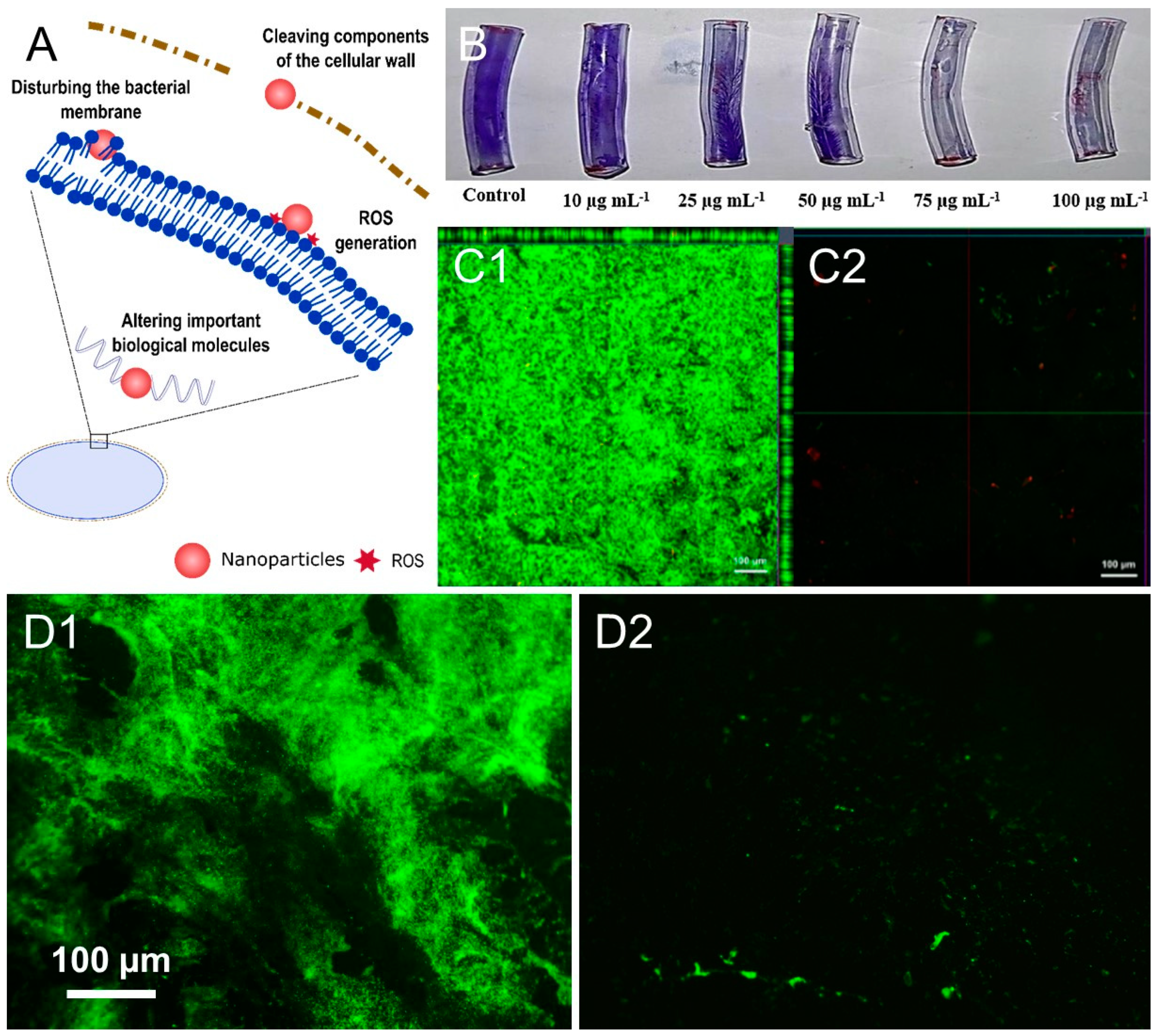
4.5. Metal and Metal Oxide Nanoparticles

Metal and metal oxide NPs are stronger antimicrobial agents, compared to ionic metals or metal macrostructures. Their small size and high area-to-volume ratio improve the penetration and interaction with the bacterial membrane, enhancing their antimicrobial properties. The unspecific mechanisms of antibacterial action such as disruption of the membrane stability, ROS production, and release of metal ions generally hinder the appearance of AMR. However, it is important to note that metal NPs have been reported to induce toxicity in mammalian cells. The most used metals for NPs synthesis are silver, gold, and zinc oxide. Metal NPs have been coated on catheters and implants to avoid biofilm formation or loaded in wound dressings for elimination of established biofilms in chronic wounds (Figure 3B) [125,126,127,128,129,130,131,132,133,134,135,136,137,138,139,140]. Selenium NPs were able to eradicate S. aureus, P. aeruginosa, and Salmonella typhi, disrupting their cell walls, in addition, cotton fabrics functionalized with Se NPs avoid the formation of E. coli and S. aureus biofilm on their surfaces (Table 1) [141,142]. Other metal NPs that have displayed antibiofilm properties are titanium dioxide, copper, and nickel [143,144,145,146,147]. Our group has a wide experience in the coating of medical devices with metal and metal oxide NPs, individually or in combination with other bioactive macromolecules. This is a proven strategy to reduce the intrinsic metal toxicity and/or to enhance the antimicrobial performance. Zinc oxide NPs were sonochemically coated on cotton medical textiles using laccase-oxidized gallic acid as a bioadhesive. The coating exhibited antimicrobial activity towards S. aureus, which was not affected even after 20 washing cycles at 75 °C [148]. Similarly, contact lenses were coated using ultrasound with zinc oxide NPs, chitosan, and gallic acid, displaying contact-killing capacities towards S. aureus, without affecting the optical properties [149]. Finally, silver NPs decorated with aminocellulose or chitosan were used to synthesize layer-by-layer hyaluronic acid coatings and membranes that inhibited completely bacterial growth and reduced biofilm formation (Table 1) [30].
Figure 3. (A) Schematic representation of the antimicrobial mechanisms of action of bactericidal NPs covered in this review. (B) Cristal violet staining of P. aeruginosa biofilm grown on catheters coated with different concentrations of AgNPs (Reprinted from Prog. Org. Coati, 151, LewisOscar et al., ‘In vitro analysis of green fabricated silver nanoparticles (AgNPs) against Pseudomonas aeruginosa PA14 biofilm formation, their application on urinary catheter,’ 106058, Copyright 2021, with permission from Elsevier [126]). (C) Confocal microscopy images of untreated biofilm (C1) and treated with chitosan-cellobiose dehydrogenase-DNase NPs (C2) (Reprinted from Mater. Sci. Eng. C, 108, Tan et al., ‘Co-Immobilization of Cellobiose Dehydrogenase and Deoxyribonuclease I on Chitosan Nanoparticles against Fungal/Bacterial Polymicrobial Biofilms Targeting Both Biofilm Matrix and Microorganisms,’ 110499, Copyright 2019, with permission from Elsevier [124]). (D) Fluorescence microscopy images showing the grown of P. aeruginosa biofilm on pristine silicone catheters (D1) and its inhibition when the catheters were coated with self-assembled pSBMA-polymyixin B NPs (D2) (reproduced from [100] under the terms of the Creative Commons Attribution International License (CC BY 4.0)).
Antibiotics 12 00310 g003
Table 1. Relevant examples of nanomaterials and coatings against biofilms.
StrategyActivesNanoparticleAntibiofilm ActivityApplicationRef.
Quorum sensing inhibitorsQuorum sensing inhibitorsGold-silica-cinnamaldehydeS. aureusColloidal suspension[38]
Acid-poly (lactic-co-glycolic acid)-azithromycinP. aeruginosaColloidal suspension[44]
Eugenol nanoemulsionE. coliNanocomposite hydrogel[48]
Quorum-quenching enzymesSilver-aminocellulose-acylaseP. aeruginosaColloidal suspension[33]
Acylase-gentamicine[28]
Gold-lactonase[56]
Metal and metal oxidesGold, nickel oxide, tellurium and seleniumP. aeruginosaColloidal suspension[59,60,62]
Mimics of QS machineryCAI-1 (autoinducer)V. choleraColloidal suspension[64]
Molecularly imprinted NPsP. aeruginosa[66]
Anti-adhesionNO donorsNO-releasing silica NPsP. aeruginosa, E. coli,
S. aureus, S. epidermidis
Colloidal suspension[70]
Polymeric stars-spermineP. aeruginosaColloidal suspension[71]
Silver-NO donorP. aeruginosa, E. coli,
S. aureus, K. pneumoniae
NP-containing PVA/PEG-films[73]
ZwitterionsPSBMA-silver, PCBDA-silverP. aeruginosa, E. coli,
S. aureus
Membranes and gauzes coatings[77,80]
Matrix degrading enzymesSilver-amylaseS. aureus, E. coliColloidal suspension[34]
Zinc oxide-amylaseCatheters coatings[31]
Shellac NPs-proteaseS. aureusColloidal suspension[88]
BactericidalPeptidesRBRBR-chitosan, IVFK-silverS. aureus, E. coliColloidal suspension[95,96]
Polymyxin B-PSBMAP. aeruginosaCatheters coatings[100]
Antibacterial enzymesLysozyme-magnetite-chitosanS. aureus, P. aeruginosaColloidal suspension[121]
Lysozyme-polydopamineE. coliHydrogel dressing[122]
Cellobiose dehydrogenaseS. aureusSurface coatings[123]
Metal and metal oxidesSilverS. aureus, E. coli,
P. aeruginosa
Catheters and implants coatings[125,126,127]
Silver-chitosanS. aureus, E. coliCoatings[30]
SeleniumS. aureus, P. aeruginosa and S. typhiColloidal suspension[141]
Zinc oxide-chitosan-gallic acidS. aureusContact lenses coatings[149]

5. Conclusions

Different nanotechnological approaches have been used for highly efficient strategies against biofilms. We have presented how antimicrobial and antibiofilm actives can be engineered for three different strategies: (i) disrupting the QS, (ii) preventing the bacterial attachment and promoting the biofilm detachment, and (iii) killing the bacteria encased inside the biofilm. Enzymes, depending on their type and activity, can quench QS, destroy selected components of the EPM, or directly exercise an antimicrobial role; however, their stability and possible side effects in vivo may limit their use. Metal NPs are excellent antimicrobial agents and have been reported to interfere in the intercellular communication; however, their intrinsic toxicity requires innovative approaches for their formulation. Zwitterions are able to prevent bacterial colonization of surfaces, while other molecules such as lipids and peptides directly disrupt the bacterial membrane, regardless of whether in planktonic or biofilm form. We showed how the combination of antibiofilm and antimicrobial agents and their synergistic activity are critical for the efficacy. However, although the numerous examples in this fruitful field display highly promising results, they are usually restricted to in vitro models in idealized laboratory conditions. Thus, the immense progress in the lab still needs to be validated in relevant environments such as in vivo experiments and realistic settings, provided the diverse nature of clinical isolates and their capacity to develop AMR in short time. At the same time, further factors such as scale of production, economic viability, and environmental impact have to be addressed before translation into actual practice.

Author Contributions

Design of the review structure: G.F., K.I. and T.T. Literature searches and data extraction: G.F. and K.I. interpretation of data: G.F. and K.I. Drafting the article: G.F., K.I. and I.I. Critical revision of the article: I.I. and T.T. All authors have read and agreed to the published version of the manuscript.

Funding

This research was funded by the Spanish Ministry of Economy and Competitiveness Projects (MINECO) CoatToSave—Nanoenabled hydrogel coatings against antimicrobial-resistant catheter-related infections, PID2019-104111RB-I00, European project AMROCE—Nanoenabled strategies to reduce the presence of contaminants of emergent concern in aquatic environments, PCI2021-121926, and the European project TARDIS Nano-enabled stimuli-responsive scaffolds for targeted antimicrobials delivery to treat Staphylococcus aureus infections and restore skin homeostasis, PCI2022-13292. G. F acknowledges Universitat Politècnica de Catalunya and Banco Santander for his PhD grant (113 FPI-UPC 2018).

Institutional Review Board Statement

Not applicable.

Informed Consent Statement

Not applicable.

Data Availability Statement

Not applicable.

Conflicts of Interest

The authors declare no conflict of interest.

References

  1. Flemming, H.C.; Wingender, J.; Szewzyk, U.; Steinberg, P.; Rice, S.A.; Kjelleberg, S. Biofilms: An emergent form of bacterial life. Nat. Rev. Microbiol. 2016, 14, 563–575. [Google Scholar] [CrossRef] [PubMed]
  2. Toyofuku, M.; Inaba, T.; Kiyokawa, T.; Obana, N.; Yawata, Y.; Nomura, N. Environmental factors that shape biofilm formation. Biosci. Biotechnol. Biochem. 2016, 80, 7–12. [Google Scholar] [CrossRef] [PubMed]
  3. De Kievit, T.R.; Iglewski, B.H. Bacterial Quorum Sensing in Pathogenic Relationships. Infect. Immun. 2000, 68, 4839–4849. [Google Scholar] [CrossRef]
  4. Winstanley, C.; Fothergill, J.L. The role of quorum sensing in chronic cystic fibrosis Pseudomonas aeruginosa infections. FEMS Microbiol. Lett. 2009, 290, 1–9. [Google Scholar] [CrossRef] [PubMed]
  5. Rand, J.D.; Danby, S.G.; Greenway, D.L.A.; England, R.R. Increased expression of the multidrug efflux genes acrAB occurs during slow growth of Escherichia coli. FEMS Microbiol. Lett. 2002, 207, 91–95. [Google Scholar] [CrossRef]
  6. Wright, G.D. Molecular mechanisms of antibiotic resistance. Chem. Comm. 2011, 47, 4055–4061. [Google Scholar] [CrossRef]
  7. Olsen, I. Biofilm-specific antibiotic tolerance and resistance. Eur. J. Clin. Microbiol. Infect. Dis. 2015, 34, 877–886. [Google Scholar] [CrossRef]
  8. Høiby, N.; Bjarnsholt, T.; Givskov, M.; Molin, S.; Ciofu, O. Antibiotic resistance of bacterial biofilms. Int. J. Antimicrob. Agents 2010, 35, 322–332. [Google Scholar] [CrossRef]
  9. Vuong, C.; Voyich, J.M.; Fischer, E.R.; Braughton, K.R.; Whitney, A.R.; Deleo, F.R.; Otto, M. Polysaccharide intercellular adhesin (PIA) protects Staphylococcus epidermidis against major components of the human innate immune system. Cell. Microbiol. 2004, 6, 269–275. [Google Scholar] [CrossRef]
  10. Malaekeh-nikouei, B.; Sedigheh, B.; Bazzaz, F.; Mirhadi, E.; Sadat, A.; Khameneh, B. The role of nanotechnology in combating biofilm-based antibiotic resistance. J. Drug Deliv. Sci. Technol. 2020, 60, 101880. [Google Scholar] [CrossRef]
  11. Madsen, J.S.; Burmølle, M.; Hansen, L.H.; Sørensen, S.J. The interconnection between biofilm formation and horizontal gene transfer. FEMS Immunol. Med. Microbiol. 2012, 65, 183–195. [Google Scholar] [CrossRef] [PubMed]
  12. Davies, D. Understanding Biofilm Resistance To Antibacterial Agents. Nat. Rev. Drug Discov. 2003, 2, 114–122. [Google Scholar] [CrossRef] [PubMed]
  13. Macià, M.D.; del Pozo, J.L.; Díez-Aguilar, M.; Guinea, J. Microbiological diagnosis of biofilm-related infections. Enferm. Infecc. Microbiol. Clin. 2018, 36, 375–381. [Google Scholar] [CrossRef] [PubMed]
  14. Elnagdy, S.; Raptopoulos, M.; Kormas, I.; Pedercini, A.; Wolff, L.F. Local Oral Delivery Agents withAnti-BiofilmProperties for the Treatment of Periodontitis and Peri-Implantitis. A Narrative Review. Molecules 2021, 26, 5661. [Google Scholar] [CrossRef] [PubMed]
  15. Ciofu, O.; Rojo-Molinero, E.; Macià, M.D.; Oliver, A. Antibiotic treatment of biofilm infections. Apmis 2017, 125, 304–319. [Google Scholar] [CrossRef]
  16. Lynch, A.S.; Robertson, G.T. Bacterial and Fungal Biofilm Infections. Annu. Rev. Med. 2014, 59, 415–428. [Google Scholar] [CrossRef]
  17. Percival, S.L.; Suleman, L.; Vuotto, C.; Donelli, G. Healthcare-associated infections, medical devices and biofilms: Risk, tolerance and control. J. Med. Microbiol. 2015, 64, 323–334. [Google Scholar] [CrossRef]
  18. Guzella Carvalho, L.; Alfonso Alvim, M.M.; Luiz Fabri, R.; Morais Apolonio, A.C. Staphylococcus aureus biofilm formation in Minas Frescal cheese packaging. Dairy Technol. 2021, 74, 575–580. [Google Scholar] [CrossRef]
  19. Zimmermann, K.; Li, S.; Mohseni, M. Biological Activity Offsets Diffusion Limitations of Biofilms in Biological Ion Exchange Water Filters. ACS EST Water 2022, 2, 2441–2449. [Google Scholar] [CrossRef]
  20. Barravecchia, I.; DeCesari, C.; Pyankova, O.V.; Scebba, F.; Carlo, M.; Vecchione, A.; Tavanti, A.; Tedeschi, L.; Angeloni, D. Pitting Corrosion Within Bioreactors for Space Cell-Culture Contaminated by Paenibacillus glucanolyticus, a Case Report. Microgravity-Sci. Technol. 2018, 30, 309–319. [Google Scholar] [CrossRef]
  21. Stipanicev, M.; Turcu, F.; Esnault, L.; Schweitzer, E.W.; Kilian, R.; Basseguy, R. Corrosion behavior of carbon steel in presence of sulfate-reducing bacteria in seawater environment. Electrochim. Acta 2013, 113, 390–406. [Google Scholar] [CrossRef]
  22. Bockmühl, D.P. Laundry hygiene—How to get more than clean. J. Appl. Microbiol. 2017, 122, 1124–1133. [Google Scholar] [CrossRef] [PubMed]
  23. Clatworthy, A.E.; Pierson, E.; Hung, D.T. Targeting virulence: A new paradigm for antimicrobial therapy. Nat. Chem. Biol. 2007, 3, 541–548. [Google Scholar] [CrossRef]
  24. Wu, H.; Moser, C.; Wang, H.Z.; Høiby, N.; Song, Z.J. Strategies for combating bacterial biofilm infections. Int. J. Oral Sci. 2014, 7, 1–7. [Google Scholar] [CrossRef] [PubMed]
  25. Bi, Y.; Xia, G.; Shi, C.; Wan, J.; Liu, L.; Chen, Y.; Wu, Y.; Zhang, W.; Zhou, M.; He, H.; et al. Therapeutic strategies against bacterial biofilms. Fundam. Res. 2021, 1, 193–212. [Google Scholar] [CrossRef]
  26. Blaser, M. Stop the killing of beneficial bacteria. Nature 2011, 476, 393–394. [Google Scholar] [CrossRef] [PubMed]
  27. Rigo, S.; Cai, C.; Gunkel-Grabole, G.; Maurizi, L.; Zhang, X.; Xu, J.; Palivan, C.G. Nanoscience-Based Strategies to Engineer Antimicrobial Surfaces. Adv. Sci 2018, 5, 1700892. [Google Scholar] [CrossRef]
  28. Ivanova, K.; Ivanova, A.; Hoyo, J.; Pérez-Rafael, S.; Tzanov, T. Nano-Formulation Endows Quorum Quenching Enzyme-Antibiotic Hybrids with Improved Antibacterial and Antibiofilm Activities against Pseudomonas aeruginosa. Int. J. Mol. Sci. 2022, 23, 7632. [Google Scholar] [CrossRef]
  29. Zhang, L.; Gu, F.X.; Chan, J.M.; Wang, A.Z.; Langer, R.S.; Farokhzad, O.C. Nanoparticles in Medicine: Therapeutic Applications and Developments. Clin. Pharmacol. Ther. 2008, 83, 761–769. [Google Scholar] [CrossRef]
  30. Francesko, A.; Ivanova, K.; Hoyo, J.; Pérez-Rafael, S.; Petkova, P.; Fernandes, M.M.; Heinze, T.; Mendoza, E.; Tzanov, T. Bottom-up Layer-by-Layer Assembling of Antibacterial Freestanding Nanobiocomposite Films. Biomacromolecules 2018, 19, 3628–3636. [Google Scholar] [CrossRef]
  31. Ivanova, A.; Ivanova, K.; Perelshtein, I.; Gedanken, A.; Todorova, K.; Milcheva, R.; Dimitrov, P.; Popova, T.; Tzanov, T. Sonochemically engineered nano-enabled zinc oxide/amylase coatings prevent the occurrence of catheter-associated urinary tract infections. Mater. Sci. Eng. C 2021, 131, 112518. [Google Scholar] [CrossRef]
  32. Soule, L.D.; Chomorro, N.P.; Chuong, K.; Mellott, N.; Hammer, N.; Hankenson, K.D.; Chatzistavrou, X. Sol−Gel-Derived Bioactive and Antibacterial Multi-Component Thin Films by the Spin-Coating Technique. ACS Biomater. Sci. Eng. 2020, 5549–5562. [Google Scholar] [CrossRef]
  33. Ivanova, A.; Ivanova, K.; Tied, A.; Heinze, T.; Tzanov, T. Layer-By-Layer Coating of Aminocellulose and Quorum Quenching Acylase on Silver Nanoparticles Synergistically Eradicate Bacteria and Their Biofilms. Adv. Funct. Mater. 2020, 30, 2001284. [Google Scholar] [CrossRef]
  34. Ferreres, G.; Bassegoda, A.; Hoyo, J.; Torrent-Burgués, J.; Tzanov, T. Metal-Enzyme Nanoaggregates Eradicate Both Gram-Positive and Gram-Negative Bacteria and Their Biofilms. ACS Appl. Mater. Interfaces 2018, 10, 40434–40442. [Google Scholar] [CrossRef]
  35. Ivanova, K.; Ivanova, A.; Ramon, E.; Hoyo, J.; Sanchez-Gomez, S.; Tzanov, T. Antibody-Enabled Antimicrobial Nanocapsules for Selective Elimination of Staphylococcus aureus. ACS Appl. Mater. Interfaces 2020, 12, 35918–35927. [Google Scholar] [CrossRef]
  36. Da Silva Filho, P.M.; Andrade, A.L.; Cruz Lopes, J.B.A.; de Azevedo Pinheiro, A.; Alve de Vesconcelos, M.; da Cruz Fonseca, S.G.; de Lopes, F.L.G.; Silva Sousa, E.H.; Teixera, E.H.; Longhinotti, E. The biofilm inhibition activity of a NO donor nanosilica with enhanced antibiotics action. Int. J. Pharm. 2021, 610, 121220. [Google Scholar] [CrossRef]
  37. Niu, C.; Afre, S.; Gilbert, E.S. Subinhibitory concentrations of cinnamaldehyde interfere with quorum sensing. Lett. Appl. Microbiol. 2006, 43, 489–494. [Google Scholar] [CrossRef]
  38. Ramasamy, M.; Lee, J.; Jintae, L. Development of gold nanoparticles coated with silica containing the antibiofilm drug cinnamaldehyde and their effects on pathogenic bacteria. Int. J. Nanomed. 2017, 12, 2813–2828. [Google Scholar] [CrossRef]
  39. Subhaswaraj, P.; Barik, S.; Macha, C.; Venkata, P. Anti quorum sensing and anti biofilm efficacy of cinnamaldehyde encapsulated chitosan nanoparticles against Pseudomonas aeruginosa PAO1. LWT Food Sci. Technol. 2018, 97, 752–759. [Google Scholar] [CrossRef]
  40. Ouyang, J.; Sun, F.; Feng, W.; Sun, Y.; Qiu, X.; Xiong, L.; Liu, Y.; Chen, Y. Quercetin is an effective inhibitor of quorum sensing, biofilm formation and virulence factors in Pseudomonas aeruginosa. J. Appl. Microbiol. 2016, 120, 966–974. [Google Scholar] [CrossRef] [Green Version]
  41. Tran, T.; Hadinoto, K. A Potential Quorum-Sensing Inhibitor for Bronchiectasis Therapy: Quercetin—Chitosan Nanoparticle Complex Exhibiting Superior Inhibition of Biofilm Formation and Swimming Motility of Pseudomonas aeruginosa to the Native Quercetin. Int. J. Mol. Sci. 2021, 22, 1541. [Google Scholar] [CrossRef]
  42. Yu, L.; Shang, F.; Chen, X.; Ni, J.; Yu, L.; Zhang, M. The anti-biofilm effect of silver- nanoparticle-decorated quercetin nanoparticles on a multi-drug resistant Escherichia coli strain isolated from a dairy cow with mastitis. PeerJ 2018, 6, e5711. [Google Scholar] [CrossRef]
  43. Tateda, K.; Comte, R.; Pechere, J.-C.; Köhler, T.; Yamaguchi, K.; van Delden, C. Azithromycin Inhibits Quorum Sensing in Pseudomonas aeruginosa. Antimicrob. Agents Chemother. 2001, 45, 1930–1933. [Google Scholar] [CrossRef]
  44. Kłodzinska, S.N.; Wan, F.; Jumaa, H.; Sternberg, C.; Rades, T.; Nielsen, H.M. Utilizing nanoparticles for improving anti-biofilm effects of azithromycin: A head-to-head comparison of modified hyaluronic acid nanogels and coated poly (lactic-co-glycolic acid) nanoparticles. J. Colloid Interface Sci. 2019, 555, 595–606. [Google Scholar] [CrossRef]
  45. Gao, Y.; Wang, J.; Chai, M.; Li, X.; Deng, Y.; Jin, Q.; Ji, J. Size and Charge Adaptive Clustered Nanoparticles Targeting the Biofilm Microenvironment for Chronic Lung Infection Management. ACS Nano 2020, 14, 5686–5699. [Google Scholar] [CrossRef]
  46. Ashrafudoulla, M.; Rahaman, F.; Ha, A.J.; Hong, S.; Ha, S. Antibacterial and antibio film mechanism of eugenol against antibiotic resistance Vibrio parahaemolyticus. J. Food Microbiol. 2020, 91, 103500. [Google Scholar] [CrossRef]
  47. Lou, Z.; Letsididi, K.S.; Yu, F.; Pei, Z.; Wang, H. Inhibitive Effect of Eugenol and Its Nanoemulsion on Quorum Sensing–Mediated Virulence Factors and Biofilm Formation by Pseudomonas aeruginosa. J. Food Prot. 2019, 82, 379–389. [Google Scholar] [CrossRef]
  48. Prateeksha; Barik, S.K.; Singh, B.N. Nanoemulsion-loaded hydrogel coatings for inhibition of bacterial virulence and biofilm formation on solid surfaces. Sci. Rep. 2019, 9, 6520. [Google Scholar] [CrossRef]
  49. Antunes, J.C.; Tavares, T.D.; Teixeira, M.A.; Teixeira, M.O.; Homem, C.; Amorim, M.T.P.; Felgueiras, H.P. Eugenol-Containing Essential Oils Loaded onto Chitosan/Polyvinyl Alcohol Blended Films and Their Ability to Eradicate Staphylococcus aureus or Pseudomonas aeruginosa from Infected Microenvironments. Pharmaceutics 2021, 13, 195. [Google Scholar] [CrossRef]
  50. Nabawy, A.; Marie, J.; Schmidt-malan, S.; Park, J.; Li, C.; Huang, R.; Fedeli, S.; Nath, A.; Patel, R.; Rotello, V.M. Dual antimicrobial-loaded biodegradable nanoemulsions for synergistic treatment of wound biofilms. J. Control. Release 2022, 347, 379–388. [Google Scholar] [CrossRef]
  51. Sethuram, L.; Thomas, J.; Mukherjee, A.; Chandrasekaran, N. Eugenol micro-emulsion reinforced with silver nanocomposite electrospun mats for wound dressing strategies. Mater. Adv. 2021, 2, 2971–2988. [Google Scholar] [CrossRef]
  52. Sio, C.F.; Otten, L.G.; Cool, R.H.; Diggle, S.P.; Braun, P.G.; Bos, R.; Daykin, M.; Ca, M.; Williams, P.; Quax, W.J. Quorum Quenching by an N-Acyl-Homoserine Lactone Acylase from Pseudomonas aeruginosa PAO1. Infect. Immun. 2006, 74, 1673–1682. [Google Scholar] [CrossRef]
  53. Lee, B.; Yeon, K.; Shim, J.; Kim, S.; Lee, C.; Lee, J.; Kim, J. Effective Antifouling Using Quorum-Quenching Acylase Stabilized in Magnetically-Separable Mesoporous Silica. Biomacromolecules 2014, 15, 1153–1159. [Google Scholar] [CrossRef]
  54. Zhu, Z.; Wang, L.; Li, Q. A bioactive poly (vinylidene fluoride)/graphene oxide@acylase nanohybrid membrane: Enhanced anti-biofouling based on quorum quenching. J. Membr. Sci. 2018, 547, 110–122. [Google Scholar] [CrossRef]
  55. Anandan, K.; Rai, R. Microbial Pathogenesis Quorum quenching activity of AiiA lactonase KMMI17 from endophytic Bacillus thuringiensis KMCL07 on AHL-mediated pathogenic phenotype in Pseudomonas aeruginosa. Microb. Pthogenes. 2019, 132, 230–242. [Google Scholar] [CrossRef]
  56. Gupta, K.; Chhibber, S. Biofunctionalization of Silver Nanoparticles With Lactonase Leads to Altered Antimicrobial and Cytotoxic Properties. Front. Mol. Biosci. 2019, 6, 63. [Google Scholar] [CrossRef]
  57. Vinoj, G.; Pati, R.; Sonawane, A. In Vitro Cytotoxic Effects of Gold Nanoparticles Coated with Functional Acyl Homoserine Lactone Lactonase Protein from Bacillus licheniformis and Their Antibiofilm Activity against Proteus Species. Antimicrob Agents Chemother. 2014, 59, 763–771. [Google Scholar] [CrossRef]
  58. Singh, B.R.; Singh, B.N.; Singh, A.; Khan, W.; Naqvi, A.H. Mycofabricated biosilver nanoparticles interrupt Pseudomonas aeruginosa quorum sensing systems. Sci. Rep. 2015, 5, 13719. [Google Scholar] [CrossRef]
  59. Qais, F.A.; Ahmad, I.; Altaf, M.; Alotaibi, S.H. Biofabrication of Gold Nanoparticles Using Capsicum annuum Extract and Its Antiquorum Sensing and Antibio fi lm Activity against Bacterial Pathogens. ACS Omega 2021, 6, 16670–16682. [Google Scholar] [CrossRef]
  60. Maruthupandy, M.; Nadar, G.; Quero, F.; Li, W. Anti-quorum sensing and anti-biofilm activity of nickel oxide nanoparticles against Pseudomonas aeruginosa. J. Environ. Chem. Eng. 2020, 8, 104533. [Google Scholar] [CrossRef]
  61. Singh, N.; Paknikar, K.M.; Rajwade, J. Gene expression is influenced due to ‘nano’ and ‘ionic’ copper in pre-formed Pseudomonas aeruginosa biofilms. Environ. Res. 2019, 175, 367–375. [Google Scholar] [CrossRef]
  62. Gómez-Gómez, B.; Arregui, L.; Serrano, S.; Santos, A.; Pérez-Corona, T.; Madrid, Y. Selenium and tellurium-based nanoparticles as interfering factors in quorum sensing-regulated processes: Violacein production and bacterial biofilm formation. Metallomics 2019, 11, 1104–1114. [Google Scholar] [CrossRef] [PubMed]
  63. Gómez-gómez, B.; Arregui, L.; Serrano, S.; Santos, A.; Pérez-corona, T.; Madrid, Y. Unravelling mechanisms of bacterial quorum sensing disruption by metal-based nanoparticles. Sci. Total Environ. 2019, 696, 133869. [Google Scholar] [CrossRef] [PubMed]
  64. Lu, H.D.; Spiegel, A.C.; Hurley, A.; Perez, L.J.; Maisel, K.; Ensign, L.M.; Hanes, J.; Bassler, B.L.; Semmelhack, M.F.; Prud, R.K. Modulating Vibrio cholerae Quorum-Sensing-Controlled Communication Using Autoinducer-Loaded Nanoparticles. Nano Lett. 2015, 15, 2235–2241. [Google Scholar] [CrossRef]
  65. Piletska, E.V.; Stavroulakis, G.; Karim, K.; Whitcombe, M.J.; Chianella, I.; Sharma, A.; Eboigbodin, K.E.; Robinson, G.K.; Piletsky, S.A. Attenuation of vibrio fischeri quorum sensing using rationally designed polymers. Biomacromolecules 2010, 11, 975–980. [Google Scholar] [CrossRef]
  66. Lopez, J.G.; Piletska, E.V.; Whitcombe, M.J.; Czulak, J.; Piletsky, S.A. Application of molecularly imprinted polymer nanoparticles for degradation of the bacterial autoinducer N-hexanoyl homoserine lactone. Chem. Comm. 2019, 55, 2664–2667. [Google Scholar] [CrossRef] [PubMed]
  67. Fa, S.; Zhao, Y. Synthetic nanoparticles for selective hydrolysis of bacterial autoinducers in quorum sensing. Bioorg. Med. Chem. Lett. 2019, 29, 978–981. [Google Scholar] [CrossRef] [PubMed]
  68. Barraud, N.; Schleheck, D.; Klebensberger, J.; Webb, J.S.; Hassett, D.J.; Rice, S.A.; Kjelleberg, S. Nitric Oxide Signaling in Pseudomonas aeruginosa Biofilms Mediates Phosphodiesterase Activity, Decreased Cyclic Di-GMP Levels, and Enhanced Dispersal. J. Bacteriol. 2009, 191, 7333–7342. [Google Scholar] [CrossRef]
  69. Weller, R.B. Nitric oxide-containing nanoparticles as an antimicrobial agent and enhancer of wound healing. J. Investig. Dermatol. 2009, 129, 2335–2337. [Google Scholar] [CrossRef]
  70. Hetrick, E.M.; Shin, J.H.; Paul, H.S.; Schoenfisch, M.H. Anti-biofilm efficacy of nitric oxide-releasing silica nanoparticles. Biomaterials 2009, 30, 2782–2789. [Google Scholar] [CrossRef] [Green Version]
  71. Duong, H.T.T.; Jung, K.; Kutty, S.K.; Agustina, S.; Adnan, N.N.M.; Basuki, J.S.; Kumar, N.; Davis, T.P.; Barraud, N.; Boyer, C. Nanoparticle (Star Polymer) Delivery of Nitric Oxide Effectively Negates Pseudomonas aeruginosa Biofilm Formation. Biomacromolecules 2014, 15, 2583–2589. [Google Scholar] [CrossRef] [PubMed]
  72. Slomberg, D.L.; Lu, Y.; Broadnax, A.D.; Hunter, R.A.; Carpenter, A.W.; Schoen, M.H. Role of Size and Shape on Biofilm Eradication for Nitric Oxide-Releasing Silica Nanoparticles. ACS Appl. Mater. Interfaces 2013, 5, 9322–9329. [Google Scholar] [CrossRef] [PubMed]
  73. Rolim, W.R.; Pieretti, J.C.; Reno, L.S.; Lima, B.A.; Nascimento, M.H.M.; Ambrosio, F.N.; Lombello, C.B.; Brocchi, M.; De Souza, A.C.S.; Seabra, A.B. Antimicrobial Activity and Cytotoxicity to Tumor Cells of Nitric Oxide Donor and Silver Nanoparticles Containing PVA/PEG Films for Topical Applications. ACS Appl. Mater. Interfaces 2019, 11, 6589–6604. [Google Scholar] [CrossRef] [PubMed]
  74. Urzedo, A.L.; Gonçalves, M.C.; Nascimento, M.H.M.; Lombello, C.B.; Nakazato, G.; Seabra, A.B. Cytotoxicity and Antibacterial Activity of Alginate Hydrogel Containing Nitric Oxide Donor and Silver Nanoparticles for Topical Applications ̧. ACS Biomater. Sci. Eng. 2020, 6, 2117–2134. [Google Scholar] [CrossRef] [PubMed]
  75. He, M.; Gao, K.; Zhou, L.; Jiao, Z.; Wu, M.; Cao, J.; You, X.; Cai, Z.; Su, Y.; Jiang, Z. Zwitterionic materials for antifouling membrane surface construction. Acta Biomater. 2016, 40, 142–152. [Google Scholar] [CrossRef]
  76. Neoh, K.G.; Li, M.; Kang, E.-T.; Chiong, E.; Tambyah, A. Surface modification strategies for combating catheter-related complications: Recent advances and challenges. J. Mater. Chem. B 2017, 5, 2045–2067. [Google Scholar] [CrossRef]
  77. Liu, C.; Faria, A.F.; Elimelech, M. Mitigation of Biofilm Development on Thin-Film Composite Membranes Functionalized with Zwitterionic Polymers and Silver Nanoparticles. Environ. Sci. Technol. 2017, 51, 182–191. [Google Scholar] [CrossRef] [PubMed]
  78. Xin, X.; Li, P.; Zhu, Y.; Shi, L.; Yuan, J.; Shen, J. Mussel-Inspired Surface Functionalization of PET with Zwitterions and Silver Nanoparticles for the Dual-Enhanced Antifouling and Antibacterial Properties. Langmuir 2019, 35, 1788–1797. [Google Scholar] [CrossRef]
  79. Ma, L.; Li, K.; Xia, J.; Chen, C.; Liu, Y.; Lang, S.; Yu, L.; Liu, G. Commercial soft contact lenses engineered with zwitterionic silver nanoparticles for effectively treating microbial keratitis. J. Colloid Interface Sci. 2022, 610, 923–933. [Google Scholar] [CrossRef]
  80. Xiang, J.; Zhu, R.; Lang, S.; Yan, H.; Liu, G.; Peng, B. Mussel-inspired immobilization of zwitterionic silver nanoparticles toward antibacterial cotton gauze for promoting wound healing. Chem. Eng. J. 2021, 409, 128291. [Google Scholar] [CrossRef]
  81. Flemming, H.C.; Wingender, J. The biofilm matrix. Nat. Rev. Microbiol. 2010, 8, 623–633. [Google Scholar] [CrossRef] [PubMed]
  82. Ivanova, K.; Fernandes, M.M.; Francesko, A.; Mendoza, E.; Guezguez, J.; Burnet, M.; Tzanov, T. Quorum-Quenching and Matrix-Degrading Enzymes in Multilayer Coatings Synergistically Prevent Bacterial Biofilm Formation on Urinary Catheters. ACS Appl. Mater. Interfaces 2015, 7, 27066–27077. [Google Scholar] [CrossRef] [PubMed]
  83. Abeleda, H.E.P.; Javier, A.P.; Murillo, A.Q.M.; Baculi, R.Q. Alpha-amylase conjugated biogenic silver nanoparticles as innovative strategy against biofilm-forming multidrug resistant bacteria. Biocatal. Agric. Biotechnol. 2020, 29, 101784. [Google Scholar] [CrossRef]
  84. Lamppa, J.W.; Griswold, K.E. Alginate Lyase Exhibits Catalysis-Independent Biofilm Dispersion and Antibiotic Synergy. Antimicrob. Agents Chemother. 2013, 57, 137–145. [Google Scholar] [CrossRef]
  85. Li, S.; Wang, Y.; Li, X.; Lee, B.S.; Jung, S.; Lee, M. Enhancing the Thermo-Stability and Anti-Biofilm Activity of Alginate Lyase by Immobilization on Low Molecular Weight Chitosan Nanoparticles. Int. J. Mol. Sci. 2019, 20, 4565. [Google Scholar] [CrossRef]
  86. Kumar, K.; Tripathi, M.; Pandey, N.; Kumar, A. Alginate lyase immobilized chitosan nanoparticles of ciprofloxacin for the improved antimicrobial activity against the biofilm associated mucoid P. aeruginosa infection in cystic fibrosis. Int. J. Pharm. 2019, 563, 30–42. [Google Scholar] [CrossRef]
  87. Wan, B.; Zhu, Y.; Tao, J.; Zhu, F.; Chen, J.; Li, L.; Zhao, J.; Wang, L.; Sun, S.; Yang, Y.; et al. Alginate Lyase Guided Silver Nanocomposites for Eradicating Pseudomonas aeruginosa from Lungs. ACS Appl. Mater. Interfaces 2020, 12, 9050–9061. [Google Scholar] [CrossRef]
  88. Weldrick, P.J.; Hardman, M.J. Smart active antibiotic nanocarriers with protease surface functionality can overcome biofilms of resistant bacteria. Mater. Chem. Front. 2021, 5, 961–972. [Google Scholar] [CrossRef]
  89. Weldrick, P.J.; Hardman, M.J.; Paunov, V.N. Enhanced Clearing of Wound-Related Pathogenic Bacterial Biofilms Using Protease-Functionalized Antibiotic Nanocarriers. ACS Appl. Mater. Interfaces 2019, 11, 43902–43919. [Google Scholar] [CrossRef]
  90. Li, W.; Geng, X.; Liu, D.; Li, Z. Near-Infrared Light-Enhanced Protease-Conjugated Gold Nanorods As A Photothermal Antimicrobial Agent For Elimination Of Exotoxin And Bio fi lms. Int. J. Nanomed. 2019, 14, 8047–8058. [Google Scholar] [CrossRef] [Green Version]
  91. Kumar, K.; Ashish, P.; Agrawal, K.; Anjum, M.; Tripathi, M.; Pandey, N. Dnase-I functionalization of ciprofloxacin-loaded chitosan nanoparticles overcomes the biofilm-mediated resistance of Pseudomonas aeruginosa. Appl. Nanosci. 2020, 10, 563–575. [Google Scholar] [CrossRef]
  92. Tasia, W.; Lei, C.; Cao, Y.; Ye, Q.; He, Y.; Xu, C. Enhanced eradication of bacterial biofilms with DNase I-loaded silver-doped mesoporous silica nanoparticles. Nanoscale 2020, 12, 2328. [Google Scholar] [CrossRef] [PubMed]
  93. Fang, J.-Y.; Chou, W.; Lin, C.; Sung, C.T.; Alalaiwe, A.; Yang, S. Facile Biofilm Penetration of Cationic Liposomes Loaded with DNase I/Proteinase K to Eradicate Cutibacterium acnes for Treating Cutaneous and Catheter Infections. Int. J. Nanomedicine 2021, 16, 8121–8138. [Google Scholar] [CrossRef] [PubMed]
  94. Ivanova, K.; Ramon, E.; Hoyo, J. Innovative Approaches for Prevention of Clinically Relevant Biofilms: Current Trends and Future Prospects. Curr. Top. Med. Chem. 2017, 17, 1889–1914. [Google Scholar] [CrossRef]
  95. Almaaytah, A.; Mohammed, G.K.; Abualhaijaa, A.; Al-balas, Q. Development of novel ultrashort antimicrobial peptide nanoparticles with potent antimicrobial and antibiofilm activities against multidrug- resistant bacteria. Drug Des. Devel. Ther. 2017, 11, 3159–3170. [Google Scholar] [CrossRef]
  96. Seferji, K.A.; Susapto, H.H.; Khan, B.K.; Rheman, Z.U.; Abbas, M.; Emwas, A.; Hauser, C.A.E. Green Synthesis of Silver-Peptide Nanoparticles Generated by the Photoionization Process for Anti-Biofilm Application. ACS Appl. Bio Mater. 2021, 4, 8522–8535. [Google Scholar] [CrossRef]
  97. Zhang, P.; Wu, S.; Li, J.; Bu, X.; Dong, X.; Chen, N.; Li, F.; Zhu, J.; Sang, L.; Zeng, Y.; et al. Dual-sensitive antibacterial peptide nanoparticles prevent dental caries. Theranostics 2022, 12, 4818–4833. [Google Scholar] [CrossRef]
  98. Yu, Q.; Deng, T.; Lin, F.; Zhang, B.; Zink, J.I. Supramolecular Assemblies of Heterogeneous Mesoporous Silica Nanoparticles to Co-deliver Antimicrobial Peptides and Antibiotics for Synergistic Eradication of Pathogenic Biofilm. ACS Nano 2020, 14, 5926–5937. [Google Scholar] [CrossRef]
  99. Gupta, A.; Landis, R.F.; Li, C.; Schnurr, M.; Das, R.; Lee, Y.; Yazdani, M.; Liu, Y.; Kozlova, A.; Rotello, V.M. Engineered Polymer Nanoparticles with Unprecedented Antimicrobial Efficacy and Therapeutic Indices against Multidrug- Resistant Bacteria and Biofilms. JACS 2018, 140, 12137–12143. [Google Scholar] [CrossRef]
  100. Ivanova, A.; Ivanova, K.; Tzanov, T. Simultaneous Ultrasound-Assisted Hybrid Polyzwitterion/Antimicrobial Peptide Nanoparticles Synthesis and Deposition on Silicone Urinary Catheters for Prevention of Biofilm-Associated Infections. Nanomaterials 2021, 11, 3143. [Google Scholar] [CrossRef]
  101. Tan, P.; Fu, H.; Ma, X. Design, optimization, and nanotechnology of antimicrobial peptides: From exploration to applications. Nano Today 2021, 39, 101229. [Google Scholar] [CrossRef]
  102. Wnorowska, U.; Fiedoruk, K.; Piktel, E.; Prasad, S.V.; Sulik, M.; Janion, M.; Daniluk, T.; Savage, P.B.; Bucki, R. Nanoantibiotics containing membrane-active human cathelicidin LL-37 or synthetic ceragenins attached to the surface of magnetic nanoparticles as novel and innovative therapeutic tools: Current status and potential future applications. J. Nanobiotechnol. 2020, 18, 3. [Google Scholar] [CrossRef]
  103. Chmielewska, S.J.; Skłodowski, K.; Depciuch, J.; Deptuła, P.; Piktel, E.; Fiedoruk, K.; Kot, P.; Paprocka, P.; Fortunka, K.; Wollny, T.; et al. Bactericidal properties of rod-, peanut-, and star-shaped gold nanoparticles coated with ceragenin CSA-131 against multidrug-resistant bacterial strains. Pharmaceutics 2021, 13, 425. [Google Scholar] [CrossRef] [PubMed]
  104. Tokajuk, J.; Deptuła, P.; Chmielewska, S.J.; Skłodowski, K.; Mierzejewska, Ż.A.; Grądzka-Dahlke, M.; Tołstoj, A.; Daniluk, T.; Paprocka, P.; Savage, P.B.; et al. Ceragenin CSA-44 as a Means to Control the Formation of the Biofilm on the Surface of Tooth and Composite Fillings. Pathogens 2022, 11, 491. [Google Scholar] [CrossRef] [PubMed]
  105. Epand, R.F.; Pollard, J.E.; Wright, J.O.; Savage, P.B.; Epand, R.M. Depolarization, bacterial membrane composition, and the antimicrobial action of ceragenins. Antimicrob. Agents Chemother. 2010, 54, 3708–3713. [Google Scholar] [CrossRef] [PubMed]
  106. Paprocka, P.; Durnaś, B.; Mańkowska, A.; Skłodowski, K.; Król, G.; Zakrzewska, M.; Czarnowski, M.; Kot, P.; Fortunka, K.; Góźdź, S.; et al. New β-Lactam Antibiotics and Ceragenins—A Study to Assess Their Potential in Treatment of Infections Caused by Multidrug-Resistant Strains of Pseudomonas aeruginosa. Infect. Drug Resist. 2021, 14, 5681–5698. [Google Scholar] [CrossRef] [PubMed]
  107. Paprocka, P.; Mańkowska, A.; Skłodowski, K.; Król, G.; Wollny, T.; Lesiak, A.; Głuszek, K.; Savage, P.B.; Durnaś, B.; Bucki, R. Bactericidal Activity of Ceragenin in Combination with Ceftazidime, Levofloxacin, Co-Trimoxazole, and Colistin against the Opportunistic Pathogen Stenotrophomonas maltophilia. Pathogens 2022, 11, 621. [Google Scholar] [CrossRef]
  108. Hoppens, M.A.; Sylvester, C.B.; Qureshi, A.T.; Scherr, T.; Czapski, D.R.; Duran, R.S.; Savage, P.B.; Hayes, D. Ceragenin mediated selectivity of antimicrobial silver nanoparticles. ACS Appl. Mater. Interfaces 2014, 6, 13900–13908. [Google Scholar] [CrossRef]
  109. Nagant, C.; Pitts, B.; Stewart, P.S.; Feng, Y.; Savage, P.B.; Dehaye, J.P. Study of the effect of antimicrobial peptide mimic, CSA-13, on an established biofilm formed by Pseudomonas aeruginosa. Microbiologyopen 2013, 2, 318–325. [Google Scholar] [CrossRef]
  110. Niemirowicz, K.; Durnaś, B.; Tokajuk, G.; Piktel, E.; Michalak, G.; Gu, X.; Kułakowska, A.; Savage, P.B.; Bucki, R. Formulation and candidacidal activity of magnetic nanoparticles coated with cathelicidin LL-37 and ceragenin CSA-13. Sci. Rep. 2017, 7, 4610. [Google Scholar] [CrossRef] [Green Version]
  111. Gu, X.; Jennings, J.D.; Snarr, J.; Chaudhary, V.; Pollard, J.E.; Savage, P.B. Optimization of ceragenins for prevention of bacterial colonization of hydrogel contact lenses. Investig. Ophthalmol. Vis. Sci. 2013, 54, 6217–6223. [Google Scholar] [CrossRef] [PubMed]
  112. Williams, D.L.; Sinclair, K.D.; Jeyapalina, S.; Bloebaum, R.D. Characterization of a novel active release coating to prevent biofilm implant-related infections. J. Biomed. Mater. Res. Part B Appl. Biomater. 2013, 101 B, 1078–1089. [Google Scholar] [CrossRef]
  113. Hashemi, M.M.; Holden, B.S.; Taylor, M.F.; Wilson, J.; Coburn, J.; Hilton, B.; Nance, T.; Gubler, S.; Genberg, C.; Deng, S.; et al. Antibacterial and antifungal activities of poloxamer micelles containing ceragenin CSA-131 on ciliated tissues. Molecules 2018, 23, 596. [Google Scholar] [CrossRef] [PubMed]
  114. Stefanović, O.D. Synergistic activity of antibiotics and bioactive plant extracts: A study against gram-positive and gram-negative bacteria. In Bacterial Pathogenesis and Antibacterial Control; IntechOpen: London, UK, 2018; pp. 23–48. [Google Scholar]
  115. Alves, E.; Dias, M.; Lopes, D.; Almeida, A.; Domingues, M.D.R.; Rey, F. Antimicrobial lipids from plants and marine organisms: An overview of the current state-of-the-art and future prospects. Antibiotics 2020, 9, 441. [Google Scholar] [CrossRef] [PubMed]
  116. Desbois, A.P.; Lawlor, K.C. Antibacterial activity of long-chain polyunsaturated fatty acids against Propionibacterium acnes and Staphylococcus aureus. Mar. Drugs 2013, 11, 4544–4557. [Google Scholar] [CrossRef] [PubMed]
  117. Cheow, W.S.; Chang, M.W.; Hadinoto, K. The roles of lipid in anti-biofilm efficacy of lipid-polymer hybrid nanoparticles encapsulating antibiotics. Colloids Surfaces A Physicochem. Eng. Asp. 2011, 389, 158–165. [Google Scholar] [CrossRef]
  118. Fazly Bazzaz, B.S.; Khameneh, B.; Zarei, H.; Golmohammadzadeh, S. Antibacterial efficacy of rifampin loaded solid lipid nanoparticles against Staphylococcus epidermidis biofilm. Microb. Pathog. 2016, 93, 137–144. [Google Scholar] [CrossRef] [PubMed]
  119. Rozenbaum, R.T.; Su, L.; Umerska, A.; Eveillard, M.; Håkansson, J.; Mahlapuu, M.; Huang, F.; Liu, J.; Zhang, Z.; Shi, L.; et al. Antimicrobial synergy of monolaurin lipid nanocapsules with adsorbed antimicrobial peptides against Staphylococcus aureus biofilms in vitro is absent in vivo. J. Control. Release 2019, 293, 73–83. [Google Scholar] [CrossRef]
  120. Xu, C.; He, Y.; Li, Z.; Nor, Y.A.; Ye, Q. Nanoengineered hollow mesoporous silica nanoparticles for the delivery of antimicrobial proteins into biofilms. J. Mater. Chem. B 2018, 6, 1899–1902. [Google Scholar] [CrossRef]
  121. Spirescu, V.A.; Niculescu, A.; Slave, S.; Birca, A.C.; Dorcioman, G.; Grumezescu, V.; Holban, A.M.; Oprea, O.-C.; Vasile, B.S.; Grumezescu, A.M.; et al. Anti-Biofilm Coatings Based on Chitosan and Lysozyme Functionalized Magnetite Nanoparticles. antibiotics 2021, 10, 1269. [Google Scholar] [CrossRef]
  122. Wang, Y.; Lv, Q.; Chen, Y.; Xu, L.; Feng, M.; Xiong, Z.; Li, J.; Ren, J.; Liu, J.; Liu, B. Bilayer hydrogel dressing with lysozyme- enhanced photothermal therapy for biofilm eradication and accelerated chronic wound repair. Acta Pharm. Sin. B 2022. [Google Scholar] [CrossRef]
  123. Lipovsky, A.; Thallinger, B.; Perelshtein, I.; Ludwig, R.; Sygmund, C.; Nyanhongo, G.S.; Guebitz, G.M.; Gedanken, A. Ultrasound coating of polydimethylsiloxanes with antimicrobial enzymes. J. Mater. Chem. B 2015, 3, 7014–7019. [Google Scholar] [CrossRef]
  124. Tan, Y.; Ma, S.; Leonhard, M.; Moser, D.; Ludwig, R. Co-immobilization of cellobiose dehydrogenase and deoxyribonuclease I on chitosan nanoparticles against fungal/bacterial polymicrobial biofilms targeting both biofilm matrix and microorganisms. Mater. Sci. Eng. C 2020, 108, 110499. [Google Scholar] [CrossRef] [PubMed]
  125. Roe, D.; Karandikar, B.; Bonn-savage, N.; Gibbins, B.; Roullet, J. Antimicrobial surface functionalization of plastic catheters by silver nanoparticles. J. Antimicrob. Chemother. 2008, 61, 869–876. [Google Scholar] [CrossRef] [PubMed]
  126. Lewisoscar, F.; Nithya, C.; Vismaya, S.; Arunkumar, M.; Thajuddin, N. In vitro analysis of green fabricated silver nanoparticles (AgNPs) against Pseudomonas aeruginosa PA14 biofilm formation, their application on urinary catheter. Prog. Org. Coat. 2021, 151, 106058. [Google Scholar] [CrossRef]
  127. Zepon, K.M.; Souza, M.; Witt, A.; Fetzner, A.; Dal, F.; Morisso, P.; Luiza, A.; Heriberto, J.; Faverzani, R.; Alberto, L. Polymer-based wafers containing in situ synthesized gold nanoparticles as a potential wound-dressing material. Mater. Sci. Eng. C 2020, 109, 1–8. [Google Scholar] [CrossRef] [PubMed]
  128. Van Hengel, I.A.J.; Riool, M.; Fratila-apachitei, L.E.; Witte-bouma, J.; Farrell, E.; Zadpoor, A.A.; Zaat, S.A.J.; Apachitei, I. Selective laser melting porous metallic implants with immobilized silver nanoparticles kill and prevent bio fi lm formation by methicillin- resistant Staphylococcus aureus. Biomaterials 2017, 140, 1–15. [Google Scholar] [CrossRef]
  129. Tran, H.A.; Tran, P.A. In Situ Coatings of Silver Nanoparticles for Biofilm Treatment in Implant-Retention Surgeries: Antimicrobial Activities in Monoculture and Coculture. ACS Appl. Mater. Interfaces 2021, 13, 41435–41444. [Google Scholar] [CrossRef]
  130. Wickramasinghe, S.; Ju, M.; Milbrandt, N.B.; Tsai, Y.H.; Navarreto-lugo, M.; Visperas, A.; Klika, A.; Barsoum, W.; Higuera-rueda, C.A.; Samia, A.C.S. Photoactivated Gold Nanorod Hydrogel Composite Containing. ACS Appl. Nano Mater. 2020, 3, 5862–5873. [Google Scholar] [CrossRef]
  131. Abdulkareem, E.H.; Memarzadeh, K.; Allaker, R.P.; Huang, J.; Pratten, J.; Spratt, D. Anti-biofilm activity of zinc oxide and hydroxyapatite nanoparticles as dental implant coating materials. J. Dent. 2015, 43, 1462–1469. [Google Scholar] [CrossRef]
  132. Taylor, P.; Kulshrestha, S.; Khan, S.; Meena, R.; Singh, B.R.; Khan, A.U. A graphene/zinc oxide nanocomposite film protects dental implant surfaces against cariogenic Streptococcus mutans. Biofouling 2014, 30, 37–41. [Google Scholar] [CrossRef]
  133. Neethu, S.; Midhun, S.J.; Radhakrishnan, E.K.; Jyothis, M. Surface functionalization of central venous catheter with mycofabricated silver nanoparticles and its antibio fi lm activity on multidrug resistant Acinetobacter baumannii. Microb. Pthogenes. 2020, 138, 103832. [Google Scholar] [CrossRef] [PubMed]
  134. Kumari, P.; Bharti, A.; Agarwal, V.; Kumar, N. ZnO coating on silicon rubber urinary catheter to reduce the biofilm infections. Adv. Mater. Process. Technol. 2021, 8, 423–433. [Google Scholar] [CrossRef]
  135. Ballo, M.K.S.; Rtimi, S.; Pulgarin, C.; Hopf, N.; Berthet, A.; Kiwi, J.; Moreillon, P.; Entenza, J.M. In Vitro and In Vivo Effectiveness of an Innovative Silver-Copper Nanoparticle Coating of Catheters To Prevent Methicillin-Resistant Staphylococcus aureus Infection. Antimicrob. Agents Chemother. 2016, 60, 5349–5356. [Google Scholar] [CrossRef]
  136. Pérez-díaz, M.; Alvarado-gomez, E.; Magaña-aquino, M.; Sánchez-sánchez, R.; Velasquillo, C.; Gonzalez, C. Anti-biofilm activity of chitosan gels formulated with silver nanoparticles and their cytotoxic effect on human fi broblasts. Mater. Sci. Eng. C 2016, 60, 317–323. [Google Scholar] [CrossRef]
  137. Haidari, H.; Bright, R.; Garg, S.; Vasilev, K.; Cowin, A.J.; Kopecki, Z. Eradication of Mature Bacterial Biofilms with Concurrent Improvement in Chronic Wound Healing Using Silver Nanoparticle Hydrogel Treatment. Biomedicines 2021, 9, 1182. [Google Scholar] [CrossRef]
  138. Velázquez-velázquez, J.L.; Santos-flores, A.; Araujo-meléndez, J.; Sánchez-sánchez, R.; Velasquillo, C.; González, C.; Martínez-castañon, G.; Martinez-gutierrez, F. Anti-biofilm and cytotoxicity activity of impregnated dressings with silver nanoparticles. Mater. Sci. Eng. C 2015, 49, 604–611. [Google Scholar] [CrossRef]
  139. Khan, R.; Huang, K.; Jinzhong, Z.; Zhu, T. Exploration of the antibacterial and wound healing potential of a PLGA/silk fibroin based electrospun membrane loaded with zinc oxide nanoparticles. J. Mater. Chem. B 2021, 9, 1452–1465. [Google Scholar] [CrossRef]
  140. Gong, C.; Luo, Y.; Pan, Y. Novel synthesized zinc oxide nanoparticles loaded alginate-chitosan biofilm to enhanced wound site activity and anti-septic abilities for the management of complicated abdominal wound dehiscence. J. Photochem. Photobiol. B Biol. 2019, 192, 124–130. [Google Scholar] [CrossRef]
  141. Ullah, A.; Mirani, Z.A.; Binbin, S.; Wang, F.; Chan, M.W.H.; Aslam, S.; Yonghong, L.; Hasan, N.; Naveed, M.; Hussain, S.; et al. An elucidative study of the anti-biofilm effect of selenium nanoparticles (SeNPs) on selected biofilm producing pathogenic bacteria: A disintegrating effect of SeNPs on bacteria. Process Biochem. 2023, 126, 98–107. [Google Scholar] [CrossRef]
  142. Mirza, K.; Naaz, F.; Ahmad, T.; Manzoor, N.; Sardar, M. Development of Cost-Effective, Ecofriendly Selenium Nanoparticle-Functionalized Cotton Fabric for Antimicrobial and Antibiofilm Activity. Fermentation 2023, 9, 18. [Google Scholar] [CrossRef]
  143. Taylor, P.; Lewisoscar, F.; Mubarakali, D. One pot synthesis and anti-biofilm potential of copper nanoparticles ( CuNPs ) against clinical strains of Pseudomonas aeruginosa. Biofouling 2015, 31, 371–391. [Google Scholar] [CrossRef]
  144. Altaf, M.; Zeyad, T.; Hashmi, A.; Abdulaziz, M.; Almuzaini, M. Efective inhibition and eradication of pathogenic biofilms by titanium dioxide nanoparticles synthesized using Carum copticum extract. RSC Adv. 2021, 11, 19248–19257. [Google Scholar] [CrossRef] [PubMed]
  145. Rajivgandhi, G.; Ramachandran, G.; Chackaravarthi, G.; Kanisha, C.; Maruthupandy, M.; Quero, F.; Al-mekhlafi, F.A.; Wadaan, M.A.; Li, W. Preparation of antibacterial Zn and Ni substituted cobalt ferrite nanoparticles for efficient biofilm eradication. Anal. Biochem. 2022, 653, 114787. [Google Scholar] [CrossRef] [PubMed]
  146. Khan, S.T.; Ahamed, M.; Al-khedhairy, A.; Musarrat, J. Biocidal effect of copper and zinc oxide nanoparticles on human oral microbiome and biofilm formation. Mater. Lett. 2013, 97, 67–70. [Google Scholar] [CrossRef]
  147. Seo, Y.; Hwang, J.; Lee, E.; Kim, Y.J.; Lee, K.; Park, C.; Choi, Y.; Jeon, H.; Choi, J. Engineering copper nanoparticles synthesized on the surface of carbon nanotubes for anti-microbial and anti-biofilm applications. Nanoscale 2018, 10, 15529–15544. [Google Scholar] [CrossRef]
  148. Salat, M.; Petkova, P.; Hoyo, J.; Perelshtein, I.; Gedanken, A.; Tzanov, T. Durable antimicrobial cotton textiles coated sonochemically with ZnO nanoparticles embedded in an in-situ enzymatically generated bioadhesive. Carbohydr. Polym. 2018, 189, 198–203. [Google Scholar] [CrossRef]
  149. Hoyo, J.; Ivanova, K.; Guaus, E.; Tzanov, T. Multifunctional ZnO NPs-chitosan-gallic acid hybrid nanocoating to overcome contact lenses associated conditions and discomfort. J. Colloid Interface Sci. 2019, 543, 114–121. [Google Scholar] [CrossRef]
Disclaimer/Publisher’s Note: The statements, opinions and data contained in all publications are solely those of the individual author(s) and contributor(s) and not of MDPI and/or the editor(s). MDPI and/or the editor(s) disclaim responsibility for any injury to people or property resulting from any ideas, methods, instructions or products referred to in the content.

