Article

Chemical Profile, Anti-Microbial and Anti-Inflammaging Activities of Santolina rosmarinifolia L. Essential Oil from Portugal

by
Jorge M. Alves-Silva
1,2,
Maria José Gonçalves
2,3,
Ana Silva
4,
Carlos Cavaleiro
2,3,
Maria Teresa Cruz
2,4 and
Lígia Salgueiro
2,3,*
1
Institute for Clinical and Biomedical Research, Health Sciences Campus, University of Coimbra, Azinhaga de S. Comba, 3000-548 Coimbra, Portugal
2
Faculty of Pharmacy, Health Sciences Campus, University of Coimbra, Azinhaga de S. Comba, 3000-548 Coimbra, Portugal
3
Chemical Process Engineering and Forest Products Research Centre, Department of Chemical Engineering, Faculty of Sciences and Technology, University of Coimbra, 3030-790 Coimbra, Portugal
4
Center for Neuroscience and Cell Biology, Faculty of Medicine, University of Coimbra, Rua Larga, 3004-504 Coimbra, Portugal
*
Author to whom correspondence should be addressed.
Antibiotics 2023, 12(1), 179; https://doi.org/10.3390/antibiotics12010179
Submission received: 29 December 2022 / Revised: 8 January 2023 / Accepted: 11 January 2023 / Published: 15 January 2023
(This article belongs to the Special Issue Antimicrobial Activities of Plant Extracts)

Abstract

:
Fungal infections and the accompanying inflammatory responses are associated with great morbidity and mortality due to the frequent relapses triggered by an increased resistance to antifungal agents. Furthermore, this inflammatory state can be exacerbated during inflammaging and cellular senescence. Essential oils (EO) are receiving increasing interest in the field of drug discovery due to their lipophilic nature and complex composition, making them suitable candidates in the development of new antifungal drugs and modulators of numerous molecular targets. This work chemically characterized the EO from Santolina rosmarinifolia L., collected in Setúbal (Portugal), and assessed its antifungal potential by determining its minimum inhibitory (MIC) and minimum lethal (MLC) concentration in accordance with the Clinical Laboratory Standard Guidelines (CLSI) guidelines, as well as its effect on several Candida albicans virulence factors. The anti-inflammatory effect was unveiled using lipopolysaccharide (LPS)-stimulated macrophages by assessing several pro-inflammatory mediators. The wound healing and anti-senescence potential of the EO was also disclosed. The EO was mainly characterized by β-pinene (29.6%), borneol (16.9%), myrcene (15.4%) and limonene (5.7%). It showed a strong antifungal effect against yeasts and filamentous fungi (MIC = 0.07–0.29 mg/mL). Furthermore, it inhibited dimorphic transition (MIC/16), decreased biofilm formation with a preeminent effect after 24 h (MIC/2) and disrupted preformed biofilms in C. albicans. Additionally, the EO decreased nitric oxide (NO) release (IC50 = 0.52 mg/mL) and pro-IL-1β and inducible nitric oxide synthase (iNOS) expression in LPS-stimulated macrophages, promoted wound healing (91% vs. 81% closed wound) and reduced cellular senescence (53% vs. 73% β-galactosidase-positive cells). Overall, this study highlights the relevant pharmacological properties of S. rosmarinifolia, opening new avenues for its industrial exploitation.

1. Introduction

Despite their high mortality rates, fungal diseases are still neglected by health authorities [1]. These infections are more commonly found in the vaginal and mouth mucosae [2], with immunocompromised individuals being more susceptible [1,3]. Fungi from the Candida, Aspergillus and Cryptococcus genera are the principal agents that cause invasive infections [1,3]. Candida spp. virulence is tightly related to several factors, such dimorphic transition and biofilms [4], which are often associated with resistance to antifungal drugs [5,6]. Indeed, the concentrations needed to inhibit biofilms are much higher when compared to those for planktonic cells [6,7,8]. Dermatophytes such as those from the Trichophyton, Epidermophyton and Microsporum genera are often associated with superficial mycoses [9]; however, in immunocompromised hosts, they can also lead to invasive infections [10]. Cryptococcus neoformans are also relevant etiological agents due to their capacity to infect the central nervous system [11]. The yeast Yarrowia lipolytica is an opportunistic pathogen usually associated with catheter-related candidemia [12].
Fungal infections are often associated with an inflammatory response by the host due to the activation of Toll-like receptors 2/4 [13,14,15], which in turn activates several pro-inflammatory cascades. The nuclear factor kappa B (NF-κB) pathway is one of the most relevant pro-inflammatory pathways triggering the production several of pro-inflammatory factors, such as inducible nitric oxide synthase (iNOS) [16], an enzyme associated with nitric oxide (NO) production and often associated with chronic inflammation. In addition, the activation of the NF-κB pathway is also associated with the senescence-associated secretory phenotype (SASP) as well as with several aging phenotypes in the skin and blood [17], thus contributing to inflammaging. Accordingly, SASP is usually linked to a pro-inflammatory state, which would perpetuate the inflammatory response in the individual [18].
Furthermore, fungi are often associated with wound infection [19,20,21] and can induce wounds such as those formed during dermatophyte infections [22]. In addition, Candida biofilms can also colonize chronic wounds [23,24]. Therefore, wound healing is of the uttermost importance in avoiding the occurrence of systemic infections. The wound healing process requires different stages that must occur at the correct time in order to promote the resolution of the open wound [25].
Fungal infections are managed by antifungal agents; however, they are often associated with reduced activity due to several drawbacks, such as drug safety, pharmacokinetics, undesirable side effects, a narrow activity spectrum and a small number of cellular targets [26,27]. Worsening this scenario, resistant fungal strains are emerging [28,29,30,31,32] and therefore increasing the fungal infection-associated mortality rate [11]. In addition, some antifungal classes show increased toxicity [28,33], while fluconazole and other azoles only have a fungistatic effect and hence are highly predisposed to resistance [33]. Furthermore, inflammatory drugs, particularly non-steroid anti-inflammatory drugs (NSAIDs), are often associated with several adverse effects, namely nephrotoxicity, hepatotoxicity and gastric ulcers [34,35]. Thus, it is imperative to discover new anti-inflammatory and antifungal agents showing fewer side effects.
Essential oils (EOs) are emerging as a source for new therapeutic agents due to their biological properties. Indeed, EOs appear as interesting antifungal agents due to their lipophilic nature, complex composition and capacity to strike at several targets, such as the integration of membrane structures, leading to increased permeability, the leaking of intracellular components and enzyme inactivation [36,37]. In addition, due to their highly heterogenous composition, synergic effects might be evoked, thereby promoting the biological effects of the essential oils [37], particularly as antimicrobial, anti-inflammatory, anti-senescent or wound healing agents.
Asteraceae is a botanical aromatic family that includes around 1000 genera and over 20000 species [38] and is well known for its richness in bioactive secondary metabolites [39]. Santolina is a highly important genus belonging to the Asteraceae family that is widely distributed in the Mediterranean basin [40] and has several traditional uses, including as anti-inflammatory and an antimicrobial agent, among several others, ascribed to it [40]. Some species of this genus, such as Santolina chamaecyparissus, are traditionally used in the Mediterranean basin for the treatment of dermatitis and also as anti-inflammatory and antiseptic agents [40]. S. semidentata, S. rosmarinifolia and S. impressa are the sole representatives of this genus in Portugal. In Spain, S. rosmarinifolia’s flowerheads are traditionally used as antipyretic, anti-inflammatory, antihypertensive and hepatoprotective [40]. The same uses are also reported in Serra da Arrábida, Portugal [41]. In Algeria, this plant is used for the treatment of dermatoses [42], sometimes caused by bacteria and fungi, thus highlighting antimicrobial effects of this species.
Having this in mind, the aim of this work was to chemically characterize the essential oil from Santolina rosmarinifolia, an Iberian endemism [43] collected in the Setubal region (Portugal), and to report its antimicrobial and anti-inflammatory properties, thus validating its traditional uses. In addition, and to further promote the interest in this species, other pharmacological activities were pointed out, namely its wound healing capacity and anti-senescence potential.

2. Results

2.1. Chemical Composition of the Essential Oil

Hydrodistillation produced EO with a 1.3% yield. The EO is predominantly rich in monoterpenes hydrocarbons (56.8%) and oxygen-containing monoterpenes (29.1%). Regarding individual compounds, as shown in Table 1, S. rosmarinifolia EO is characterized by high amounts of β-pinene (29.6%), borneol (16.9%), myrcene (15.4%) and limonene (5.7%). Sesquiterpenes were present in very low amounts, 1% for sesquiterpenes hydrocarbons and 2.7% for oxygen-containing sesquiterpenes.

2.2. Antifungal Activity

The antifungal activity of S. rosmarinifolia EO was determined against yeasts and filamentous fungi, particularly dermatophytes and Aspergillus spp. In addition, the effect of the EO on several relevant virulence factors of Candida spp. was also disclosed, with special focus on the yeast-to-hypha transition, the inhibition of biofilm formation and the disruption of preformed biofilms.

2.2.1. S. rosmarinifolia Inhibited the Growth of Several Pathogenic Fungi

As shown in Table 2, all tested strains were susceptible to the activity of S. rosmarinifolia EO. Dermatophytes were the most susceptible (Minimum inhibitory concentration, MIC = 0.07–0.14 mg/mL), followed by yeasts (MIC = 0.14–0.29 mg/mL). The EO also inhibited the growth of Aspergillus spp. For C. albicans, C. krusei, T. mentagrophytes, T. rubrum and E. floccosum, a fungicidal effect was observed.

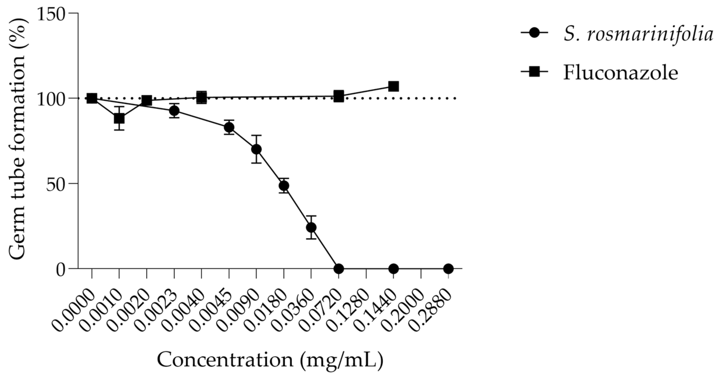
2.2.2. S. rosmarinifolia Essential Oil Inhibited Germ Tube Formation by C. albicans

Considering the relevance of dimorphic transition in C. albicans pathogenicity, we assessed whether the EO from S. rosmarinifolia could inhibit this transition. As observed in Figure 1, the EO inhibits around 50% of germ tube formation at 0.018 mg/mL (MIC/16), while fluconazole, a widely used antifungal agent in the clinic, had no effect on this virulence factor even at concentrations 200 times higher than the MIC (0.200 mg/mL). At values close to the MIC, the EO completely inhibited germ tube formation.

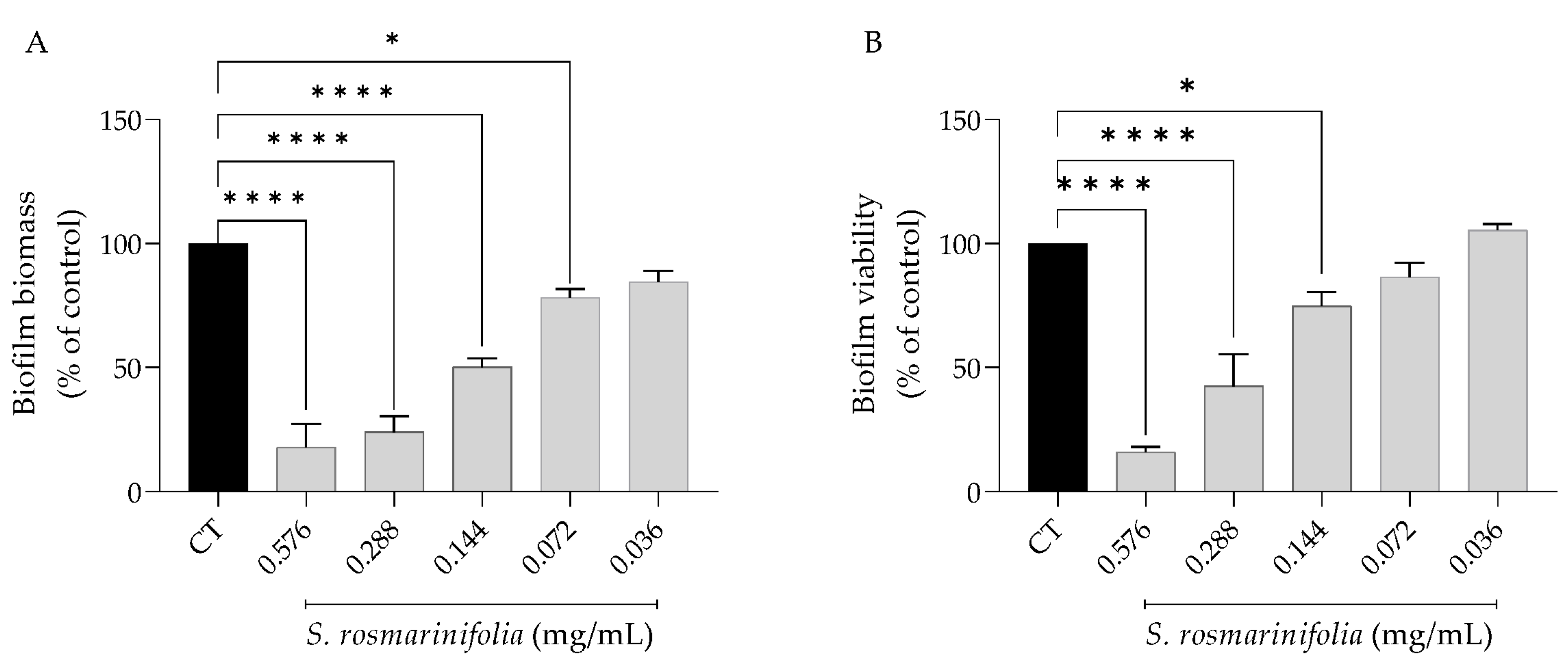
2.2.3. S. rosmarinifolia Inhibited C. albicans Biofilm Formation

Fungi resistance to antifungals is often linked to the formation of biofilms [8]; therefore, inhibiting biofilm formation would prevent this resistance. Considering its promising fungicidal effect and dimorphic transition inhibition, we next assessed the effect of the EO on biofilm formation for 24 h and 48 h. As seen in Figure 2, the presence of the EO had a stronger effect on the biofilm’s mass than on its viability, since at 0.14 mg/mL (MIC/2) the EO decreased biofilm mass by approximately 50% while decreasing viability by only 25%.
When the effect of the EO on biofilm formation was assessed 48 h post-seeding, it was observed that the effect was overall weaker (Figure 3). Indeed, at the same dose of 0.14 mg/mL, the EO inhibited biofilm mass by 35% and viability by 40%.

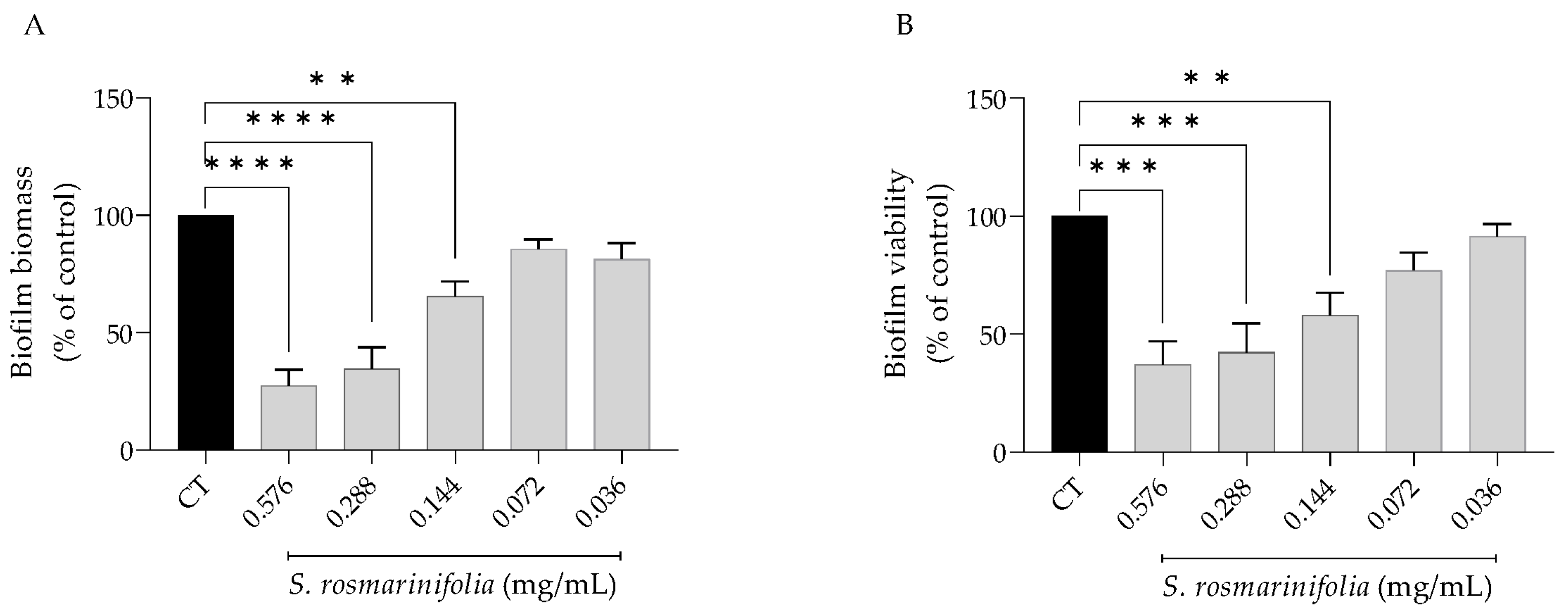
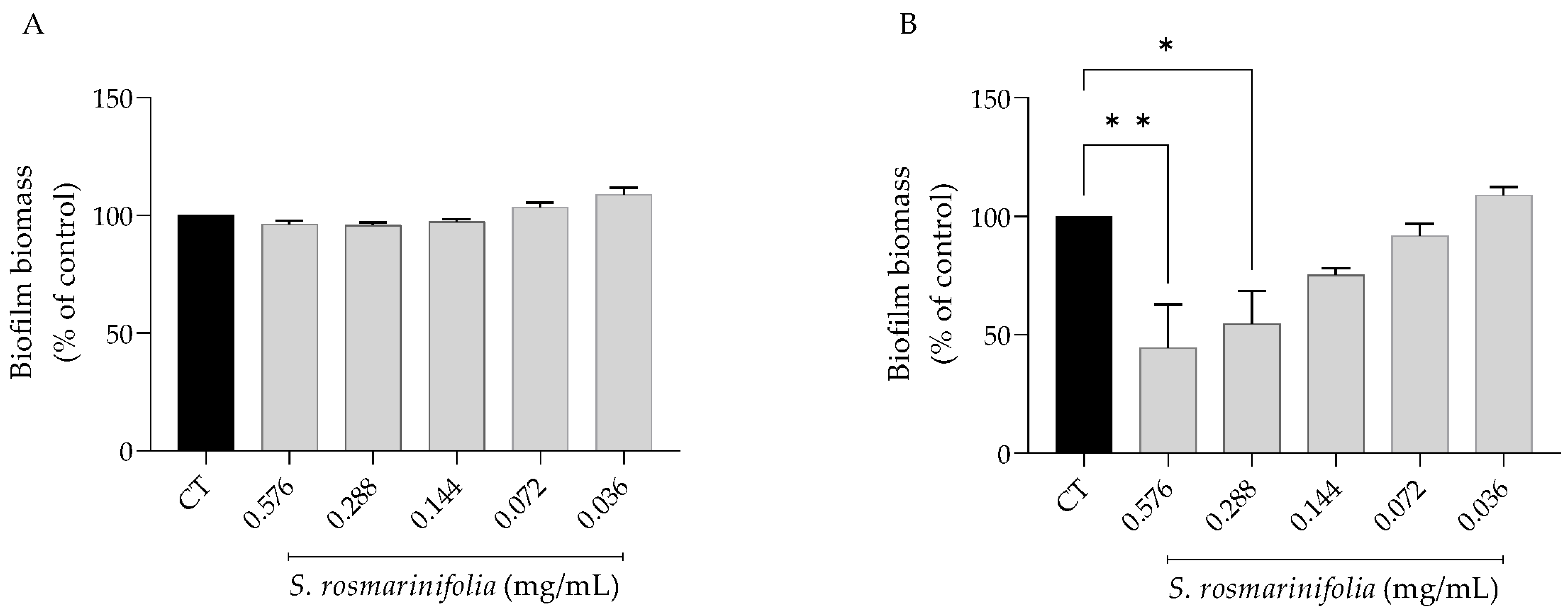
2.2.4. S. rosmarinifolia Disrupted C. albicans Preformed Biofilms

Due to their capacity to induce ecologic advantages, biofilms are difficult to eliminate, leading to refractory infections [8]; therefore, the effect of the EO on preformed C. albicans biofilms was assessed. As expected, the preformed biofilms were more resistant to the activity of the EO (Figure 4), as evidenced by a significant effect only on biofilm viability at 0.58 and 0.29 mg/mL (2xMIC and MIC).

2.3. Anti-Inflammatory Effect

Fungal infections, including those caused by dermatophytes [6,15], C. albicans [14,44] and Aspergillus [13], are often associated with an inflammatory response from the host system. Considering the relevant antifungal effect of the S. rosmarinifolia EO, we disclosed its anti-inflammatory potential by assessing the production of nitric oxide (NO) induced by lipopolysaccharide (LPS), a Toll-like receptor 4 (TLR4) agonist, in macrophages. Furthermore, the underlying mechanism of action comprised the assessment of the protein levels of inducible nitric oxide synthase (iNOS) and pro-IL1β, key pro-inflammatory mediators.

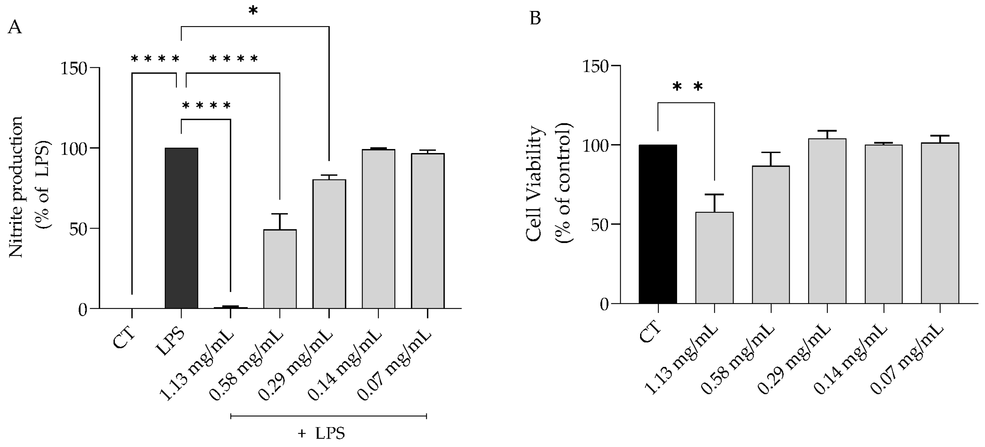
2.3.1. S. rosmarinifolia Inhibited Nitric Oxide Production on LPS-Stimulated Macrophages

As expected, the stimulation of macrophages with LPS induced an exacerbated production of nitric oxide ([NO] = 63.5 ± 13.2 µM). Pre-treatment with the EO for 1h before the stimulation with LPS led to a dose-dependent decrease in NO production (IC50 = 0.52 mg/mL) (Figure 5A). To further deepen and elucidate the underlying mechanism of action, we selected the dose of 0.58 mg/mL, as we wanted to point out a dose that could present fungicidal effect and concomitantly inhibit the C. albicans virulence factors and, importantly, was devoid of toxicity to macrophages (Figure 5B).

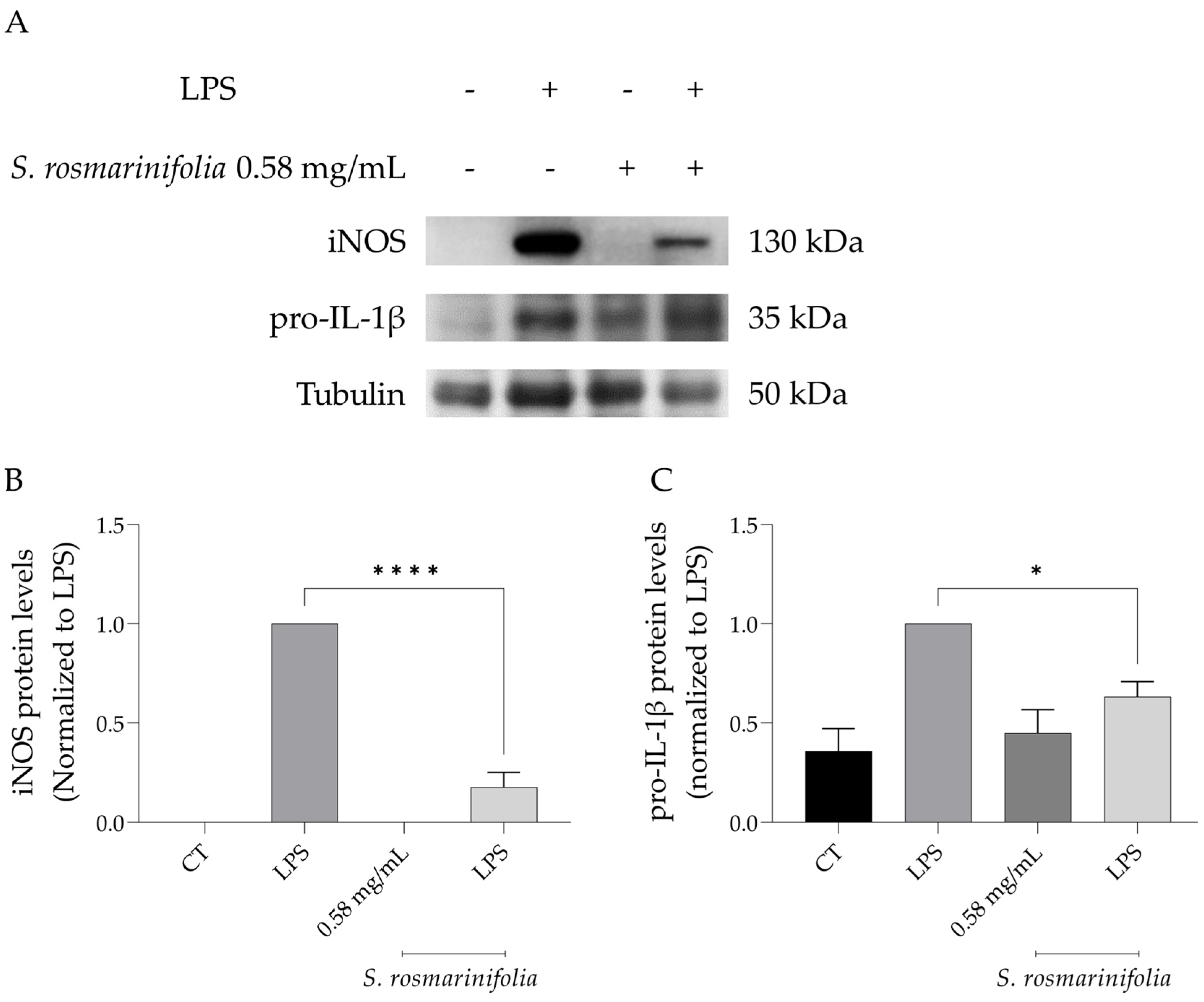
2.3.2. S. rosmarinifolia Inhibited Protein Levels of iNOS and Pro-IL1β

Among the signaling cascades associated with inflammation, NF-κB assumes a relevant role, and it is activated by the binding of LPS, or other ligands, to TLR4 and other pattern-recognition receptors (PPRs). The phosphorylation of NF-κB inhibitor alpha (IκB-α) by IκB kinase (IκK) leads to its proteasomal degradation, causing the release of p65 and p50 NF-κB subunits that translocate to the nucleus, triggering the transcription of several genes related to inflammation, namely Il1b, Il6, Nos2, Ptgs2 and Tnfα. This leads to the overproduction of several pro-inflammatory cytokines, including, but not limited to, NO, resulting from the increased expression of iNOS [45,46]. As expected, stimulating the macrophages with LPS for 24h led to an increase in the protein levels of both iNOS and pro-IL-1β, the precursor form of IL-1β, the expression of which is also controlled by NF-κB [47] (Figure 6A–C). In the presence of S. rosmarinifolia EO, the levels of both proteins were significantly reduced, particularly for iNOS (Figure 6A–C), suggesting that the EO modulates the expression of Nos2 and Il1b genes via inhibition of the NF-κB activation.

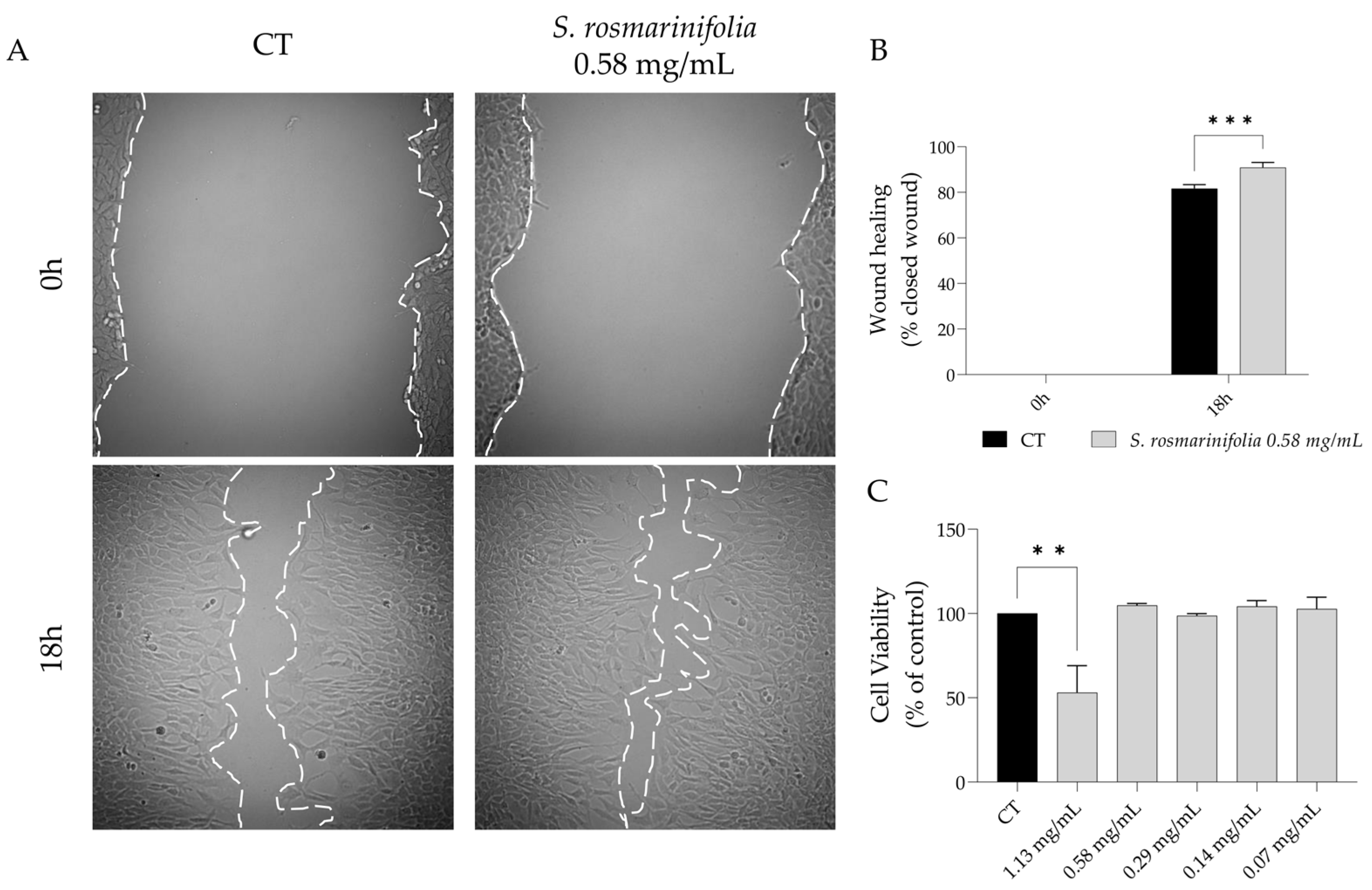
2.4. S. rosmarinifolia Promoted Wound Healing

Candida spp. are one of the most prevalent fungi found in wounds [21]. Furthermore, biofilm formation is also frequent in open wounds, which would compromise wound healing, thus perpetuating the wound and inducing chronic inflammation in the host [23,24]. Since the EO from S. rosmarinifolia inhibits and disrupts Candida spp. biofilms and decrease inflammation, we hypothesize that the EO could promote wound healing, which would decrease the risk of infection induced by biofilm formation. As seen in Figure 7, the presence of the EO significantly increased NIH 3T3 fibroblast migration in the scratch wound healing assay (91% vs. 81% of closed wound, Figure 7A) at concentrations without toxicity, thus presenting a safety profile (Figure 7B).

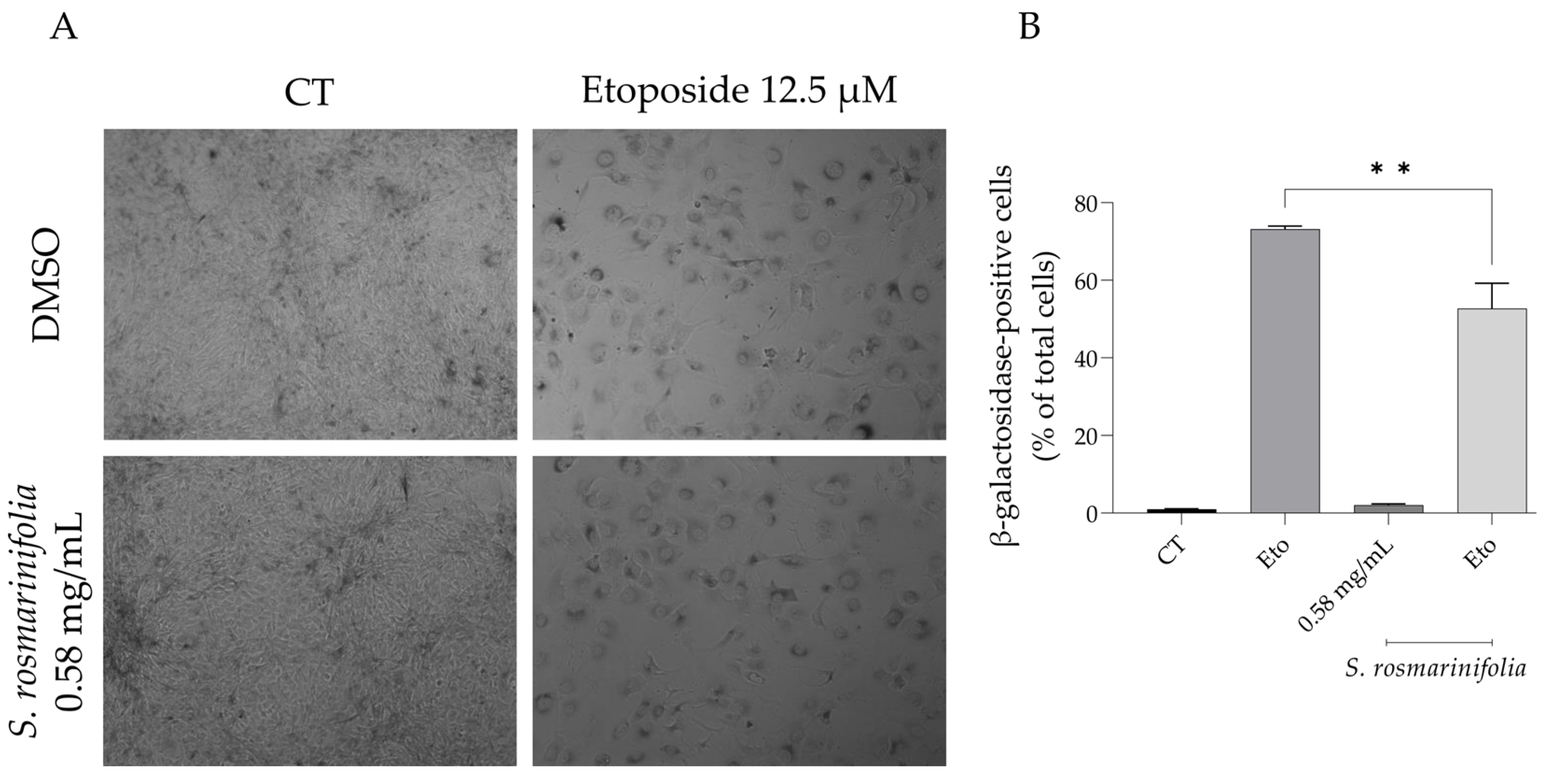
2.5. S. rosmarinifolia Decreased Etoposide-Induced Cell Senescence

It is known that inflammation leads to cell senescence [17], and that senescence itself can trigger an inflammatory response in the individual [18]. Having this in mind, we assessed whether the EO from S. rosmarinifolia could decrease cell senescence using etoposide as a senescence-inducing agent.
As expected, etoposide (for 24 h followed by 72 h in culture medium alone) triggered a large percentage of β-galactosidase-positive cells (Figure 8A,B); interestingly, the addition of the EO during the recovery phase significantly reduced the number of β-galactosidase-positive cells (53% vs. 73% of β-galactosidase-positive cells).

3. Discussion

The present study reports the chemical composition of the EO and validates the anti-inflammatory and antimicrobial properties attributed to this species. Furthermore, we report, for the first time, its capacity to inhibit C. albicans virulence factors, and we also disclose the molecular mechanisms underlying the reported anti-inflammatory activity. In addition, we also highlight the wound healing capacities and anti-senescence properties of the EO.
Herein, we report that the EO from S. rosmarinifolia collected from Portugal is mainly characterized by β-pinene, borneol, myrcene and limonene. No previous studies have been carried out on the chemical composition of the EO from Portuguese S. rosmarinifolia. However, some studies have evaluated the compositions of EOs from other Portuguese species, namely S. semidentata and S. impressa, which have distinct chemical profiles. S. semidentata presents high amounts of α-pinene, E-pinocarveol and pinocarvone [48], while S. impressa is characterized by β-pinene, 1,8-cineole, limonene, camphor and β-phellandrene [49], thus highlighting a high chemical variability among these Portuguese species. Furthermore, a recent review compared the chemical profiles of ten Santolina species, reinforcing the high variability among these species [40]. Regarding S. rosmarinifolia, it was reported that the EO from plants collected in Spain (July) has a chemical profile similar to that of the Portuguese one, namely concerning the high levels of β-pinene, limonene and myrcene. However, some differences exist, especially in the content of borneol and β-phellandrene [50]. Interestingly, a different sample from Spain, collected in April, presented high amounts of capillene, followed by β-phellandrene, myrcene, β-pinene and sabinene [51]. It is known that the season and developmental stage of the plant, as well as the genetic variability between plants, are determinant factors in the EO composition [52,53,54]. Therefore, the chemical differences between the two samples might be due to these factors. Although S. rosmarinifolia is an Iberian endemism [43], some studies have evaluated the composition of this species in other regions, particularly Algeria and Romania. These samples have different chemical profiles with high levels of sesquiterpenes, mainly germacrene D [55] and β-eudesmol or ar-curcumene [56], respectively. Another Algerian sample also showed a distinct composition, being rich in capillene [57]. The distinct chemical profiles of these samples may be due to the fact that S. rosmarinifolia is not indigenous to these countries. In addition, the soil composition and climatic conditions also impact chemical composition of EOs [54]. Furthermore, post-harvest treatment, storage conditions and EO extraction methods are known to affect EO composition [52,58]. Therefore, the observed differences might be attributed to any of these factors.
Considering the emergence of fungi resistance to conventional antifungal drugs [28,29,30,31,32], we aimed to disclose the antifungal properties of S. rosmarinifolia essential oil. We showed that the EO was able to inhibit the growth of all tested strains, with dermatophytes being the most susceptible. Previous studies have shown that S. rosmarinifolia from Algeria exerts an antibacterial effect against several bacterial strains, with Escherichia coli and Enterobacter aerogenes being the most susceptible bacteria (MIC = 0.16 mg/mL) [55]. When compared with other Portuguese Santolina species [48,49], this species shows a more potent antifungal effect, since it was active against all tested strains. Regarding its major compounds, it was shown that β-pinene inhibited the growth of Candida spp., C. neoformans, Trichophyton spp. and A. fumigatus [59,60,61,62,63]. Several studies reported the antifungal effect of limonene against Candida spp., A. fumigatus, T. rubrum and several other fungi [60,64,65,66,67,68,69,70]. In contrast, borneol presented a very weak activity against several fungal strains [60,71,72,73,74]. Similarly, myrcene presents low antifungal activity [75,76,77,78,79]. The minor compounds found in the essential oil seem to play a minimal role on the antifungal activity of S. rosmarinifolia due to their weak antimicrobial activity. Indeed, camphor [80,81,82], 1,8-cineole [83], camphene [83,84,85,86,87,88] and α-pinene [88] are reported to exert weak inhibitory effects on the growth of several fungi. These reports suggest that the inhibitory effect of S. rosmarinifolia on fungal growth might be attributed predominantly to the presence of β-pinene and limonene. Considering its interesting inhibitory effects on planktonic form, we also assessed the effect of the EO of S. rosmarinifolia on the yeast-to-hypha transition, biofilm formation and disruption of preformed biofilms. Our results show that the EO inhibits both germ tube and biofilm formation, and, interestingly, disrupts preformed biofilms, thus reinforcing its strong antifungal effect. To the best of our knowledge, no studies have been conducted on the mechanism of action underlying the antifungal properties of S. rosmarinifolia. When compared with the Portuguese S. impressa, the EO from S. rosmarinifolia presents a stronger effect on germ tube inhibition [49]. Interestingly, regarding the capacity to inhibit biofilm formation, S. rosmarinifolia was able to affect this feature at concentrations lower than the MIC, while fluconazole is ineffective even at doses 200xMIC (data not shown). Regarding isolated compounds, a review compared the effects of different terpenes on several virulence factors and reported that β-pinene and myrcene are able to inhibit germ tube formation and biofilm development, but have, however, no effect on mature biofilms [89]. The ability of β-pinene to inhibit biofilm development was reinforced by its capacity to prevent biofilm adhesion [61]. Limonene also prevents the dimorphic transition in C. parapsilosis [70]. For C. albicans, limonene was able to inhibit germ tube formation as well as the adhesion, formation and maturation of biofilms [67]. Borneol was also able to inhibit the yeast-to-hypha transition in C. albicans and disrupts preformed biofilms [73]. Regarding the effect of minor compounds on C. albicans virulence factors, it was reported that 1,8-cineole, camphene and α-pinene were able to inhibit the dimorphic transition but had weak activity against biofilm formation and disruption [89]. Camphor is also reported to inhibit biofilm formation in both C. albicans and non-albicans Candida, as well as germ tube formation [84,90]. The results herein reported suggest that the effect of S. rosmarinifolia on C. albicans virulence factors might be due to synergic effects between the major and minor compounds of the EO.
Considering the link between fungal infections and inflammation, we assessed whether the EO from S. rosmarinifolia could inhibit the inflammatory response evoked by LPS in macrophages. The results demonstrated that the EO led to a dose-dependent decrease in NO production, thus validating the traditional uses ascribed to this plant. To the best knowledge of the authors, no scientific studies have been conducted yet confirming the anti-inflammatory effects of S. rosmarinifolia. Regarding other Santolina species, the anti-inflammatory potential of the EOs from S. impressa, S. corsica, S. insularis and S. africana has been previously reported. S. africana inhibited lipoxygenase activity with an IC50 of 0.065 mg/mL [91]. On other hand, S. corsica showed anti-inflammatory potential by decreasing the inflammatory response in bronchoalveolar lavage fluid [92]. The anti-inflammatory effects of both S. insularis and S. impressa were disclosed using the NO production evoked by LPS in macrophages, with IC50 of 0.55 and 0.35 mg/mL, respectively [49,93]. Regarding isolated compounds, β-pinene significantly reduced NO production in both LPS-stimulated macrophages [94] and IL-1β-activated chondrocytes [95]. Furthermore, this monoterpene is also able to inhibit the chemotaxis in neutrophils [96]. The supplementation of the mice diet with borneol significantly decreased the levels of the pro-inflammatory cytokines IL-1β and IL-6 [97]. Accordingly, several other studies reported the anti-inflammatory effect of borneol, by showing its capacity to inhibit pro-inflammatory mediators such as NO, TNF-α and IL-6 in LPS-stimulated macrophages and by decreasing fever in a model of endotoxic fever [98]. The anti-inflammatory potential of borneol was validated in animal models of mucositis [99], acne [100] and carrageenan-induced leukocyte migration [101]. Myrcene and limonene, also found in high amounts in S. rosmarinifolia, decreased NO production in IL-1β-activated human chondrocytes (IC50 = 37.3 and 85.3 µg/mL, respectively) [102] and LPS-activated macrophages, as well as in a model of pleurisy [103]. Considering the reported effects of these compounds, we hypothesize that the reduction of NO production evoked by the S. rosmarinifolia EO might be due to the synergistic effects between them. Having in mind the relevant activity of the EO in decreasing NO production, we further assessed its effect on the protein levels of relevant pro-inflammatory enzymes and cytokines. The results obtained demonstrated that the EO decreased the protein levels of iNOS and pro-IL-1β triggered by LPS, suggesting that S. rosmarinifolia might be able to inhibit the nuclear translocation of NF-κB, thus inhibiting the gene expression of pro-inflammatory mediators. Regarding the major compounds, Rufino and colleagues demonstrated that β-pinene [95] and myrcene [102] inhibited the expression of iNOS. Additionally, borneol inhibits IκBα degradation and the consequent NF-κB translocation to the nucleus [104], thus suggesting that it could inhibit the expression of pro-inflammatory mediators. Another relevant monoterpene, limonene, also decreased the protein levels of iNOS [102,105,106]. The anti-inflammatory effects reported for S. rosmarinifolia seem to be also attributable to the presence of minor compounds. Indeed, α-pinene inhibited NO release in IL-1β-stimulated chondrocytes [95] and LPS-stimulated macrophages [94] to a larger extent relative to the β isomer, thus suggesting that the reported activity might include a significant contribution from this minor compound. Other minor compounds, particularly 1,8-cineole [107,108,109,110,111,112], camphor [113,114,115] and camphene [116,117], are widely described as exerting anti-inflammatory effects both in vivo and in vitro. On the other hand, bornyl acetate is devoid of anti-inflammatory activity on LPS-stimulated macrophages [94].
Considering that fungal infections are often associated with chronic wounds, we hypothesized that the EO from S. rosmarinifolia could promote wound healing, thus decreasing the risk of wound-associated infections. Our results show, for the first time, that S. rosmarinifolia EO promotes wound healing, and, as far as the authors know, this is the first report on the wound healing properties of Santolina spp. Regarding the wound healing activity of the major compounds found in the S. rosmarinifolia EO, several studies have reported this activity for borneol [99,118,119] and limonene [120,121,122]. Regarding β-pinene, an essential oil from Teucrium polium subsp. capitatum rich in this compound increased wound healing [123]. Myrcene’s effect on cell migration is of a dual nature, with inhibitory effects on oral cancer cell lines [124] and stimulation of cell migration on HaCaT cells [125]. The minor compounds 1,8-cineole [112,126,127], camphor [128] and α-pinene [126] are known to promote wound healing, and thus they may also contribute to the herein reported activity of S. rosmarinifolia. Considering these results, we suggest that the reported activity of S. rosmarinifolia might be attributed to the presence of these compounds.
Since inflammation and aging/senescence are closely related, and considering the promising anti-inflammatory effects observed for S. rosmarinifolia, we then assessed the anti-senescence potential of the EO. The presence of the EO reduced the senescence-associated β-galactosidase activity, thus showing anti-aging effects. To the best of our knowledge, no prior studies have reported the anti-senescence effect of Santolina spp. Regarding isolated compounds, few studies have addressed the anti-aging effects of the major compounds found in S. rosmarinifolia. Myrcene improves skin aging by reducing the production of metalloproteinases and increasing the secretion of TGF-1 and type I collagen [129], and limonene increased the lifespan of the Mediterranean fruit fly and Drosophila melanogaster [130,131]. These results suggest that the presence of limonene and myrcene might explain the anti-senescence effect reported for S. rosmarinifolia. Nevertheless, the contribution of other minor compounds cannot be ruled out. Indeed, camphor, a monoterpene found in low amounts in the EO of S. rosmarinifolia, prevents the increased activity of senescence-associated β-galactosidase [128]. The anti-aging potential of α-pinene is also known [132]. In contrast, 1,8-cineole, another minor compound of S. rosmarinifolia, induced cell senescence [133]; therefore, it is likely that this compound does not contribute to and is not involved in the anti-senescence activity of S. rosmarinifolia.

4. Materials and Methods

4.1. Essential Oil Distillation and Analysis

4.1.1. Plant Collection and Essential Oil Distillation

Aerial parts from Santolina rosmarinifolia were collected in July 2021, during the flowering stage, at Alto das Necessidades, Palmela, Setúbal (Portugal, 38°31′54.4″ N 8°59′19.4″ W). A voucher specimen (L. Salgueiro 87) was deposited at the Herbarium of the Faculty of Pharmacy of the University of Coimbra, Portugal. The species authenticity was validated by a taxonomist at the University of Coimbra, Doutor Jorge Paiva. The plant name was confirmed at http://www.worldfloraonline.org/taxon/wfo-0000014722 (accessed on 29 December 2022).
Prior to hydrodistillation, the plant material was air-dried for three days while protected from light. Afterwards, the material was submitted to hydrodistillation for 3 h using a Clevenger-type apparatus, as recommended by the European Pharmacopoeia [134] for isolation of EOs.

4.1.2. Essential Oil Analysis

The composition of the essential oil was determined by a combination of gas chromatography with flame ionization detection (GC-FID) and gas chromatography-mass spectrometry (GC-MS) as previously reported [135]. The essential oil components were identified by their retention indices on both the SPB-1 and Supelcowax-10 columns and by their mass spectra. The retention indices, calculated by linear interpolation to the retention times of C8–C23 n-alkanes, were compared with those of reference samples included in the laboratory database or reported in the literature [136]. The acquired mass spectra were compared with reference spectra from the laboratory’s database, the Wiley/National Institute of Standards and Technology (NIST) library [137] and data in the literature [138]. Relative amounts of individual components were calculated based on the GC raw data areas without FID response factor correction.

4.2. Antifungal Activity

4.2.1. Fungal Strains

The antifungal activity of the EO of S. rosmarinifolia was evaluated against filamentous fungi and yeasts. The filamentous fungi included dermatophytes from clinical origin (Epidermophyton floccosum FF9, Trichophyton mentagrophytes FF7 and Microsporum canis FF1) and from collection (T. mentagrophytes var. interdigitale CECT 2958, T. rubrum CECT 2794, T. verrucosum CECT 2992, and M. gypseum CECT 2908) and Aspergillus spp. (A. flavus F44, A. fumigatus ATCC 46645 and A. niger ATCC 16404). The yeasts were represented by a Cryptococcus neoformans type strain (C. neoformans YPO186); by clinical Candida strains (C. krusei LF33, C. guilliermondii MAT23) and Candida collection strains (C. albicans ATCC 10231, C. tropicalis YPO128 and C. parapsilopsis ATCC 90018) and by a Yarrowia lipolitica type strain (Y. lipolytica ISA1774).

4.2.2. Macrodilution Broth Assay

The MIC and MLC of the EO were discovered using the CLSI reference protocols M27-A3 [139] and M38-A2 [140] for yeasts and filamentous fungi, respectively, as previously reported [85]. A negative control (non-inoculated medium) and a positive control (inoculated medium treated with DMSO) were included. The DMSO concentration never exceeded 1% (v/v).

4.2.3. C. albicans Germ Tube Formation

To assess the effect of the EO on the yeast-to-hypha transition, cell suspensions of C. albicans ATCC 10231 from overnight cultures on SDA were prepared in NYP medium [141], as reported [142]. Sub-inhibitory concentrations of the EO and fluconazole were used. Germ tubes were considered when the hypha was at least as long as the diameter of the blastopore. DMSO in a maximum concentration of 1% (v/v) was used as a positive control.

4.2.4. C. albicans Biofilm Formation and Disruption of Preformed Biofilm

C. albicans biofilm formation was assessed according to the method described by Taweechaisupapong et al. [143] with slight modifications, as previously reported [142]. Briefly, 100 µL of a C. albicans suspension (1 × 10⁶ cells/mL) made in RPMI-1640 were added to 96-well pre-sterilized, polystyrene and flat-bottom microtiter plates, previously loaded with 100 µL of S. rosmarinifolia EO (0.036–0.576 mg/mL) serial dilutions made in RPMI. The plates were then incubated for 24h or 48h at 37 °C to allow the biofilm formation. C. albicans biofilm disruption assays were performed as reported [144]. Succinctly, 100 µL of C. albicans suspension (1 × 10⁶ cells/mL) made in RPMI was added to 96-well polystyrene microtiter plates and incubated for 24h at 37 °C. After medium removal, the wells were washed with PBS and 100 µL of serial dilutions of S. rosmarinifolia EO (0.036–0.576 mg/mL) made in RPMI were added and further incubated for 24 h at 37 °C. Before biofilm viability, the wells were washed three times with PBS to remove non-adherent cells. Negative (non-inoculated medium) and positive (inoculated medium treated with 1% DMSO) controls were also included. The biofilm viability was determined using the XTT/menadione metabolic assay as described [145] with some modifications, as reported [83]. The biofilm mass was quantified using the crystal violet assay as reported [89].

4.3. Anti-Inflammatory Assays

4.3.1. Cell Culture

RAW 264.7 macrophages from the American Type Culture Collection (ATCC TIB-71) were cultured as previously reported [117].

4.3.2. Nitric Oxide Production

The NO production was measured by quantifying the accumulation of nitrites in the culture supernatants, using the Griess reagent [146] stabilization, the macrophages were pre-treated for 1h with the EO (0.07–1.13 mg/mL) diluted in DMSO and then activated with 50 ng/mL of LPS over a period of 24h. Positive (LPS-stimulated macrophages) and negative controls (untreated macrophages) were performed. After this incubation period, equal volumes of the culture supernatants and Griess reagent [1:1 of 0.1% (w/v) N-(1-naphthyl) ethylenediaminedihydrochloride and 1% (w/v) sulphanilamide containing 5% (w/v) H3PO4] were mixed and incubated for 30 min in the dark. The absorbance at 550 nm was registered in an automated plate reader (SLT, Austria), and the nitrite concentration was determined from a sodium nitrite standard curve. DMSO at the maximum concentration used (0.4%) was already demonstrated by our group to be devoid of anti-inflammatory and cytotoxicity effects (data not shown).

4.3.3. Expression of Pro-Inflammatory Proteins, iNOS and pro-IL-1β

RAW 264.7 (1 × 10⁶ cells/mL) were cultured in 6-well plates and allowed to stabilize overnight. Then, the cells were incubated with the EO (0.58 mg/mL) for 1 h, followed by LPS (50 ng/mL) activation for a period of 24 h. A negative control (untreated cells) and a positive control (LPS-only treated cells) were considered. The cell lysates were prepared as previously described [117].
A Western blot analysis was carried out to measure the protein levels of inducible nitric oxide synthase (iNOS) and pro-IL-1β. The separation, transference and membrane blocking with 5% milk made in TBS-T were performed as previously reported [147]. Afterwards, the membranes were incubated overnight at 4 °C with antibodies against iNOS (1:500; R & D Systems, MAB9502) or pro-IL-1β (1:1000; Abcam, ab9722). The membranes were then washed and incubated with secondary antibodies (1:40,000; Santa Cruz Biotechnology) conjugated with horseradish peroxidase for 1h at room temperature. After a new set of washes, the immunocomplexes were detected using a chemiluminescence scanner (Image Quant LAS 500, GE). Tubulin was used as loading control. A semi-quantitative analysis was carried out using ImageLab version 6.1.0 (Bio-Rad Laboratories Inc.).

4.4. Cell Migration

4.4.1. Cell Culture

NIH 3T3, a mouse embryonic fibroblast cell line (ATCC CRL-1658), was cultured as previously described in our group [147].

4.4.2. Cell Migration Assay

The cell migration was carried out using the scratch wound assay as reported [148] with slight modifications. Briefly, NIH 3T3 fibroblasts were seeded at 2.5 × 10⁵ cells/mL in 12-well plates. After 24h of growth, a scratch was made in the cell monolayer using a 20–200 µL pipette tip. The detached cells were removed by washing the cells with sterile PBS 1x. DMEM with 2% serum was added to all plates, in the presence or absence of the EO (0.58 mg/mL). Using a phase-contrast microscope, images were acquired 0 and 18 h post-scratch, and the wound area was measured using ImageJ/Fiji software. The results presented were obtained using the following equation:
wound   closure   ( % ) = A t = 0 h   A t = xh A t = 0 h × 100
where At=0h is the area of the wound 0h after the scratch and At=xh is the area at the different time post-scratch (0 h and 18 h).

4.5. Cell Viability

The study of the effects of different concentrations of the essential oil on the viability of both macrophages and fibroblasts was carried out using the resazurin reduction assay. Briefly, macrophages (0.6 × 10⁶ cells/mL) or fibroblasts (1.25 × 10⁵ cells/mL) were seeded in 48-well plates. After an overnight stabilization, different concentrations of the EO (0.07–1.13 mg/mL), diluted in DMSO, were added for 24 h. At the end of the experiment, the medium was removed and fresh medium containing resazurin (1:10) was added for 1h or 4h, for macrophages and fibroblasts, respectively. The absorbance at 570 nm with a reference filter of 620 nm was registered in an automated plate reader (SLT, Austria). Cell viability was determined using the following equation:
Cell   viability   ( % ) = Abs Exp Abs CT × 100
where AbsExp is the absorbance (difference between 570 and 620 nm) in the different experimental conditions and AbsCT is the absorbance in the control cells (no essential oil).

4.6. Etoposide-Induced Senescence

Senescence was assessed using the methodology previously published [149], with slight modifications. Briefly, after etoposide incubation for 24 h, cells were left to recover in the presence or absence of S. rosmarinifolia EO (0.58 mg/mL) for 72 h. Afterwards, cells were washed with PBS 1x, and beta-galactosidase staining was performed as described in the manufacturer’s protocol (Cell Signaling Technology). Eight images per condition were randomly acquired using a bright-field microscope. Cells showing a distinct blue color of staining were senescent. The number of positive and total cells was counted, and the percentage of senescence was calculated.

4.7. Statistical Analysis

Results are shown as mean values ± SEM (standard error of the mean) from a minimum of three independent experiments made in duplicate. For cell migration assays, two-way ANOVA followed by Sydák’s multiple comparison test were performed to determine statistical significance. For the remaining assays, statistical significance was determined using one-way ANOVA followed by Dunnett’s post-hoc test using GraphPad Prism version 9.5.0 (GraphPad Software, San Diego, CA, USA). Significance was considered when the p value was lower than 0.05.

5. Conclusions

This study validates the traditional uses ascribed to S. rosmarinifolia, particularly its antimicrobial and anti-inflammatory activities. Indeed, we report that the EO inhibits the growth of several pathogenic fungi and mitigates relevant C. albicans virulence factors, particularly the yeast-to-hypha transition and biofilm formation. Of relevance, we also report that the EO is able to disrupt preformed biofilms, thus strengthening the antifungal potential of this species. Regarding the anti-inflammatory activity, the EO significantly inhibits NO production as well as iNOS and pro-IL1β protein levels, probably through NF-κB pathway modulation. Interestingly, other pharmacological properties were also highlighted, specifically wound healing and anti-senescence.
Overall, this study emphasizes the industrial interest of S. rosmarinifolia by showing its antimicrobial and anti-inflammatory properties and concomitantly shedding light on new pharmacological activities. Furthermore, S. rosmarinifolia could be of interest for the management of fungal infections and the associated inflammatory response, as well as for inflammaging.

Author Contributions

Conceptualization, L.S.; validation, A.S., M.J.G. and M.T.C.; formal analysis, J.M.A.-S., M.J.G. and C.C.; investigation, J.M.A.-S. and C.C.; resources, M.T.C. and L.S.; writing—original draft preparation, J.M.A.-S.; writing—review and editing, M.T.C. and L.S.; visualization, J.M.A.-S.; supervision, L.S.; project administration, L.S.; funding acquisition, L.S. and M.T.C. All authors have read and agreed to the published version of the manuscript.

Funding

This work was financed by COMPETE 2020—Operational Programme for Competitiveness and Internationalisation and Portuguese national funds via FCT—Fundação para a Ciência e a Tecnologia, under projects UIDB/04539/2020, UIDP/04539/2020 and LA/P/0058/2020.

Institutional Review Board Statement

Not applicable.

Informed Consent Statement

Not applicable.

Data Availability Statement

Data will be available upon request.

Conflicts of Interest

The authors declare no conflict of interest. The funders had no role in the design of the study; in the collection, analyses or interpretation of data; in the writing of the manuscript, or in the decision to publish the results.

References

  1. Bongomin, F.; Gago, S.; Oladele, R.O.; Denning, D.W. Global and Multi-National Prevalence of Fungal Dis-eases-Estimate Precision. J. Fungi 2017, 3, 57. [Google Scholar] [CrossRef]
  2. Pierce, C.; Lopez-Ribot, J.L. Candidiasis Drug Discovery and Development: New Approaches Targeting Vir-ulence for Discovering and Identifying New Drugs. Expert Opin. Drug Discov. 2013, 8, 1117–1126. [Google Scholar] [CrossRef] [PubMed] [Green Version]
  3. Campoy, S.; Adrio, J.L. Antifungals. Biochem. Pharm. 2017, 133, 86–96. [Google Scholar] [CrossRef]
  4. Dadar, M.; Tiwari, R.; Karthik, K.; Chakraborty, S.; Shahali, Y.; Dhama, K. Candida Albicans - Biology, Mo-lecular Characterization, Pathogenicity, and Advances in Diagnosis and Control—An Update. Microb. Pathog. 2018, 117, 128–138. [Google Scholar] [CrossRef] [PubMed]
  5. Gulati, M.; Nobile, C.J. Candida Albicans Biofilms: Development, Regulation, and Molecular Mechanisms. Microbes Infect. 2016, 18, 310–321. [Google Scholar] [CrossRef] [PubMed] [Green Version]
  6. Kernien, J.F.; Snarr, B.D.; Sheppard, D.C.; Nett, J.E. The Interface between Fungal Biofilms and Innate Im-munity. Front. Immunol. 2018, 8, 1968. [Google Scholar] [CrossRef] [Green Version]
  7. Lee, J.-H.; Kim, Y.-G.; Gupta, V.K.; Manoharan, R.K.; Lee, J. Suppression of Fluconazole Resistant Candida Albicans Biofilm Formation and Filamentation by Methylindole Derivatives. Front. Microbiol. 2018, 9, 2641. [Google Scholar] [CrossRef]
  8. Silva, S.; Rodrigues, C.; Araújo, D.; Rodrigues, M.; Henriques, M. Candida Species Biofilms’ Antifungal Resis-tance. J. Fungi 2017, 3, 8. [Google Scholar] [CrossRef] [Green Version]
  9. Gupta, A.K.; Cooper, E.A. Update in Antifungal Therapy of Dermatophytosis. Mycopathologia 2008, 166, 353–367. [Google Scholar] [CrossRef]
  10. Piggott, C.D.S.; Friedlander, S.F. Etiologic Agents of Infections Diseases. In Principles and Practice of Pediatric Infections Diseases; Elsevier: Amsterdam, The Netherlands, 2012. [Google Scholar]
  11. Cardoso, N.N.R.; Alviano, C.S.; Blank, A.F.; Romanos, M.T.V.; Fonseca, B.B.; Rozental, S.; Rodrigues, I.A.; Al-viano, D.S. Synergism Effect of the Essential Oil from Ocimum Basilicum Var. Maria Bonita and Its Major Components with Fluconazole and Its Influence on Ergosterol Biosynthesis. Evid. Based Complement. Altern. Med. 2016, 2016, 5647182. [Google Scholar] [CrossRef]
  12. Desnos-Ollivier, M.; Letscher-Bru, V.; Neuvéglise, C.; Dromer, F. Yarrowia Lipolytica Causes Sporadic Cases and Local Outbreaks of Infections and Colonisation. Mycoses 2020, 63, 737–745. [Google Scholar] [CrossRef]
  13. Yan, W.; Zhao, Y.; Xie, K.; Xing, Y.; Xu, F. Aspergillus Fumigatus Influences Gasdermin-D-Dependent Pyrop-tosis of the Lung via Regulating Toll-Like Receptor 2-Mediated Regulatory T Cell Differentiation. J. Immunol. Res. 2021, 2021, 5538612. [Google Scholar] [CrossRef]
  14. Wang, W.; Deng, Z.; Wu, H.; Zhao, Q.; Li, T.; Zhu, W.; Wang, X.; Tang, L.; Wang, C.; Cui, S.-Z.; et al. A Small Secreted Protein Triggers a TLR2/4-Dependent Inflammatory Response during Invasive Candida Albicans Infection. Nat. Commun. 2019, 10, 1015. [Google Scholar] [CrossRef] [PubMed] [Green Version]
  15. Celestrino, G.A.; Reis, A.P.C.; Criado, P.R.; Benard, G.; Sousa, M.G.T. Trichophyton Rubrum Elicits Phagocytic and Pro-Inflammatory Responses in Human Monocytes Through Toll-Like Receptor 2. Front. Microbiol. 2019, 10, 2589. [Google Scholar] [CrossRef] [Green Version]
  16. Sun, S.-C. The Non-Canonical NF-B Pathway in Immunity and Inflammation. Nat. Rev. Immunol. 2017, 17, 545–558. [Google Scholar] [CrossRef] [PubMed]
  17. Zhu, X.; Chen, Z.; Shen, W.; Huang, G.; Sedivy, J.M.; Wang, H.; Ju, Z. Inflammation, Epigenetics, and Metabo-lism Converge to Cell Senescence and Ageing: The Regulation and Intervention. Signal Transduct Target 2021, 6, 245. [Google Scholar] [CrossRef]
  18. Stojanović, S.D.; Fiedler, J.; Bauersachs, J.; Thum, T.; Sedding, D.G. Senescence-Induced Inflammation: An Important Player and Key Therapeutic Target in Atherosclerosis. Eur. Heart J. 2020, 41, 2983–2996. [Google Scholar] [CrossRef] [Green Version]
  19. Malani, P.N.; McNeil, S.A.; Bradley, S.F.; Kauffman, C.A. Candida Albicans Sternal Wound Infections: A Chronic and Recurrent Complication of Median Sternotomy. Clin. Infect. Dis. 2002, 35, 1316–1320. [Google Scholar] [CrossRef] [PubMed] [Green Version]
  20. Gil, J.; Solis, M.; Higa, A.; Davis, S.C. Candida Albicans Infections: A Novel Porcine Wound Model to Evalu-ate Treatment Efficacy. BMC Microbiol. 2022, 22, 45. [Google Scholar] [CrossRef]
  21. Kalan, L.; Grice, E.A. Fungi in the Wound Microbiome. Adv. Wound Care 2018, 7, 247–255. [Google Scholar] [CrossRef]
  22. Sharma, A.; Gupta, S. Protective Manifestation of Herbonanoceuticals as Antifungals: A Possible Drug Can-didate for Dermatophytic Infection. Health Sci. Rep. 2022, 5, e775. [Google Scholar] [CrossRef]
  23. Wu, Y.-K.; Cheng, N.-C.; Cheng, C.-M. Biofilms in Chronic Wounds: Pathogenesis and Diagnosis. Trends Bio-technol. 2019, 37, 505–517. [Google Scholar] [CrossRef]
  24. Percival, S.L. Importance of Biofilm Formation in Surgical Infection. Br. J. Surg. 2017, 104, e85–e94. [Google Scholar] [CrossRef] [PubMed] [Green Version]
  25. Guo, S.; DiPietro, L.A. Factors Affecting Wound Healing. J. Dent. Res. 2010, 89, 219–229. [Google Scholar] [CrossRef]
  26. Pfaller, M.A. Antifungal Drug Resistance: Mechanisms, Epidemiology, and Consequences for Treatment. Am. J. Med. 2012, 125, S3–S13. [Google Scholar] [CrossRef] [PubMed]
  27. Fuentefria, A.M.; Pippi, B.; Dalla Lana, D.F.; Donato, K.K.; de Andrade, S.F. Antifungals Discovery: An Insight into New Strategies to Combat Antifungal Resistance. Lett. Appl. Microbiol. 2018, 66, 2–13. [Google Scholar] [CrossRef] [Green Version]
  28. Pozzatti, P.; Scheid, L.A.; Spader, T.B.; Atayde, M.L.; Santurio, J.M.; Alves, S.H. In Vitro Activity of Essential Oils Extracted from Plants Used as Spices against Fluconazole-Resistant and Fluconazole-Susceptible Can-dida Spp. Can J. Microbiol. 2008, 54, 950–956. [Google Scholar] [CrossRef]
  29. Mourad, A.; Perfect, J.R. The War on Cryptococcosis: A Review of the Antifungal Arsenal. Mem. Inst. Oswaldo. Cruz. 2018, 113, e170391. [Google Scholar] [CrossRef] [PubMed]
  30. Chowdhary, A.; Sharma, C.; Meis, J.F. Azole-Resistant Aspergillosis: Epidemiology, Molecular Mechanisms, and Treatment. J. Infect. Dis. 2017, 216, S436–S444. [Google Scholar] [CrossRef] [Green Version]
  31. Martinez-Rossi, N.M.; Bitencourt, T.A.; Peres, N.T.A.; Lang, E.A.S.; Gomes, E.V.; Quaresemin, N.R.; Martins, M.P.; Lopes, L.; Rossi, A. Dermatophyte Resistance to Antifungal Drugs: Mechanisms and Prospectus. Front. Microbiol. 2018, 9, 1108. [Google Scholar] [CrossRef]
  32. Colombo, A.L.; Júnior, J.N.; de Almeida Júnior, J.N.; Guinea, J. Emerging Multidrug-Resistant Candida Species. Curr. Opin Infect. Dis. 2017, 30, 528–538. [Google Scholar] [CrossRef] [PubMed]
  33. Khan, A.; Ahmad, A.; Akhtar, F.; Yousuf, S.; Xess, I.; Khan, L.A.; Manzoor, N. Ocimum Sanctum Essential Oil and Its Active Principles Exert Their Antifungal Activity by Disrupting Ergosterol Biosynthesis and Mem-brane Integrity. Res. Microbiol. 2010, 161, 816–823. [Google Scholar] [CrossRef] [PubMed]
  34. Attiq, A.; Jalil, J.; Husain, K.; Ahmad, W. Raging the War Against Inflammation With Natural Products. Front. Pharm. 2018, 9, 976. [Google Scholar] [CrossRef] [PubMed] [Green Version]
  35. Vega, R.J.S.; Xolalpa, N.C.; Castro, A.J.A.; Pérez González, C.; Pérez Ramos, J.; Pérez Gutiérrez, S. Terpenes from Natural Products with Potential Anti-Inflammatory Activity. In Terpenes and Terpenoids; IntechOpen: London, UK, 2018. [Google Scholar]
  36. Serra, E.; Hidalgo-Bastida, L.; Verran, J.; Williams, D.; Malic, S. Antifungal Activity of Commercial Essential Oils and Biocides against Candida Albicans. Pathogens 2018, 7, 15. [Google Scholar] [CrossRef] [Green Version]
  37. Bakkali, F.; Averbeck, S.; Averbeck, D.; Idaomar, M. Biological Effects of Essential Oils—A Review. Food Chem. Toxicol. 2008, 46, 446–475. [Google Scholar] [CrossRef]
  38. Abad, M.J.; Bedoya, L.M.; Apaza, L.; Bermejo, P. The Artemisia L. Genus: A Review of Bioactive Essential Oils. Molecules 2012, 17, 2542–2566. [Google Scholar] [CrossRef] [Green Version]
  39. Babaei, G.; Aliarab, A.; Abroon, S.; Rasmi, Y.; Aziz, S.G.-G. Application of Sesquiterpene Lactone: A New Promising Way for Cancer Therapy Based on Anticancer Activity. Biomed. Pharmacother. 2018, 106, 239–246. [Google Scholar] [CrossRef]
  40. Tundis, R.; Loizzo, M.R. A Review of the Traditional Uses, Phytochemistry and Biological Activities of the Genus Santolina. Planta Med. 2018, 84, 627–637. [Google Scholar] [CrossRef] [Green Version]
  41. Novais, M.H.; Santos, I.; Mendes, S.; Pinto-Gomes, C. Studies on Pharmaceutical Ethnobotany in Arrabida Natural Park (Portugal). J. Ethnopharmacol. 2004, 93, 183–195. [Google Scholar] [CrossRef]
  42. Beniston, N.T.; Beniston, W.S. Fleurs d’Algérie; Entreprise Nationale du Livre: Alger, Algérie, 1984. [Google Scholar]
  43. RIVERO-GUERRA, A.O. Phenotypic Differentiation of Peripheral Populations of Santolina Rosmarinifolia (Asteraceae). Bot. J. Linn. Soc. 2008, 158, 650–668. [Google Scholar] [CrossRef]
  44. Stappers, M.H.T.; Brown, G.D. Host Immune Responses During Infections with Candida Albicans. In Candida albicans: Cellular and Molecular Biology; Springer International Publishing: Cham, Switzerland, 2017; pp. 145–183. [Google Scholar]
  45. Guijarro-Muñoz, I.; Compte, M.; Álvarez-Cienfuegos, A.; Álvarez-Vallina, L.; Sanz, L. Lipopolysaccharide Activates Toll-like Receptor 4 (TLR4)-Mediated NF-ΚB Signaling Pathway and Proinflammatory Response in Human Pericytes. J. Biol. Chem. 2014, 289, 2457–2468. [Google Scholar] [CrossRef] [PubMed] [Green Version]
  46. Liu, T.; Zhang, L.; Joo, D.; Sun, S.-C. NF-ΚB Signaling in Inflammation. Signal Transduct. Target. 2017, 2, 17023. [Google Scholar] [CrossRef] [PubMed] [Green Version]
  47. Basak, C.; Pathak, S.K.; Bhattacharyya, A.; Mandal, D.; Pathak, S.; Kundu, M. NF-ΚB- and C/EBPβ-Driven Interleukin-1β Gene Expression and PAK1-Mediated Caspase-1 Activation Play Essential Roles in Interleu-kin-1β Release from Helicobacter Pylori Lipopolysaccharide-Stimulated Macrophages. J. Biol. Chem. 2005, 280, 4279–4288. [Google Scholar] [CrossRef] [PubMed] [Green Version]
  48. Gomes, A.; Pimpão, R.C.; Fortalezas, S.; Figueira, I.; Miguel, C.; Aguiar, C.; Salgueiro, L.; Cavaleiro, C.; Gon-çalves, M.J.; Clemente, A.; et al. Chemical Characterization and Bioactivity of Phytochemicals from Iberian Endemic Santolina Semidentata and Strategies for Ex Situ Propagation. Ind. Crop. Prod. 2015, 74, 505–513. [Google Scholar] [CrossRef] [Green Version]
  49. Alves-Silva, J.M.; Zuzarte, M.; Gonçalves, M.J.; Cruz, M.T.; Cavaleiro, C.; Salgueiro, L. Unveiling the Bioac-tive Potential of the Essential Oil of a Portuguese Endemism, Santolina Impressa. J. Ethnopharmacol. 2019, 244, 112–120. [Google Scholar] [CrossRef]
  50. Palá-Paúl, J.; Pérez-Alonso, M.J.; Velasco-Negueruela, A.; Palá-Paúl, R.; Sanz, J.; Conejero, F. Seasonal Varia-tion in Chemical Constituents of Santolina rosmarinifolia L. Ssp. Rosmarinifolia. Biochem. Syst. Ecol 2001, 29, 663–672. [Google Scholar] [CrossRef]
  51. Palá-Paúl, J.; Pérez-Alonso, M.J.; Velasco-Negueruela, A.; Ramos-Vázquez, P.; Gómez-Contreras, F.; Sanz, J. Essential Oil OfSantolina Rosmarinifolia L. Ssp.Rosmarinifolia: First Isolation of Capillene, a Diacetylene Derivative. Flavour Fragr. J. 1999, 14, 131–134. [Google Scholar] [CrossRef]
  52. Mohammadhosseini, M.; Sarker, S.D.; Akbarzadeh, A. Chemical Composition of the Essential Oils and Ex-tracts of Achillea Species and Their Biological Activities: A Review. J. Ethnopharmacol. 2017, 199, 257–315. [Google Scholar] [CrossRef]
  53. Raut, J.S.; Karuppayil, S.M. A Status Review on the Medicinal Properties of Essential Oils. Ind. Crop. Prod. 2014, 62, 250–264. [Google Scholar] [CrossRef]
  54. Tabanca, N.; Demirci, B.; Gürbüz, İ.; Demirci, F.; Becnel, J.J.; Wedge, D.E.; Can Başer, K.H. Essential Oil Com-position of Five Collections of Achillea Biebersteinii from Central Turkey and Their Antifungal and Insecti-cidal Activity. Nat. Prod. Commun. 2011, 6, 1934578X1100600526. [Google Scholar]
  55. Chibani, S.; Amira, L.; Kabouche, A.; Semra, Z.; Smati, F.; Aburjai, T.; Kabouche, Z. Antibacterial Activity and Chemical Composition of Essential Oil of Santolina Rosmarinifolia L. (Asteraceae) from Algeria. Pharm. Lett. 2013, 5, 238–241. [Google Scholar]
  56. Ioannou, E.; Poiata, A.; Hancianu, M.; Tzakou, O. Chemical Composition and in Vitro Antimicrobial Activity of the Essential Oils of Flower Heads and Leaves of Santolina rosmarinifolia L. Form. Romania. Nat. Prod. Res. 2007, 21, 18–23. [Google Scholar] [CrossRef] [PubMed]
  57. DJAMEL, S.; HENDEL, N.; HADJER, F.; GİUSEPPE, R.; MADANİ, S. Chemical Constituents of the Aerial Parts of Santolina Rosmarinifolia L. a Wild Algerian Medicinal Plant. Nat. Volatiles Essent. Oils 2021, 8, 22–28. [Google Scholar] [CrossRef]
  58. Baptiste Hzounda Fokou, J.; Michel Jazet Dongmo, P.; Fekam Boyom, F. Essential Oil’s Chemical Composition and Pharmacological Properties. In Essential Oils—Oils of Nature; IntechOpen: London, UK, 2020. [Google Scholar]
  59. da Silva, A.C.R.; Lopes, P.M.; de Azevedo, M.M.B.; Costa, D.C.M.; Alviano, C.S.; Alviano, D.S. Biological Ac-tivities of A-Pinene and -Pinene Enantiomers. Molecules 2012, 17, 6305–6316. [Google Scholar] [CrossRef] [PubMed] [Green Version]
  60. Jang, S.-K.; Lee, S.-Y.; Kim, S.-H.; Hong, C.-Y.; Park, M.-J.; Choi, I.-G. Antifungal Activities of Essential Oils from Six Conifers against Aspergillus Fumigatus. J. Korean Wood Sci. Technol. 2012, 40, 133–140. [Google Scholar] [CrossRef] [Green Version]
  61. de Macêdo Andrade, A.C.; Rosalen, P.L.; Freires, I.A.; Scotti, L.; Scotti, M.T.; Aquino, S.G.; de Castro, R.D. An-tifungal Activity, Mode of Action, Docking Prediction and Anti-Biofilm Effects of (+)-β-Pinene Enantiomers against Candida Spp. Curr. Top. Med. Chem. 2018, 18, 2481–2490. [Google Scholar] [CrossRef] [PubMed]
  62. Shin, S. Antifungal Activities of Essential Oils from Glehnia Littoralis Alone and in Combination with Keto-conazole. Nat. Prod. Sci. 2005, 11, 92–96. [Google Scholar]
  63. Iraji, A.; Yazdanpanah, S.; Alizadeh, F.; Mirzamohammadi, S.; Ghasemi, Y.; Pakshir, K.; Yang, Y.; Zomorodi-an, K. Screening the Antifungal Activities of Monoterpenes and Their Isomers against Candida Species. J. Appl. Microbiol. 2020, 129, 1541–1551. [Google Scholar] [CrossRef]
  64. Yu, H.; Lin, Z.-X.; Xiang, W.-L.; Huang, M.; Tang, J.; Lu, Y.; Zhao, Q.-H.; Zhang, Q.; Rao, Y.; Liu, L. Antifungal Activity and Mechanism of D-Limonene against Foodborne Opportunistic Pathogen Candida Tropicalis. LWT 2022, 159, 113144. [Google Scholar] [CrossRef]
  65. Cai, R.; Hu, M.; Zhang, Y.; Niu, C.; Yue, T.; Yuan, Y.; Wang, Z. Antifungal Activity and Mechanism of Citral, Limonene and Eugenol against Zygosaccharomyces Rouxii. LWT 2019, 106, 50–56. [Google Scholar] [CrossRef]
  66. Chee, H.Y.; Kim, H.; Lee, M.H. In Vitro Antifungal Activity of Limonene against Trichophyton Rubrum. Myco-Biol. 2009, 37, 243. [Google Scholar] [CrossRef] [Green Version]
  67. Thakre, A.; Zore, G.; Kodgire, S.; Kazi, R.; Mulange, S.; Patil, R.; Shelar, A.; Santhakumari, B.; Kulkarni, M.; Kharat, K.; et al. Limonene Inhibits Candida Albicans Growth by Inducing Apoptosis. Med. Mycol. 2018, 56, 565–578. [Google Scholar] [CrossRef] [PubMed]
  68. ÜNAL, M.Ü.; UÇAN, F.; ŞENER, A.; DİNÇER, S. Research on Antifungal and Inhibitory Effects of DL-Limonene on Some Yeasts. Turk. J. Agric. For. 2012, 36, 576–582. [Google Scholar] [CrossRef]
  69. Muñoz, J.E.; Rossi, D.C.P.; Jabes, D.L.; Barbosa, D.A.; Cunha, F.F.M.; Nunes, L.R.; Arruda, D.C.; Pelleschi Taborda, C. In Vitro and In Vivo Inhibitory Activity of Limonene against Different Isolates of Candida Spp. J. Fungi 2020, 6, 183. [Google Scholar] [CrossRef] [PubMed]
  70. Leite-Andrade, M.C.; de Araújo Neto, L.N.; Buonafina-Paz, M.D.S.; de Assis Graciano dos Santos, F.; da Silva Alves, A.I.; de Castro, M.C.A.B.; Mori, E.; de Lacerda, B.C.G.V.; Araújo, I.M.; Coutinho, H.D.M.; et al. Anti-fungal Effect and Inhibition of the Virulence Mechanism of D-Limonene against Candida Parapsilosis. Molecules 2022, 27, 8884. [Google Scholar] [CrossRef]
  71. Lee, S.Y.; Kim, S.H.; Hong, C.Y.; Park, M.-J.; Choi, I.-G. Effects of (−)-Borneol on the Growth and Morphology of Aspergillus Fumigatus and Epidermophyton Floccosom. Flavour Fragr. J. 2013, 28, 129–134. [Google Scholar] [CrossRef]
  72. Cheng, S.-S.; Lin, C.-Y.; Gu, H.-J.; Chang, S.-T. Antifungal Activities and Chemical Composition of Wood and Leaf Essential Oils from Cunninghamia Konishii. J. Wood Chem. Technol. 2011, 31, 204–217. [Google Scholar] [CrossRef]
  73. Wang, Y.; Liu, H.; Zhan, F. Effects of Natural Borneol on Germ Tube Formation and Preformed Biofilm Ac-tivity in Candida Albicans. Nat. Prod. Commun. 2022, 17, 1934578X2211291. [Google Scholar] [CrossRef]
  74. Roxo, M.; Zuzarte, M.; Gonçalves, M.J.; Alves-Silva, J.M.; Cavaleiro, C.; Cruz, M.T.; Salgueiro, L. Antifungal and Anti-Inflammatory Potential of the Endangered Aromatic Plant Thymus Albicans. Sci. Rep. 2020, 10, 18859. [Google Scholar] [CrossRef]
  75. CHANG, H.; CHENG, Y.; WU, C.; CHANG, S.; CHANG, T.; SU, Y. Antifungal Activity of Essential Oil and Its Constituents from Calocedrus Macrolepis Var. Formosana Florin Leaf against Plant Pathogenic Fungi. Biore-Sour Technol. 2008, 99, 6266–6270. [Google Scholar] [CrossRef]
  76. Jain, N.; Sharma, M. Inhibitory Effect of Some Selected Essential Oil Terpenes on Fungi Causing Superficial Infection in Human Beings. J. Essent. Oil Bear. Plants 2020, 23, 862–869. [Google Scholar] [CrossRef]
  77. Tavares, A.C.; Gonçalves, M.J.; Cruz, M.T.; Cavaleiro, C.; Lopes, M.C.; Canhoto, J.; Salgueiro, L.R. Essential Oils from Distichoselinum Tenuifolium: Chemical Composition, Cytotoxicity, Antifungal and An-ti-Inflammatory Properties. J. Ethnopharmacol. 2010, 130, 593–598. [Google Scholar] [CrossRef] [PubMed]
  78. Betancur-Galvis, L.; Zapata, B.; Baena, A.; Bueno, J.; Ruíz-Nova, C.A.; Stashenko, E.; Mesa-Arango, A.C. An-tifungal, Cytotoxic and Chemical Analyses of Essential Oils of Lippia Origanoides H.B.K Grown in Colombia. Revista de la Universidad Industrial de Santander. Salud 2011, 43, 141–148. [Google Scholar]
  79. Hammer, K.A.; Carson, C.F.; Riley, T.V. Antifungal Activity of the Components of Melaleuca Alternifolia (Tea Tree) Oil. J. Appl. Microbiol. 2003, 95, 853–860. [Google Scholar] [CrossRef] [PubMed]
  80. Huo, H.; Gu, Y.; Cao, Y.; Liu, N.; Jia, P.; Kong, W. Antifungal Activity of Camphor against Four Phytopatho-gens of Fusarium. S. Afr. J. Bot. 2021, 148, 437–445. [Google Scholar] [CrossRef]
  81. Wu, K.; Lin, Y.; Chai, X.; Duan, X.; Zhao, X.; Chun, C. Mechanisms of Vapor-phase Antibacterial Action of Essential Oil from Cinnamomum Camphora Var. Linaloofera Fujita against Escherichia Coli. Food Sci. Nutr. 2019, 7, 2546–2555. [Google Scholar] [CrossRef] [Green Version]
  82. Magiatis, P.; Skaltsounis, A.-L.; Chinou, I.; Haroutounian, S.A. Chemical Composition and In-Vitro Antimicrobial Activity of the Essential Oils of Three Greek Achillea Species. Z. Für Nat. C 2002, 57, 287–290. [Google Scholar] [CrossRef] [Green Version]
  83. Alves, M.; Gonçalves, M.J.; Zuzarte, M.; Alves-Silva, J.M.; Cavaleiro, C.; Cruz, M.T.; Salgueiro, L. Unveiling the Antifungal Potential of Two Iberian Thyme Essential Oils: Effect on C. Albicans Germ Tube and Pre-formed Biofilms. Front. Pharm. 2019, 10, 446. [Google Scholar] [CrossRef]
  84. Manoharan, R.K.; Lee, J.-H.; Lee, J. Antibiofilm and Antihyphal Activities of Cedar Leaf Essential Oil, Cam-phor, and Fenchone Derivatives against Candida Albicans. Front. Microbiol. 2017, 8, 1476. [Google Scholar] [CrossRef] [Green Version]
  85. Thakre, A.D.; Mulange, S.V.; Kodgire, S.S.; Zore, G.B.; Karuppayil, S.M. Effects of Cinnamaldehyde, Ocimene, Camphene, Curcumin and Farnesene on Candida albicans. Adv. Microbiol. 2016, 6, 627–643. [Google Scholar] [CrossRef] [Green Version]
  86. Mulyaningsih, S.; Youns, M.; El-Readi, M.Z.; Ashour, M.L.; Nibret, E.; Sporer, F.; Herrmann, F.; Reichling, J.; Wink, M. Biological Activity of the Essential Oil of Kadsura Longipedunculata (Schisandraceae) and Its Major Components. J. Pharm. Pharmacol. 2010, 62, 1037–1044. [Google Scholar] [CrossRef] [PubMed]
  87. Marei, G.I.K.; Abdel Rasoul, M.A.; Abdelgaleil, S.A.M. Comparative Antifungal Activities and Biochemical Effects of Monoterpenes on Plant Pathogenic Fungi. Pestic Biochem. Physiol. 2012, 103, 56–61. [Google Scholar] [CrossRef]
  88. Rajput, S.B.; Karuppayil, S.M. Small Molecules Inhibit Growth, Viability and Ergosterol Biosynthesis in Can-dida Albicans. Springerplus 2013, 2, 1. [Google Scholar] [CrossRef] [PubMed] [Green Version]
  89. Raut, J.S.; Shinde, R.B.; Chauhan, N.M.; Karuppayil, S.M. Terpenoids of Plant Origin Inhibit Morphogenesis, Adhesion, and Biofilm Formation ByiCandida Albicans/i. Biofouling 2013, 29, 87–96. [Google Scholar] [CrossRef] [PubMed]
  90. Ivanov, M.; Kannan, A.; Stojković, D.S.; Glamočlija, J.; Calhelha, R.C.; Ferreira, I.C.F.R.; Sanglard, D.; Soković, M. Camphor and Eucalyptol—Anticandidal Spectrum, Antivirulence Effect, Efflux Pumps Interference and Cytotoxicity. Int. J. Mol. Sci. 2021, 22, 483. [Google Scholar] [CrossRef]
  91. Malti, C.E.W.; Baccati, C.; Mariani, M.; Hassani, F.; Babali, B.; Atik-Bekkara, F.; Paoli, M.; Maury, J.; Tomi, F.; Bekhechi, C. Biological Activities and Chemical Composition of Santolina Africana Jord. et Fourr. Aerial Part Essential Oil from Algeria: Occurrence of Polyacetylene Derivatives. Molecules 2019, 24, 204. [Google Scholar] [CrossRef] [Green Version]
  92. Foddai, M.; Marchetti, M.; Ruggero, A.; Juliano, C.; Usai, M. Evaluation of Chemical Composition and An-ti-Inflammatory, Antioxidant, Antibacterial Activity of Essential Oil of Sardinian Santolina Corsica Jord. & Fourr. Saudi. J. Biol. Sci. 2019, 26, 930–937. [Google Scholar] [CrossRef]
  93. Alves-Silva, J.M.; Piras, A.; Porcedda, S.; Falconieri, D.; Maxia, A.; Gonçalves, M.J.; Cruz, M.T.; Salgueiro, L. Chemical Characterization and Bioactivity of the Essential Oil from Santolina Insularis, a Sardinian Ende-mism. Nat. Prod. Res. 2021, 36, 445–449. [Google Scholar] [CrossRef]
  94. Coté, H.; Boucher, M.-A.; Pichette, A.; Legault, J. Anti-Inflammatory, Antioxidant, Antibiotic, and Cytotoxic Activities of Tanacetum vulgare L. Essential Oil and Its Constituents. Medicines 2017, 4, 34. [Google Scholar] [CrossRef]
  95. Rufino, A.T.; Ribeiro, M.; Judas, F.; Salgueiro, L.; Lopes, M.C.; Cavaleiro, C.; Mendes, A.F. Anti-Inflammatory and Chondroprotective Activity of ()--Pinene: Structural and Enantiomeric Selectivity. J. Nat. Prod. 2014, 77, 264–269. [Google Scholar] [CrossRef]
  96. Schepetkin, I.A.; Kushnarenko, S.V.; Özek, G.; Kirpotina, L.N.; Utegenova, G.A.; Kotukhov, Y.A.; Danilova, A.N.; Özek, T.; Başer, K.H.C.; Quinn, M.T. Inhibition of Human Neutrophil Responses by the Essential Oil of Artemisia Kotuchovii and Its Constituents. J. Agric. Food Chem. 2015, 63, 4999–5007. [Google Scholar] [CrossRef] [PubMed] [Green Version]
  97. Juhás, Š.; Čikoš, Š.; Czikková, S.; Veselá, J.; Il’Ková, G.; Hájek, T.; Domaracká, K.; Domaracký, M.; Bujňáková, D.; Rehák, P.; et al. Effects of Borneol and Thymoquinone on TNBS-Induced Colitis in Mice. Folia. Biol. 2008, 54, 1–7. [Google Scholar] [PubMed]
  98. Zou, L.; Zhang, Y.; Li, W.; Zhang, J.; Wang, D.; Fu, J.; Wang, P. Comparison of Chemical Profiles, An-ti-Inflammatory Activity, and UPLC-Q-TOF/MS-Based Metabolomics in Endotoxic Fever Rats between Syn-thetic Borneol and Natural Borneol. Molecules 2017, 22, 1446. [Google Scholar] [CrossRef] [PubMed] [Green Version]
  99. do Nascimento-Júnior, B.J.; Brito, L.; de Souza Brito, L.; Barros, W.N.; Gonçalves, D.M.; de Souza Matos, L.; Nascimento, C.R.B.; de Araújo Ribeiro, L.A.; de Lima, R.S.; Silva-Júnior, R.G.C.; et al. Anti-Inflammatory and Healing Action of Oral Gel Containing Borneol Monoterpene in Chemotherapy-Induced Mucositis in Rats (Rattus Norvegicus). Braz. J. Pharm. Sci. 2017, 53. [Google Scholar] [CrossRef]
  100. Ji, J.; Zhang, R.; Li, H.; Zhu, J.; Pan, Y.; Guo, Q. Analgesic and Anti-Inflammatory Effects and Mechanism of Action of Borneol on Photodynamic Therapy of Acne. Environ. Toxicol. Pharm. 2020, 75, 103329. [Google Scholar] [CrossRef]
  101. da Silva Almeida, J.R.G.; Souza, G.R.; Silva, J.C.; de Lima Saraiva, S.R.G.; de Oliveira Júnior, R.G.; de Souza Siqueira Quintans, J.; de Souza Siqueira Barreto, R.; Bonjardim, L.R.; de Holanda Cavalcanti, S.C.; Junior, L.J.Q. Borneol, a Bicyclic Monoterpene Al-cohol, Reduces Nociceptive Behavior and Inflammatory Response in Mice. Sci. World J. 2013, 2013, 1–5. [Google Scholar] [CrossRef] [Green Version]
  102. Rufino, A.T.; Ribeiro, M.; Sousa, C.; Judas, F.; Salgueiro, L.; Cavaleiro, C.; Mendes, A.F. Evaluation of the An-ti-Inflammatory, Anti-Catabolic and pro-Anabolic Effects of E-Caryophyllene, Myrcene and Limonene in a Cell Model of Osteoarthritis. Eur. J. Pharm. 2015, 750, 141–150. [Google Scholar] [CrossRef]
  103. Souza, M.C.; Siani, A.C.; Ramos, M.F.S.; Menezes-de-Lima, O.J.; Henriques, M.G.M.O. Evaluation of An-ti-Inflammatory Activity of Essential Oils from Two Asteraceae Species. Die Pharm. -Int. J. Pharm. Sci. 2003, 58, 582–586. [Google Scholar]
  104. Liu, R.; Zhang, L.; Lan, X.; Li, L.; Zhang, T.-T.; Sun, J.-H.; Du, G.-H. Protection by Borneol on Cortical Neurons against Oxygen-Glucose Deprivation/Reperfusion: Involvement of Anti-Oxidation and Anti-Inflammation through Nuclear Transcription Factor ΚappaB Signaling Pathway. Neuroscience 2011, 176, 408–419. [Google Scholar] [CrossRef]
  105. Rehman, M.U.; Tahir, M.; Khan, A.Q.; Khan, R.; Oday-O-Hamiza; Lateef, A.; Hassan, S.K.; Rashid, S.; Ali, N.; Zeeshan, M.; et al. d-Limonene Suppresses Doxorubicin-Induced Oxidative Stress and Inflam-mation via Repression of COX-2, INOS, and NFκB in Kidneys of Wistar Rats. Exp. Biol. Med. 2014, 239, 465–476. [Google Scholar] [CrossRef]
  106. Yoon, W.-J.; Lee, N.H.; Hyun, C.-G. Limonene Suppresses Lipopolysaccharide-Induced Production of Nitric Oxide, Prostaglandin E2, and Pro-Inflammatory Cytokines in RAW 264.7 Macrophages. J. Oleo. Sci. 2010, 59, 415–421. [Google Scholar] [CrossRef] [PubMed] [Green Version]
  107. Yadav, N.; Chandra, H. Suppression of Inflammatory and Infection Responses in Lung Macrophages by Eu-calyptus Oil and Its Constituent 1,8-Cineole: Role of Pattern Recognition Receptors TREM-1 and NLRP3, the MAP Kinase Regulator MKP-1, and NFκB. PLoS ONE 2017, 12, e0188232. [Google Scholar] [CrossRef] [PubMed] [Green Version]
  108. AM, B.; P, Z.; Filipova M, D.; J, L. Effects of 1,8-Cineole on the Activity of Cyclooxygenase and Cyclooxygen-ase 1 and Cyclooxygenase 2 Isoforms. Nat. Prod. Chem. Res. 2017, 5. [Google Scholar] [CrossRef]
  109. Bastos, V.P.D.; Gomes, A.S.; Lima, F.J.B.; Brito, T.S.; Soares, P.M.G.; Pinho, J.P.M.; Silva, C.S.; Santos, A.A.; Souza, M.H.L.P.; Magalhães, P.J.C. Inhaled 1,8-Cineole Reduces Inflammatory Parameters in Airways of Ovalbumin-Challenged Guinea Pigs. Basic. Clin. Pharm. Toxicol. 2011, 108, 34–39. [Google Scholar] [CrossRef]
  110. Juergens, L.J.; Racké, K.; Tuleta, I.; Stoeber, M.; Juergens, U.R. Anti-Inflammatory Effects of 1,8-Cineole (Eu-calyptol) Improve Glucocorticoid Effects in Vitro: A Novel Approach of Steroid-Sparing Add-on Therapy for COPD and Asthma? Synergy 2017, 5, 1–8. [Google Scholar] [CrossRef]
  111. Santos, F.A.; Rao, V.S.N. Antiinflammatory and Antinociceptive Effects of 1,8-Cineole a Terpenoid Oxide Present in Many Plant Essential Oils. Phytother. Res. 2000, 14, 240–244. [Google Scholar] [CrossRef]
  112. Mohammed, H.A.; Mohammed, S.A.A.; Khan, O.; Ali, H.M. Topical Eucalyptol Ointment Accelerates Wound Healing and Exerts Antioxidant and Anti-Inflammatory Effects in Rats’ Skin Burn Model. J. Oleo. Sci. 2022, 71, ess22214. [Google Scholar] [CrossRef]
  113. dos Santos, E.; Leitão, M.M.; Aguero Ito, C.N.; Silva-Filho, S.E.; Arena, A.C.; Silva-Comar, F.M.; de SouzaSilva-Comare, F.M.; Naka-mura Cuman, R.K.; Oliveira, R.J.; Nazari Formagio, A.S.; et al. Analgesic and An-ti-Inflammatory Articular Effects of Essential Oil and Camphor Isolated from Ocimum Kilimandscharicum Gürke Leaves. J. Ethnopharmacol. 2021, 269, 113697. [Google Scholar] [CrossRef]
  114. Adhikari, A.; Bhandari, S.; Pandey, D.P. Anti-Inflammatory Compounds Camphor and Methylsalicylate from Traditionally Used Pain Curing Plant Equisetum arvense L. J. Nepal. Chem. Soc. 2019, 40, 1–4. [Google Scholar] [CrossRef]
  115. Silva-Filho, S.; de Souza Silva-Comar, F.; Wiirzler, L.; do Pinho, R.; Grespan, R.; Bersani-Amado, C.; Cuman, R. Effect of Camphor on the Behavior of Leukocytes In Vitro and In Vivo in Acute Inflammatory Response. Trop. J. Pharm. Res. 2015, 13, 2031. [Google Scholar] [CrossRef] [Green Version]
  116. Tiwari, M.; Kakkar, P. Plant Derived Antioxidants – Geraniol and Camphene Protect Rat Alveolar Macro-phages against t-BHP Induced Oxidative Stress. Toxicol. Vitr. 2009, 23, 295–301. [Google Scholar] [CrossRef] [PubMed]
  117. Zuzarte, M.; Alves-Silva, J.M.; Alves, M.; Cavaleiro, C.; Salgueiro, L.; Cruz, M.T. New Insights on the An-ti-Inflammatory Potential and Safety Profile of Thymus Carnosus and Thymus Camphoratus Essential Oils and Their Main Compounds. J. Ethnopharmacol. 2018, 225, 10–17. [Google Scholar] [CrossRef] [PubMed]
  118. Serena, T.; Parnall, L.K.S.; Knox, C.; Vargo, J.; Oliver, A.; Merry, S.; Klugh, A.; Bubar, N.; Anderson, N.; Rie-man, L.; et al. Bismuth Subgallate/Borneol (Suile) Is Superior to Bacitracin in the Human Forearm Biopsy Model for Acute Wound Healing. Adv. Ski. Wound Care 2007, 20, 485–492. [Google Scholar] [CrossRef] [PubMed]
  119. Barreto, R.S.; Quintans, J.S.; Barreto, A.S.; Albuquerque-Júnior, R.L.; Galvão, J.G.; Gonsalves, J.K.; Nunes, R.S.; Camargo, E.A.; Lucca-Júnior, W.; Soares, R.C.; et al. Improvement of Wound Tissue Repair by Chitosan Films Containing (-)-Borneol, a Bicyclic Monoterpene Alcohol, in Rats. Int. Wound J. 2016, 13, 799–808. [Google Scholar] [CrossRef] [PubMed]
  120. Keskin, I.; Gunal, Y.; Ayla, S.; Kolbasi, B.; Sakul, A.; Kilic, U.; Gok, O.; Koroglu, K.; Ozbek, H. Effects of Foenic-ulum Vulgare Essential Oil Compounds, Fenchone and Limonene, on Experimental Wound Healing. Biotech. Histochem. 2017, 92, 274–282. [Google Scholar] [CrossRef]
  121. d’Alessio, P.; Mirshahi, M.; Bisson, J.-F.; Bene, M. Skin Repair Properties of D-Limonene and Perillyl Alcohol in Murine Models. Antiinfla. Antiallergy Agents Med. Chem. 2014, 13, 29–35. [Google Scholar] [CrossRef] [Green Version]
  122. Ahmad, M.; Khan, T.H.; Ansari, M.N.; Ahmad, S.F. Enhanced Wound Healing by Topical Administration of D-Limonene in Alloxan Induced Diabetic Mice through Reduction of pro-Inflammatory Markers and Chemokine Expression. BMC Genom. 2014, 15, P29. [Google Scholar] [CrossRef] [Green Version]
  123. Chabane, S.; Boudjelal, A.; Napoli, E.; Benkhaled, A.; Ruberto, G. Phytochemical Composition, Antioxidant and Wound Healing Activities of Teucrium Polium Subsp. Capitatum (L.) Briq. Essent. Oil. J. Essent. Oil Res. 2021, 33, 143–151. [Google Scholar] [CrossRef]
  124. Wu, Z.; Li, Z.; Liang, Y. Myrcene Exerts Anti-Tumor Effects on Oral Cancer Cells in Vitro via Induction of Apoptosis. Trop. J. Pharm. Res. 2022, 21, 933–938. [Google Scholar] [CrossRef]
  125. Xanthis, V.; Fitsiou, E.; Voulgaridou, G.-P.; Bogadakis, A.; Chlichlia, K.; Galanis, A.; Pappa, A. Antioxidant and Cytoprotective Potential of the Essential Oil Pistacia Lentiscus Var. Chia and Its Major Components Myrcene and α-Pinene. Antioxidants 2021, 10, 127. [Google Scholar] [CrossRef]
  126. Salas-Oropeza, J.; Jimenez-Estrada, M.; Perez-Torres, A.; Castell-Rodriguez, A.E.; Becerril-Millan, R.; Rodri-guez-Monroy, M.A.; Jarquin-Yañez, K.; Canales-Martinez, M.M. Wound Healing Activity of α-Pinene and α-Phellandrene. Molecules 2021, 26, 2488. [Google Scholar] [CrossRef]
  127. Rocha Caldas, G.F.; Oliveira, A.R.; da Silva Oliveira, A.R.; Araújo, A.V.; Lafayette, S.S.L.; Albuquerque, G.S.; Silva-Neto, J.; da Costa Silva-Neto, J.; Costa-Silva, J.H.; Ferreira, F.; et al. Gastroprotective Mechanisms of the Mono-terpene 1,8-Cineole (Eucalyptol). PLoS ONE 2015, 10, e0134558. [Google Scholar] [CrossRef] [Green Version]
  128. Tran, T.A.; Ho, M.T.; Song, Y.W.; Cho, M.; Cho, S.K. Camphor Induces Proliferative and Anti-Senescence Ac-tivities in Human Primary Dermal Fibroblasts and Inhibits UV-Induced Wrinkle Formation in Mouse Skin. Phytother. Res. 2015, 29, 1917–1925. [Google Scholar] [CrossRef] [PubMed]
  129. Hwang, E.; Ngo, H.T.T.; Park, B.; Seo, S.-A.; Yang, J.-E.; Yi, T.-H. Myrcene, an Aromatic Volatile Compound, Ameliorates Human Skin Extrinsic Aging via Regulation of MMPs Production. Am. J. Chin. Med. 2017, 45, 1113–1124. [Google Scholar] [CrossRef]
  130. Papanastasiou, S.A.; Bali, E.-M.D.; Ioannou, C.S.; Papachristos, D.P.; Zarpas, K.D.; Papadopoulos, N.T. Toxic and Hormetic-like Effects of Three Components of Citrus Essential Oils on Adult Mediterranean Fruit Flies (Ceratitis Capitata). PLoS ONE 2017, 12, e0177837. [Google Scholar] [CrossRef] [PubMed]
  131. Fernández-Bedmar, Z.; Anter, J.; de La Cruz-Ares, S.; Muñoz-Serrano, A.; Alonso-Moraga, Á.; Pérez-Guisado, J. Role of Citrus Juices and Distinctive Components in the Modulation of Degenerative Processes: Genotoxi-city, Antigenotoxicity, Cytotoxicity, and Longevity in Drosophila. J. Toxicol. Env. Health A 2011, 74, 1052–1066. [Google Scholar] [CrossRef] [PubMed]
  132. Karthikeyan, R.; Kanimozhi, G.; Madahavan, N.R.; Agilan, B.; Ganesan, M.; Prasad, N.R.; Rathinaraj, P. Al-pha-Pinene Attenuates UVA-Induced Photoaging through Inhibition of Matrix Metalloproteinases Expres-sion in Mouse Skin. Life Sci. 2019, 217, 110–118. [Google Scholar] [CrossRef]
  133. Rodenak-Kladniew, B.; Castro, A.; Stärkel, P.; Galle, M.; Crespo, R. 1,8-Cineole Promotes G0/G1 Cell Cycle Arrest and Oxidative Stress-Induced Senescence in HepG2 Cells and Sensitizes Cells to Anti-Senescence Drugs. Life Sci. 2020, 243, 117271. [Google Scholar] [CrossRef]
  134. Council of Europe European Pharmacopoeia, 7th ed.; Directorate for the Quality of Medicines & HealthCare of the Council of Europe: Strasbourg, France, 2010; ISBN 978-92-871-6700-2.
  135. Cavaleiro, C.; Salgueiro, L.R.; Miguel, M.G.; Proença da Cunha, A. Analysis by Gas Chromatography–Mass Spectrometry of the Volatile Components of Teucrium Lusitanicum and Teucrium Algarbiensis. J. Chromatogr. A 2004, 1033, 187–190. [Google Scholar] [CrossRef] [Green Version]
  136. Linstrom, P.; Mallard, W.G. NIST Chemistry WebBook. Available online: https://webbook.nist.gov/chemistry/ (accessed on 23 November 2022).
  137. McLafferty, F.W. Wiley Registry of Mass Spectral Data 9th/NIST 08; Mass Spectral Libraly: Gaithersburg, MD, USA, 2009. [Google Scholar]
  138. Adams, R.P. Identification of Essential Oil Components by Gas Chromatography/Quadrupole Mass Spectroscopy, 4th ed.; Allured Publishing Corporation: Carol Stream, IL, USA, 2007. [Google Scholar]
  139. Clinical and Laboratory Standards Institute Reference Method for Broth Dilution Antifungal Susceptibility Test-ing of Yeasts; Approved Standard M27-A3, 3rd ed.; Clinical and Laboratory Standards Institute: Wanye, PA, USA, 2008; ISBN 1-56238-666-2.
  140. Clinical and Laboratory Standards Institute Reference Method for Broth Dilution Antifungal Susceptibility Test-ing of Filamentous Fungi; Approved Standard M38-A2, 2nd ed.; Clinical and Laboratory Standards Institute: Wayne, PA, USA, 2008; ISBN 1-56238-668-9.
  141. Marichal, P.; Gorrens, J.; Cutsem, J.; Bossche, H. Vanden Culture Media for the Study of the Effects of Azole Derivatives on Germ Tube Formation and Hyphal Growth of C. Albicans/Nährböden Zur Untersuchung Der Wirkungen von Azolderivaten Auf Die Keimschlauchbildung Und Das Hyphenwachstum Bei C. Albicans. Mycoses. 2009, 29, 76–81. [Google Scholar] [CrossRef] [PubMed]
  142. Alves-Silva, J.M.; Guerra, I.; Gonçalves, M.J.; Cavaleiro, C.; Cruz, M.T.; Figueirinha, A.; Salgueiro, L. Chemi-cal Composition of Crithmum Maritimum L. Essential Oil and Hydrodistillation Residual Water by GC-MS and HPLC-DAD-MS/MS, and Their Biological Activities. Ind. Crop. Prod. 2020, 149, 112329. [Google Scholar] [CrossRef]
  143. Taweechaisupapong, S.; Singhara, S.; Lertsatitthanakorn, P.; Khunkitti, W. Antimicrobial Effects of Boesen-bergia Pandurata and Piper Sarmentosum Leaf Extracts on Planktonic Cells and Biofilm of Oral Pathogens. Pak. J. Pharm. Sci. 2010, 23, 224–231. [Google Scholar] [PubMed]
  144. Taweechaisupapong, S.; Ngaonee, P.; Patsuk, P.; Pitiphat, W.; Khunkitti, W. Antibiofilm Activity and Post Antifungal Effect of Lemongrass Oil on Clinical Candida Dubliniensis Isolate. S. Afr. J. Bot. 2012, 78, 37–43. [Google Scholar] [CrossRef] [Green Version]
  145. Saharkhiz, M.J.; Motamedi, M.; Zomorodian, K.; Pakshir, K.; Miri, R.; Hemyari, K. Chemical Composition, Antifungal and Antibiofilm Activities of the Essential Oil of Mentha piperita L. ISRN Pharm. 2012, 2021, 718645. [Google Scholar] [CrossRef] [PubMed] [Green Version]
  146. Green, L.C.; Wagner, D.A.; Glogowski, J.; Skipper, P.L.; Wishnok, J.S.; Tannenbaum, S.R. Analysis of Nitrate, Nitrite, and [15N]Nitrate in Biological Fluids. Anal. Biochem. 1982, 126, 131–138. [Google Scholar] [CrossRef]
  147. Piras, A.; Maccioni, A.; Falconieri, D.; Porcedda, S.; Gonçalves, M.J.; Alves-Silva, J.M.; Silva, A.; Cruz, M.T.; Salgueiro, L.; Maxia, A. Chemical Composition and Biological Activity of Essential Oil of Teucrium Scordium L. Subsp. Scordioides (Schreb.) Arcang. (Lamiaceae) from Sardinia Island (Italy). Nat. Prod. Res. 2021, 36, 1–8. [Google Scholar] [CrossRef]
  148. Martinotti, S.; Ranzato, E. Scratch Wound Healing Assay. In Epidermal Cells. Methods in Molecular Biology; Turksen, K., Ed.; Humana: New York, NY, USA, 2019; Volume 2109, pp. 225–229. [Google Scholar]
  149. Moreira, P.; Sousa, F.J.; Matos, P.; Brites, G.S.; Gonçalves, M.J.; Cavaleiro, C.; Figueirinha, A.; Salgueiro, L.; Batista, M.T.; Branco, P.C.; et al. Chemical Composition and Effect against Skin Alterations of Bioactive Ex-tracts Obtained by the Hydrodistillation of Eucalyptus Globulus Leaves. Pharmaceutics 2022, 14, 561. [Google Scholar] [CrossRef]
Figure 1. Effect of S. rosmarinifolia essential oil on germ tube formation of C. albicans ATCC 10231. Results show the mean ± SD of three independent assays made in duplicate.
Antibiotics 12 00179 g001
Figure 2. Santolina rosmarinifolia essential oil inhibited biofilm formation 24 h post-seeding. (A) Biofilm mass as measured by crystal violet assay. (B) Biofilm viability as assessed by XTT metabolization assay. Results show the mean ± SEM of five independent assays made in duplicate. * p < 0.05 and **** p < 0.0001 when compared to control (CT).
Antibiotics 12 00179 g002
Figure 3. Santolina rosmarinifolia essential oil inhibited biofilm formation 48 h post-seeding. (A) Biofilm mass as measured by crystal violet assay. (B) Biofilm viability as assessed by XTT metabolization assay. Results show the mean ± SEM of five independent assays made in duplicate. ** p < 0.01, *** p < 0.001 and **** p < 0.0001 when compared to control (CT).
Antibiotics 12 00179 g003
Figure 4. Santolina rosmarinifolia essential oil disrupted preformed C. albicans biofilms. (A) Biofilm mass as measured by crystal violet assay. (B) Biofilm viability as assessed by XTT metabolization assay. Results show the mean ± SEM of five independent assays made in duplicate. * p < 0.05 and ** p < 0.01 when compared to control (CT).
Antibiotics 12 00179 g004
Figure 5. Santolina rosmarinifolia essential oil prevented RAW264.7 macrophage’s activation evoked by lipopolysaccharide (LPS). (A) Nitric oxide was quantified by measuring the amount of nitrites in the cell cultures’ supernatant using the Griess reaction, after 1h pre-treatment with the essential oil followed by 24h stimulation with LPS. (B) Cell viability after 24h treatment with the essential oil. Results show the mean ± SEM of three independent assays made in duplicate. * p < 0.05, ** p < 0.01 and **** p < 0.0001 when compared to control (CT) or LPS.
Antibiotics 12 00179 g005
Figure 6. Santolina rosmarinifolia essential oil decreased iNOS and pro-IL-1β protein levels. (A) Representative western blot for iNOS and pro-IL-1β. (B) Relative protein levels of iNOS. (C) Relative protein levels of pro-IL-1β. Results show the mean ± SEM of three independent assays made in duplicate. Protein levels were normalized to tubulin and to LPS. * p < 0.05 and **** p < 0.0001 when compared to LPS (Lipopolysaccharide).
Antibiotics 12 00179 g006
Figure 7. Santolina rosmarinifolia promoted NIH 3T3 fibroblasts migration using the scratch wound healing assay. (A) Representative bright-field images of NIH 3T3 fibroblasts 0 and 18h post-scratch. White line limits wound edges. (B) Percentage of closed wound at 0 h and 18 h post-scratch. (C) Cell viability of NIH 3T3 fibroblasts after 24 h of treatment with the essential oil. Results show the mean ± SEM of three independent assays made in duplicate. ** p < 0.01 and *** p < 0.001 when compared to control (CT).
Antibiotics 12 00179 g007
Figure 8. Santolina rosmarinifolia reduced senescence-induced β-galactosidase activity. (A) Representative bright-field images of NIH 3T3 cells treated with 24h of etoposide (Eto, 12.5 µM) followed by 72h in the presence or absence of S. rosmarinifolia (0.58 mg/mL). (B) Percentage of senescence-induced β-galactosidase-positive cells. Results show the mean ± SEM of three independent assays made in duplicate. ** p < 0.01 when compared to etoposide (Eto).
Antibiotics 12 00179 g008
Table 1. Chemical composition of the essential oil of Santolina rosmarinifolia.
Exp. RIaRef. RIaExp. RIaRef. RIbCompound *%
91992010301030Tricyclene0.2
92893010301030α-Pinene2.3
94394310751075Camphene2.6
96897011171118β-Pinene29.6
97898011601162Myrcene15.4
1011101112751275p-Cymene0.3
10181019121512151,8-Cineole2.1
1018101912041205Limonene5.7
1033103512461245E-β-Ocimene0.3
1044104612491249γ-Terpinene0.2
1074107612881088Terpinolene0.2
1118111815171515Camphor3.2
1119111916471647E-Pinocarveol0.9
1144114416951695Borneol16.9
1159115815951597Terpinene-4-ol1.0
1166116516241622Myrtenal0.9
1171116916941692α-terpineol0.5
1176117617861786Myrtenol0.2
1199119618311830trans-Carveol0.0
1231123218491852Geraniol0.4
12371235n.d.-Chrysanthenyl acetate0.2
1261126415751574Bornyl acetate2.6
1362135917501746Geranyl acetate0.2
1369136814871487α-Copaene0.1
1442144516371637allo-Aromadendrene0.3
1470146617031699Germacrene D0.1
1477148217351735Bicyclogermacrene0.1
1507150817511751δ-Cadinene0.3
1522152117631763β-Sesquiphellandrene0.1
1526152520702073Elemol0.2
1550155321132113Spathulenol0.5
1555155719771975Caryophyllene oxide0.4
1574157820742077Guaiol0.4
1621161921802187α-Muurolol1.0
Monoterpene hydrocarbons56.8
Oxygen-containing monoterpenes29.1
Sesquiterpene hydrocarbons1.0
Oxygen-containing sesquiterpenes2.7
Total identified89.6
Exp. RIa: Experimental retention indices on the SPB-1 column. Ref. RIa: Reference retention indices in non-polar column. Exp. RIb: Experimental retention on the SupelcoWax-10 column. Ref. RIb: Reference retention indices in polar column. * Compounds listed according to their elution order on the SPB-1 column.
Table 2. Minimum inhibitory (MIC) and lethal (MLC) concentrations of Santolina rosmarinifolia essential oil against Candida, dermatophyte and Aspergillus strains.
StrainsS. rosmarinifolia Essential Oil
MIC (a)MLC (a)
Candida albicans ATCC 102310.290.29
C. tropicalis YPO1280.290.57
C. krusei LF330.290.29
C. guillermondii MAT230.140.29
C. parapsilosis ATCC 900180.290.57
Cryptococcus neoformans YPO1860.140.29
Yarrowia lipolytica ISA 17740.140.29
Trichophyton mentagrophytes FF70.070.07
T. rubrum CECT 27940.140.14
T. mentagrophytes var. interdigitale CECT 29580.070.14
T. verrucosum CECT 29920.070.29
Microsporum canis FF10.070.14
M. gypseum CECT 29080.070.14
Epidermophyton floccosum FF90.140.14
Aspergillus niger ATCC164040.291.13
A. fumigatus ATCC 466450.140.57
A. flavus F440.290.57
a MIC and MLC are expressed in mg/mL (w/v).
Disclaimer/Publisher’s Note: The statements, opinions and data contained in all publications are solely those of the individual author(s) and contributor(s) and not of MDPI and/or the editor(s). MDPI and/or the editor(s) disclaim responsibility for any injury to people or property resulting from any ideas, methods, instructions or products referred to in the content.

