
Bacteriophage-Enriched Galenic for Intrapericardial Ventricular Assist Device Infection

 , ,
, , 
Abstract
:1. Introduction
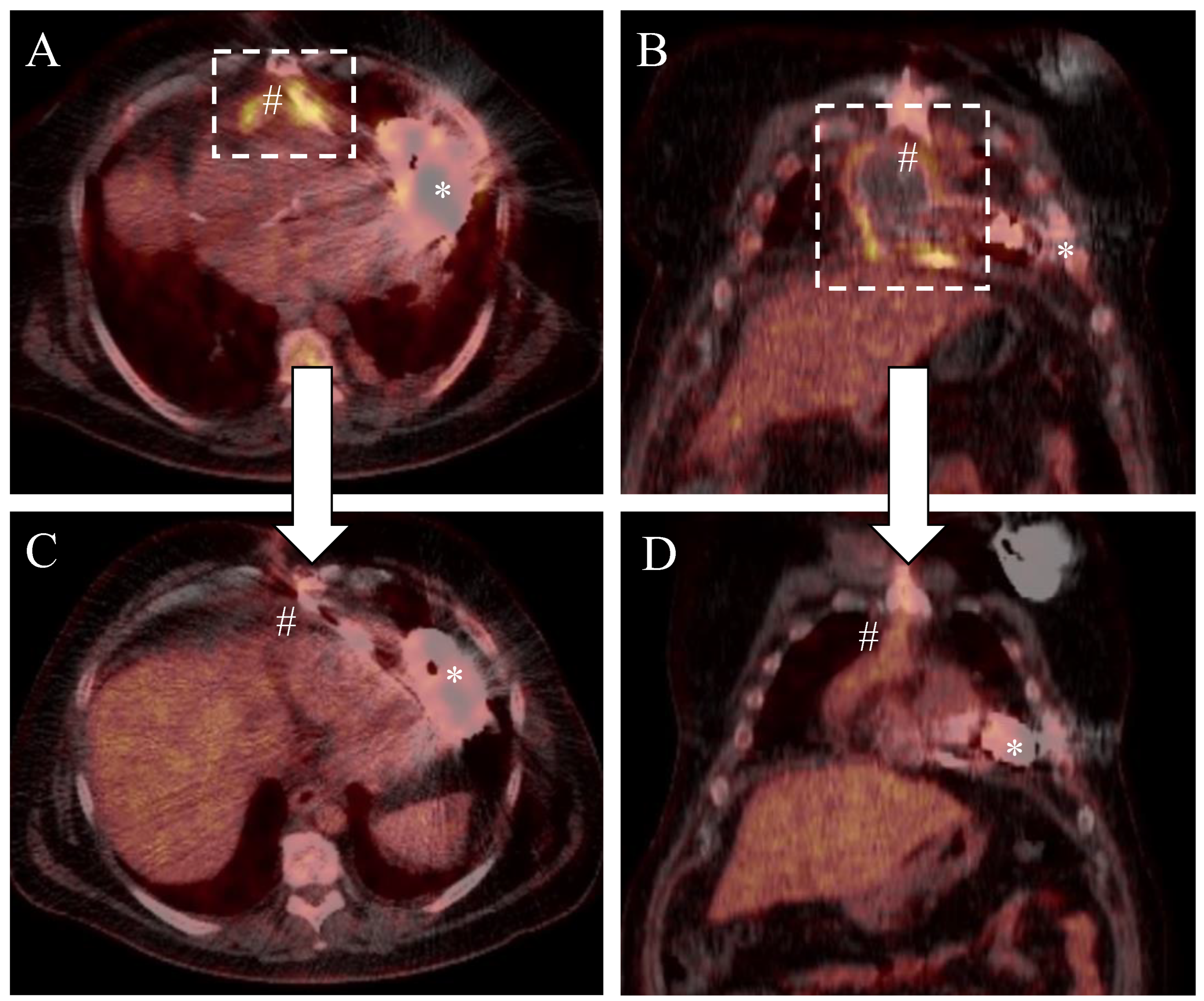
2. Medical History
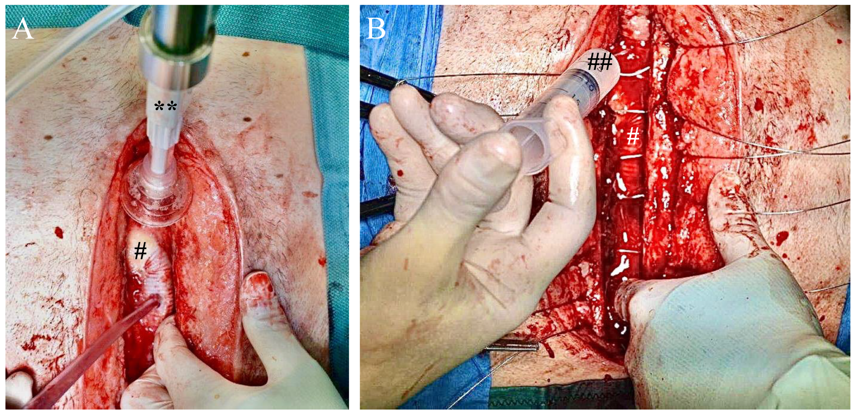
3. Surgical Wound Management and Postoperative Outcome
4. Discussion
Author Contributions
Funding
Institutional Review Board Statement
Informed Consent Statement
Data Availability Statement
Acknowledgments
Conflicts of Interest
References
- Shore, S.; Pienta, M.J.; Watt, T.M.F.; Yost, G.; Townsend, W.A.; Cabrera, L.; Fetters, M.D.; Chenoweth, C.; Aaronson, K.D.; Pagani, F.D.; et al. Non-Patient Factors Associated with Infections in LVAD Recipients: A Scoping Review. J. Hear Lung Transplant. 2021, 41, 1–16. [Google Scholar] [CrossRef]
- Pienta, M.J.; Shore, S.; Pagani, F.D.; Likosky, D.S.; Investigators, M.C.H.F. Rates and Types of Infections in Left Ventricular Assist Device Recipients: A Scoping Review. JTCVS Open 2021, 8, 405–411. [Google Scholar] [CrossRef]
- Bernhardt, A.M.; Schlöglhofer, T.; Lauenroth, V.; Mueller, F.; Mueller, M.; Schoede, A.; Klopsch, C.; Group, the Driveline Expert STagINg and carE DESTINE study group, a Ventricular Assist Device Driveline Infection Study Group. Prevention and Early Treatment of Driveline Infections in Ventricular Assist Device Patients—The DESTINE Staging Proposal and the First Standard of Care Protocol. J Crit. Care 2019, 56, 106–112. [Google Scholar] [CrossRef] [PubMed]
- Hanff, T.C.; Birati, E.Y. Left Ventricular Assist Device as Destination Therapy: A State of the Science and Art of Long-Term Mechanical Circulatory Support. Curr. Hear Fail Rep. 2019, 16, 168–179. [Google Scholar] [CrossRef] [PubMed]
- Krzelj, K.; Petricevic, M.; Gasparovic, H.; Biocina, B.; McGiffin, D. Ventricular Assist Device Driveline Infections: A Systematic Review. Thorac. Cardiovasc. Surg. 2021; Online ahead of print. [Google Scholar] [CrossRef]
- Tkhilaishvili, T.; Potapov, E.; Starck, C.; Mulzer, J.; Falk, V.; Trampuz, A.; Schoenrath, F. Bacteriophage Therapy as a Treatment Option for Complex Cardiovascular Implant Infection: The German Heart Center Berlin Experience. J. Hear Lung Transplant. 2022, 41, 551–555. [Google Scholar] [CrossRef] [PubMed]
- Tkhilaishvili, T.; Merabishvili, M.; Pirnay, J.-P.; Starck, C.; Potapov, E.; Falk, V.; Schoenrath, F. Successful Case of Adjunctive Intravenous Bacteriophage Therapy to Treat Left Ventricular Assist Device Infection. J. Infect. 2021, 83, e1–e3. [Google Scholar] [CrossRef] [PubMed]
- Mulzer, J.; Trampuz, A.; Potapov, E. Treatment of Chronic Left Ventricular Assist Device Infection with Local Application of Bacteriophages. J. Hear Lung Transplant. 2020, 39, S487. [Google Scholar] [CrossRef]
- Mulzer, J.; Trampuz, A.; Potapov, E.V. Treatment of Chronic Left Ventricular Assist Device Infection with Local Application of Bacteriophages. Eur. J. Cardio-Thorac. Surg. 2019, 57, 1003–1004. [Google Scholar] [CrossRef] [PubMed]
- Rubalskii, E.; Ruemke, S.; Salmoukas, C.; Aleshkin, A.; Bochkareva, S.; Modin, E.; Mashaqi, B.; Boyle, E.C.; Boethig, D.; Rubalsky, M.; et al. Fibrin Glue as a Local Drug-Delivery System for Bacteriophage PA5. Sci. Rep. 2019, 9, 2091. [Google Scholar] [CrossRef] [PubMed] [Green Version]
- Rojas, S.V.; Hanke, J.S.; Haverich, A.; Schmitto, J.D. Chronic Ventricular Assist Device Support: Surgical Innovation. Curr. Opin. Cardiol. 2016, 31, 308–312. [Google Scholar] [CrossRef] [PubMed]
- Yost, G.; Coyle, L.; Gallagher, C.; Cotts, W.; Pappas, P.; Tatooles, A. Outcomes Following Left Ventricular Assist Device Exchange: Focus on the Impacts of Device Infection. Asaio J. Am. Soc. Artif. Intern. Organs 2021, 67, 642–649. [Google Scholar] [CrossRef] [PubMed]
- Rubalskii, E.; Ruemke, S.; Salmoukas, C.; Boyle, E.C.; Warnecke, G.; Tudorache, I.; Shrestha, M.; Schmitto, J.D.; Martens, A.; Rojas, S.V.; et al. Bacteriophage Therapy for Critical Infections Related to Cardiothoracic Surgery. Antibiotics 2020, 9, 232. [Google Scholar] [CrossRef] [PubMed]


Publisher’s Note: MDPI stays neutral with regard to jurisdictional claims in published maps and institutional affiliations. |
© 2022 by the authors. Licensee MDPI, Basel, Switzerland. This article is an open access article distributed under the terms and conditions of the Creative Commons Attribution (CC BY) license (https://creativecommons.org/licenses/by/4.0/).
Share and Cite
Rojas, S.V.; Junghans, S.; Fox, H.; Lazouski, K.; Schramm, R.; Morshuis, M.; Gummert, J.F.; Gross, J. Bacteriophage-Enriched Galenic for Intrapericardial Ventricular Assist Device Infection. Antibiotics 2022, 11, 602. https://doi.org/10.3390/antibiotics11050602
Rojas SV, Junghans S, Fox H, Lazouski K, Schramm R, Morshuis M, Gummert JF, Gross J. Bacteriophage-Enriched Galenic for Intrapericardial Ventricular Assist Device Infection. Antibiotics. 2022; 11(5):602. https://doi.org/10.3390/antibiotics11050602
Chicago/Turabian StyleRojas, Sebastian V., Simon Junghans, Henrik Fox, Kanstantsin Lazouski, Rene Schramm, Michiel Morshuis, Jan F. Gummert, and Justus Gross. 2022. "Bacteriophage-Enriched Galenic for Intrapericardial Ventricular Assist Device Infection" Antibiotics 11, no. 5: 602. https://doi.org/10.3390/antibiotics11050602