Article

Citrus bergamia: Kinetics of Antimicrobial Activity on Clinical Isolates

1
Department of Health Sciences, Institute of Clinical Microbiology, “Magna Graecia” University of Catanzaro, 88100 Catanzaro, Italy
2
Institute of Research for Food Safety & Health (IRC-FSH), “Magna Graecia” University of Catanzaro, 88100 Catanzaro, Italy
*
Author to whom correspondence should be addressed.
These authors contributed equally to this work.
Antibiotics 2022, 11(3), 361; https://doi.org/10.3390/antibiotics11030361
Submission received: 1 February 2022 / Revised: 24 February 2022 / Accepted: 4 March 2022 / Published: 8 March 2022

Abstract

:
Background: The inappropriate use of antibiotics has increased selective pressure and the spread of multi-drug-resistant (MDR) pathogens, which reduces the possibility of effective treatment. A potential alternative therapeutic approach may be represented by essential oils, such as the distilled extract of bergamot (Citrus bergamia Risso et Poiteau). Such natural products exercise numerous biological activities, including antimicrobial effects. Methods: This work aimed to evaluate the kinetics of the bactericidal and fungicidal activity of the distilled extract of bergamot on MDR bacteria and fungi from clinical specimens using the time-kill assay. Furthermore, the antimicrobial activity of the distilled extract of bergamot on the morphology and cellular organization of clinical pathogens was evaluated by confocal laser scanning microscopy. Results: Our results demonstrated that the distilled extract of bergamot exhibited significant antimicrobial activity and a specific bactericidal effect against the bacterial and fungal strains tested. Furthermore, confocal microscope images clearly showed compromised membrane integrity, damage and cell death in bacterial samples treated with the distilled extract of bergamot. In addition, progressive alterations in cell-wall composition, cytoplasmic material and nucleus structure triggered by exposure to the distilled extract of bergamot were identified in the fungal samples considered. Conclusions: Our data suggest that the use of essential oils, such as distilled extract of bergamot (Citrus bergamia Risso et Poiteau), can represent a valid alternative therapeutic strategy to counteract antibiotic resistance of pathogens.

Graphical Abstract

1. Introduction

The continued and often inappropriate use of antibiotics has increased selective pressure, favoring the emergence, growth and spread of resistant microorganism strains [1]. The appearance of multiple antibiotic-resistant pathogens (multi-drug Resistance, MDR) further reduces the possibility of effective treatment. The World Health Organization (WHO) reported a list of MDR microorganisms responsible for high mortality, especially in healthcare settings [2]. Among the WHO priority pathogens, methicillin-resistant Staphylococcus aureus (MRSA) recently developed resistance to three widely used antibiotics (linezolid, vancomycin, and daptomycin), forming a drug-tolerant biofilm [3]. Few antimicrobial drugs are effective against carbapenem-resistant Acinetobacter baumannii (CRAB), classified as critical by the WHO, such as ampicillin-sulbactam in monotherapy or in combination [4]. The prevalence of carbapenem-resistant Klebsiella pneumoniae (CRKP) and ceftazidime/avibactam (CZA) resistance is increasing, with a negative impact on nosocomial infection [5]. Finally, Candida species (the most common opportunistic pathogen) are increasingly resistant to azole drugs with potential fungicidal effects [6]. Therefore, the therapeutic challenge to treating these infections requires the development of novel antimicrobial agents. A potential alternative therapeutic approach for the treatment of some infectious diseases can be represented by essential oils, which, with their complex and varied chemical composition, exercise a number of biological activities [7,8,9].
Several studies showed the in vitro antimicrobial properties of bergamot (Citrus bergamia Risso et Poiteau) on several microorganisms, including Gram-positive and Gram-negative bacteria and yeasts. In particular, the distilled extract can contrast emerging pathogens responsible for healthcare-related infections that are increasingly difficult to control and treat. Scientific interest also turned to evaluating the potential of the individual components of bergamot distilled extract [10]. Quirino et al. [11] highlighted the bactericidal and fungicidal activity of this natural substance using plate microdilution assays. The results showed that the distilled bergamot extract had a significant effect on all microorganisms tested [11]. In a recent study, the major components of the distilled extract of bergamot (limonene, linalool, linalyl acetate, and γ-terpinene) were tested both individually and in combination on a wide range of bacteria and Candida species. From in vitro testing, it emerged that the best antibacterial and antifungal capacity was given by the synergism between the components rather than by a single molecule [12].
The aim of this work was to evaluate the kinetics of the bactericidal and fungicidal activity of the distilled extract of bergamot on multi-drug-resistant bacteria and fungi of clinical isolates using the time-kill assay. The morphology and cellular organization of clinical isolates were evaluated by confocal laser scanning microscopy to understand the possible antimicrobial mechanisms of the distilled extract of bergamot.

2. Materials and Methods

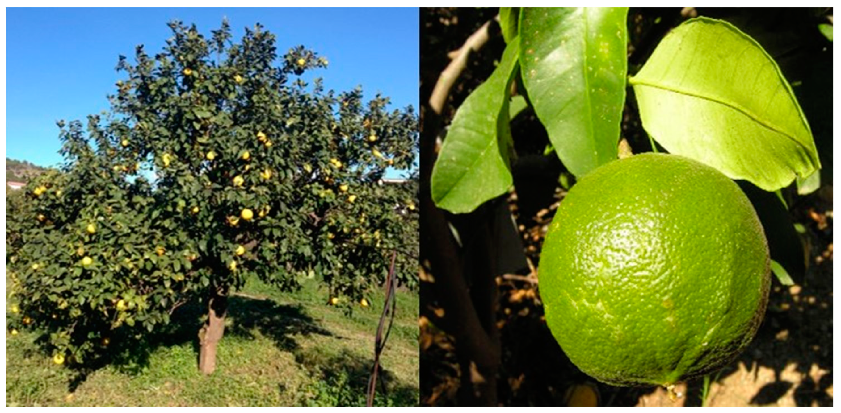
2.1. Citrus Bergamia Risso et Poiteau

Citrus bergamia Risso et Poiteau belongs to the family Rutaceae, genus Citrus and it grows almost exclusively in Calabria, Italy, along the southern east coast in the area of Reggio Calabria. Bergamot is a small evergreen tree that, in cultivation, can grow up to 5 m in height, with an erect, cylindrical, dark grayish-brown stem and irregular branches (Figure 1). The leaves have a short, sometimes winged petiole and are persistent, simple, alternate and aromatic when bruised. The lamina is ovate-oblong or lanceolate, usually with a sharp apex and a crenated margin or slightly wavy, hairless, and leathery, with a dark-green upper surface and light-green lower surface. The inflorescence is racemose and many-flowered. The flowers are fragrant, actinomorphic and pentamerous. The calyx persists in the ripened fruit. The corolla often presents five white petals inserted alternately with the sepals. Oil glands are also present in the petals. The hesperidium (Citrus spp. fruit) is slightly flattened and subglobose to pyriform. The peel is 3–6 mm thick, with numerous glands containing the essential oil, smooth to rough, adherent and shiny green turning to pale yellow when the fruit is ripe. The mesocarp is white, whilst the endocarp is divided into 10 to 15 lodges containing a sour and bitter greenish-yellow juice.

2.2. Preparation of Distilled Extract of Bergamot

The distilled extract of bergamot used in this study was a kind gift by Capua S.r.l., Reggio Calabria, Italy. Briefly, according to the manufacturer’s instructions, bergamots were peeled, and the peel was chopped and minced thoroughly. The cold expressed essential oil obtained was collected and subjected to vacuum distillation. This process involves heating the original bergamot essential oil and vaporizing the compounds, which depends on the volatility of the components. The packaging type used was “rashing rings”. The distillation temperature ranged between 20 °C and 180 °C, while the vacuum range was between 0.05 and 15 millibars.

2.3. Chemical Characterization of the Distilled Extract of Bergamot

The chemical characterization of the distilled extract of bergamot was performed by the Hewlett Packard (Agilent, Santa Clara, CA, USA) gas chromatograph (model HP 5890A)—mass spectrometer (model HP 5972A) equipped with an HP-35MS column. Helium was used as the carrier gas, and ionization was obtained by electron impact. Column temperature was maintained at 60 °C for 5 min and then raised to 280 °C in 10 °C/minute increments. Nineteen compounds were identified and quantified. Other compounds were identified by comparison of mass spectra of each peak with those of authentic samples in the NIST (National Institute of Standard and Technology) library. The qualitative and quantitative gas-chromatographic analysis of the distilled extract of bergamot showed a high level of some major aromatic compounds, as shown in Table 1.

2.4. Microorganisms Tested

Thirty-one multi-drug-resistant microorganisms isolated from clinical samples of patients admitted to the “Magna Græcia” University Hospital of Catanzaro (Italy) were selected (Table 2).
The identification and sensitivity to antibiotics of the microorganisms tested were evaluated with the automated system Vitek 2 (BioMérieux, France) according to the European Committee on Antimicrobial Susceptibility Testing (EUCAST) guidelines. Antimicrobial activities of the distilled extract of bergamot were evaluated using different methods such as a micro-well dilution assay and confocal microscopy. Moreover, the time-kill assay was carried out to evaluate the kinetics of the bactericidal and fungicidal activity of the distilled extract of bergamot on isolates.

2.5. Broth Microdilution Assay

Bactericidal and fungicidal activity was analyzed using broth microdilution assays followed by the determination of the minimum bactericidal concentration (MBC) as previously described by Quirino et al. [12]. Aliquots of 1 mL of distilled extract of bergamot were placed in sterile glass test tubes and emulsified with Tween 20, an inert, non-ionic tensioactive agent with no inherent bactericidal and fungicidal activity (900 μL of substance plus 100 μL of Tween 20). The mixture was then vortexed, and the first dilution (1:20) of the substance was performed in a liquid medium (Nutrient Broth, BioMérieux). In total, 100 μL of the first dilution was dispensed into the first well of a sterile microtitration plate, and two-fold serial dilutions were then performed. Microbial suspensions were adjusted to a final concentration of 1 × 10⁶ CFU/mL and distributed in each well of a 96-well plate. In each well, a wide range of concentrations, from 5% to 0.03% (v/v), was obtained by the two-fold serial dilution of the investigated substance. The microplate was incubated for 24 h at 37 °C. To determine the antimicrobial effect of distilled bergamot extract, subcultures of 1 µL from each dilution were inoculated onto blood agar plates (bioMérieux, Craponne, France) and incubated for 24 h at 37 °C in aerobic conditions, before colony count. The first dilution of the substance at which no microbial growth was observed represents the minimal bactericidal concentration (MBC).
All experiments included the substance alone to exclude possible contamination (negative control), while bacterial suspension without substance was used as a control sample of microbial growth. Each experiment was performed in triplicate.

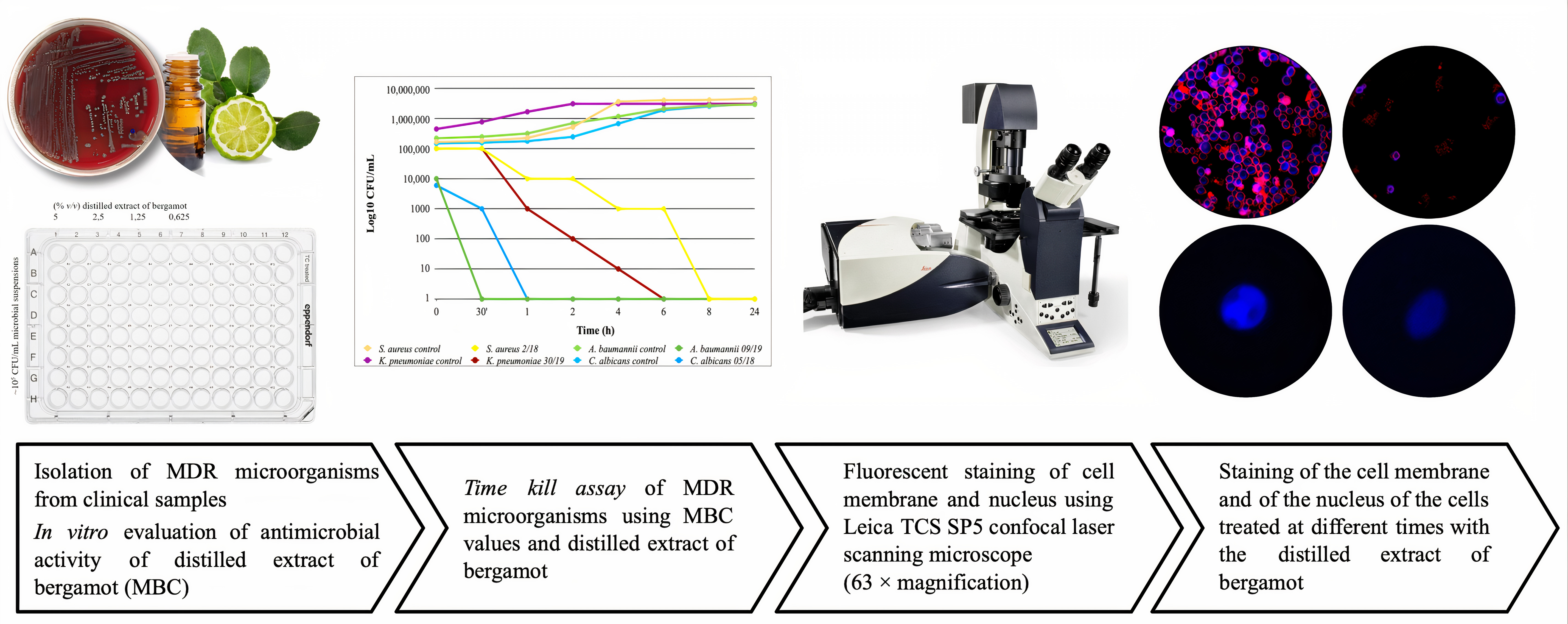
2.6. Time-Kill Assay

Data are presented in terms of log10 CFU/mL. The bactericidal effect of a substance, such as the distilled extract of bergamot, is defined as the 3 log10 CFU/mL reduction of microbial colonies compared to the initial inoculum. A reduction of less than 3 log10 CFU/mL is defined as a bacteriostatic effect [13].
A volume of 100 µL of bergamot distilled extract, at a concentration of 1 × MBC for each microorganism used, was dispensed in the wells of a 96-microwell plate. Microbial cultures in exponential growth were diluted in nutrient broth (bioMérieux, France) up to the concentration of 1 × 10⁵ CFU/mL, and 100 µL were dispensed in each well of the microplate. Subsequently, the microplate was incubated at 37 °C and, at pre-established time intervals (0, 30′, 1, 2, 4, 6, 8, 24 h), a 1 µL volume was inoculated onto blood agar plates (bioMérieux, France). After incubation at 37 °C for 24 h, colonies on individual plates were counted and expressed as the number of colony forming units/mL (CFU/mL).
All experiments were performed in triplicate. The decrease in growth was compared with a growth control of the untreated strain. The killing rate was determined by plotting the logarithm of the viable counts (CFU/mL) against time.

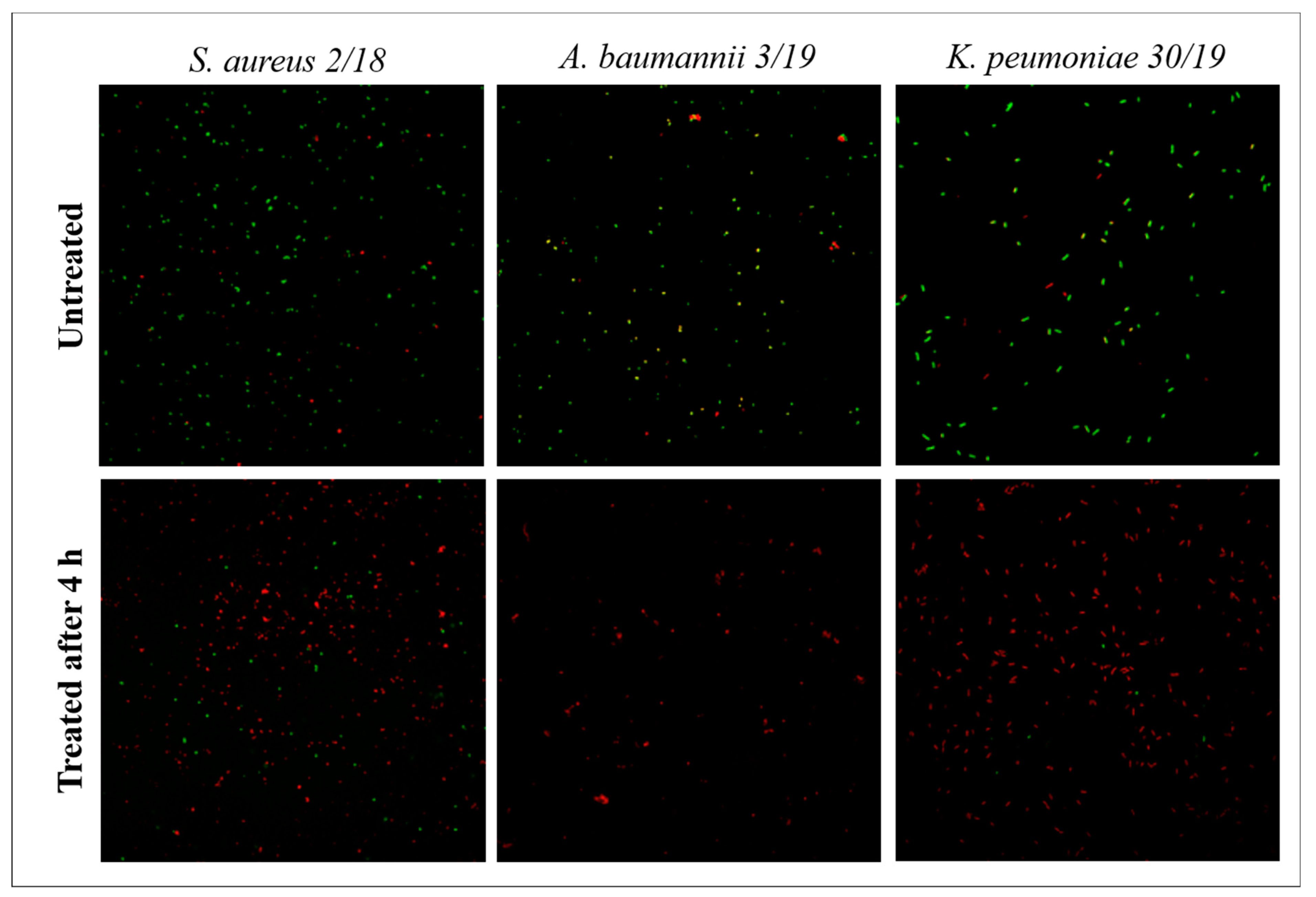
2.7. Confocal Microscopy

The interaction of the distilled extract of bergamot on multi-drug-resistant (MDR) bacteria and Candida cells was investigated using confocal laser scanning microscopy. MDR bacteria were observed under confocal microscopy during incubation with the distilled bergamot extract at a concentration of 1 × MBC. In particular, microscopic preparations of untreated strains and strains treated with the substance after at least 4 h of incubation were prepared. At different times, each preparation was washed with PBS buffer and fixed in formalin. To determine the viability of bacterial cells after treatment, as well as the bactericidal activity of the distilled extract of bergamot, 10 µL of each preparation was labeled with 10 µL of acridine orange (Sigma Aldrich, St. Louis, MO, USA), a nucleic acid selective metachromatic stain. Following the acridine orange procedure, differential staining was observed based on the presence of live or dead cells. Indeed, live cells appeared green while dead cells were stained red. Images were acquired with a 63X objective using the Leica TCS SP5 Confocal Microscopy System (Leica Microsystems, Wetzlar, Germany).
Each Candida strain was tested with a 1 × MBC of distilled extract of bergamot at three different times: immediately after the addition of the substance, at the mean time of growth reduction and at the time indicated as no growth. At the selected times, cells treated with distilled bergamot extract were washed with PBS buffer and fixed in formalin. For the determination of cell wall components, 200 µL of the microorganism suspensions were labeled with two different dyes: 50 µL of Concanavalin A (ConcA, Sigma Aldrich) and 30 µg/mL of Calcofluor White (CFW, Sigma Aldrich). ConcA selectively binds to α-mannanopyranosyl and α-glucopyranosyl residues; CFW is a non-specific fluorochrome that binds cellulose and chitin. Furthermore, 15 µL of DAPI stain (4′,6-diamidine-2-phenylindole, Sigma Aldrich) was used to evaluate the nuclear morphology. After 15 min for the DAPI stain and 60 min for the ConcA and CFW stains of incubation in a dark chamber, each specimen was observed under the confocal microscope.
One slide untreated with the substance and one unlabeled slide were added to each observation to exclude background interference or autofluorescence. Each experiment was performed in duplicate. The protocol described for the present work was adapted from Xing et al. [14].

3. Results

3.1. Antimicrobial Activity

The antimicrobial activity of the distilled extract of bergamot was determined by MBC values against 31 tested bacterial and fungal strains. Minimal bactericidal concentration is defined as 99.9%, or greater, killing efficacy at a selected time (Table 3).
The microorganisms tested showed different sensitivity to the distilled extract of bergamot. A first difference was noted within the bacteria: the bactericidal effect in Gram-positive bacteria was observed only at high concentrations of the substance (5–2.5% v/v) compared to concentrations (2.5–0.625% v/v) needed to kill Gram-negative bacteria.
The results obtained from tests on Candida species showed more significant heterogeneity: one isolate was sensitive to a distillate concentration of 0.313% v/v, while complete growth inhibition in the other isolates required distillate concentrations greater than 1.25% v/v.

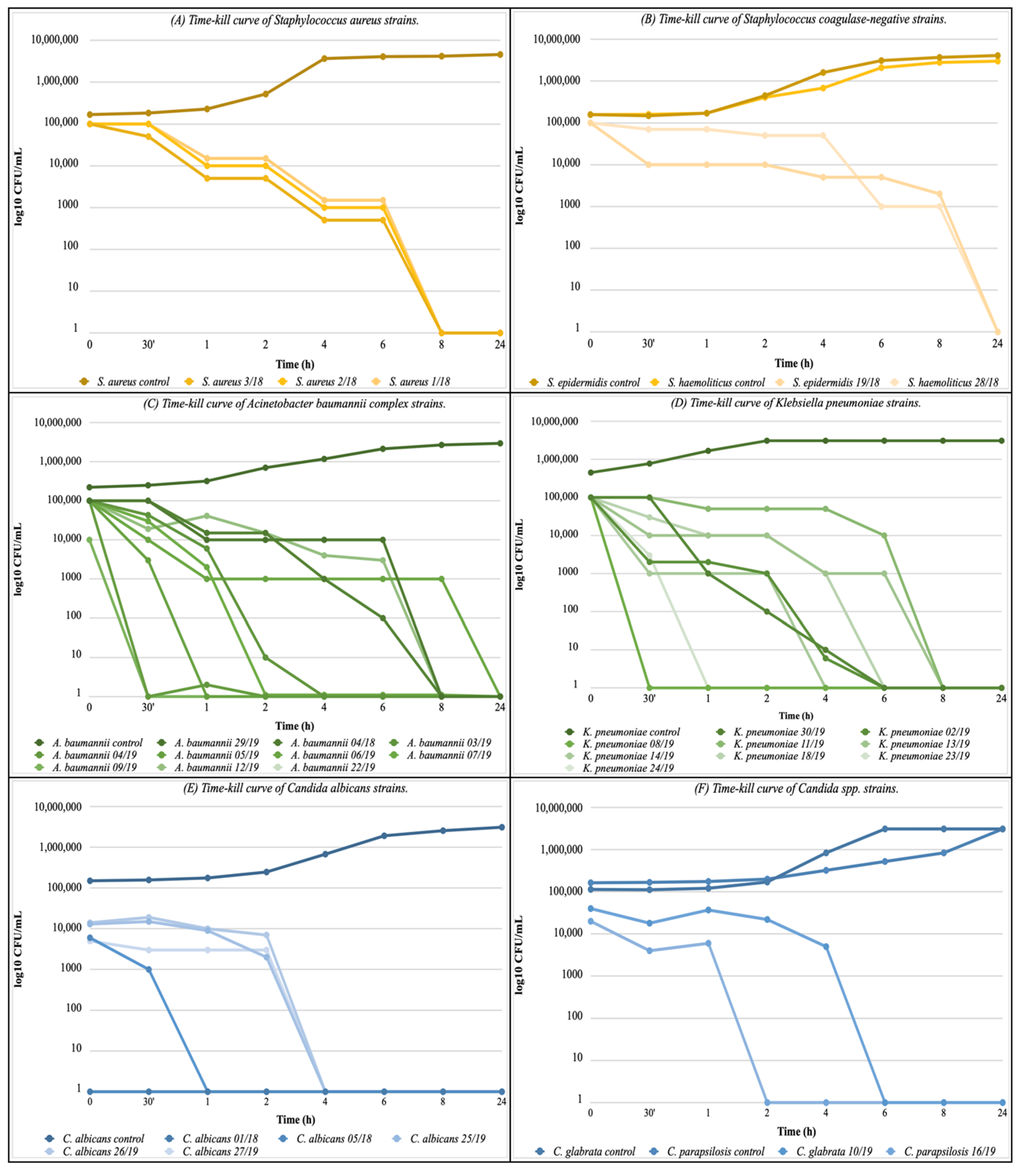
3.2. Time-Kill Assay

The bactericidal effect on the Gram-positive bacteria was obtained only at high concentrations of distilled bergamot extract (5% and 2.5% v/v) and at the longest exposure times. In the first 6 h of incubation, no significant reduction in the number of log10 CFU/mL was observed in the three S. aureus strains (Figure 2A) showing a bactericidal effect only after 8 h. S. epidermidis and S. haemolyticus showed a similar pattern to S. aureus. but the bactericidal effect occurred after a longer time of exposure (between 8 and 24 h) (Figure 2B).
More interesting results were obtained on Gram-negative bacteria. Ten strains of A. baumannii complex were monitored over 24 h using the kill curves (Figure 2C). In particular, a concentration of 0.625% v/v of the distilled extract of bergamot caused a bactericidal effect against A. baumannii 22/19, with a reduction of 5 log10 CFU/mL in just 30 min of exposure. However, in most A. baumannii strains tested, the bactericidal effect was observed between 6 to 8 h of exposure. Nine strains of K. pneumoniae multi-drug resistance (MDR) were tested (Figure 2D). A steady reduction in microbial growth of 1 log10 CFU/mL every hour was observed in K. pneumoniae 11/19; while exposure of K. pneumoniae 24/19 and K. pneumoniae 23/19 at a concentration of 1.25% (v/v) of the distilled extract of bergamot was sufficient to cause complete absence of growth after 30 and 60 min, respectively. In the other strains tested, the bactericidal effect was observed between 4 to 8 h of exposure.
The killing tests performed on Candida strains showed an inverse correlation between the distilled extract of bergamot concentration and the time of complete inhibition of growth. Indeed, the bactericidal effect on C. albicans 1/18 and C. albicans 5/18 was observed after only one hour of exposure with a distillate concentration of 2.5% (v/v), while on C. albicans 25/19, C. albicans 26/19, and C. albicans 27/19, the FBC value of 1.25% (v/v) required a longer exposure time to have the fungicidal effect. Similarly, the fungicidal effect on C. parapsilosis 16/19, with an FBC value of 1.25% v/v, was obtained after 2 h (Figure 2E). Finally, C. glabrata 10/19 was the most sensitive strain to the substance tested, with an FBC of 0.313% (v/v): the growth curve showed a similar trend to the untreated control for 4 h, and the complete absence of growth occurred after 6 h (Figure 2F).

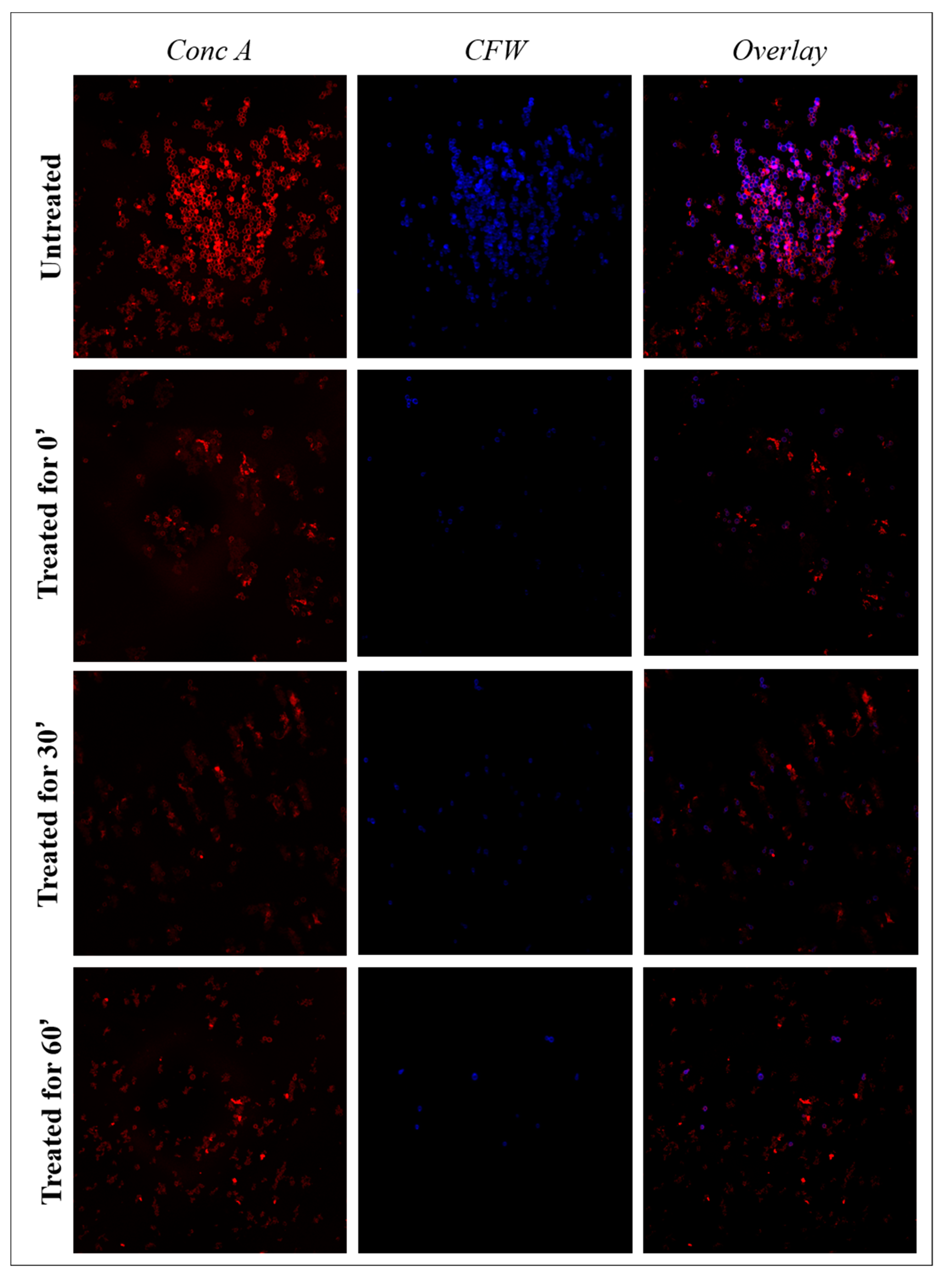
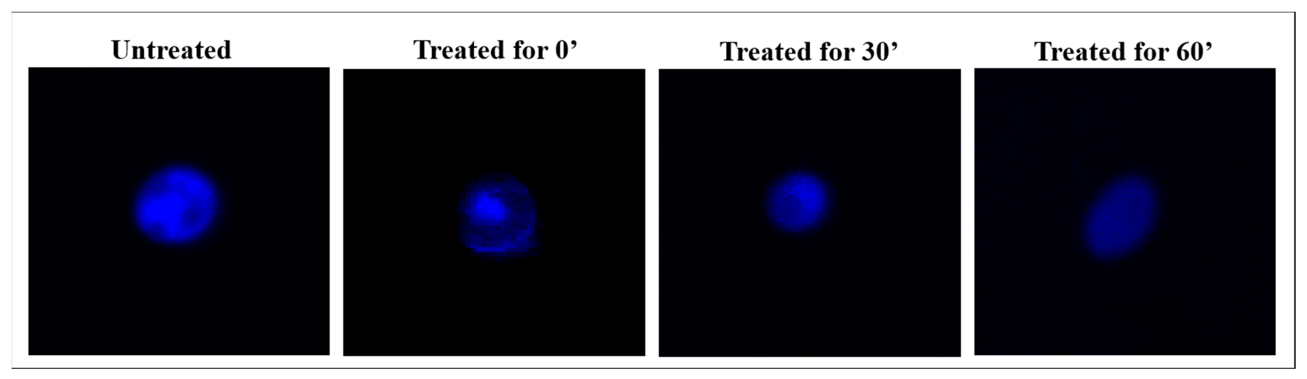
3.3. Confocal Microscopy

Orange acridine staining (Figure 3) showed dead cells after treatment with the distilled bergamot extract for at least 4 h compared to untreated cells. Confocal microscopy confirmed results obtained with the time-kill Assay and also showed a modified cell morphology after 4 h of treatment.
The main components of the cell membrane and the organization of the genetic material of C. albicans cells treated with the distilled extract of bergamot were analyzed with confocal laser scanning microscopy and fluorescent dyes, comparing them to those of untreated cells.
Chitin was directly labeled with calcofluor white (CFW) (blue color) and α-mannopyranosys with Concanavalin A (ConcA) (red color). Moreover, the spectral overlay of the two fluorescent dyes permitted simultaneous analysis. Images of C. albicans 1/18 visualized by confocal microscopy during exposure to distilled extract of bergamot are reported in Figure 4. Evaluation by confocal microscopy did not allow for the precise quantitative assessment of cell wall components, but it did facilitate imaging of changes in cell wall composition triggered by exposure to the distilled extract of bergamot. In cells not treated with the distilled extract of bergamot, there was a strong fluorescent signal that indicates the structural integrity of the cell wall, in particular of the inner layer of chitin and the outer layer of mannan. Within 60 min in the treated cells, the ConcA fluorescence intensity showed a gradual and evident reduction. Nevertheless, the fluorescent signal intensity for CFW seemed to remain stable, and a slight reduction was evident only after 60 min. Through the overlapped images, it is possible to highlight the reorganization of the treated cell wall: CFW and ConcA staining showed a heterogeneous distribution of chitin and mannan, respectively, in the cell wall. In untreated C. albicans cells, DAPI staining highlighted the rounded, blue nucleus with finely dispersed chromatin (Figure 5). The cells treated at time 0′, with the antimicrobial substance tested, showed minimal nuclear structural differences from the control cells, but the nucleus was still well defined and stained in blue. Similarly, at the median incubation time, the DAPI still detected the presence of a nuclear structure even if with a lower intensity of fluorescence, indicating lower compactness of the genetic material. Instead, cells treated for up to 60 min showed some distinct morphological differences: the typical rounded structure of the nucleus was no longer observable, while the presence of more fluorescent signals dispersed in the cell’s cytosol was evident. It is interesting to note how progressive alterations of the cell wall and cytoplasmic material of C. albicans cells, treated with distilled extract of bergamot, reflect the trend of the killing kinetic curve.
Optical Microscopy of C. albicans 1/18 clinical isolate treated for up to 60′ with the distilled extract of bergamot showed a significant cellular shrinkage highlighting the antimicrobial activity of bergamot (data not shown).

4. Discussion

The emergence of multi-drug-resistant (MDR) pathogens has become a major problem around the world with a significant clinical and economic impact. Within this scenario, the possibility of using essential oils, such as distilled extract of bergamot, represents a valid therapeutic alternative, as our data showed.
Bergamot (Citrus bergamia Risso et Poiteau) represents an ancient fruit, historically used for the production of essential oil widely applied to the cosmetic, pharmaceutical and food industries. Furthermore, more recent studies reported that bergamot juice or its enriched polyphenolic fraction (BPF), obtained from the peeled fruit, is mainly composed of flavanones (such as naringenin, hesperetin, eriodictyol glycosides), flavones (apigenin, luteolin, chrysoeriol, diosmetin glycosides) and their 3-hydroxy-3-methyl-glutaryl (HMG) derivatives [15,16,17]. Several in vitro and in vivo experiments employing these compounds clearly showed important hypolipemic, hypoglycemic, anti-inflammatory and cardioprotective effects as well as cholesterol-lowering activity by modulating hepatic HMG-CoA levels, binding bile acids and increasing the turnover rate of blood and liver cholesterol [18,19,20,21,22].
Bergamot essential oil is extracted from the peel and is rich in furocoumarins (bergapten and bergamottin, in particular), coumarins, pectins and flavonoids, which are suggested to exert important health-related properties, especially due to their antioxidant and anti-inflammatory activity [23,24]. Moreover, these compounds show a natural defensive role against invading pathogens, including bacteria, fungi, and viruses [25]. The most abundant compounds, limonene, linalool and linalyl acetate, are responsible for the synergistic effect of antimicrobial activity [12]. The antimicrobial activity of the monoterpenes was previously investigated, and is related to the lipophilicity and water solubility, then significantly influenced by their physicochemical characteristics, and the composition of bacterial membranes [26].
The distilled extract possessed the greatest antimicrobial efficacy against MDR Gram-negative bacteria (such as Acinetobacter baumannii and Klebsiella pneumoniae) and Candida albicans responsible for nosocomial infections representing a worldwide public health problem. On the contrary, Gram-positive bacteria and, in particular, Staphylococcus strains are more resistant to hydrophobic molecules due to the presence of slime [27,28]. The mechanism of interaction and entry of the distilled extract into bacterial cells is related to the molecular composition of the external membranes [29,30].
The present study evaluated the kinetics of the bactericidal and fungicidal activity of the distilled extract of bergamot on MDR bacteria and fungi of clinical isolates using the time-kill assay. This in vitro procedure is able to provide dynamic information about the decrease in microbial growth in relation to the time of exposure to the substance.
The bactericidal effect on the Gram-positive bacteria was observed only at a high concentration of distilled extract and after long-time exposure. A similar pattern of killing kinetics was analyzed on clinical MRSA, which demonstrated bactericidal response at 1% (v/v) and 2% (v/v) to tea tree oil and 0.12% (v/v) and 0.25% (v/v) to thyme oil after 24 h and 48 h of incubation, respectively [31]. Therefore, this data confirms the need for longer incubation times with the natural antimicrobial agent to achieve complete growth inhibition.
Distilled extract of bergamot showed enhanced antimicrobial activity on Gram-negative bacteria. Indeed, the bactericidal effect was observed at shorter exposure times and sometimes with a lower concentration of the substance. Similar killing curves were observed during processing with the essential oil from Zingiber cassumunar against the extensively drug-resistant (XDR) A. baumannii strains. This essential oil could completely inhibit A. baumannii 1 h after treatment [32]. Z. cassumunar was also shown to exhibit antibacterial effects against Gram-positive bacteria, such as S. aureus, with very low antibacterial activity [33], while it displayed high antimicrobial activity against dermatophytes and yeasts [34]. The antimicrobial activity of Origanum vulgare L. essential oil (OVeo) against CRKP, Serratia marcescens, and Acinetobacter baumannii was assessed by the time-kill assays, and a decrease in cell count was observed after 4 h treatment [35].
García-Salinas et al. evaluated the bactericidal mechanism of molecules derived from different essential oils, such as carvacrol, cinnamaldehyde and thymol, which were shown to be very effective against Gram-negative and Gram-positive bacteria. Indeed, confocal microscope images clearly confirmed the membrane damage exerted by the tested compounds against the bacteria, showing the compromised membrane integrity [36]. Similar antimicrobial findings caused by extracts from Thymus vulgaris and Myrthus communis were reported [37].
Candida strains, particularly C. albicans, were found to be extremely sensitive to the distilled extract of bergamot. Additionally, citral, the main phytoconstituent of other essential oils, such as Mentha piperita L. (Briq), Origanum vulgare and Zingiber officinale L. showed strong antifungal activity against Candida albicans strains. In the time-kill curve, the reduction in the growth of clinical strains was equal to 3 log10 CFU/mL after exposure of citral for 2 h [38].
In addition, Shanina et al. suggested that not only does the main compound of the substance tested have the ability to alter the integrity of the wall, but also minor compounds such as limonene, linalool and others contribute to the fungicidal action [39].
The confocal laser scanning microscopy is able to highlight the structural changes of the cell membrane, the first defensive barrier against cytotoxic substances, and at the nuclear level. In addition, the use of fluorescent dyes allows specific marking of the cell wall. It will therefore be possible to evaluate the cellular integrity of different components of Candida albicans cells and thus observe the extent of cell damage following exposure with distilled extract of bergamot in relation to the different intensity of fluorescence.
Evaluation by confocal microscopy did not allow precise quantitative assessment of cell wall components but facilitated the imaging of changes in cell wall composition triggered by exposure to distilled extract of bergamot. It is interesting to note how the progressive alterations of the cell wall and cytoplasmic material of C. albicans cells, treated with distilled extract of bergamot, reflect the trend of the killing kinetic curves. Other essential oils, such as those of Cinnamomum zeylanicum Blume bark and leaf, demonstrated fungicidal activities at very low concentrations. The mechanism of action of this oil on C. albicans and C. auris was damage to the envelope, evident by the shrinkage of the cell surfaces, reducing the cytoplasm, thus leading to cell lysis [40]. Moreover, envelope sterols of Candida spp., have a good affinity for different constituents of bergamot, suggesting a potential role for bergamot in topical treatment [11].

5. Conclusions

The emergence of multi-drug-resistant (MDR) pathogens has become a major problem worldwide with a significant clinical and economic impact. Within this scenario, the possibility of using essential oils, such as the distilled extract of bergamot (Citrus bergamia Risso et Poiteau), represents a valid therapeutic alternative, as our data showed.
Broth microdilution and time-kill assays demonstrated significant antimicrobial activity and a specific bactericidal effect against bacteria and fungi when studied with the distilled extract of bergamot. Confocal microscope images in bacterial samples treated with the natural extract clearly showed compromised membrane integrity, damage and cell death. In particular, in the fungal samples, it triggered progressive alterations in cell wall composition, cytoplasmic material and nucleus structure.
Furthermore, the combined action of the distilled extract of bergamot with antibiotics could represent both a strategy to eradicate antibiotic-resistant microorganisms and the possibility of decreasing the concentrations of antibiotics used, with a consequent reduction in the spread of drug resistance.

Author Contributions

A.Q., V.G., E.P., V.M. (Vincenzo Mollace), M.C.L. and G.M. conceptualized and designed the manuscript; A.Q., V.G. and E.P. wrote the manuscript; V.G., N.M., P.M., A.M., F.D., G.D.A. and V.T., performed the experiments; A.Q., V.G., E.P., N.M., P.M., A.M., F.D., G.D.A., V.T., S.N., M.G., V.M. (Vincenzo Musolino) C.C., V.M. (Vincenzo Mollace), M.C.L. and G.M. participated in drafting the article and revising it critically. All authors have read and agreed to the published version of the manuscript.

Funding

The work was supported by the public resources from the Italian Ministry of Research, PON-MIUR 03PE000_78_1, PONMIUR 03PE000_78_2; POR Calabria FESR FSE 2014–2020 Asse 12-Azioni 10.5.6 e 10.5.12. POR Calabria FESR FSE 2014–2020 azione 1.5.1: progetto AgrInfra Calabria.

Data Availability Statement

The present study includes all available data.

Acknowledgments

This study was supported by Programma Operativo Nazionale, PON-MIUR 03PE000_78_1 and 03PE000_78_2; POR Calabria FESR FSE 2014–2020 Asse 12-Azioni 10.5.6 e 10.5.12. POR Calabria FESR FSE 2014–2020 azione 1.5.1: progetto AgrInfra Calabria.

Conflicts of Interest

The authors have no relevant affiliations or financial involvement with any organization or entity with a financial interest in or financial conflict with the subject matter or materials discussed in the manuscript. This includes employment, consultancies, honoraria, stock ownership or options, expert testimony, grants or patents received or pending or royalties. No writing assistance was utilized in the production of this manuscript.

References

  1. Glass Whole-Genome Sequencing for Surveillance of Antimicrobial Resistance; World Health Organization: Geneva, Switzerland, 2020.
  2. WHO Publishes List of Bacteria for Which New Antibiotics Are Urgently Needed. 2017. Available online: https://www.who.int/news/item/27-02-2017-who-publishes-list-of-bacteria-for-which-new-antibiotics-are-urgently-needed (accessed on 21 February 2022).
  3. Hamad, M.; Al-Marzooq, F.; Srinivasulu, V.; Omar, H.A.; Sulaiman, A.; Zaher, D.M.; Orive, G.; Al-Tel, T.H. Antibacterial Activity of Small Molecules Which Eradicate Methicillin-Resistant Staphylococcus aureus Persisters. Front. Microbiol. 2022, 13, 823394. [Google Scholar] [CrossRef] [PubMed]
  4. Bartal, C.; Rolston, K.V.I.; Nesher, L. Carbapenem-resistant Acinetobacter baumannii: Colonization, Infection and Current Treatment Options. Infect Dis. Ther. 2022. [Google Scholar] [CrossRef] [PubMed]
  5. Liu, X.; Chu, Y.; Yue, H.; Huang, X.; Zhou, G. Risk factors for and clinical outcomes of ceftazidime-avibactam-resistant carbapenem-resistant Klebsiella pneumoniae nosocomial infections: A single-center retrospective study. Infection 2022. [Google Scholar] [CrossRef] [PubMed]
  6. Mahmoud, D.E.; Faraag, A.H.I.; Abu El-Wafa, W.M. In vitro study on the potential fungicidal effects of atorvastatin in combination with some azole drugs against multidrug resistant Candida albicans. World J. Microbiol. Biotechnol. 2021, 37, 191. [Google Scholar] [CrossRef] [PubMed]
  7. Karaman, M.; Bogavac, M.; Radovanović, B.; Sudji, J.; Tesanović, K.; Janjusević, L. Origanum vulgare essential oil affects pathogens causing vaginal infections. J. Appl. Microbiol. 2017, 122, 1177–1185. [Google Scholar] [CrossRef] [PubMed]
  8. Mancuso, M.; Catalfamo, M.; Laganà, P.; Rappazzo, A.C.; Raymo, V.; Zampino, D.; Zaccone, R. Screening of antimicrobial activity of citrus essential oils against pathogenic bacteria and Candida strains. Flavour. Fragr. J. 2019, 34, 187–200. [Google Scholar] [CrossRef]
  9. Tanhaeian, A.; Sekhavati, M.H.; Moghaddam, M. Antimicrobial activity of some plant essential oils and an antimicrobial-peptide against some clinically isolated pathogens. Chem. Biol. Technol. Agric. 2020, 7, 13. [Google Scholar] [CrossRef]
  10. Herman, A.; Tambor, K.; Herman, A. Linalool Affects the Antimicrobial Efficacy of Essential Oils. Curr. Microbiol. 2015, 72, 165–172. [Google Scholar] [CrossRef]
  11. Quirino, A.; Morelli, P.; Marano, V.; Cortese, G.; Barreca, G.S.; Liberto, M.C.; Focà, A. In vitro antimicrobial activity of natural essence and distilled extract of bergamot against Drug Resistance clinical isolates. Med. Aromat. Plants 2016, S3, 1–8. [Google Scholar] [CrossRef]
  12. Quirino, A.; Morelli, P.; Capua, G.; Arena, G.; Matera, G.; Liberto, M.C.; Focà, A. Synergistic and antagonistic effects of Citrus bergamia distilled extract and its major components on drug resistant clinical isolates. Nat. Prod. Res. 2018, 34, 1626–1629. [Google Scholar] [CrossRef]
  13. Wikler, M.A.; Clinical and Laboratory Standards Institute. Development of In Vitro Susceptibility Testing Criteria and Quality Control Parameters. Approved Giudeline, 5th ed.; CLSI Document M23; 2018; Volume 38, Available online: https://clsi.org/standards/products/microbiology/documents/m23s/ (accessed on 21 February 2022).
  14. Xing, X.; Liao, Z.; Tan, F.; Zhu, Z.; Jiang, Y.; Cao, Y. Effect of nicotinamide against Candida albicans. Front. Microbiol. 2019, 10, 595. [Google Scholar] [CrossRef] [PubMed]
  15. Gattuso, G.; Barreca, D.; Caristi, C.; Gargiulli, C.; Leuzzi, U. Distribution of flavonoids and furocoumarins in juices from cultivars of Citrus bergamia Risso. J. Agric. Food Chem. 2007, 55, 9921–9927. [Google Scholar] [CrossRef] [PubMed]
  16. Gardana, C.; Nalin, F.; Simonetti, P. Evaluation of flavonoids and furanocoumarins from Citrus bergamia (Bergamot) juice and identification of new compounds. Molecules 2008, 13, 2220–2228. [Google Scholar] [CrossRef] [PubMed] [Green Version]
  17. Salerno, R.; Casale, F.; Calandruccio, C.; Procopio, A. Characterization of flavonoids in Citrus bergamia (Bergamot) polyphenolic fraction by liquid chromatography-high resolution mass spectrometry (LC/HRMS). Pharma Nutr. 2016, 4, S1–S7. [Google Scholar] [CrossRef]
  18. Bok, S.H.; Lee, S.H.; Park, Y.B.; Bae, K.H.; Son, K.H.; Jeong, T.S. Plasma and hepatic cholesterol and hepatic activities of 3-hydroxy-3-methyl-glutaryl-CoA reductase and acyl CoA: Cholesterol transferase are lower in rats fed citrus peel extract or a mixture of citrus bioflavonoids. J. Nutr. 1999, 129, 1182–1185. [Google Scholar] [CrossRef]
  19. Carresi, C.; Musolino, V.; Gliozzi, M.; Maiuolo, J.; Mollace, R.; Nucera, S.; Maretta, A.; Sergi, D.; Muscoli, S.; Gratteri, S.; et al. Anti-oxidant effect of bergamot polyphenolic fraction counteracts doxorubicin-induced cardiomyopathy: Role of autophagy and ckitposCD45negCD31neg cardiac stem cell activation. J. Mol. Cell. Cardiol. 2018, 119, 10–18. [Google Scholar] [CrossRef]
  20. Musolino, V.; Gliozzi, M.; Scarano, F.; Bosco, F.; Scicchitano, M.; Nucera, S.; Carresi, C.; Ruga, S.; Zito, M.C.; Maiuolo, J.; et al. Bergamot Polyphenols Improve Dyslipidemia and Pathophysiological Features in a Mouse Model of Non-Alcoholic Fatty Liver Disease. Sci. Rep. 2020, 10, 2565. [Google Scholar] [CrossRef]
  21. Mollace, V.; Scicchitano, M.; Paone, S.; Casale, F.; Calandruccio, C.; Gliozzi, M.; Musolino, V.; Carresi, C.; Maiuolo, J.; Nucera, S.; et al. Hypoglycemic and Hypolipemic Effects of a New Lecithin Formulation of Bergamot Polyphenolic Fraction: A Double Blind, Randomized, Placebo Controlled Study. Endocr. Metab. Immune Disord. Drug Targets 2019, 19, 136–143. [Google Scholar] [CrossRef]
  22. Carresi, C.; Gliozzi, M.; Musolino, V.; Scicchitano, M.; Scarano, F.; Bosco, F.; Nucera, S.; Maiuolo, J.; Macrì, R.; Ruga, S.; et al. The Effect of Natural Antioxidants in the Development of Metabolic Syndrome: Focus on Bergamot Polyphenolic Fraction. Nutrients 2020, 12, 1504. [Google Scholar] [CrossRef]
  23. Dugo, P.; Mondello, L.; Dugo, L.; Stancanelli, R.; Dugo, G. LCMS for the identification of oxygen heterocyclic compounds in citrus essential oils. J. Pharm. Biomed. Anal. 2000, 24, 147–154. [Google Scholar] [CrossRef]
  24. Dugo, P.; Presti, M.L.; Ohman, M.; Fazio, A.; Dugo, G.; Mondello, L. Determination of flavonoids in citrus juices by micro HPLC-ESI/MS. J. Sep. Sci. 2005, 28, 1149–1156. [Google Scholar] [CrossRef] [PubMed]
  25. Sohn, H.Y.; Son, K.H.; Know, C.S.; Kang, S.S. Antimicrobial and cytotoxic activity of 18 prenylated flavonoids isolated from medicinal plants: Morus alba L., Morus mongolica Schneider, Broussnetia papyrifera (L.) Vent, Sophora flavescens Ait and Echinosophora koreensis Nakai. Phytomedicine 2004, 11, 666–672. [Google Scholar] [CrossRef] [PubMed]
  26. Trombetta, D.; Castelli, F.; Sarpietro, M.G.; Venuti, V.; Cristani, M.; Daniele, C.; Saija, A.; Mazzanti, G.; Bisignano, G. Mechanisms of antibacterial action of three monoterpenes. Antimicrob. Agents Chemother. 2005, 49, 2474–2478. [Google Scholar] [CrossRef] [PubMed] [Green Version]
  27. Nazzaro, F.; Fratianni, F.; De Martino, L.; Coppola, R.; De Feo, V. Effect of Essential Oils on Pathogenic Bacteria. Pharmaceuticals 2013, 6, 1451–1474. [Google Scholar] [CrossRef]
  28. Shingu-Vazquez, M.; Traven, A. Mitochondria and fungal pathogenesis: Drug tolerance, virulence, and potential for antifungal therapy. Eukaryot. Cell 2011, 10, 1376–1383. [Google Scholar] [CrossRef] [Green Version]
  29. Borges, A.; Ferreira, C.; Saavedra, M.J.; Simões, M. Antibacterial Activity and Mode of Action of Ferulic and Gallic Acids Against Pathogenic Bacteria. Microb. Drug Resist. 2013, 19, 256–265. [Google Scholar] [CrossRef]
  30. Wongsariya, K.; Phanthong, P.; Bunyapraphatsara, N.; Srisukh, V.; Chomnawang, M.T. Synergistic interaction and mode of action of Citrus hystrix essential oil against bacteria causing periodontal diseases. Pharm. Biol. 2014, 52, 273–280. [Google Scholar] [CrossRef]
  31. Sakkas, H.; Economou, V.; Gousia, P.; Bozidis, P.; Sakkas, V.A.; Petsios, S.; Mpekoulis, G.; Ilia, A.; Papadopoulou, C. Antibacterial efficacy of commercially available essential oils tested against Drug-Resistant Gram-positive pathogens. Appl. Sci. 2018, 8, 2201. [Google Scholar] [CrossRef] [Green Version]
  32. Boonyanugomol, W.; Kraisriwattana, K.; Rukseree, K.; Boonsam, K.; Narachai, P. In vitro synergistic antibacterial activity of the essential oil from Zingiber cassumunar Roxb against extensively drug-resistant Acinetobacter baumannii strains. J. Infect. Public Health 2017, 10, 586–592. [Google Scholar] [CrossRef]
  33. Habsah, M.; Amran, M.; Mackeen, M.M.; Lajis, N.H.; Kikuzaki, H.; Nakatani, N.; Rahman, A.A.; Ghafar; Ali, A.M. Screening of Zingiberaceae extracts forantimicrobial and antioxidant activities. J. Ethnopharmacol. 2000, 72, 403–410. [Google Scholar] [CrossRef]
  34. Nakamura, S.; Iwami, J.; Matsuda, H.; Wakayama, H.; Pongpiriyadacha, Y.; Yoshikawa, M. Structures of new phenylbutanoids and nitricoxide production inhibitors from the rhizomes of Zingiber cassumunar. Chem. Pharm. Bull. 2009, 57, 1267–1272. [Google Scholar] [CrossRef] [PubMed] [Green Version]
  35. Vasconcelos, N.G.; Vaz, M.S.M.; Radai, J.A.S.; Kassuya, A.L.K.; Formagio, A.S.N.; Graciani, F.S.; Leal, M.L.; Rodrigo Juliano Oliveira, R.J.; da Silva, K.E.; Croda, J.; et al. Antimicrobial activity of plant extracts against carbapenem-producing Klebsiella pneumoniae and in vivo toxicological assessment. J. Toxicol. Environ. Health Part A 2020, 83, 719–729. [Google Scholar] [CrossRef] [PubMed]
  36. García-Salinas, S.; Elizondo-Castillo, H.; Arruebo, M.; Mendoza, G.; Irusta, S. Evaluation of the antimicrobial activity and cytotoxicity of different components of natural origin present in essential oils. Molecules 2018, 23, 1399. [Google Scholar] [CrossRef] [PubMed] [Green Version]
  37. Sanguinetti, M.; Posteraro, B.; Romano, L.; Battaglia, F.; Lopizzo, T.; De Carolis, E.; Fadda, G. In vitro activity of Citrus bergamia (bergamot) oil against clinical isolates of dermatophytes. J. Antimicrob. Chemother. 2007, 59, 305–308. [Google Scholar] [CrossRef] [Green Version]
  38. Freire, J.C.P.; de Júnior, J.K.O.; de Silva, D.F.; de Sousa, J.P.; Guerra, F.Q.S.; de Oliveira Lima, E. Antifungal activity of essential oils against Candida albicans strains isolated from users of dental prostheses. Evid.-Based Complement. Altern. Med. 2017, 2017, 7158756. [Google Scholar] [CrossRef] [Green Version]
  39. Shahina, Z.; El-Ganiny, A.M.; Minion, J.; Whiteway, M.; Sultana, T.; Dahms, T.E.S. Cinnamomum zeylanicum bark essential oil induces cell wall remodelling and spindle defects in Candida albicans. Fungal Biol. Biotechnol. 2018, 5, 3. [Google Scholar] [CrossRef] [Green Version]
  40. Tran, H.N.H.; Graham, L.; Adukwu, E.C. In vitro antifungal activity of Cinnamomum zeylanicum bark and leaf essential oils against Candida albicans and Candida auris. Appl. Microbiol. Biotechnol. 2020, 104, 8911–8924. [Google Scholar] [CrossRef]
Figure 1. Citrus bergamia Risso et Poiteau.
Antibiotics 11 00361 g001
Figure 2. Time-kill curves of distilled extract of bergamot 1 × MBC against: (A) Staphylococcus aureus strains; (B) Staphylococcus coagulase-negative strains; (C) Acinetobacter baumannii complex strains; (D) Klebsiella pneumoniae strains; (E) Candida albicans strains; (F) Candida spp. strains.
Antibiotics 11 00361 g002
Figure 3. Representative images of confocal laser scanning microscopy of MDR bacteria treated with the distilled extract of bergamot and stained with acridine orange. “Untreated”: control of bacterial growth; “treated”: strains treated with the distilled extract of bergamot (MBC). Green fluorescence shows the presence of living cells; red fluorescence shows the presence of dead cells.
Antibiotics 11 00361 g003
Figure 4. Confocal laser scanning microscopy of cell wall components of C. albicans 1/18 clinical isolate treated with the distilled extract of bergamot for 60′. Double-staining of chitin and mannan fluorescently labeled with Calcofluor white (CFW) and Concanavalin A (Conc A), respectively, was performed. “Untreated”: growth control of C. albicans 1/18; “treated”: C. albicans 1/18 treated with the distilled extract of bergamot (MBC). The reduction in fluorescence intensity shows a decrease in the relative cell wall components.
Antibiotics 11 00361 g004
Figure 5. Confocal laser scanning microscopy of C. albicans 1/18 clinical isolate exposed for up to 60′ to the distilled extract of bergamot. DAPI staining for DNA identification was performed. “Untreated”: growth control of C. albicans 1/18; “treated”: C. albicans 1/18 treated with the distilled extract of bergamot.
Antibiotics 11 00361 g005
Table 1. Chemical characterization of the distilled extract of bergamot.
CompoundstR (min.)Distillate (w/w%)Bp (°C)
Cyclic monoterpenes
α-pinene4.241.03155
β-pinene5.996.56167–167
α-phellandrene6.890.04171–172
α-terpinene7.180.16173–175
Limonene7.4830.2176
p-cimene7.890.18177
ɣ-terpinene8.3711.95182
Terpinolene8.990.27184–185
Acyclic monoterpenes
Myrcene6.290.82165
Ocimene7.750.0865–66
Oxygenated acyclic monoterpenes
Linalool9.4721.82196–198
Lynalyl acetate12.0316.21220
Neral12.60.21103
Geranial13.090.11229
Neryl acetate13.910.28134 (25 mmHg)
Oxygenated cyclic monoterpenes
Terpineol11.640.87213–218
Ester
Octyl acetate11.380.10203–213
Aldehydes
Decanal11.52Trace93–95
Sesquiterpenes
Cariofillene14.380.14128–129
Table 2. Isolation site and antibiotic resistance profiles of clinical isolates.
ID StainIsolation SiteSusceptibleResistant
S. aureus 3/18MRSA nasal swabFA, CM, E, GM, LZD, MUP, RA, TEC, TE, TGC, SXT, VAP, LVX, OX
S. aureus 2/18MRSA nasal swabFA, GM, LZD, MUP, TEC, TE, TGC, SXT, VACM, E, P, LVX, OX, RA
S. aureus 1/18MRSA nasal swabFA, CM, E, GM, LZD, MUP, RA, TEC, TE, TGC, SXT, VAP, LVX, OX
S. epidermidis 19/19Wound swabLZD, RA, TE, TGC, VAFA, CM, E, GM, LVX, OX, SXT
S. haemolyticus 28/19Wound swabCM, LZD, RA, TGC, VAFA, E, GM, LVX, OX, SXT
A. baumannii 29/19Throat swabAMC, CSCTX, MEM, GM, CIP, FOS, FT, SXT
A. baumannii 04/18Throat swabCSFOX, CTX, MEM, AM, GM, CIP, FOS, FT, CS, SXT
K. pneumoniae 30/19Rectal swabGMAMC, TZP, FOX, CTX, CAZ, FEP, ETP, MEM, AM, CIP, FOS, FT, CS, SXT
K. pneumoniae 02/19Rectal swabAMC, TZP, ETP, MEM, AM, GM, FOS, CSCTX, CAZ, FEP, CIP, SXT
A. baumannii 03/19Wound swabCSMEM, AM, GM, CIP, SXT
A. baumannii 04/19Rectal swabAM, CSMEM, GM, CIP, SXT
A. baumannii 05/19Throat swabAM, CSMEM, GM, CIP, SXT
A. baumannii 06/19Rectal swabAM, CSGM, CIP, SXT
A. baumannii 07/19Throat swabAM, CSMEM, GM, CIP, SXT
K. pneumoniae 08/19Throat swabAM, FOS, CSAMC, TZP, CTX, CAZ, FEP, ETP, MEM, GM, CIP, SXT
A. baumannii 09/19Bronchial aspirateAMMEM, GM, CIP, CS, SXT
K. pneumoniae 11/19Rectal swabCS, SXTAMC, TZP, CTX, CAZ, FEP, ETP, MEM, AM, GM, CIP, FOS
A. baumannii 12/19Throat swabAM, CSMEM, GM, CIP, SXT
K. pneumoniae 13/19Rectal swabAM, FOS, CSAMC, TZP, CTX, CAZ, FEP, ETP, MEM, GM, CIP, SXT
K. pneumoniae 14/19Urinary catheterFOS, CSAMC, TZP, CTX, CAZ, FEP, ETP, MEM, AM, GM, CIP, SXT
K. pneumoniae 18/19Rectal swabMEM, AM, FOS, CSAMC, TZP, CTX, CAZ, FEP, ETP, GM, CIP, TGC, SXT
A. baumannii 22/19Throat swabCSMEM, AM, GM, CIP, SXT
K. pneumoniae 23/19Throat swabCSAMC, TZP, CTX, CAZ, FEP, ETP, MEM, AM, GM, CIP, FOS, SXT
K. pneumoniae 24/19Rectal swabCSAMC, TZP, CTX, CAZ, FEP, ETP, MEM, AM, GM, CIP, FOS, SXT
C. albicans 01/18Vaginal swabCAS, MYC, AMBFLU, VOR
C. albicans 05/18BloodFLU, VOR, CAS, MYC, AMB
C. glabrata 10/19Bronchial aspirateCAS, MYC, AMB
C. parapsilosis 16/19Urinary catheterFLU, VOR, CAS, MYC, AMB
C. albicans 25/19Foot skin swabFLU, VOR, CAS, MYC, AMB
C. albicans 26/19Nasal dischargeFLU, VOR, CAS, MYC, AMB
C. albicans 27/19Wound swabFLU, VOR, CAS, MYC, AMB
Amikacin (AM); Amoxicillin—Clavulanic acid (AMC); Amphotericin B (AMB); Benzylpenicillin (P); Caspofungin (CAS); Cefepime (FEP); Cefotaxime (CTX); Cefoxitin (FOX); Ceftazidime (CAZ); Ciprofloxacin (CIP); Clindamycin (CM); Colistin (CS); Ertapenem (ETP); Erythromycin (E); Fluconazole (FLU); Fosfomycin (FOS); Fusidic acid (FA); Gentamicin (GM); Levofloxacin (LVX); Linezolid (LZD); Meropenem (MEM); Mycafungin (MYC); Mupirocin (MUP); Nitrofurantoin (FT); Oxacillin (OX); Piperacillin—Tazobactam (TZP); Rifampicin (RA); Teicoplanin (TEC); Tetracyclin (TE); Tigecycline (TGC); Trimetoprim—Sulfamethoxazole (SXT); Vancomycin (VA); Voriconazole (VOR).
Table 3. MBC values% v/v (dilution of distilled extract of bergamot).
ID StrainMBCID StrainMBC
S. aureus 3/182.5% v/v (1:40)K. pneumoniae 11/191.25% v/v (1:80)
S. aureus 2/182.5% v/v (1:40)A. baumannii 12/191.25% v/v (1:80)
S. aureus 1/185% v/v (1:20)K. pneumoniae 13/195% v/v (1:20)
S. epidermidis 19/195 % v/v (1:20)K. pneumoniae 14/191.25% v/v (1:80)
S. epidermidis 28/195% v/v (1:20)K. pneumoniae 18/192.5% v/v (1:40)
A. baumannii 29/191.25% v/v (1:80)A. baumannii 22/190.625% v/v (1:160)
A. baumannii 04/181.25% v/v (1:80)K. pneumoniae 23/192.5% v/v (1:40)
K. pneumoniae 30/192.5% v/v (1:40)K. pneumoniae 24/192.5% v/v (1:40)
K. pneumoniae 02/192.5% v/v (1:40)C. albicans 01/182.5% v/v (1:40)
A. baumannii 03/190.625% v/v (1:160)C. albicans 05/182.5% v/v (1:40)
A. baumannii 04/191.25% v/v (1:80)C. glabrata 10/190.313% v/v (1:320)
A. baumannii 05/192.5% v/v (1:40)C. parapsilosis 16/191.25% v/v (1:80)
A. baumannii 06/190.625% v/v (1:160)C. albicans 25/191.25% v/v (1:80)
A. baumannii 07/191.25% v/v (1:80)C. albicans 26/191.25% v/v (1:80)
K. pneumoniae 08/195% v/v (1:20)C. albicans 27/191.25% v/v (1:80)
A. baumannii 09/192.5% v/v (1:40)
Publisher’s Note: MDPI stays neutral with regard to jurisdictional claims in published maps and institutional affiliations.

