Article

Evaluation of Bi-Lateral Co-Infections and Antibiotic Resistance Rates among COVID-19 Patients

1
Department of Microbiology, Faculty of Life Sciences, University of Central Punjab, Lahore 54000, Pakistan
2
Center for Transdisciplinary Research (CFTR), Saveetha Dental College & Hospitals, Saveetha Institute of Medical and Technical Sciences University, Chennai 600077, Tamil Nadu, India
3
Department of Restorative Dentistry & Endodontics, Faculty of Dentistry, University of Puthisastra, Phnom Penh 12211, Cambodia
4
Department of Pediatric Dentistry, Faculty of Dentistry, Taif University, P.O. Box 11099, Taif 21944, Saudi Arabia
5
Department of Prosthodontics, Faculty of Stomatology, Yerevan State Medical University after Mkhitar Heratsi, Str. Koryun 2, Yerevan 0025, Armenia
6
Molecular Diagnostic Laboratory, Johns Hopkins Aramco Healthcare, Dhahran 31311, Saudi Arabia
7
College of Medicine, Alfaisal University, Riyadh 11533, Saudi Arabia
8
Department of Public Health and Nutrition, The University of Haripur, Haripur 22610, Pakistan
9
School of Nursing, Wollongong University, Wollongong, NSW 2522, Australia
10
Administration of Pharmaceutical Care, Al-Ahsa Health Cluster, Ministry of Health, Al-Ahsa 31982, Saudi Arabia
11
Home Health Care Department, Ras Tanurah Hospital, Ras Tanurah 32817, Saudi Arabia
12
Microbiology Laboratory, King Faisal General Hospital, Al-Ahsa 31982, Saudi Arabia
13
Department of Pharmaceutical Chemistry, College of Pharmacy, Taif University, P.O. Box 11099, Taif 21944, Saudi Arabia
14
Immunology and Infectious Microbiology, Glasgow G1 1XQ, UK
*
Authors to whom correspondence should be addressed.
Antibiotics 2022, 11(2), 276; https://doi.org/10.3390/antibiotics11020276
Submission received: 21 January 2022 / Revised: 12 February 2022 / Accepted: 17 February 2022 / Published: 19 February 2022
(This article belongs to the Special Issue Antimicrobial Use and Stewardship during the COVID-19 Era)

Abstract

:
In addition to the pathogenesis of SARS-CoV-2, bacterial co-infection plays an essential role in the incidence and progression of SARS-CoV-2 infections by increasing the severity of infection, as well as increasing disease symptoms, death rate and antimicrobial resistance (AMR). The current study was conducted in a tertiary-care hospital in Lahore, Pakistan, among hospitalized COVID-19 patients to see the prevalence of bacterial co-infections and the AMR rates among different isolated bacteria. Clinical samples for the laboratory diagnosis were collected from 1165 hospitalized COVID-19 patients, of which 423 were found to be positive for various bacterial infections. Most of the isolated bacteria were Gram-negative rods (n = 366), followed by Gram-positive cocci (n = 57). A significant association (p < 0.05) was noted between the hospitalized COVID-19 patients and bacterial co-infections. Staphylococcus aureus (S. aureus) showed high resistance against tetracycline (61.7%), Streptococcus pyogenes against penicillin (100%), E. coli against Amp-clavulanic acid (88.72%), Klebsiella pneumoniae against ampicillin (100%), and Pseudomonas aeruginosa against ciprofloxacin (75.40%). Acinetobacter baumannii was 100% resistant to the majority of tested antibiotics. The prevalence of methicillin-resistant S. aureus (MRSA) was 14.7%. The topmost symptoms of >50% of COVID-19 patients were fever, fatigue, dyspnea and chest pain with a significant association (p < 0.05) in bacterial co-infected patients. The current study results showed a comparatively high prevalence of AMR, which may become a severe health-related issue in the future. Therefore, strict compliance of antibiotic usage and employment of antibiotic stewardship programs at every public or private institutional level are recommended.

1. Introduction

Coronavirus or SARS-CoV-2 (severe acute respiratory syndrome) belongs to the coronaviridae family and is an enveloped single-stranded positive-sense RNA virus. SARS-CoV-2 is a beta strain of the coronavirus family that causes severe acute respiratory syndrome. The virus responsible for coronavirus disease was first reported in December 2019 in Wuhan, China. Later, in March 2020, the World Health Organization (WHO) declared it a pandemic [1]. In Wuhan, it was first observed as an outbreak of atypical pneumonia. Due to this disease, many hospitalized patients’ illnesses became more serious, and COVID-19 cases doubled within a week. On further investigation, it was concluded that the coronavirus family’s beta strain was responsible for this infection, which was later identified as SARS-CoV-2 [2].
After studying the infection data from infected patients, many critical characteristics concerning the pathophysiology of SARS-CoV-2 were discovered. The most important contributors were secondary bacterial and fungal infections, as the majority of patients were infected with bacterial and fungal infections after acquiring SARS-CoV-2. In many patients, viral infection was seen to disrupt the macrophage model of activity by distorting the TLR4 and 5 pathways, which could be the reason for secondary bacterial infection by promoting bacterial attachments [3]. Furthermore, the virus may cause mucosal cell death, which compromises the pathogen purging mechanism and promotes bacterial adhesion [4]. Interactions between viruses and host cells may result in the formation of pro-inflammatory markers, such as TNF-α, a cytokine that can harm host cells and may lead to opportunistic bacterial infections. Viral infection may also activate INF1, which promotes the response of Toll-like receptors (TLR) to the ligand lipopolysaccharide [3].
The COVID-19 pandemic put a massive burden on the healthcare system, leading to changes in standard patient care approaches, increasing the incidences of hospital-acquired infections (HAIs). This additional COVID-19 burden, and long-term treatment of the associated patients, may have a significant association with HAIs [5]. The research community of AMR observed the transmission of SARS-CoV-2 and its interaction with other diseases worldwide, especially with secondary bacterial infections. It was concluded that there was a significant association between the usage of antibiotics and COVID-19 interventions that increased AMR rates [6]. COVID-19 patients receive antibiotic therapy for two reasons. The first one is COVID pneumonia symptoms with bacterial infection, and the second is a secondarily-acquired bacterial co-infection [7]. These reasons can determine the AMR evaluation in a specific population, the emergence of a pathogen, its transmission and infection burden [8].
Antibiotic stewardship programs and infection prevention and control practices may vary with locality. Empirical therapy is used for the primary treatment of critically ill patients in which a wide range of pathogens need to be covered, which may lead to the prescription of broad-spectrum antibiotics, like carbapenem and vancomycin. During the first phase of COVID-19, clinicians were faced with whether to use antibiotics as a treatment choice or not since they can have a significant impact on high AMR rates [9]. Antibiotic usage in the COVID-19 pandemic may change the AMR scenario to the worst situation ever. Keeping in view the current pandemic and threat of high AMR rates, the current study was conducted to determine the prevalence of bilateral bacterial co-infections in COVID-19 patients and the AMR patterns among each of the isolated bacteria.

2. Materials and Methods

2.1. Study Setting and Duration

This study was conducted at a tertiary care hospital in Lahore, Pakistan, collaborating with the Department of Microbiology, Faculty of Life Sciences (FoLS), University of Central Punjab (UCP), Lahore, Pakistan, from 16 October 2021 to 21 December 2021. All of the patients suffering from COVID-19 infection were admitted to different COVID-19 setting wards (medical ward, nephrology ward, gastroenterology ward, hepatology ward and general ward), and surgical intensive care units (SICUs) of the hospital were included in the study. The outpatient department (OPD) patients who were not suffering from COVID-19 infection were excluded from the study. The patients who were referred to any other hospital due to a treatment facility not being available in our hospital, and those who did not sign the consent form for the study, were also excluded from the study. Ethical approval was granted before starting the current study.

2.2. Collection of Patient Data

A total of 1165 COVID-19 patients were included in the current study. The demographical characteristics (age and sex) of studied patients were recorded from the patient medical record. The patients’ symptoms, baseline comorbidities, clinical characteristics and outcomes (discharge or death) were also recorded, apart from their demographic characteristics.

2.3. Collection of Clinical Samples

Keeping the testing facility available, each patient was proceeded for one type of sample only to include and study a more diverse group of patients. The clinical samples, including blood (n = 391), urine (n = 273), sputum (n = 123), throat swabs (n = 87), tracheal aspirate (n = 113), bronchoalveolar lavage (n = 63) and pus (n = 115) were collected under strict sterile conditions followed by standard operating procedures (SOPs) for COVID-19 infection. After collecting the samples, these were immediately transported to the Microbiology laboratory for further laboratory testing.

2.4. Isolation and Identification of Bacterial Isolates

After receiving the samples in the Microbiology laboratory, the samples were placed in the biosafety cabinet to proceed with culture inoculation. Before inoculating samples on different culture media plates, the samples (except urine) proceeded to smear preparation for Gram’s staining. After the preparation of smears on the sterilized glass slides, these samples were inoculated on MacConkey blood and chocolate agar as primary, secondary and tertiary streaking protocol. The urine samples were inoculated on cysteine electrolyte deficient (CLED) agar medium using the zig-zag streaking protocol. On each single agar plate, only one type of sample was inoculated. The blood culture vials (aerobic and anaerobic) were processed in the automated blood culture system (BectAlert 3D) for up to 7 days until declared unfavorable. After culture inoculation, the agar medium plates were incubated at 37 °C for 18 to 24 h, except for the sputum cultures, which were also incubated in the microaerophilic environment for certain specific organisms. After the first reading, if there was no growth present on the plates, they were again incubated for the next 24 h; at the second reading (48 h), the test was declared as “No growth” if there was still no growth.
For bacterial identification, first, the Gram’s staining slides were observed under the microscope’s 100X lens, and results were recorded on the tested sample file. After the incubation period, the agar medium plates were observed for the appearance of bacterial colonies and correlated with the Gram’s staining results. The bacteria were identified based on the morphology characteristics of colonies, Gram’s staining results and their biochemical profiling. The list of biochemical tests and their description is given in Table 1.

2.5. Antimicrobial Susceptibility Testing

Isolated organisms were tested for particular antibiotics as per the clinical laboratory standards (CLSI) recommendations. Both the Kirby Bauer (also known as the disc diffusion method) and serial dilution methods were used for antimicrobial activity testing of isolated strains. To validate the performance accuracy of the test, the American Type Culture Collection (ATCC) E. coli 25,922 strain was tested.
A bacterial colony was suspended in sterile normal saline with MacFarland standard, 0.5% for the Kirby Bauer disk diffusion method. The prepared suspension was then lawned on Muller Hinton (MH) agar plates, and the antibiotic discs were placed as per CLSI guidelines. After placing the discs, the agar plates were incubated for 24 h at 37 °C, then observed for the zone of inhibitions around the disks.
For the serial dilution method, the prepared MacFarland standard was added with gradually increasing antibiotic concentrations in 10 different tubes and incubated for 24 h at 37 °C. The results were checked based on the turbidity and reported as minimum inhibitory concentration (MIC).

2.6. Statistical Analysis

Data were recorded in Microsoft Excel and SPSS (Version 26.0). The association between the hospitalized COVID-19 patients with secondary bacterial infections and the appearance of symptoms in COVID-19 patients was compared with those infected with bi-lateral co-infections using the Chi-square test. A p-value < 0.05 was considered significant.

3. Results

3.1. Demographical Characteristics of Coronavirus Disease 2019 (COVID-19) Patients

Of the 1165 recruited patients (confirmed positive for COVID-19 by real-time PCR using nasopharyngeal swabs), the majority were male (n = 652), and the remaining (n = 513 were female). However, most of the patients co-infected with various bacterial infections were female (n = 224). Detailed information regarding the general characteristics of patients is given in Table 2.

3.2. Sample-Wise Positive Ratio of Bacterial Cultures

Each patient was processed for one sample type only to collect a more diverse group of samples and cover more patients. Most of the collected samples in the current study were blood followed by urine. Sputum, wound swabs, tracheal aspirate, throat swabs and bronchoalveolar lavage. A significant association (p < 0.05) was noted between the hospitalized COVID-19 patients and the bacterial co-infections. The positive ratios for bacterial cultures in different samples are shown in Table 3.

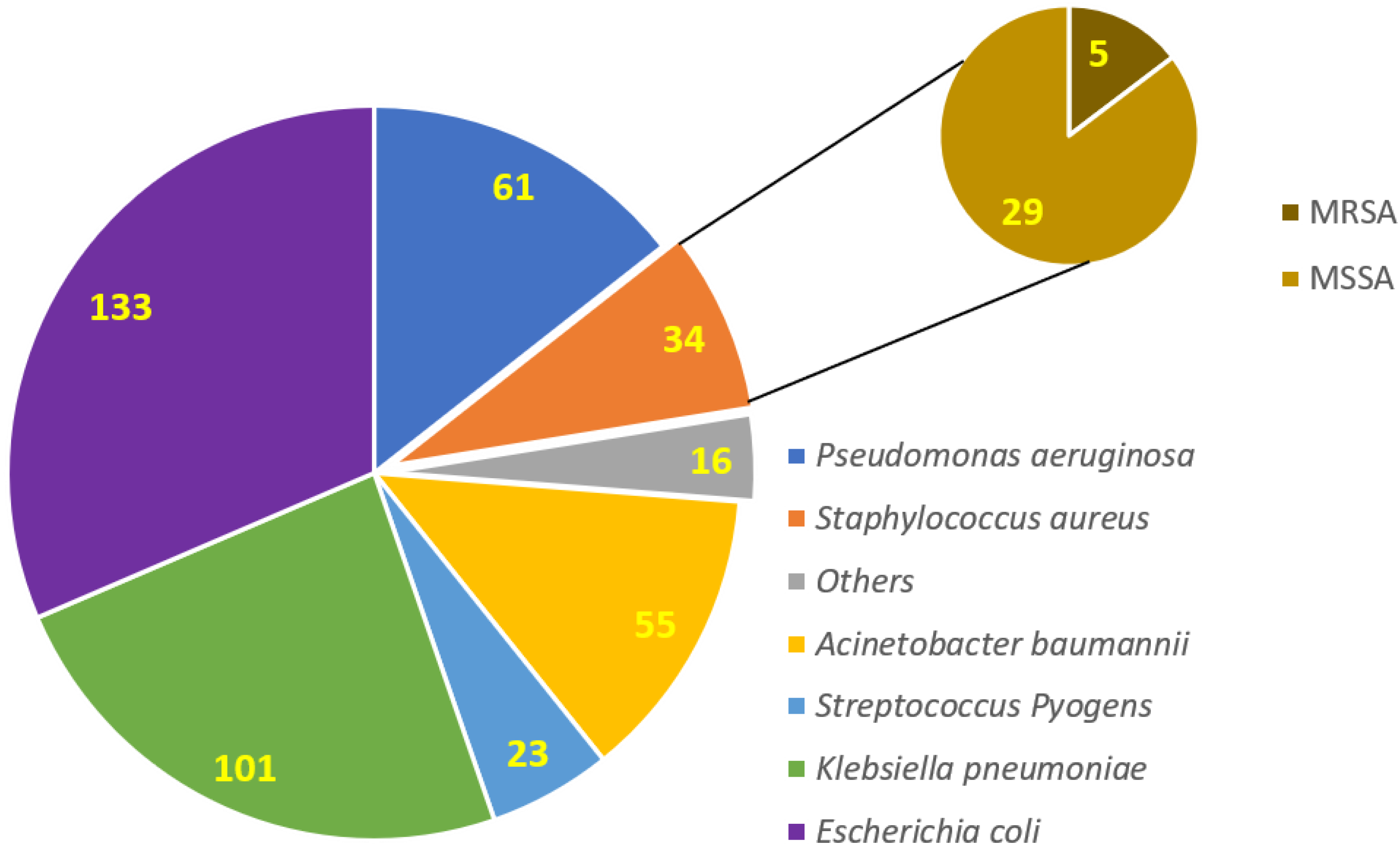
3.3. Isolation and Identification of Clinical Bacterial Isolates among COVID-19 Patients

Most of the isolated bacteria were Gram-negative rods (n = 366), followed by Gram-positive cocci (n = 57). Among the Gram-positive bacteria, n = 34 showed a positive reaction to the catalase biochemical test, which then proceeded to the coagulase test, and all showed positive reactions, which confirmed the identification of Staphylococcus aureus. The colony characteristics of S. aureus on the blood agar plate are shown in Supplementary Figure S1. A total of 23 Gram-positive bacterial isolates showed adverse reactions for the catalase biochemical test, further proceeded with the Streptococcus grouping and identified as Streptococcus pyogenes. Among the Gram-negative rods, 234 were lactose fermenters, as shown in Supplementary Figure S2, and 132 were non-lactose fermenters (Supplementary Figure S3). In the lactose fermenters, 133 bacteria showed pink mucoid colonies and were indole positive (Escherichia coli), while the remaining 101 showed flat pink colonies and were indole negative, and therefore, proceeded further for citrate testing, which showed positive reactions (Klebsiella pneumoniae). The non-lactose fermenters first proceeded for the oxidase test, among which 61 isolates showed positive reactions (Pseudomonas aeruginosa). The Enterobacteriaceae were counter-confirmed for identification using API 20E strips, which showed the exact identification, as confirmed by the manual biochemical tests, as shown in Supplementary Figure S4. The prevalence of each bacterium among the studied COVID-19 patients is shown in Figure 1.

3.4. Antibiotic Susceptibility Patterns of Individual Isolated Bacterial Isolates

A high prevalence of S. aureus resistance against tetracycline (61.7%) was noted, followed by gentamycin (50%), ciprofloxacin (47%), levofloxacin (47%), clindamycin (47%) and erythromycin (47%). The prevalence of the methicillin-resistant S. aureus (MRSA) strain was 14.7%. At the same time, no case of vancomycin-resistant S. aureus (VRSA) was found. The antibiotic resistance patterns of each isolated Gram-negative and Gram-positive bacteria are shown in Table 4 and Table 5, respectively.
The sensitivity pattern of Pseudomonas aeruginosa on the MH agar plate is shown in Supplementary Figure S5. The MIC testing of colistin (CT) on the MH agar plate is shown in Supplementary Figure S6. The MIC of vancomycin (VA) on the MH agar plate with the sensitivity pattern of VA (sensitive) and cefoxitin (sensitive) discs is shown in Supplementary Figure S7. In comparison, the MIC of VA on the MH agar plate with the sensitivity pattern of VA (sensitive) and cefoxitin (resistant), oxacillin (resistant) and tetracycline (sensitive) discs are shown in Supplementary Figure S8.

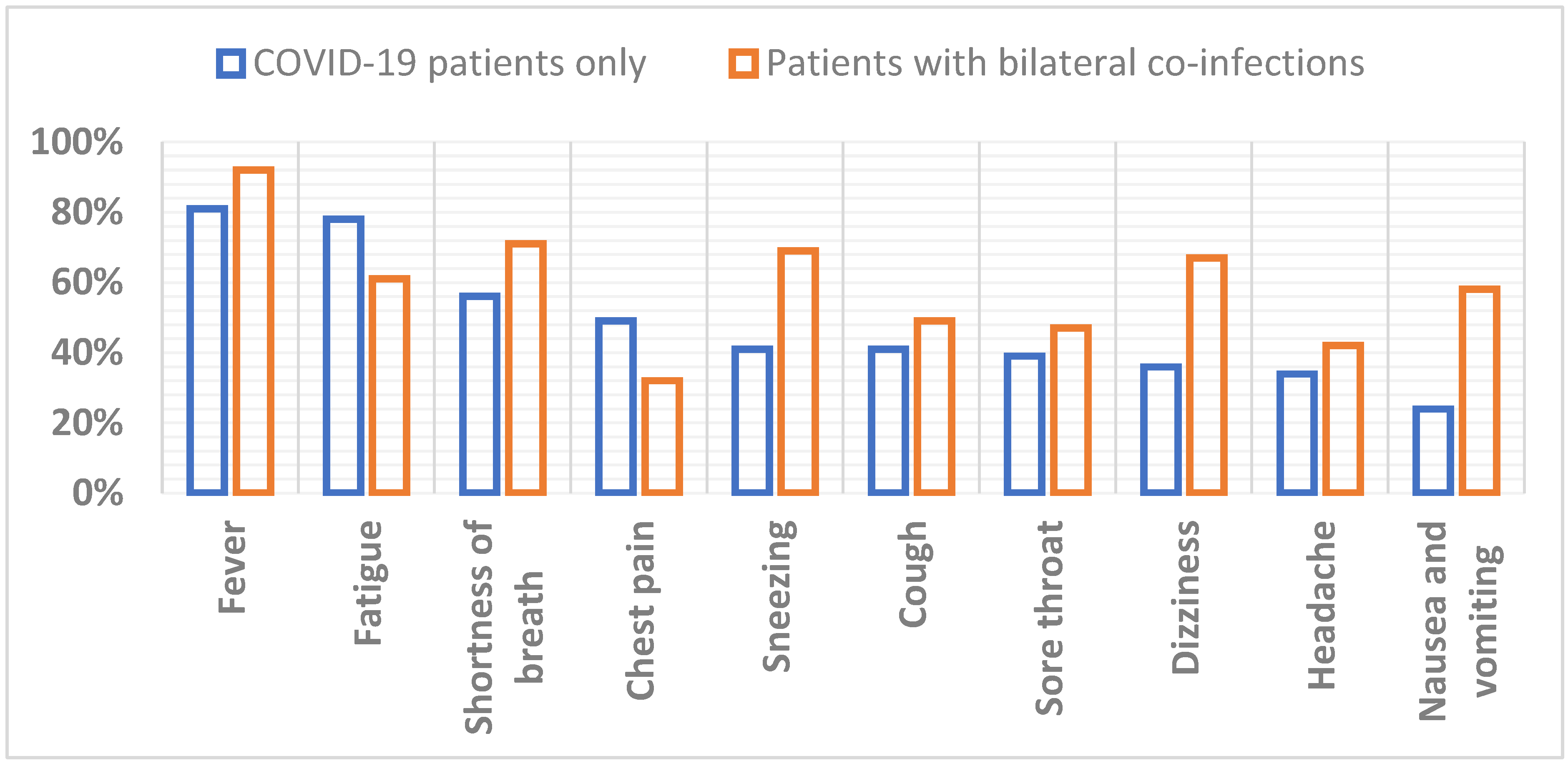
3.5. Appearance of Symptoms in COVID-19 Patients

At the time of admission to the hospital, patients faced different symptoms. The most common symptoms faced by critically ill patients were fever followed by fatigue, dyspnea, chest pain, sneezing, cough, sore throat, dizziness, headache and vomiting. Patients with different comorbidities faced more complicated symptoms. Fever followed by shortness of breath was the most commonly occurring onset symptom in ICU patients, as well as a cough that might be dry. These three symptoms were faced by more than 50% of patients admitted to the ICU. According to their immune system weakness and related comorbidities, rare symptoms were also prevalent in many patients. A significant correlation (p < 0.001) was found between the appearance of symptoms in COVID-19 patients and those who were infected with bi-lateral co-infections. The top 10 observed symptoms that appeared in COVID-19 patients and those co-infected with different bacterial infections are shown in Figure 2.

4. Discussion

Antimicrobial resistance (AMR) is a global health problem that poses a severe threat to treating a wide range of bacterial infections. The high AMR rates are a significant threat for patients admitted to different hospital wards, and patients admitted to intensive care units (ICUs) [10]. For public health and life sciences, finding strategies to combat the advancement of antibiotic resistance is a significant challenge. There has been a rapid increase in multidrug-resistant (MDR) pathogenic bacteria worldwide in the past few decades, especially in the current COVID-19 pandemic. More infections caused by MDR micro-organisms do not affect regular treatment, and the last choice of antibiotic may lose its effect [6]. Keeping in mind the current scenario of the pandemic situation, the study was conducted to determine the prevalence of bacterial infection and their antibiotic susceptibility patterns.
Nosocomial infections are serious issues for all patients admitted to the hospital, especially those admitted to the medical ICU [11]. Most patients admitted to the medical ICU are immunocompromised, putting them at higher risk of getting a nosocomial infection. Most infections in the medical ICU are catheter-related infections [12]. At present, 5 to 10% of patients entering emergency hospitals get at least one infection, and the risk has increased during the last decade [13]. The ICU accounts for 5 to 15% of hospital beds and 10 to 25% of medical expenses, equivalent to 1 to 2% of the clinical expense of the gross public product of the USA [14]. Nosocomial infections have a significant impact in the United States; more than 2 million people are admitted to ICU every year. Of these, 5 to 35% of patients admitted to the ICU get a nosocomial infection [15]. A previous study from Singapore showed that 14.8% of COVID-19 patients were co-infected with various nosocomial infections [16]. The results of the current study showed that COVID-19 patients admitted to the hospital were at high risk of getting co-infected with hospital-acquired infection. The most common causes of infection were Gram-negative bacteria. The main reason for these infections might be patients’ immune-suppressive status.
The infectious agent (SARS-CoV-2) that causes COVID-19 is exceptionally contagious, spreading mainly via droplets and close contact [17]. Several familial infection clusters have been recorded, and some of the verified individuals were infected in hospitals [18]. A recent study from Belgium has reported bacterial co-infection in 40.6% of the patients. The most common bacteria with multiple genome copies were S. aureus, H. influenzae and Moraxella catarrhalis [19]. In Italy, the prevalence of HAIs among COVID-19 patients was 56.73% [20]. However, a study conducted in Germany reported 34% of bacterial co-infections [21]. In the current study, 36% of studied patients were found to be co-infected with various bacterial infections.
The AMR also becomes an economic burden due to extensive usage of antibiotics, an extended stay in the hospital, expensive antibiotics and additional laboratory tests. Activities expected to control the issue of AMR are the responsibilities of local governments, medical care suppliers, specialists and the general population [22]. Patients admitted to ICUs have more chances of becoming resistant to drugs because of several factors, including starting empirical antibiotic treatment without doing the bacterial cultures and antimicrobial susceptibility testing, effective use of the invasive devices, and use of the drugs, which leads to a decrease in immunity and several nosocomial infections. Inappropriate antibiotic use and extensive use of broad-spectrum antibiotics are the most significant factors which lead to high AMR rates [23]. In our study, we have found that S. aureus has high resistance against tetracycline. E. coli and Klebsiella pneumoniae were highly resistant to amoxicillin, Acinetobacter baumannii to amikacin, while the P. aeruginosa was highly resistant to ciprofloxacin.
In recently published studies, it was observed that MDR bacterial prevalence has increased [24]. A retrospective study showed increased carbapenem resistance as almost more than double [25]. There were multiple underlying reasons for increased AMR rates, one of them being prolonged hospital stay. In a previous study in China, the incidences of Acinetobacter baumannii, Klebsiella pneumonia and Stenotrophomonas maltophilia were the same in both COVID and non-COVID patients [26]. It was shown that carbapenem resistance among Acinetobacter baumannii was 91.2% and 75% for Klebsiella pneumoniae. Another reason for increased AMR rates was the high usage of antibiotics, which was more than 99%, as compared to previous years [27]. Although COVID-19 is a pandemic, AMR could be an unnoticed pandemic, especially during this period, as described in a previous study where more than 30,000 deaths occurred because of AMR in Europe alone [27]. A recent study from the USA reported 44% more cases of MRSA [28]. A recent study from Pakistan [9] showed that the prevalence of MRSA was 20% in COVID-19 patients admitted to ICUs. However, in the current study, the prevalence of MRSA was 14%, with no reported cases of VRSA.

5. Study Limitations

A comparatively small number of patients were recruited because of the single study protocol. The patient’s data about the previous history of consuming antibiotics and the dosage of antibiotics could not be obtained because of ethical issues from the institution. Further large-scale and multi-institutional studies are recommended.

6. Conclusions

The current study reported a significant prevalence of bacterial co-infections among the hospitalized COVID-19 patients and a high AMR rate. The immune-suppressive status of these patients could play an essential role in acquiring different hospital-acquired infections. These high AMR rates threaten the loss of antibiotic effectiveness, especially in hospitalized patients. To monitor the compliance of antibiotic usage, periodic audits and antibiotic stewardship programs are required at the institutional level.

Supplementary Materials

The following are available online at https://www.mdpi.com/article/10.3390/antibiotics11020276/s1: Figure S1. Growth of catalase and coagulase positive (Staphylococcus aureus) bacteria on the blood agar plate; Figure S2. Growth of Gram-negative bacteria (Lactose fermenter) on blood and MacConkey agar plates after 18 h of incubation at 37 °C; Figure S3. Growth of oxidase-positive bacteria (probably Pseudomonas aeruginosa) on blood and CLED agar plates; Figure S4. API 20E strip, on the reading page, all results of wells are given. Based on the results, a unique number was produced, matched with the data bank available on the BIOMEUREX website; Figure S5. Sensitivity pattern of Pseudomonas aeruginosa on MH agar plates. Figure S6. MIC of Colistin on the MH agar plate. Figure S7. MIC of vancomycin on MH agar plate with a Sensitivity pattern of vancomycin (sensitive) and cefoxitin (sensitive) discs; Figure S8. MIC of vancomycin on MH agar plate with a sensitivity pattern of vancomycin (sensitive) and cefoxitin (resistant), oxacillin (resistant), and tetracycline (sensitive) discs.

Author Contributions

Conceptualization, N.A., M.I.K. and W.S.; methodology, W.S.; software, M.K. and A.H.; validation, A.A.R., A.A.M. and S.A.; formal analysis, M.K. and W.S.; investigation, M.K. and W.S.; resources, R.N.M. and A.H.; data curation, N.A., W.S. and M.K.; writing—original draft preparation, N.A., W.S. and M.I.K.; writing—review and editing, M.I.K., A.H., S.A.A., A.S.B. and E.Y.S.; visualization, R.N.M., A.H. and A.S.B.; supervision, M.I.K. and A.A.R.; project administration, N.A. and M.I.K.; funding acquisition, A.A.M., S.A. and J.H.A. All authors have read and agreed to the published version of the manuscript.

Funding

This research received no external funding.

Institutional Review Board Statement

The study was conducted according to the guidelines of the Declaration of Helsinki and approved by the Institutional Review Board (Human Research Ethics Committee) of the University of Central Punjab.

Informed Consent Statement

Informed consent was obtained from all subjects involved in the study.

Data Availability Statement

The data will be shared upon reasonable request to the corresponding author.

Acknowledgments

The present research work was supported by Taif University Researchers support project Number (TURSP-2020/102), Taif University, P.O Box-11099, Taif-21944, Saudi Arabia.

Conflicts of Interest

The authors declare no conflict of interest.

References

  1. Bashir, M.F.; Jiang, B.; Komal, B.; Bashir, M.A.; Farooq, T.H.; Iqbal, N.; Bashir, M. Correlation between environmental pollution indicators and COVID-19 pandemic: A brief study in Californian context. Environ. Res. 2020, 187, 109652. [Google Scholar] [CrossRef] [PubMed]
  2. Li, Q.; Guan, X.; Wu, P.; Wang, X.; Zhou, L.; Tong, Y.; Ren, R.; Leung, K.S.; Lau, E.H.; Wong, J.Y.; et al. Early transmission dynamics in Wuhan, China, of novel coronavirus–infected pneumonia. N. Engl. J. Med. 2020, 382, 1199–1207. [Google Scholar] [CrossRef] [PubMed]
  3. Hendaus, M.A.; Jomha, F.A. COVID-19 induced superimposed bacterial infection. J. Biomol. Struct. Dyn. 2020, 39, 4185–4191. [Google Scholar] [CrossRef] [PubMed]
  4. Yeo, A.J.; Henningham, A.; Fantino, E.; Galbraith, S.; Krause, L.; Wainwright, C.E.; Sly, P.D.; Lavin, M.F. Increased susceptibility of airway epithelial cells from ataxia-telangiectasia to S. pneumoniae infection due to oxidative damage and impaired innate immunity. Sci. Rep. 2019, 9, 2627. [Google Scholar] [CrossRef]
  5. Baker, M.A.; Sands, K.E.; Huang, S.S.; Kleinman, K.; Septimus, E.J.; Varma, N.; Blanchard, J.; Poland, R.E.; Coady, M.H.; Yokoe, D.S.; et al. The Impact of Coronavirus Disease 2019 (COVID-19) on Healthcare-Associated Infections. Clin. Infect. Dis. 2021, 9, 1–7. [Google Scholar] [CrossRef]
  6. Knight, G.M.; Glover, R.E.; McQuaid, C.F.; Olaru, I.D.; Gallandat, K.; Leclerc, Q.J.; Fuller, N.M.; Willcocks, S.J.; Hasan, R.; van Kleef, E.; et al. Antimicrobial resistance and COVID-19: Intersections and implications. Elife 2021, 10, e64139. [Google Scholar] [CrossRef]
  7. Lansbury, L.; Lim, B.; Baskaran, V.; Lim, W.S. Co-infections in people with COVID-19: A systematic review and meta-analysis. J. Infect. 2020, 81, 266–275. [Google Scholar] [CrossRef]
  8. Allawy, M.Y.; Al-Taie, B.S.; Al-Tikreeti, H.M. A Review Article: Resistant Bacterial Strains “Real Threat”. Int. J. Psychosoc. Rehabil. 2020, 24, 4206–4211. [Google Scholar] [CrossRef]
  9. Zeshan, B.; Karobari, M.I.; Afzal, N.; Siddiq, A.; Basha, S.; Basheer, S.N.; Peeran, S.W.; Mustafa, M.; Daud, N.H.A.; Ahmed, N.; et al. The Usage of Antibiotics by COVID-19 Patients with Comorbidities: The Risk of Increased Antimicrobial Resistance. Antibiotics 2022, 11, 35. [Google Scholar] [CrossRef]
  10. Ventola, C.L. The antibiotic resistance crisis: Part 1: Causes and threats. Pharm. Ther. 2015, 40, 277. [Google Scholar]
  11. Bardi, T.; Pintado, V.; Gomez-Rojo, M.; Escudero-Sanchez, R.; Lopez, A.A.; Diez-Remesal, Y.; Castro, N.M.; Ruiz-Garbajosa, P.; Pestaña, D. Nosocomial infections associated to COVID-19 in the intensive care unit: Clinical characteristics and outcome. Eur. J. Clin. Microbiol. Infect. Dis. 2021, 40, 495–502. [Google Scholar] [CrossRef] [PubMed]
  12. Ji, H.; Liu, L.; Huang, T.; Zhu, Y. Nosocomial infections in psychiatric hospitals during the COVID-19 outbreak. Eur. J. Psychiatry 2020, 34, 177–179. [Google Scholar] [CrossRef] [PubMed]
  13. Carter, B.; Collins, J.; Barlow-Pay, F.; Rickard, F.; Bruce, E.; Verduri, A.; Quinn, T.J.; Mitchell, E.; Price, A.; Vilches-Moraga, A.; et al. Nosocomial COVID-19 infection: Examining the risk of mortality. The COPE-Nosocomial Study (COVID in Older PEople). J. Hosp. Infect. 2020, 106, 376–384. [Google Scholar] [CrossRef] [PubMed]
  14. Rhee, C.; Baker, M.; Vaidya, V.; Tucker, R.; Resnick, A.; Morris, C.A.; Klompas, M.; Program, C.P.E. Incidence of nosocomial COVID-19 in patients hospitalized at a large US academic medical center. JAMA Netw. Open 2020, 3, e2020498. [Google Scholar] [CrossRef] [PubMed]
  15. Iyer, P.; Aziz, K.; Ojcius, D.M. Impact of COVID-19 on dental education in the United States. J. Dent. Educ. 2020, 84, 718–722. [Google Scholar] [CrossRef] [PubMed]
  16. Ong, C.C.H.; Farhanah, S.; Linn, K.Z.; Tang, Y.W.; Poon, C.Y.; Lim, A.Y.; Tan, H.R.; Binte Hamed, N.H.; Huan, X.; Puah, S.H.; et al. Nosocomial infections among COVID-19 patients: An analysis of intensive care unit surveillance data. Antimicrob. Resist. Infect. Control 2021, 10, 119. [Google Scholar] [CrossRef]
  17. Ahmed, N.; Rizvi, A.; Naeem, A.; Saleem, W.; Ahmed, A.; Parveen, S.; Ilyas, M. COVID-19 and public awareness. Prof. Med. J. 2020, 27, 1710–1716. [Google Scholar] [CrossRef]
  18. Yusof, W.; Irekeola, A.A.; Wada, Y.; Engku Abd Rahman, E.N.S.; Ahmed, N.; Musa, N.; Khalid, M.F.; Rahman, Z.A.; Hassan, R.; Yusof, N.Y.; et al. A Global Mutational Profile of SARS-CoV-2: A Systematic Review and Meta-Analysis of 368,316 COVID-19 Patients. Life 2021, 11, 1224. [Google Scholar] [CrossRef]
  19. Verroken, A.; Scohy, A.; Gérard, L.; Wittebole, X.; Collienne, C.; Laterre, P.-F. Co-infections in COVID-19 critically ill and antibiotic management: A prospective cohort analysis. Crit. Care 2020, 24, 410. [Google Scholar] [CrossRef]
  20. Baccolini, V.; Migliara, G.; Isonne, C.; Dorelli, B.; Barone, L.; Giannini, D.; Marotta, D.; Marte, M.; Mazzalai, E.; Alessandri, F.; et al. The impact of the COVID-19 pandemic on healthcare-associated infections in intensive care unit patients: A retrospective cohort study. Antimicrob. Resist. Infect. Control 2021, 10, 87. [Google Scholar] [CrossRef]
  21. Rothe, K.; Feihl, S.; Schneider, J.; Wallnöfer, F.; Wurst, M.; Lukas, M.; Treiber, M.; Lahmer, T.; Heim, M.; Dommasch, M.; et al. Rates of bacterial co-infections and antimicrobial use in COVID-19 patients: A retrospective cohort study in light of antibiotic stewardship. Eur. J. Clin. Microbiol. Infect. Dis. 2021, 40, 859–869. [Google Scholar] [CrossRef] [PubMed]
  22. Tomczyk, S.; Taylor, A.; Brown, A.; De Kraker, M.E.; El-Saed, A.; Alshamrani, M.; Hendriksen, R.S.; Jacob, M.; Löfmark, S.; Perovic, O.; et al. Impact of the COVID-19 pandemic on the surveillance, prevention and control of antimicrobial resistance: A global survey. J. Antimicrob. Chemother. 2021, 76, 3045–3058. [Google Scholar] [CrossRef] [PubMed]
  23. Rizvi, S.G.; Ahammad, S.Z. COVID-19 and antimicrobial resistance: A cross-study. Sci. Total Environ. 2022, 807, 150873. [Google Scholar] [CrossRef] [PubMed]
  24. Lai, C.-C.; Chen, S.-Y.; Ko, W.-C.; Hsueh, P.-R. Increased antimicrobial resistance during the COVID-19 pandemic. Int. J. Antimicrob. Agents 2021, 57, 106324. [Google Scholar] [CrossRef]
  25. Tiri, B.; Sensi, E.; Marsiliani, V.; Cantarini, M.; Priante, G.; Vernelli, C.; Martella, L.A.; Costantini, M.; Mariottini, A.; Andreani, P.; et al. Antimicrobial stewardship program, COVID-19, and infection control: Spread of carbapenem-resistant Klebsiella pneumoniae colonization in ICU COVID-19 patients. What did not work? J. Clin. Med. 2020, 9, 2744. [Google Scholar] [CrossRef] [PubMed]
  26. Li, J.; Wang, J.; Yang, Y.; Cai, P.; Cao, J.; Cai, X.; Zhang, Y. Etiology and antimicrobial resistance of secondary bacterial infections in patients hospitalized with COVID-19 in Wuhan, China: A retrospective analysis. Antimicrob. Resist. Infect. Control 2020, 9, 153. [Google Scholar] [CrossRef] [PubMed]
  27. Monnet, D.L.; Harbarth, S. Will coronavirus disease (COVID-19) have an impact on antimicrobial resistance? Eurosurveillance 2020, 25, 2001886. [Google Scholar] [CrossRef] [PubMed]
  28. Baker, M.A.; Sands, K.; Huang, S.S.; Kleinman, K.; Septimus, E.; Varma, N.; Blanchard, E.J.; Poland, R.; Coady, M.H.; Yokoe, D.S.; et al. 171. The Impact of COVID-19 on Healthcare-Associated Infections. In Open Forum Infectious Diseases; Oxford University: Oxford, UK, 2021; pp. S102–S103. [Google Scholar]
Figure 1. Prevalence of bacterial co-infections in COVID-19 patients. MRSA: methicillin-resistant S. aureus; MSSA: methicillin-sensitive S. aureus.
Antibiotics 11 00276 g001
Figure 2. Top 10 symptoms observed in COVID-19 patients vs. COVID-19 patients with bilateral co-infections.
Antibiotics 11 00276 g002
Table 1. List of biochemical tests and their description.
SR Number.Test NameDescription
1CatalaseUsed to differentiate Staphylococcus spp. and Streptococcus spp.
2CoagulaseUsed to differentiate Staphylococcus aureus from others
3OxidaseUsed to identify Pseudomonas spp.
4IndoleUsed to differentiate E. coli from other lactose fermenters
5APIA strip of biochemical tests with reference database, different types were used to identify almost all kinds of organisms
6DNAseUsed to identify Staphylococcus aureus
Table 2. General characteristics of patients included in the current study.
CharacteristicsNumber (n)Percentage (%)
GenderMale65255.96
Female51344.03
Age (Years)<3015213.04
30–5058149.87
>5043237.08
ComorbiditiesKidney diseases15213.04
Hypertension12610.81
Liver disease1149.78
Hepatitis B423.60
Hepatitis C867.38
Meningoencephalitis242.06
Diabetes mellitus978.32
Gastrointestinal disorders20117.25
None32327.72
SmokersYes55647.72
No60952.27
Admission ward (COVID-19 unit)Intensive care unit19716.90
Gastro ward18615.96
Nephrology ward14612.53
Hepatology ward23620.25
General medical ward28424.37
Emergency1169.95
Table 3. Prevalence of positive bacterial cultures in different collected samples from COVID-19 patients.
Serial NumberSpecimenFrequency
(n = 1165)
Positive for Bacterial
Cultures (n = 423)
1Blood391146
2Urine273114
3Sputum12356
4Throat Swab8711
5Tracheal Aspirate11341
6Broncho alveolar lavage 6323
7Pus/Wound swab11532
Table 4. The antibiotic resistance patterns in Gram-positive bacteria.
AntibioticsResistance Percentage (%)
Staphylococcus aureus
(n = 34)
Streptococcus pyogenes
(n = 23)
Amikacin11.76NT
Chloramphenicol *17.6482.60
Cefoxitin14.70NT
Ciprofloxacin47.05NT
Co-trimoxazole35.29NT
Clindamycin47.05NT
Erythromycin *47.0560.86
Fusidic acid *35.29NT
Gentamicin50.00NT
Linezolid0NT
PenicillinNT100
Tetracycline61.7682.60
Teicoplanin0NT
Tobramycin44.11NT
CeftriaxoneNT82.60
Levofloxacin47.0582.60
Vancomycin00
* Not reported in urinary isolates. NT: not tested.
Table 5. The antibiotic resistance patterns in Gram-negative bacteria.
AntibioticsResistance Percentage (%)
E. coli
(n = 133)
Klebsiella spp.
(n = 101)
Acinetobacter baumannii
(n = 55)
P. aeruginosa
(n = 61)
Ampicillin84.21100.00NTNT
Amp-clavulanic acid88.7290.09NTNT
Amikacin12.0316.83100.0013.11
Ceftriaxone42.1084.15NTNT
Cefuroxime56.3989.10NTNT
Cefixime56.3987.12NTNT
CeftazidimeNTNT10024.59
Chloramphenicol *58.6487.12NTNT
Ciprofloxacin72.1887.1210075.40
LevofloxacinNTNT100NT
Co-trimoxazole81.2073.26100NT
Gentamicin38.3427.7296.3619.67
Imipenem6.0116.8392.7227.86
Meropenem6.7616.8392.7229.50
Piperacillin-tazobactam18.0417.82100.009.83
Tetracycline85.7170.29100.00NT
Tigecycline0074.54NT
Tobramycin51.87090.9029.50
Colistin000NT
Polymyxin B000NT
Cefepime050.49100.0052.45
Nitrofurantoin **13.5342.57NTNT
Fosfomycin **11.27NTNTNT
* Not reported in urinary isolates. ** Only reported in urinary isolates. NT: not tested.
Publisher’s Note: MDPI stays neutral with regard to jurisdictional claims in published maps and institutional affiliations.

