Article

Identification and Characterization of Genes Related to Ampicillin Antibiotic Resistance in Zymomonas mobilis

State Key Laboratory of Biocatalysis and Enzyme Engineering, Environmental Microbial Technology Center of Hubei Province, School of Life Sciences, Hubei University, Wuhan 430062, China
*
Authors to whom correspondence should be addressed.
These authors contributed equally to this work.
Antibiotics 2022, 11(11), 1476; https://doi.org/10.3390/antibiotics11111476
Submission received: 10 September 2022 / Revised: 18 October 2022 / Accepted: 24 October 2022 / Published: 25 October 2022
(This article belongs to the Special Issue Synthetic Biology Brings New Opportunity for Antibiotics Discovery)

Abstract

:
Antibiotics can inhibit or kill microorganisms, while microorganisms have evolved antibiotic resistance strategies to survive antibiotics. Zymomonas mobilis is an ideal industrial microbial chassis and can tolerate multiple antibiotics. However, the mechanisms of antibiotic resistance and genes associated with antibiotic resistance have not been fully analyzed and characterized. In this study, we investigated genes associated with antibiotic resistance using bioinformatic approaches and examined genes associated with ampicillin resistance using CRISPR/Cas12a−based genome−editing technology. Six ampicillin−resistant genes (ZMO0103, ZMO0893, ZMO1094, ZMO1650, ZMO1866, and ZMO1967) were identified, and five mutant strains ZM4∆0103, ZM4∆0893, ZM4∆1094, ZM4∆1650, and ZM4∆1866 were constructed. Additionally, a four−gene mutant ZM4∆ARs was constructed by knocking out ZMO0103, ZMO0893, ZMO1094, and ZMO1650 continuously. Cell growth, morphology, and transformation efficiency of mutant strains were examined. Our results show that the cell growth of ZM4∆0103 and ZM4∆ARs was significantly inhibited with 150 μg/mL ampicillin, and cells changed to a long filament shape from a short rod shape. Moreover, the transformation efficiencies of ZM4∆0103 and ZM4∆ARs were decreased. Our results indicate that ZMO0103 is the key to ampicillin resistance in Z. mobilis, and other ampicillin−resistant genes may have a synergetic effect with it. In summary, this study identified and characterized genes related to ampicillin resistance in Z. mobilis and laid a foundation for further study of other antibiotic resistance mechanisms.

1. Introduction

Antibiotics are natural secondary metabolites or artificially synthesized analogs produced by organisms, such as bacteria, animals, and plants during the metabolic process to kill pathogens [1]. They are commonly used during plasmid and strain construction in genetic engineering and are usually used as the feed additive for the growth and disease resistance of plants and animals [2]. In the environment, competition exists among microorganisms for living space and nutrients. Some microorganisms compete with others by producing antibiotics that are sensitive to antibiotics. For example, streptomyces can produce 80% of the antibiotics currently known [3]. At the same time, various microorganisms naturally have certain resistance to different antibiotics, which brings challenges to the genetic engineering of strains [4].
At present, four classes of antibiotics are mainly classified according to their inhibition pathway: (i) inhibition of cell wall synthesis, such as β−lactam antibiotics and glycopeptides; (ii) inhibition of protein synthesis including macrolides, oxazolidinones, amphenicols, lincosamides, tetracyclines, and aminoglycosides [5]; (iii) inhibition of DNA synthesis by targeting gyrase or DNA, such as fluoroquinolones and nitroimidazoles [6,7]; and (iv) inhibition of membrane integrity, such as lipopeptides (e.g., daptomycin) [8] and polymyxins (e.g., colistin) [9]. β−Lactams are the most widely used antibiotics with the potential to interrupt bacterial cell wall formation as a result of covalent binding to essential penicillin−binding protein (PBP) enzymes that are involved in the terminal steps of peptidoglycan cross−linking in both Gram−negative and Gram−positive bacteria [10]. Ampicillin is the representative β−lactam antibiotic and commonly used in genetic engineering to screen colonies with ampicillin resistance.
To survive in an environment containing antibiotics, microorganisms have developed a variety of antibiotic resistance (AR) mechanisms during evolution. Based on the biochemical route involved in resistance, the mechanisms of antibiotic resistance currently are classified into four types. The first one is to modify the antimicrobial molecule. Some enzymes are capable of chemical alterations to inactivate the antibiotics with acetylation, phosphorylation, and adenylation of the antibiotics, such as aminoglycosides, chloramphenicol, and streptogramins [11]. Some enzymes can destroy the antibiotic molecule, such as β−lactamases, rendering the antibiotic unable to interact with the target sites [11]. The second is to prevent antibiotics from reaching their target by decreasing penetration or actively extruding the antimicrobial compound. A reduced number or differential expression of porins, such as the OprD porin protein in some microorganisms, prevented the entry of carbapenems [12]. Extruding the toxic compound out of the cell through an efflux pump [13,14] and the formation of biofilm [15] are also effective to prevent the entrance of antibiotics. The third one is to change or bypass target sites by avoiding the antibiotic to reach its binding site or to modify the target sites that results in decreased affinity for the antibiotic molecule. The last type is a global cell adaptive response to the antibacterial attack.
Z. mobilis is a facultative anaerobic Gram−negative bacterium with a unique Entner–Doudoroff (ED) pathway and many excellent physiological characteristics for industrial bioethanol production, such as the highly efficient utilization of sugar and high ethanol yield and ethanol tolerance [16,17]. Currently, the available antibiotics used for genetic engineering in Z. mobilis include ampicillin, kanamycin, spectinomycin, chloramphenicol, tetracycline, streptomycin, and gentamicin [18,19]. Among them, some antibiotics are naturally resisted by Z. mobilis at low concentrations, such as ampicillin < 300 μg/mL, kanamycin < 350 μg/mL, streptomycin < 300 μg/mL, gentamicin < 100 μg/mL, and tetracycline and chloramphenicol < 25 μg/mL [18,19]. In addition, different subspecies of Z. mobilis have variable susceptibility to different antibiotics. For example, the working concentrations of ampicillin, chloramphenicol, tetracycline, and kanamycin that are used for genetics studies were 300 vs. 500, 100 vs. 100, 25 vs. 25, and 350 vs. 250 μg/mL in Z. mobilis ZM4 and CP4, respectively [19].
Although Z. mobilis is tolerant to ampicillin, only one work reported that ZMO0103 probably is an ampicillin−resistant gene, which reported the results of a heterologous protein expression and an enzymatic kinetic analysis [20]. The genome sequence of Z. mobilis ZM4 was published, and the genome annotation was further improved [21,22,23]. Moreover, the genome−editing tools including the native type I−F CRISPR−Cas system and the CRISPR−Cas12 system as well as the platform to identify and characterize biological parts have been established in Z. mobilis [24,25,26]. Therefore, we attempted to explore the ampicillin tolerance mechanism of Z. mobilis by identifying potential resistance genes using bioinformatics approaches and constructing ampicillin−sensitive mutant strains by genome engineering to verify its function.

2. Results

2.1. In Silico Analysis of the AR Genes of Z. mobilis ZM4

A total of 100 candidate AR genes in Z. mobilis ZM4 were predicted using the databases of CARD and MEGARes. The results demonstrate that Z. mobilis ZM4 contains 9 putative lactamase−related genes (ZMO0103, ZMO0108, ZMO0598, ZMO0675, ZMO0781, ZMO1336, ZMO1574, ZMO1914, and ZMO1967), 7 putative transferase−related genes (ZMO0111, ZMO0183, ZMO1143, ZMO1306, ZMO1355, ZMO1452, and ZMO1577), 8 putative porin−related genes (ZMO0079, ZMO0257, ZMO0478, ZMO1124, ZMO1164, ZMO1177, ZMO1322, and ZMO1387), and 76 putative efflux pump−related genes (See Supplementary Materials Table S1). In addition, 68 β−lactamase genes were predicted in ZM4 by BLASTP with the gene sequences of the β−lactam class in the UniProt database (See Supplementary Materials Table S2). Among them, six genes were annotated as β−lactamase−encoding genes (ZMO0103, ZMO0893, ZMO1094, ZMO1650, ZMO1866, ZMO1967), two candidate genes in list 1 (ZMO0103, ZMO1967), and five genes in list 2 (ZMO0103, ZMO0893, ZMO1650, ZMO1866, and ZMO1967).
Five putative ampicillin−resistant (AR) candidate genes of ZMO0103, ZMO0893, ZMO1650, ZMO1866, and ZMO1967 were predicted as the β−lactamase genes, and ZMO1094 was annotated as metallo−beta−lactamase−like protein−encoding gene in ZM4. Multiple sequence alignment revealed that ZMO0103, ZMO0893, and ZMO1650 belong to the AmpC superfamily (Supplementary Materials Figure S1); ZMO1967 and ZMO1094 belong to the PenP superfamily (β−lactamase class A), while ZMO1866 belongs to the RnjA superfamily (Supplementary Materials Figure S1) according to the conserved domain search [27]. Moreover, ZMO0103, ZMO0893, and ZMO1650 have similar conserved structures based on the multisequence alignment results (Supplementary Materials Figure S2).

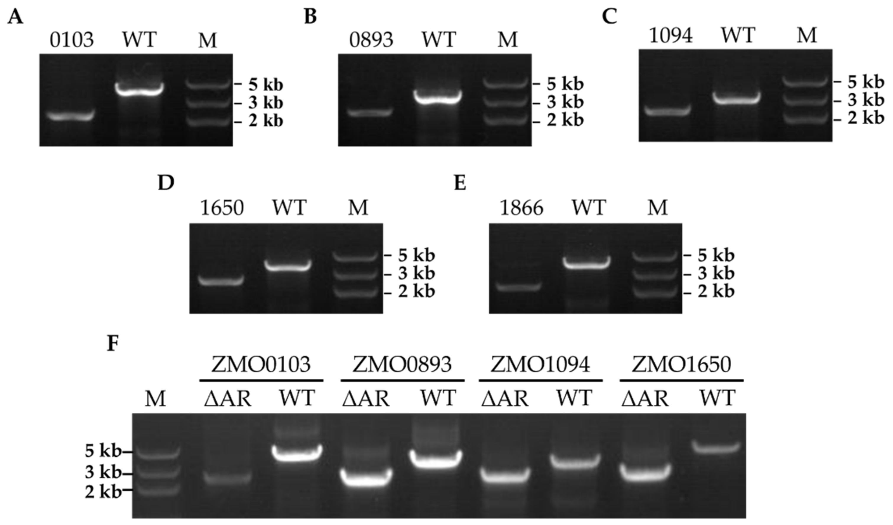
2.2. Ampicillin Resistance−Related Gene Knockout in Z. mobilis ZM4

Subsequently, six ampicillin−resistant (AR) candidate genes based on the above bioinformatics study were selected for knockout using the CRISPR−Cas12a genome−editing system. Except for ZMO1967, which may be an essential gene and cannot be knocked out, five other genes of ZMO0103, ZMO0893, ZMO1094, ZMO1650, and ZMO1866 were successfully knocked out with the CRISPR−Cas12a system. As demonstrated in Figure 1, the expected sizes of the amplicon of ZMO0103, ZMO0893, ZMO1094, ZMO1650, and ZMO1866 in the wild type (WT) strain were obtained, which were ~4021, 3314, 3122, 3746, and 3768 bp, respectively. As a contrast, the corresponding amplified fragments in the knockout strains were ~2247, 2232, 2361, 2221, and 2080 bp, respectively (Figure 1). The inactivation of the AR genes was further confirmed by Sanger sequencing of the PCR products. Then, the knockout strains harboring the editing plasmids were cultured in RMG5 for several passages to obtain the final strains with the editing plasmid cured. Except for ZMO1866, ZMO0103, ZMO0893, ZMO1094, and ZMO1650, the knockout strains lost the editing plasmid successfully, which can only grow in RMG5 after 24 h cultivation, but not in the medium with chloramphenicol (Supplementary Materials Figure S2).
To obtain the strain with all the ampicillin−resistant (AR) candidate genes knocked out, we further conducted six rounds of genome editing continuously with the CRISPR−Cas12a system. Consistent with the above single−gene deletion experiments, we only obtained a mutant strain, ZM4∆Ars, with four ampicillin−resistant (AR) genes of ZMO0103, ZMO0893, ZMO1094, and ZMO1650 knocked out continuously, while ZMO1967 and ZMO1866 were not able to be deleted. The ZM4∆ARs was further identified by colony PCR using the primers for each gene. The results of the correct PCR products of four genes indicate that these four ampicillin−resistant (AR) genes were knocked out successfully in ZM4∆ARs (Figure 1F).

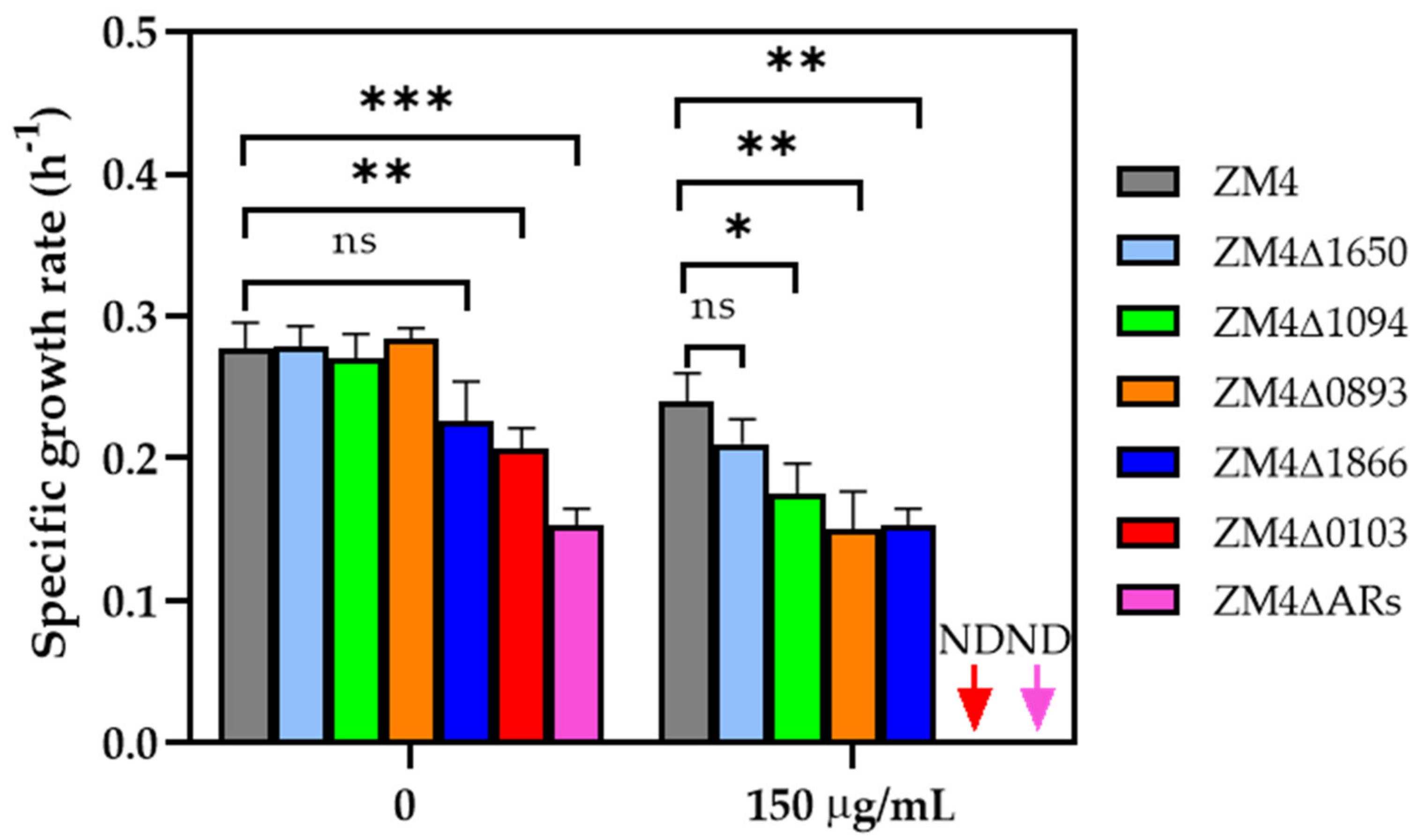
2.3. Antibiotic Tolerance of Ampicillin−Resistant (AR) Gene Knockout Strains

Previous studies reported that the concentration of ampicillin required for plate screening and liquid culture of the transformants in genetic engineering manipulation of Z. mobilis ZM4 was 300 μg/mL [19]. In this study, we set three ampicillin concentration gradients of 0, 150, and 300 μg/mL to cultivate the ampicillin−resistant (AR) gene knockout strains. As demonstrated in the Supplementary Materials Figure S4, the deletion of ZMO1094 and ZMO1650 individually did not decrease the ampicillin resistance. Specifically, the growth of ZM4∆1094 and ZM4∆1650 under different ampicillin concentrations was not inhibited compared with ZM4, whereas the deletion of ZMO0893 and ZMO1866 individually were effective. The growth of ZM4∆0893 and ZM4∆1866 was inhibited under 150 μg/mL ampicillin with a slower growth rate of 0.15 ± 0.03 h−1 and 0.05 ± 0.01 h−1 compared with the value of 0.24 ± 0.02 h−1 for wild−type ZM4 (Figure 2). The growth of the ZMO0103 mutant ZM4∆0103 was the slowest among all strains with the lowest growth rate of 0.21 ± 0.01 h−1 under 0 μg/mL ampicillin, and ZM4∆0103 was inhibited by all concentrations of ampicillin, such as 150 μg/mL (Figure 2, Supplementary Materials Figure S4).
The same experiment was also carried out for ZM4∆ARs, a mutant strain with four ampicillin−resistant (AR) genes deleted. Under 0 μg/mL ampicillin, a long lag phase was observed with a lower growth rate of 0.15 ± 0.01 h−1 in ZM4∆ARs compared to ZM4∆0103 (Figure 2, Supplementary Materials Figure S4), and ZM4∆ARs also cannot grow at 150 μg/mL ampicillin. Combined with the growth of single−gene knockout strains under RMG5, we speculated that the deletion of ZMO0103 is one of the major reasons for the poor growth of ZM4∆ARs, and other ampicillin−resistant (AR) genes may have a synergetic effect on ampicillin resistance. However, as previously reported, when the ampicillin concentration increased to 300 μg/mL, nearly all the strains could not grow. These results also demonstrate that the mutants ZM4∆0103 and ZM4∆ARs had a lower ampicillin tolerance concentration—150 μg/mL.

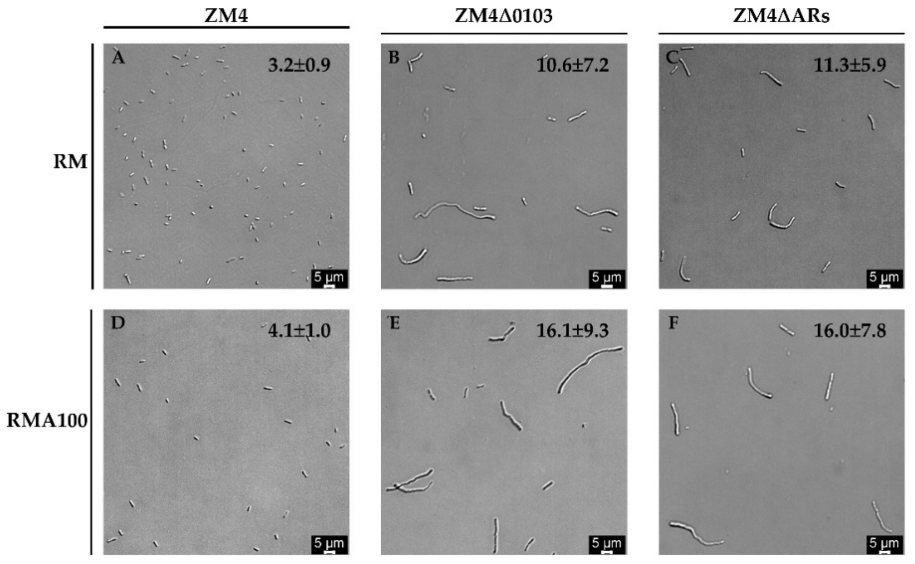
2.4. Cell Morphology of Mutants Treated with Ampicillin

Previous studies demonstrated that cell cultures in the presence of antibiotics resulted in abnormal cellular morphology in various degrees, such as elongated or distorted cell shapes [28,29]. To evaluate the morphological changes of ampicillin−resistant (AR) gene knockout strains during the ampicillin treatment, cell morphologies of three Z. mobilis strains (ZM4, ZM4∆0103, and ZM4∆Ars) were observed under light microscopy. The results show that ZM4∆0103 and ZM4∆ARs had longer rod shapes with various cell lengths and widths. The lengths of ZM4∆0103 and ZM4∆ARs were around 10.6 ± 7.2 μm and 11.3 ± 5.9 μm compared to that of wild−type ZM4 (3.2 ± 0.9 μm) in RMG5 without ampicillin (Figure 3). However, when strains were cultured in RMG5 with 100 μg/mL ampicillin (RMA100), the cellular morphology of ZM4∆0103 (16.1 ± 9.3 μm) and ZM4∆ARs (16.0 ± 7.8 μm) changed and was longer with a filament shape, while the length of ZM4 did not change much (4.1 ± 1.0 μm). This demonstrated that ampicillin was more stressful to ZM4∆0103 and ZM4∆ARs during cell growth, and ZM4∆0103 and ZM4∆ARs were more sensitive to ampicillin than ZM4. Similar morphological changes of Z. mobilis have been previously described when the cells were exposed to different stresses, such as high temperature [30], lignocellulosic hydrolysate inhibitory [31], high concentration of xylose [32], and salt conditions [33].

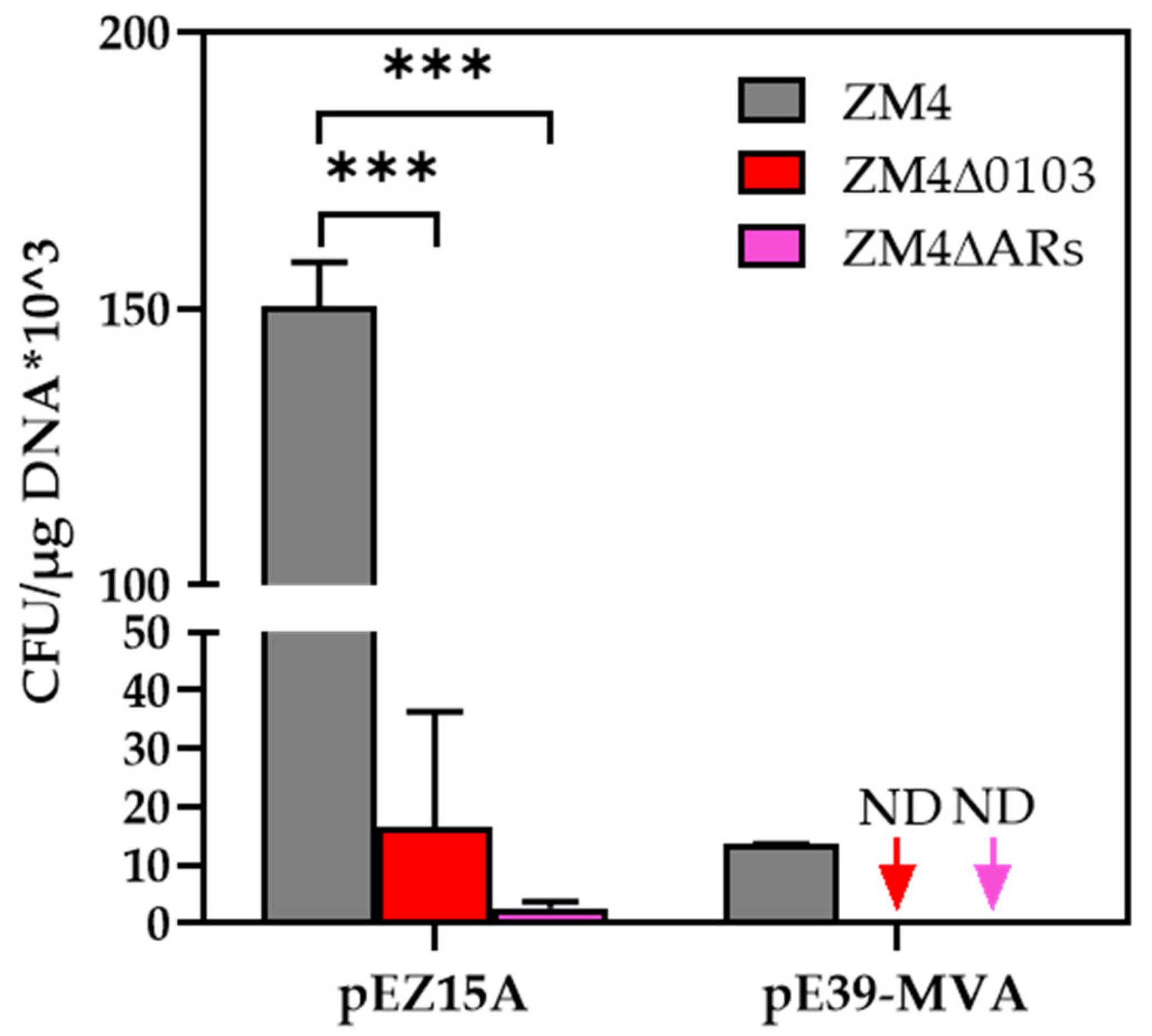
2.5. Effects of Ampicillin−Resistant (AR) Gene Mutagenesis on Genetic Transformation Efficiency

The transformation efficiencies of both ZM4∆0103 and ZM4∆ARs were significantly reduced using the plasmids of pEZ15A (~3 kb) and pE39−MVA (~10 kb). The electroporation efficiency of plasmid pEZ15A in ZM4 was (1.50 ± 0.08) × 105 CFU/μg DNA, which was decreased to (1.66 ± 1.98) × 104 and (2.66 ± 1.11) × 103 CFU/μg DNA in ZM4∆0103 and ZM4∆ARs, respectively (Figure 4, Supplementary Materials Figure S5), with the transformation efficiencies reduced ca 10~100 fold. Few colonies grew on the plate after the electroporation of pE39−MVA to ZM4∆0103 and ZM4∆ARs, while the efficiency of (1.35 ± 0.01) × 104 CFU/μg DNA in ZM4 was observed (Figure 4, Supplementary Materials Figure S6). The transformation efficiency of ZM4∆0103 and ZM4∆ARs cannot be calculated when the pE39−MVA plasmid with a large size was used. These results suggest that the predicted β−lactamase genes influenced the genetic transformation efficiency and significantly reduced the electroporation efficiency especially with the larger plasmid.

3. Discussion

In this study, we first predicted six functional β−lactamase genes that may cause ampicillin antibiotic resistance in Z. mobilis ZM4 by bioinformatics analysis. ZMO0103, ZMO0893, and ZMO1650 belong to the AmpC superfamily containing similar conserved structures, indicating that they may have similar functions in cellular processes. ZMO1967 belongs to the PenP superfamily, which is associated with β−lactamase class A. ZMO1094 and ZMO1866 are not closely related to the function of β−lactams. We attempted to knock out all six of them in ZM4; unfortunately, only 5 of them were deleted individually using the CRISPR−Cas12a genome−editing system. The ZMO1967 protein has a transmembrane domain at the N−terminus with a probability of 0.98 by TMHMM−2.0 (https://dtu.biolib.com/DeepTMHMM, accessed on 24 August 2022). In addition, ZMO1967 is probably an essential gene according to the results of the genome−wide CRISPRi (unpublished data), which may be the reason that it failed to be knocked out in this study. We successfully deleted five β−lactamase genes individually in ZM4, but the editing plasmid for ZMO1866 deletion was not able to be eliminated.
Our study demonstrated that in addition to ampicillin resistance, these genes annotated as ampicillin−resistant (AR) genes had other effects on cell morphology, cell growth, and transformation efficiency. The knockouts ZMO0893, ZMO1094, and ZMO1866 slightly inhibited the growth under the addition of 150 μg/mL. Especially, when ZMO0103 was deleted, the growth of the strain was inhibited mostly with different concentrations of ampicillin. The biomass of ZM4∆0103 hardly increased under ampicillin ≥ 150 μg/mL. In addition, similar growth inhibition was also observed in four ampicillin−resistant (AR) genes of ZMO0103, ZMO0893, ZMO1094 and the ZMO1650 knockout strain ZM4∆ARs. The previous study reported that ZMO0103 was a β−lactamase gene, which contains a 55% amino acid sequence identity with class C β−lactamase genes [20]. A higher expression level [30,34,35,36] of ZMO0103 in ZM4 may be ascribed to the absence of the AmpR that can inhibit the expression of ZMO0103. The result of multiple sequence alignment shows that the homologous protein of β−lactamase ZMO0103 was only found in Sphingomonas (Supplementary Materials Figures S7 and S8). Combining the result of the improved sensitivity of ZM4∆0103 to ampicillin in this study, we speculated that ZMO0103 is a unique protein and the most important β−lactamase in Z. mobilis ZM4, resulting in the high resistance of ZM4 to β−lactam antibiotics, such as ampicillin [17].
In addition, we also found that the growth of ZM4∆0103 and ZM4∆ARs was inhibited even without the addition of ampicillin. Considering the existence of transmembrane helices and signal peptides at the N−terminus (https://services.healthtech.dtu.dk/service.php?SignalP, accessed on 28 August 2022) of ZMO0103, ZMO0103 located on the cell membrane may directly hydrolyze ampicillin in the periplasmic space, and the integrity of the membrane could be disrupted by knocking out ZMO0103 leading to the defective growth of ZM4∆0103 and ZM4∆ARs. Furthermore, cell sizes of ZM4∆0103 and ZM4∆ARs became longer and further lengthened with the addition of ampicillin indicating that the AmpC family lactamase protein ZMO0103 is related to cell wall biosynthesis and deconstruction and crucial for cell morphology and growth. Microscopic observations under RMG5 showed that the longest cells in ZM4∆0103 and ZM4∆ARs increased in length by 13.7 μm and 13.1 μm, respectively, compared to the longest cells in the wild−type ZM4. In addition, when 100 μg/mL of ampicillin (RMA100) was added, the cell lengths of the longest cells in ZM4∆0103 and ZM4∆ARs could increase by 7.6 μm and 6.6 μm, respectively, compared to the lengths of ZM4∆0103 and ZM4∆ARs under RM. These results suggest that the deletion of ZMO0103 affected the cell wall structure and the cell membrane and therefore led to changes in the intracellular osmotic pressure, thus enlarging the cells similar to the result of the previous study [37].
Based on our speculation that the deletion of ZMO0103 may affect cell membrane and cell wall structures, we expected that the exogenous DNA could be more easily transferred into the cells. So, we further tested the transform efficiency of ZM4∆0103 and ZM4∆ARs using two plasmids of pEZ15A and pE39−MVA with different sizes. However, lower transformation efficiencies were observed when pEZ15A and pE39−MVA were electroporated into ZM4∆0103 and ZM4∆ARs compared with ZM4. Particularly, in the case of pE39−MVA, almost no transformants were obtained, which suggested that the transformation efficiency of the knockout strains was affected by plasmids sizes. However, the mechanisms of how ZMO0103 influences the transformation efficiency remain to be investigated including the constructing a truncated mutant of ZMO0103 by deleting the catalytic domain (amino acids from position 52 to 405) or the C−terminal domain (amino acids from position 406 to 520) that may influence the transformation of exogenous DNA. In addition, the restriction–modification (R–M) system genes (ZMO0028, ZMO1933, and ZMOp32x025_028) could be knocked out in ampicillin−resistant gene knockout strains to further improve the transformation efficiency [38,39,40].

4. Materials and Methods

4.1. Strains and Cultural Conditions

E. coli DH5α was stored in our laboratory and used for plasmid maintenance and construction. During culturing, 50 μg/mL of chloramphenicol was added to Luria−Bertani (LB) medium (10 g/L tryptone, 5 g/L yeast extract, and 10 g/L NaCl, pH 7.0) for E. coli and cultured at 37 °C. Z. mobilis ZM4 was used as the parental strain for the construction of derived mutants and cultured with Rich Medium (RMG5) (50 g/L glucose, 10 g/L yeast extract, and 2 g/L KH2PO4) at 30 °C. When required, 50 μg/mL of chloramphenicol and 100 μg/mL of spectinomycin were added to the LB and RMG5. All E. coli, Z. mobilis, and their derivative strains used in this study are listed in the Supplementary Materials Table S3.

4.2. In Silico Analysis of the AR Genes of Z. mobilis ZM4

A BLASTP analysis of the Z. mobilis ZM4 proteome was performed using two databases for resistance genes: CARD (Comprehensive antibiotic resistance database) and MEGARes database. In addition to using BLASTP to investigate the relatedness of the sequences to those contained within the CARD and MEGARes databases, we further aligned with the gene sequences of the β−lactamase class in the UniProt database with the Z. mobilis ZM4 protein sequences. We chose the common genes of candidate β−lactamase genes after BLASTP with CARD and MEGARes databases and genes annotated as β−lactamase as candidate gene list 1. Then, we chose the common genes of candidate β−lactamase genes after BLASTP with the UniProt database and genes annotated as β−lactamase as candidate gene list 2. The common genes within both candidate gene list 1 and list 2 were the final candidate β−lactamase genes used for investigation in this work. The β−lactamase genes with a significant E−value of 2 × 10⁻¹⁰ were selected.

4.3. Construction of Editing Plasmids of Ampicillin−Resistant (AR) Genes in Z. mobilis ZM4

The spacers were designed to bear the entire 23 bp sequences containing a 5′−NTTN−3′ PAM for six genes of ZMO0103, ZMO0893, ZMO1094, ZMO1358, ZMO1866, and ZMO1967. The oligonucleotides of spacers were ordered from TsingKe Biotechnology Co., Ltd. (Beijing, China). CRISPR−Cas12a−editing plasmids were constructed following the previous description [25]. The Cas12a−targeting gRNA sequence was annealed using two single−stranded oligonucleotides by first heating the reaction mixture to 95 °C for 5 min and subsequently cooling down gradually to room temperature. Then, the annealed spacer was ligated into Bsa I−linearized pEZ−sgr by T4 ligase at 22 °C for 3 h. The resulting plasmids were named as pEZ−sgr−0103, pEZ−sgr−0893, pEZ−sgr−1094, pEZ−sgr−1385, pEZ−sgr−1866, and pEZ−sgr−1967.
Gibson assembly method was utilized for donor construction. Donor sequences including extra ~800 bp upstream and downstream flank sequences of the candidate gene were amplified using Primer STAR polymerase (Takara, Japan) from the genomic DNA of Z. mobilis ZM4 and then cloned into pEZ−sgr vector by T5 exonuclease (NEB, WA, USA). The resulting plasmids were named as pEZ−sgr−0103−D, pEZ−sgr−0893−D, pEZ−sgr−1094−D, pEZ−sgr−1385−D, pEZ−sgr−1866−D, and pEZ−sgr−1967−D. All plasmids used in this work are provided in the Supplementary Materials Table S4. The sequences of primers used in this study are shown in the Supplementary Materials Table S5.

4.4. Curing of Editing Plasmids

Transformants harboring editing plasmids were cultured in RMG5 broth without the supplement of antibiotics. After 6 consecutive passages in the nonresistant RMG5 liquid medium, 100 μL cultures were diluted and plated on nonresistant RMG5 plates. Then, single colonies were picked to conduct colony PCR using primers pEZ15A−F/R for amplifying the editing plasmid. Editing plasmids were lost if no PCR product was amplified compared with the control. Single colonies with correct PCR results were then inoculated on RMG5 with or without chloramphenicol for further verification. The knockout strains losing the editing plasmids can only grow in RMG5, but not in the medium with the supplementation of chloramphenicol.

4.5. Electroporation of Editing Plasmids to Z. mobilis ZM4

Transformation of Z. mobilis ZM4 with the editing plasmid was achieved by preparing electrocompetent cells using a modified protocol as previously described. Z. mobilis ZM4 was inoculated with 40 mL RMG5 in a 100 mL flask and was grown to an OD600 nm = 0.4~0.6. Cells were harvested by centrifugation at 4000× g for 10 min at room temperature. The supernatant was discarded, and the cell pellet was washed with Milli−Q® ultrapure water and 10% (v/v) glycerol before being resuspended in a final volume of 400 μL of 10% (v/v) glycerol. Cells were stored as 50 μL aliquots on ice for immediate use in electroporation experiments. Then, 500 ng plasmid DNAs was used for electroporation (Bio−Rad, CA, USA). After pulsing at 16 kV/cm, 25 μF and 200 Ω, 1 mL of RMG5 was added to the electroporated solutions and then incubated at 30 °C for 4~6 h. Finally, the cells were plated and selected on RM plates supplemented with chloramphenicol until colonies were visible (≤2 d).

4.6. Construction of ZM4∆ARs by Continuous Gene Editing

We constructed ZM4∆ARs with four genes of ZMO0103, ZMO0893, ZMO1094, and ZMO1650 knocked out following these steps:
Step 1. A CRISPR−Cas12a−editing plasmid pEZ−sgr−0103−D was transformed into ZM4−Cas12a. Colony PCR was then performed with the primers 0103−out−F and 0103−out−R.
Step 2. The positive single clone with the correct PCR size was cultured in RMG5, and the editing plasmid was then cured following Section 4.4.
Step 3. After curing the editing plasmid, the electrocompetent cells of knockout strain ZM4∆0103 were then prepared for the next−round knockout.
Knockout of ZMO0893, ZMO1094, ZMO1650, ZMO1866, and ZMO1967 followed the above steps 1 to 3. After each gene was edited and the corresponding editing plasmid was lost, the competent cells of the corresponding mutant were prepared for transferring to the next editing plasmid.

4.7. Genetic Transformation Efficiency of β−lactamase Mutants

To calculate the electroporation efficiencies of the β−lactamase mutants, a 3−kb shuttle plasmid pEZ15A and a 10−kb shuttle plasmid pE39−MVA were prepared from E. coli DH5α and introduced into ZM4, ZM4∆0103, and ZM4∆Ars. Electroporation efficiency was presented by the colony forming units (CFUs) on selective plates when 50 μg plasmid DNA was introduced and 100 μL recovery culture was plated. The calculating formula is described below:
CFU/μg−1 DNA = (Cp/Tp) × (Vt/Vp);
where Cp is the colony number counted on selective plates; Tp is the total amount of plasmid DNA (μg) used here; Vt is the total transformation volume (μL); Vp is the volume (μL) plated.

4.8. Confirmation of Ampicillin−Resistant Gene Deletion in Z. mobilis ZM4

A pair of primers was used to confirm the disruption of each AR gene in Z. mobilis transformants: the primers were used to amplify the region including upstream of the corresponding gene, targeting gene, and downstream of the corresponding gene (see primer pairs in the Supplementary Materials Table S5, e.g., 0103−out−F and 0103−out−R).

4.9. Growth Studies and Analysis of Ampicillin−Resistant Gene Mutants

Cell growth was monitored by measuring the cell optical density (OD) values using a Bioscreen C high−throughput growth measurement instrument (Bioscreen C MBR, Helsinki, Finland). The single colony was inoculated in 1 mL of RMG5 at 30 °C overnight as the seed culture. Then, the seed culture of Z. mobilis was transferred to RMG5 until reaching the exponential phase. Subsequently, the cultures were inoculated to the well of the Bioscreen C plate containing 300 μL bacterium suspension with an initial OD600 nm value of 0.05. Three technical replicates were used for each condition.

4.10. Cell Morphology Observation

Z. mobilis strains were cultured in RMG5 or RMG5 with 150 μg/mL ampicillin overnight at 30 °C. Bacterial pellets were collected at the exponential phase, washed twice with 1× phosphate buffer saline (1× PBS), resuspended in the same buffer, and observed under a light microscope (Leica Dmi8, Buffalo Grove, IL, USA) at 400 magnifications. Each image was taken using the image−based autofocus system LAS X software of the Leica Dmi8 system, and the cell size was measured using ImageJ software [41].

5. Conclusions

In summary, genes associated with antibiotic resistance in Z. mobilis were systematically investigated, and six ampicillin−resistant genes in Z. mobilis were identified. Five of them were knocked out individually, and four ampicillin−resistant gene deletion mutant ZM4∆Ars was also constructed to verify their functions. The strains of ZM4∆0103 and ZM4∆Ars were sensitive to ampicillin at a lower concentration of 150 μg/mL with a long and filament shape. The putative membrane protein ZMO0103 is probably the essential β−lactamase, and other ampicillin−resistant genes may have a synergetic effect with it. This study not only identified the ampicillin−resistant genes in Z. mobilis ZM4, but also verified their functions on cell growth, morphology, and transformation efficiency. In addition, ampicillin−sensitive mutants can serve as the parental strains for metabolic engineering practices in Z. mobilis enabling the usage of antibiotics, such as ampicillin, that are routinely used for genetic engineering.

Supplementary Materials

The following supporting information can be downloaded at: https://www.mdpi.com/article/10.3390/antibiotics11111476/s1, Figure S1: Secondary structure prediction of ZMO0103, ZMO0893, ZMO1094, ZMO1650, ZMO1866, and ZMO1967 in NCBI database; Figure S2: Multiple sequence alignment of the AmpC superfamily and β−lactamase class A protein in Z. mobilis; Figure S3: Loss of editing plasmids in ampicillin−resistant (AR) gene knockout strains; Figure S4: Cell growth of single and multiple ampicillin−resistant gene knockout strains cultured under 0 (A), 150 (B), and 300 (C) μg/mL of ampicillin; Figure S5: Electroporation efficiency of ZM4∆0103, ZM4∆Ars, and ZM4 by pEZ15A; Figure S6: Electroporation efficiency of ZM4∆0103, ZM4∆Ars, and ZM4 by pEZ15A and pE39−MVA; Figure S7: Distance tree of ZMO0103 after multiple sequence alignment with other proteins collected in NCBI using BALSTP; Figure S8: The detailed result of multiple sequence alignment with other proteins collected in NCBI using BALSTP; Table S1: List of the putative efflux pump−type antibiotic−resistant genes by BLASTP with CARD and MEGARes databases; Table S2: List of the putative β−lactamase genes by BLASTP in the UniProt database; Table S3: Strains used in this study; Table S4: Plasmids used in this study; Table S5: Primers used in this study.

Author Contributions

Conceptualization, S.Y.; methodology, B.G., X.H. and Y.W.; validation, X.H.; formal analysis, B.G. and X.H.; investigation, B.G., X.H., Y.W., Q.H. and S.Y.; data curation, B.G. and X.H.; writing—original draft preparation, B.G. and X.H.; writing— review and editing, Q.H. and S.Y.; visualization, Y.W.; supervision and project administration, S.Y. All authors have read and agreed to the published version of the manuscript.

Funding

Supported by National Key Research and Development Program of China (2018YFC1801200), the National Natural Science Foundation of China (21978071 and U1932141), Leading Innovative and Entrepreneur Team Introduction Program of Zhejiang Province (2018R01014), and the Innovation Base for Introducing Talents of Discipline of Hubei Province (2019BJH021).

Institutional Review Board Statement

Ethical review and approval were waived for this study as the study did not involve animal interventions.

Informed Consent Statement

Not applicable.

Data Availability Statement

No new data were created or analyzed in this study. Data sharing does not apply to this article.

Acknowledgments

We acknowledge the support from the State Key Laboratory of Biocatalysis and Enzyme Engineering.

Conflicts of Interest

The authors declare no conflict of interest.

References

  1. Awad, H.; El–Shahed, K.Y.I.; Aziz, R.; Sarmidi, M.R.; El–Enshasy, H.A. Antibiotics as microbial secondary metabolites: Production and application. J. Teknol. 2012, 59, 1–11. [Google Scholar] [CrossRef] [Green Version]
  2. Parker, D.S.; Armstrong, D.G. Antibiotic feed additives and livestock production. Proc. Nutr. Soc. 1987, 46, 415–421. [Google Scholar] [CrossRef] [Green Version]
  3. Procopio, R.E.; Silva, I.R.; Martins, M.K.; Azevedo, J.L.; Araujo, J.M. Antibiotics produced by Streptomyces. Braz. J. Infect. Dis. 2012, 16, 466–471. [Google Scholar] [CrossRef] [Green Version]
  4. Urban−Chmiel, R.; Marek, A.; Stepien−Pysniak, D.; Wieczorek, K.; Dec, M.; Nowaczek, A.; Osek, J. Antibiotic resistance in bacteria−a review. Antibiotics 2022, 11, 1079. [Google Scholar] [CrossRef]
  5. Singh, S.B.; Young, K.; Silver, L.L. What is an “ideal” antibiotic? Discovery challenges and path forward. Biochem. Pharmacol. 2017, 133, 63–73. [Google Scholar] [CrossRef]
  6. Ezelarab, H.A.A.; Abbas, S.H.; Hassan, H.A.; Abuo−Rahma, G.E.A. Recent updates of fluoroquinolones as antibacterial agents. Arch Pharm 2018, 351, e1800141. [Google Scholar] [CrossRef]
  7. Ang, C.W.; Jarrad, A.M.; Cooper, M.A.; Blaskovich, M.A.T. Nitroimidazoles: Molecular fireworks that combat a broad spectrum of infectious diseases. J. Med. Chem. 2017, 60, 7636–7657. [Google Scholar] [CrossRef]
  8. Vilhena, C.; Bettencourt, A. Daptomycin: A review of properties, clinical use, drug delivery and resistance. Mini Rev. Med. Chem. 2012, 12, 202–209. [Google Scholar] [CrossRef]
  9. El−Sayed Ahmed, M.A.E.; Zhong, L.L.; Shen, C.; Yang, Y.; Doi, Y.; Tian, G.B. Colistin and its role in the Era of antibiotic resistance: An extended review (2000–2019). Emerg. Microbes Infect. 2020, 9, 868–885. [Google Scholar] [CrossRef] [Green Version]
  10. Bush, K.; Bradford, P.A. β−Lactams and β−Lactamase Inhibitors: An Overview. Cold Spring Harb. Perspect. Med. 2016, 6, a025247. [Google Scholar] [CrossRef]
  11. Wilson, D.N. Ribosome−targeting antibiotics and mechanisms of bacterial resistance. Nat. Rev. Microbiol. 2014, 12, 35–48. [Google Scholar] [CrossRef]
  12. Fernandez, L.; Hancock, R.E. Adaptive and mutational resistance: Role of porins and efflux pumps in drug resistance. Clin. Microbiol. Rev. 2012, 25, 661–681. [Google Scholar] [CrossRef] [PubMed] [Green Version]
  13. McMurry, L.; Petrucci, R.E., Jr.; Levy, S.B. Active efflux of tetracycline encoded by four genetically different tetracycline resistance determinants in Escherichia coli. Proc. Natl. Acad. Sci. USA 1980, 77, 3974–3977. [Google Scholar] [CrossRef] [Green Version]
  14. Poole, K. Efflux−mediated antimicrobial resistance. J. Antimicrob. Chemother. 2005, 56, 20–51. [Google Scholar] [CrossRef] [PubMed] [Green Version]
  15. Hoiby, N.; Bjarnsholt, T.; Givskov, M.; Molin, S.; Ciofu, O. Antibiotic resistance of bacterial biofilms. Int. J. Antimicrob. Agents 2010, 35, 322–332. [Google Scholar] [CrossRef] [Green Version]
  16. Wang, X.; He, Q.N.; Yang, Y.F.; Wang, J.W.; Haning, K.; Hu, Y.; Wu, B.; He, M.X.; Zhang, Y.P.; Bao, J.; et al. Advances and prospects in metabolic engineering of Zymomonas mobilis. Metab. Eng. 2018, 50, 57–73. [Google Scholar] [CrossRef]
  17. Yang, Y.F.; Geng, B.N.; Song, H.Y.; He, Q.N.; He, M.X.; Bao, J.; Bai, F.W.; Yang, S.H. Progress and perspectives on developing Zymomonas mobilis as a chassis cell. Synth. Biol. J. 2021, 2, 59–90. [Google Scholar] [CrossRef]
  18. Yang, S.H.; Pelletier, D.A.; Lu, T.Y.; Brown, S.D. The Zymomonas mobilis regulator hfq contributes to tolerance against multiple lignocellulosic pretreatment inhibitors. BMC Microbiol. 2010, 10, 135. [Google Scholar] [CrossRef] [Green Version]
  19. Zou, S.L.; Zhang, K.; Jing, X.; Wang, Z.R.; Zhang, M.H. Studies on selectable marker for genetic engineering of Zymomonas mobilis ZM4 and CP4 strain. Ind. Microbiol. 2012, 42, 72–77. [Google Scholar]
  20. Rajnish, K.N.; Asraf, S.A.; Manju, N.; Gunasekaran, P. Functional characterization of a putative beta−lactamase gene in the genome of Zymomonas mobilis. Biotechnol. Lett. 2011, 33, 2425–2430. [Google Scholar] [CrossRef]
  21. Yang, S.; Pappas, K.M.; Hauser, L.J.; Land, M.L.; Chen, G.L.; Hurst, G.B.; Pan, C.; Kouvelis, V.N.; Typas, M.A.; Pelletier, D.A.; et al. Improved genome annotation for Zymomonas mobilis. Nat. Biotechnol. 2009, 27, 893–894. [Google Scholar] [CrossRef]
  22. Yang, S.; Vera, J.M.; Grass, J.; Savvakis, G.; Moskvin, O.V.; Yang, Y.; McIlwain, S.J.; Lyu, Y.; Zinonos, I.; Hebert, A.S.; et al. Complete genome sequence and the expression pattern of plasmids of the model ethanologen Zymomonas mobilis ZM4 and its xylose−utilizing derivatives 8b and 2032. Biotechnol. Biofuels 2018, 11, 125. [Google Scholar] [CrossRef]
  23. Seo, J.S.; Chong, H.; Park, H.S.; Yoon, K.O.; Jung, C.; Kim, J.J.; Hong, J.H.; Kim, H.; Kim, J.H.; Kil, J.I.; et al. The genome sequence of the ethanologenic bacterium Zymomonas mobilis ZM4. Nat. Biotechnol. 2005, 23, 63–68. [Google Scholar] [CrossRef] [Green Version]
  24. Zheng, Y.L.; Han, J.M.; Wang, B.Y.; Hu, X.Y.; Li, R.X.; Shen, W.; Ma, X.D.; Ma, L.X.; Yi, L.; Yang, S.H.; et al. Characterization and repurposing of the endogenous Type I−F CRISPR−Cas system of Zymomonas mobilis for genome engineering. Nucleic Acids Res. 2019, 47, 11461–11475. [Google Scholar] [CrossRef] [Green Version]
  25. Shen, W.; Zhang, J.; Geng, B.N.; Qiu, M.Y.; Hu, M.M.; Yang, Q.; Bao, W.W.; Xiao, Y.B.; Zheng, Y.L.; Peng, W.F.; et al. Establishment and application of a CRISPR−Cas12a assisted genome−editing system in Zymomonas mobilis. Microb. Cell Fact. 2019, 18, 162. [Google Scholar] [CrossRef] [Green Version]
  26. Yang, Y.F.; Shen, W.; Huang, J.; Li, R.X.; Xiao, Y.B.; Wei, H.; Chou, Y.C.; Zhang, M.; Himmel, M.E.; Chen, S.W.; et al. Prediction and characterization of promoters and ribosomal binding sites of Zymomonas mobilis in system biology era. Biotechnol. Biofuels 2019, 12, 52. [Google Scholar] [CrossRef] [Green Version]
  27. Lu, S.; Wang, J.; Chitsaz, F.; Derbyshire, M.K.; Geer, R.C.; Gonzales, N.R.; Gwadz, M.; Hurwitz, D.I.; Marchler, G.H.; Song, J.S.; et al. CDD/SPARCLE: The conserved domain database in 2020. Nucleic Acids Res. 2020, 48, D265–D268. [Google Scholar] [CrossRef] [Green Version]
  28. Chan, E.L.; Harris, R.C.; Dalton, H.P. The effect of antibiotics on the cell morphology of Legionella pneumophila. J. Med. Microbiol. 1987, 23, 149–154. [Google Scholar] [CrossRef] [Green Version]
  29. Rodgers, F.G.; Tzianabos, A.O.; Elliott, T.S. The effect of antibiotics that inhibit cell−wall, protein, and DNA synthesis on the growth and morphology of Legionella pneumophila. J. Med. Microbiol. 1990, 31, 37–44. [Google Scholar] [CrossRef]
  30. Li, R.X.; Shen, W.; Yang, Y.F.; Du, J.; Li, M.; Yang, S.H. Investigation of the impact of a broad range of temperatures on the physiological and transcriptional profiles of Zymomonas mobilis ZM4 for high−temperature−tolerant recombinant strain development. Biotechnol. Biofuels 2021, 14, 146. [Google Scholar] [CrossRef]
  31. Mohagheghi, A.; Linger, J.G.; Yang, S.H.; Smith, H.; Dowe, N.; Zhang, M.; Pienkos, P.T. Improving a recombinant Zymomonas mobilis strain 8b through continuous adaptation on dilute acid pretreated corn stover hydrolysate. Biotechnol. Biofuels 2015, 8, 55. [Google Scholar] [CrossRef]
  32. Lou, J.Y.; Wang, J.W.; Yang, Y.F.; Yang, Q.; Li, R.X.; Hu, M.M.; He, Q.N.; Du, J.; Wang, X.; Li, M.; et al. Development and characterization of efficient xylose utilization strains of Zymomonas mobilis. Biotechnol. Biofuels 2021, 14, 231. [Google Scholar] [CrossRef]
  33. Fuchino, K.; Bruheim, P. Increased salt tolerance in Zymomonas mobilis strain generated by adaptative evolution. Microb. Cell Fact. 2020, 19, 147. [Google Scholar] [CrossRef]
  34. Yang, S.; Pan, C.; Hurst, G.B.; Dice, L.; Davison, B.H.; Brown, S.D. Elucidation of Zymomonas mobilis physiology and stress responses by quantitative proteomics and transcriptomics. Front. Microbiol. 2014, 5, 246. [Google Scholar] [CrossRef] [Green Version]
  35. Yang, Q.; Yang, Y.; Tang, Y.; Wang, X.; Chen, Y.; Shen, W.; Zhan, Y.; Gao, J.; Wu, B.; He, M.; et al. Development and characterization of acidic−pH−tolerant mutants of Zymomonas mobilis through adaptation and next−generation sequencing−based genome resequencing and RNA−Seq. Biotechnol. Biofuels 2020, 13, 144. [Google Scholar] [CrossRef]
  36. He, M.X.; Wu, B.; Shui, Z.X.; Hu, Q.C.; Wang, W.G.; Tan, F.R.; Tang, X.Y.; Zhu, Q.L.; Pan, K.; Li, Q.; et al. Transcriptome profiling of Zymomonas mobilis under furfural stress. Appl. Microbiol. Biotechnol. 2012, 95, 189–199. [Google Scholar] [CrossRef]
  37. Mack, K.; Titball, R.W. Transformation of Burkholderia pseudomallei by electroporation. Anal. Biochem. 1996, 242, 73–76. [Google Scholar] [CrossRef]
  38. Wu, B.; He, M.; Feng, H.; Zhang, Y.; Hu, Q.; Zhang, Y. Construction and characterization of restriction−modification deficient mutants in Zymomonas mobilis ZM4. Chin. J. Appplied Environ. Biol. 2013, 19, 189–197. [Google Scholar] [CrossRef]
  39. Kerr, A.L.; Jeon, Y.J.; Svenson, C.J.; Rogers, P.L.; Neilan, B.A. DNA restriction−modification systems in the ethanologen Zymomonas mobilis ZM4. Appl. Microbiol. Biotechnol. 2011, 89, 761–769. [Google Scholar] [CrossRef]
  40. Lal, P.B.; Wells, F.; Myers, K.S.; Banerjee, R.; Guss, A.M.; Kiley, P.J. Improving mobilization of foreign DNA into Zymomonas mobilis strain ZM4 by removal of multiple restriction systems. Appl. Environ. Microbiol. 2021, 87, e0080821. [Google Scholar] [CrossRef]
  41. Rueden, C.T.; Schindelin, J.; Hiner, M.C.; DeZonia, B.E.; Walter, A.E.; Arena, E.T.; Eliceiri, K.W. ImageJ2: ImageJ for the next generation of scientific image data. BMC Bioinform. 2017, 18, 529. [Google Scholar] [CrossRef]
Figure 1. Confirmation of ampicillin−resistant (AR) knockout strains in Z. mobilis ZM4 by PCR. The mutants of ZM4∆0103 (A), ZM4∆0893 (B), ZM4∆1650 (C), ZM4∆1094 (D), ZM4∆1866 (E), and ZM4∆ARs (F) were confirmed by colony PCR using their corresponding primers. The sizes of PCR products (bp) of WT and knockout strains were 4021, 2247 (ZM4∆0103); 3314, 2232 (ZM4∆0893); 3122, 2361 (ZM4∆1650); 3746, 2221 (ZM4∆1094); 3768, 2080 (ZM4∆1866).
Antibiotics 11 01476 g001
Figure 2. The specific growth rate of ampicillin−resistant (AR) gene knockout strains cultured under 0 and 150 μg/mL of ampicillin. Three replicates were performed for the experiment. When the mutant could not grow under the condition, the sample is marked “ND” (not detected). * represents a significant difference with p−value < 0.05. ** represents a significant difference with p−value < 0.01. *** represents a significant difference with p−value < 0.001. ns represents no significant difference.
Antibiotics 11 01476 g002
Figure 3. Cell morphology of strains ZM4, ZM4∆0103, and ZM4∆ARs cultured in RM and RMA100 was observed by light microscopy. The numbers with error value in each image represent the average cell size (μm) analyzed with ImageJ software. Numbers in the lower right corner of each represent the scale. RM and RMA100 represent the different RMG5 media with 0 and 100 μg/mL of ampicillin.
Antibiotics 11 01476 g003
Figure 4. Electroporation efficiency of ZM4, ZM4∆0103, and ZM4∆ARs using plasmids of pEZ15A (~3 kb) and pE39−MVA (~10 kb). Three replicates were performed for the experiment. The error bar represents standard deviation (SD). When transformation of a plasmid was below the limit of detection (0.00001), the sample is marked “ND” (not detected). *** represents a very significant difference (p−value < 0.001).
Antibiotics 11 01476 g004
Publisher’s Note: MDPI stays neutral with regard to jurisdictional claims in published maps and institutional affiliations.

