
Clinical Implications of Helicobacter pylori Antibiotic Resistance in Italy: A Review of the Literature
 ,
,  ,
,
Abstract
:1. Introduction
Genomic and Virulence Factors
2. Resistance Mechanisms of Principal Antibiotics Used for H. pylori Eradication
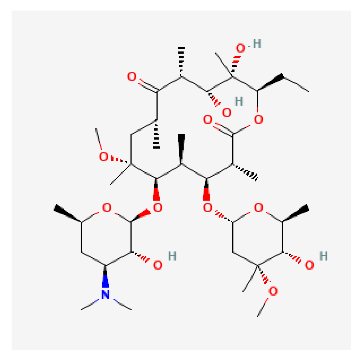
2.1. Clarithromycin
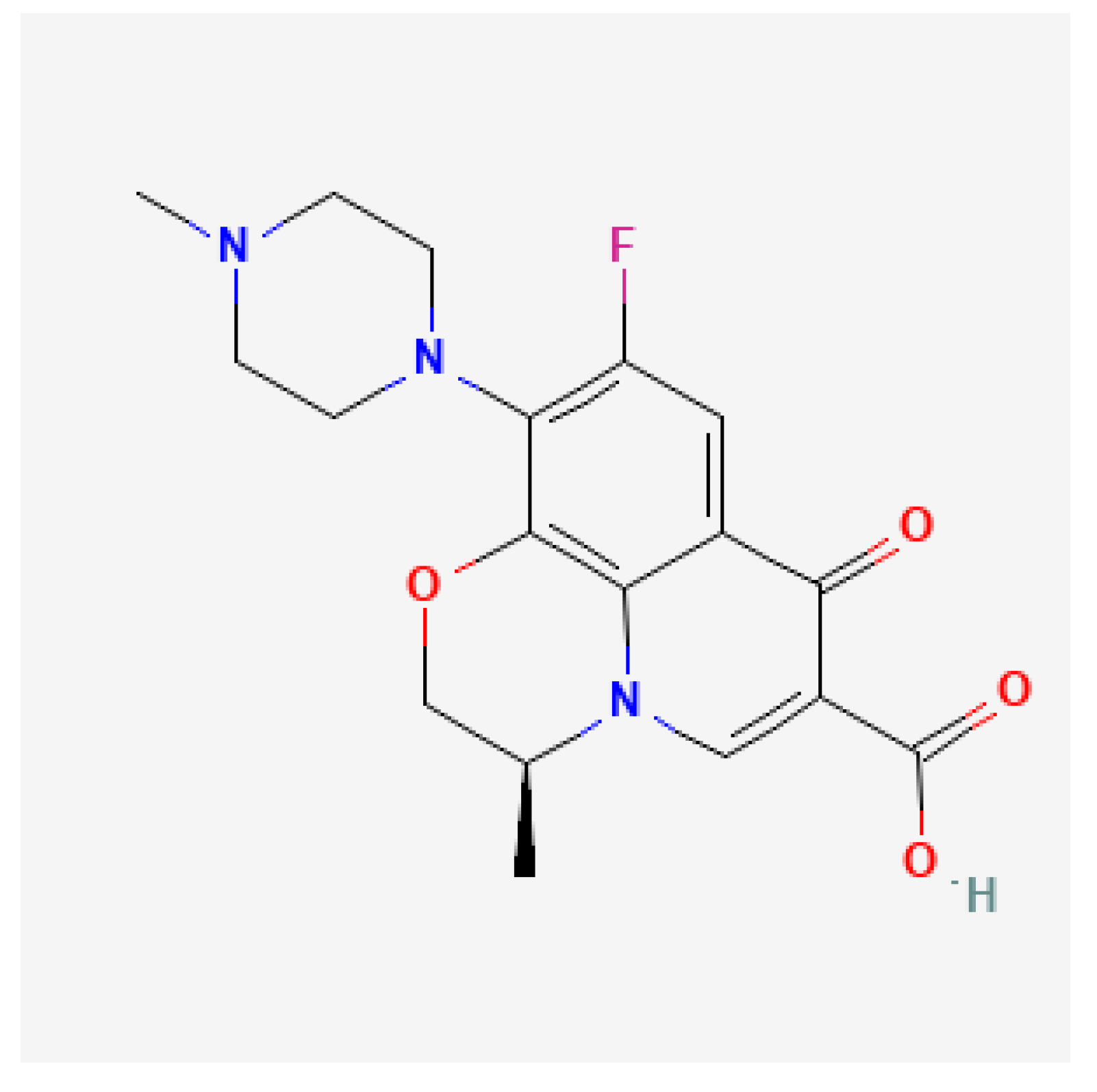
2.2. Levofloxacin
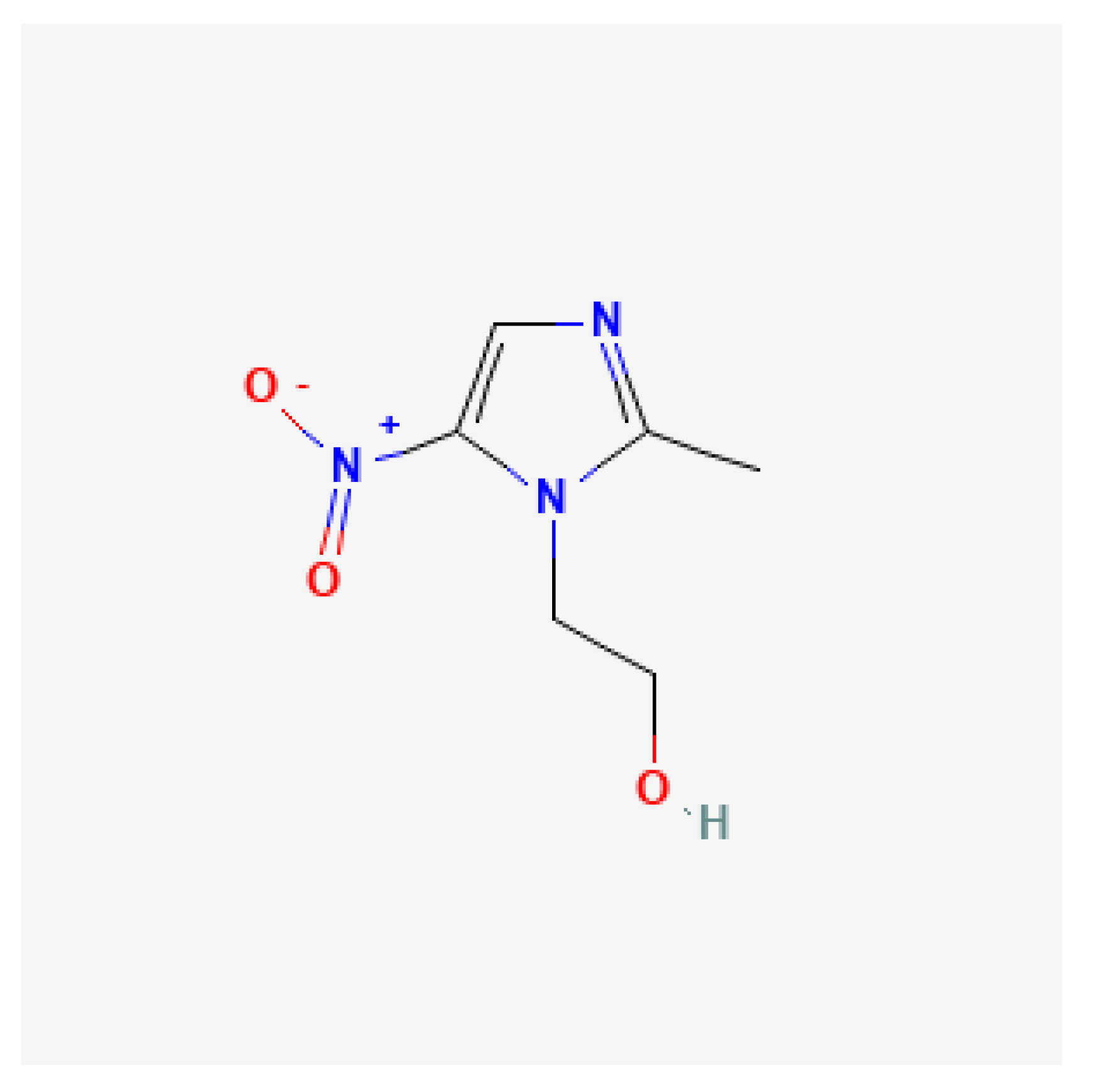
2.3. Metronidazole
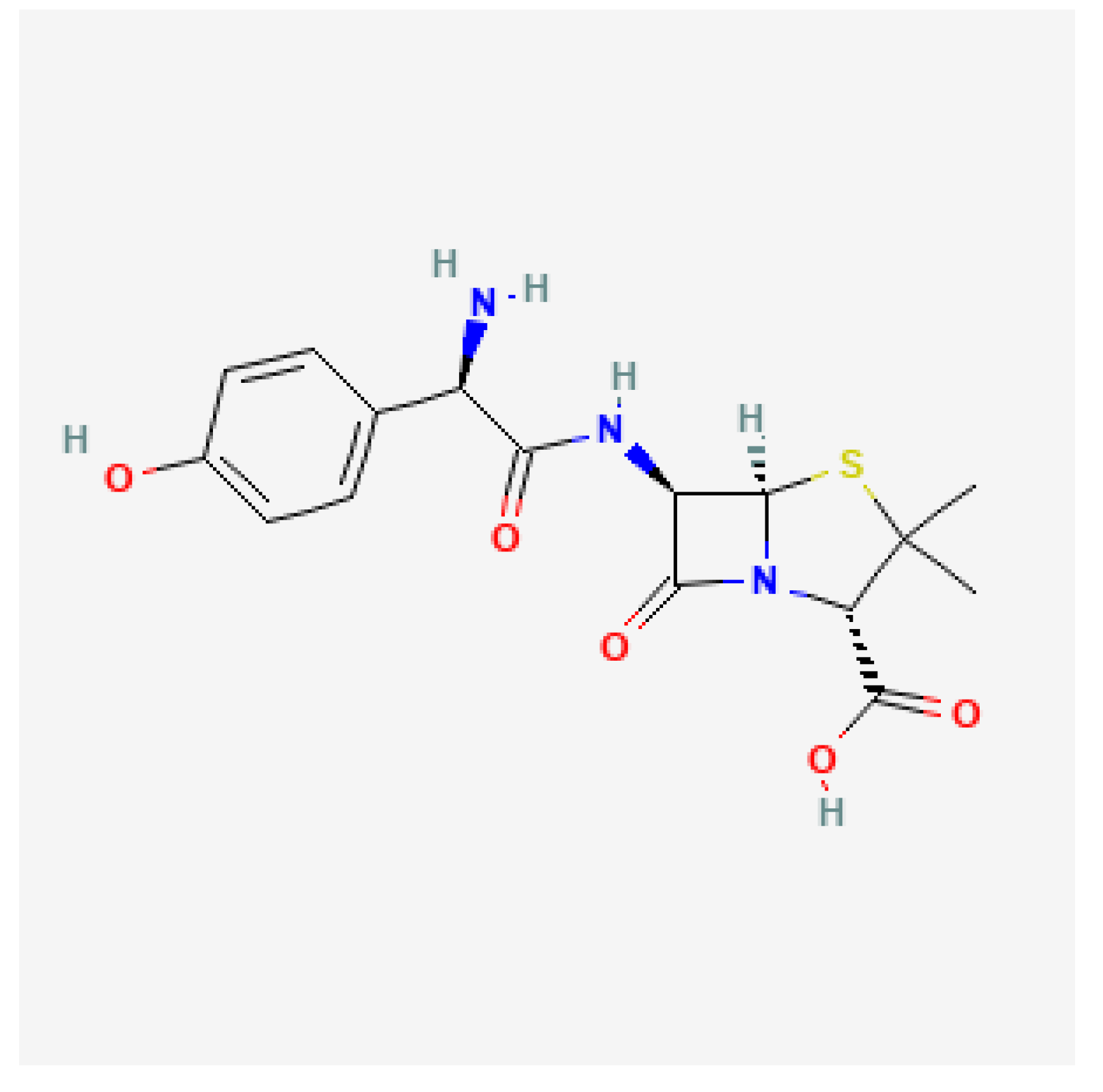
2.4. Amoxicillin
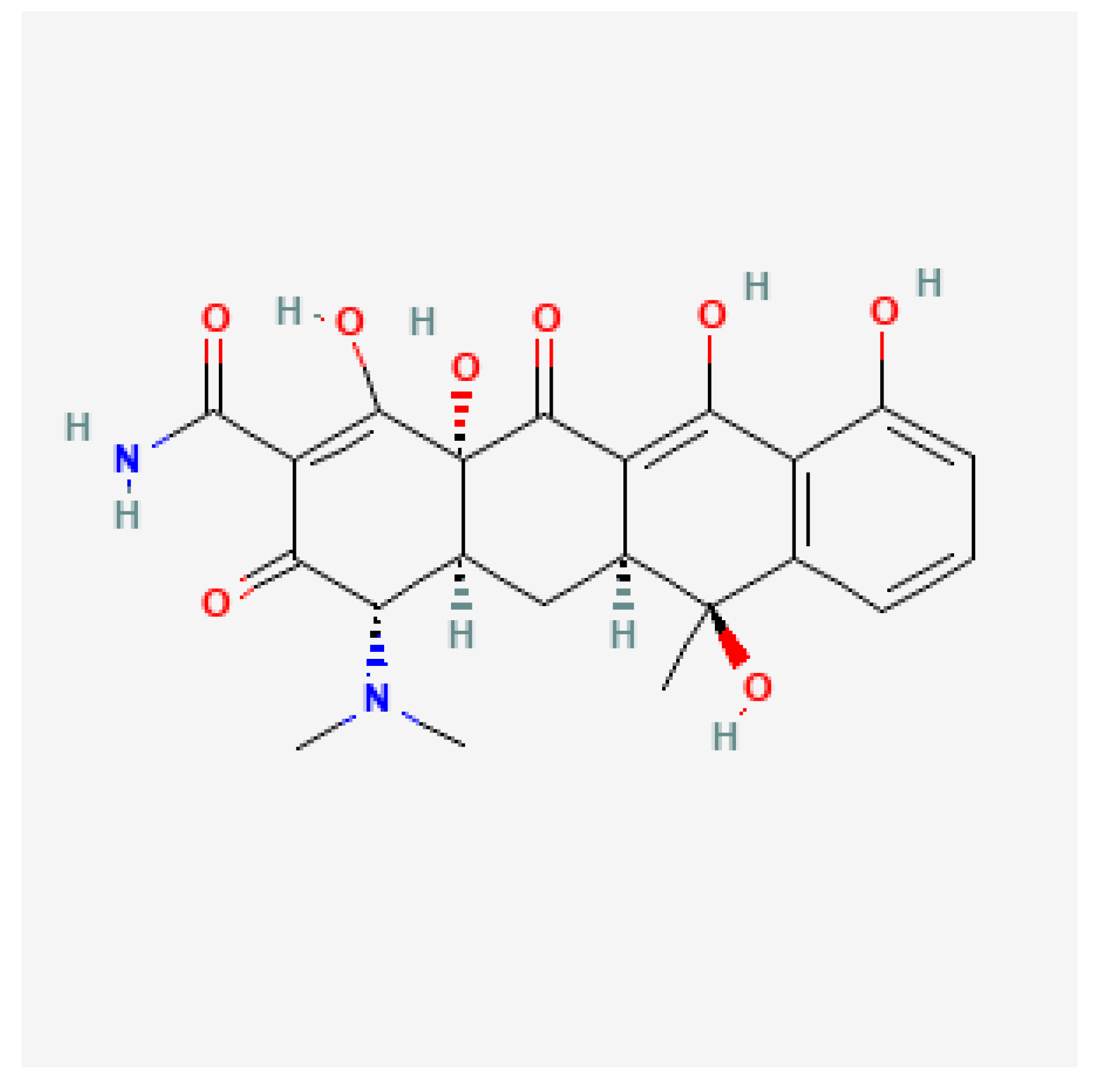
2.5. Tetracycline
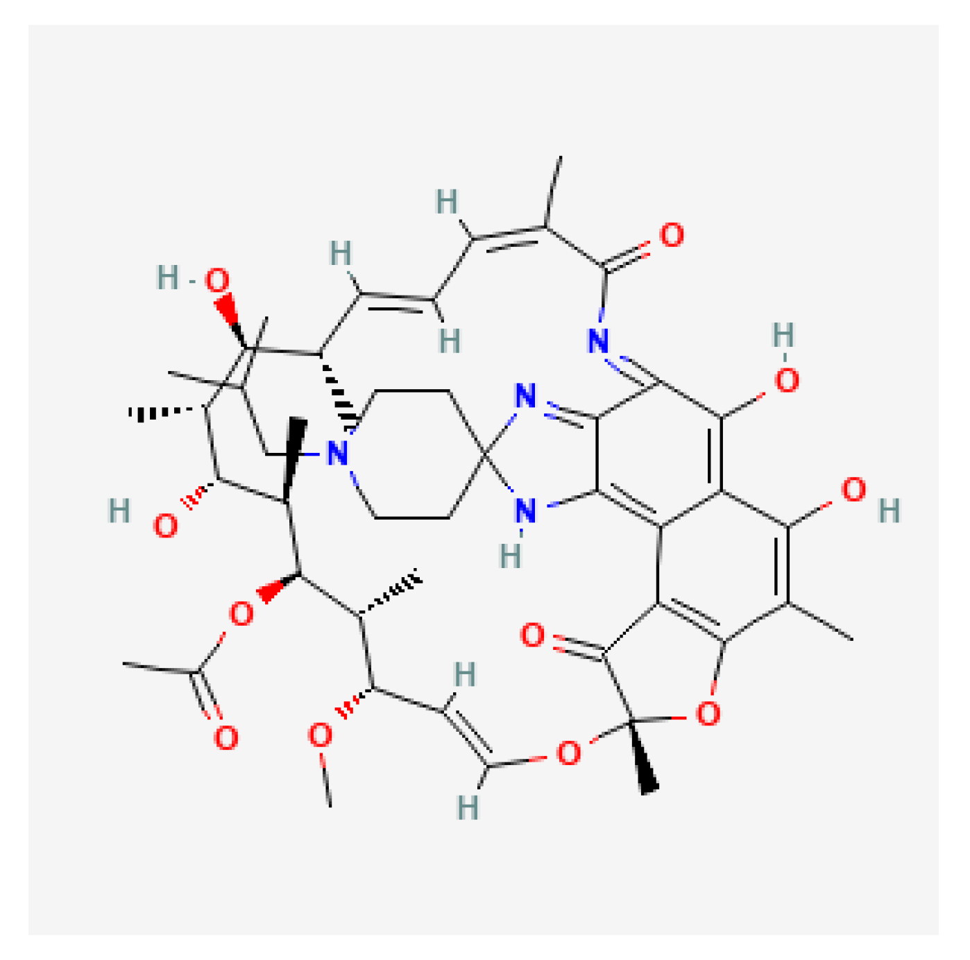
2.6. Rifabutin
3. Methods for the Detection of H. pylori Resistance to Antibiotic
4. Clinical Determinants of Antibiotic Resistance
5. Empiric vs. Tailored Therapy
5.1. First Line Therapy
5.2. Second-Line Therapy
5.3. Third-Line Therapy
6. Alternative Therapies for H. pylori
6.1. Probiotics
6.2. Vonoprazan
6.3. Vaccine
7. Rates of Antibiotic Resistance in Italy
7.1. Primary Resistance (Table 1, Table 2, Table 3 and Table 4)

| Method | Year | Resistance % | Region | Research Group | Ref. |
|---|---|---|---|---|---|
| Culture on biopsy | 2020 | 30.2% (95% CI 27.2–33.3) in 2009–2014, 37.8% (95% CI 34.2–41.4) in 2015–2019 | Emilia-Romagna | Saracino et al. | [108] |
| Culture on biopsy | 2018 | 19% in 2010, 35.6% in 2013, 35.9% in 2016 (OR not calculated) | Emilia-Romagna | Fiorini et al. | [109] |
| Culture on biopsy | 2018 | 36.1% in 2010–2015 | Emilia-Romagna | Gatta et al. | [110] |
| Culture on biopsy-PCR on isolates | 2020 | 37.7% in 2017–2018 | Puglia | Palmitessa et al. | [111] |
| RT-PCR on stools | 2020 | 27.4% in 2017–2020 | Puglia | Losurdo et al. | [112] |
| Method | Year | Resistance % | Region | Research Group | Ref. |
|---|---|---|---|---|---|
| Culture on biopsy | 2020 | 25.6% in 2010–2014 and 33.8% in 2015–2019 | Emilia- Romagna | Saracino et al. | [108] |
| Culture on biopsy | 2018 | 19% in 2019, 29.7% in 2013, 29.3% in 2016 (OR not calculated) | Emilia- Romagna | Fiorini et al. | [109] |
| Culture on biopsy | 2018 | 28.7% in 2010–2015 | Emilia- Romagna | Gatta et al. | [110] |
| Culture on biopsy-PCR on isolates | 2020 | 26.2 % in 2017–2018 | Puglia | Palmitessa et al. | [111] |
| RT-PCR on stools | 2020 | 19.2 % in 2017–2020 | Puglia | Losurdo et al. | [112] |
| Method | Year | Resistance % | Region | Research Group | Ref. |
|---|---|---|---|---|---|
| Culture on biopsy | 2020 | 33.3 % in 2010–2014 and 33.6%in 2015–2019 | Emilia- Romagna | Saracino et al. | [108] |
| Culture on biopsy | 2018 | 33.6 % in 2019, 45.3% in 2013, 40.2% in 2016 (OR not calculated) | Emilia- Romagna | Fiorini et al. | [109] |
| Culture on biopsy | 2018 | 38.6% in 2010–2015 | Emilia- Romagna | Gatta et al. | [110] |
| Culture on biopsy | 2020 | 16.4 % in 2017–2018 | Puglia | Palmitessa et al. | [111] |
| Antibiotic | Method | Year | Resistance % | Region | Research Group | Ref. |
|---|---|---|---|---|---|---|
| Amoxicillin | Culture on biopsy | 2020 | 1.6 % in 2017–2018 | Puglia | Palmitessa et al. | [111] |
| Tetracyclin | Culture on biopsy | 2020 | 0 % in 2017–2018 | Puglia | Palmitessa et al. | [111] |
| Rifabutin | Culture on biopsy | 2020 | 1.6 % in 2017–2018 | Puglia | Palmitessa et al. | [111] |
7.2. Secondary Resistance (Table 5, Table 6, Table 7 and Table 8)
| Method | Year | Resistance % | Region | Research Group | Ref. |
|---|---|---|---|---|---|
| Culture on biopsy | 2020 | 83.1% in 2009–2019 | Emilia-Romagna | Saracino et al. | [21] |
| Culture on biopsy-PCR on isolates | 2020 | 83.9% in 2017–2018 | Puglia | Palmitessa et al. | [111] |
| RT-PCR on stools | 2020 | 64.8% in 2017–2020 | Puglia | Losurdo et al. | [112] |
| Culture on biopsy-RT-PCR on isolates | 2018 | 50% (not indicated the years) | Lazio | Mascellino et al. | [113] |
| Culture on biopsy-RT-PCR on isolates | 2020 | 35% with phenotypic methods, 42.5% with genotypic methods (not indicated the years) | Lazio | Mascellino et al. | [114] |
| Method | Year | Resistance % | Region | Research Group | Ref. |
|---|---|---|---|---|---|
| Culture on biopsy | 2020 | 47.2% in 2009–2019 | Emilia-Romagna | Saracino et al. | [21] |
| Culture on biopsy-PCR on isolates | 2020 | 64.5% in 2017–2018 | Puglia | Palmitessa et al. | [111] |
| RT-PCR on stools | 2020 | 59.3% in 2017–2020 | Puglia | Losurdo et al. | [112] |
| Culture on biopsy-RT-PCR on isolates | 2018 | 25% (not indicated the years) | Lazio | Mascellino et al. | [113] |
| Culture on biopsy-RT-PCR on isolates | 2020 | 15% with phenotypic methods, 30% with genotypic methods (not indicated the years) | Lazio | Mascellino et al. | [114] |
| Method | Year | Resistance % | Region | Research Group | Ref. |
|---|---|---|---|---|---|
| Culture on biopsy | 2020 | 66.7% in 2009–2019 | Emilia- Romagna | Saracino et al. | [21] |
| Culture on biopsy-PCR on isolates | 2020 | 64.5% in 2017–2018 | Puglia | Palmitessa et al. | [111] |
| Culture on biopsy-RT-PCR on isolates | 2018 | 68% (not indicated the years) | Lazio | Mascellino et al. | [113] |
| Culture on biopsy-RT-PCR on isolates | 2020 | 61.6% (not indicated the years) | Lazio | Mascellino et al. | [114] |
| Antibiotic | Method | Year | Resistance % | Region | Research Group | Ref. |
|---|---|---|---|---|---|---|
| Amoxicillin | Culture on biopsy | 2020 | 6.5% in 2017–2018 | Puglia | Palmitessa et al. | [111] |
| Rifabutin | Culture on biopsy | 2020 | 0% in 2017–2018 | Puglia | Palmitessa et al. | [111] |
| Amoxicillin | Culture on biopsy-RT-PCR on isolates | 2018 | 4% (not indicated the years) | Lazio | Mascellino et al. | [113] |
| Amoxicillin | Culture on biopsy-RT-PCR on isolates | 2020 | 1.25% (not indicated the years) | Lazio | Mascellino et al. | [114] |
| Tetracycline | Culture on biopsy | 2020 | 0% in 2017–2018 | Puglia | Palmitessa et al. | [111] |
| Tetracycline | Culture on biopsy-RT-PCR on isolates | 2018 | 6% (not indicated the years) | Lazio | Mascellino et al. | [113] |
| Tetracycline | Culture on biopsy-RT-PCR on isolates | 2020 | 2.5% (not indicated the years) | Lazio | Mascellino et al. | [114] |
8. Eradication Rates with the Most Important Antibiotic Regimens
First Attempt (Table 9 and Table 10)
| Antibiotic Regimen | Year | Eradication Rate % | Region | Research Group | Ref. |
|---|---|---|---|---|---|
| Clarithromycin-based sequential | 2020 | 87.5% at ITT analysis, 93.4% at PP analysis, | Emilia- Romagna | Saracino et al. | [108] |
| Clarithromycin-based sequential | 2018 | 91.1% at ITT analysis, 93.7% at PP analysis, | Emilia- Romagna | Gatta et al. | [110] |
| Clarithromycin-based sequential | 2017 | 89% at ITT analysis, 89.9% at PP analysis | Puglia | Di Ciaula et al. | [116] |
| Standard Triple Therapy | 2017 | 70.7% | Puglia | Di Ciaula et al. | [116] |
| Clarithromycin-based concomitant | 2017 | 88.2% at ITT analysis, 91.2% at PP analysis, | Campania | Romano et al. | [117] |
| Clarithromycin-based sequential | 2018 | 92% at ITT analysis, 96% at PP analysis, | Emilia- Romagna | Fiorini et al. | [118] |
| Antibiotic Regimen | Year | Eradication Rate % | Region | Research Group | Ref. |
|---|---|---|---|---|---|
| Bismuth-based quadruple therapy (Pylera®) | 2017 | 100%, | Puglia | Di Ciaula et al. | [116] |
| Bismuth-based quadruple therapy Pylera®) | 2017 | 91.2% at ITT analysis, 95.8% at PP analysis | Campania | Romano et al. | [117] |
| Bismuth-based quadruple therapy (Pylera®) | 2018 | 91% at ITT analysis, 97% at PP analysis | Emilia-Romagna | Fiorini et al. | [118] |
9. Sequent Attempt (Table 11 and Table 12)
| Antibiotic Regimen | Year | Eradication Rate % | Region | Research Group | Ref. |
|---|---|---|---|---|---|
| Bismuth-based quadruple therapy (Pylera®) | 2020 | 86.8% at ITT analysis, 90% at PP analysis, | Emila Romagna | Saracino et al. | [21] |
| Bismuth-based quadruple therapy | 2020 | 90% (not indicated percentages at ITT and PP analysis) | Lazio | Mascellino et al. | [114] |
| Bismuth-based quadruple therapy (Pylera®) | 2017 | 96.7% at ITT analysis, 97.8% at PP analysis, | Puglia | Di Ciaula et al. | [116] |
| Bismuth-based quadruple therapy (Pylera®) | 2017 | 81% at ITT analysis, 87% at PP analysis, | Emilia- Romagna | Fiorini et al. | [118] |
| Bismuth-based quadruple therapy (Pylera®) | 2020 | 88.3%(not indicated percentages at ITT and PP analysis) | Emilia- Romagna | Saracino et al. | [121] |
| Bismuth-based quadruple therapy (Pylera®) | 2022 | 84.9% at ITT analysis, 86.1% at PP analysis, | Puglia | Losurdo et al. | [122] |
| Antibiotic Regimen | Year | Eradication Rate % | Region | Research Group | Ref. |
|---|---|---|---|---|---|
| Clarithromycin-based sequential | 2020 | 84.2% at ITT analysis, 91.4% at PP analysis | Emilia- Romagna | Saracino et al. | [21] |
| Levofloxacin-based triple therapy | 2020 | 80.4% at ITT analysis, 86.6% at PP analysis | Emilia- Romagna | Saracino et al. | [21] |
| Rifabutin-based triple therapy | 2020 | 75.6% at ITT analysis, 83.8% at PP analysis | Emilia- Romagna | Saracino et al. | [21] |
| Levofloxacin-based triple therapy | 2017 | 57.1% (not indicated percentages at ITT and PP analysis) | Puglia | Di Ciaula et al. | [116] |
| Rifabutin-based triple therapy | 2016 | 82.9% at ITT analysis, 88.7% at PP analysis | Emilia- Romagna | Fiorini et al. | [120] |
| Rifabutin-based triple therapy | 2020 | 61.9% (not indicated percentages at ITT and PP analysis) | Emilia- Romagna | Saracino et al. | [121] |
10. Discussion
11. Methods
12. Conclusions
Author Contributions
Funding
Institutional Review Board Statement
Informed Consent Statement
Conflicts of Interest
References
- Savoldi, A.; Carrara, E.; Graham, D.Y.; Conti, M.; Tacconelli, E. Prevalence of Antibiotic Resistance in Helicobacter pylori: A Systematic Review and Meta-analysis in World Health Organization Regions. Gastroenterology 2018, 155, 1372–1382.e17. [Google Scholar] [CrossRef] [PubMed] [Green Version]
- Hooi, J.K.Y.; Lai, W.Y.; Ng, W.K.; Suen, M.M.Y.; Underwood, F.E.; Tanyingoh, D.; Malfertheiner, P.; Graham, D.Y.; Wong, V.W.S.; Wu, J.C.Y.; et al. Global Prevalence of Helicobacter pylori Infection: Systematic Review and Meta-Analysis. Gastroenterology 2017, 153, 420–429. [Google Scholar] [CrossRef] [PubMed] [Green Version]
- Antimicrobial Resistance Collaborators. Global burden of bacterial antimicrobial resistance in 2019: A systematic analysis. Lancet 2022, 399, 629–655. [Google Scholar] [CrossRef]
- Crowe, S.E. Helicobacter pylori Infection. N. Engl. J. Med. 2019, 380, 1158–1165. [Google Scholar] [CrossRef] [PubMed]
- Gravina, A.G.; Priadko, K.; Ciamarra, P.; Granata, L.; Facchiano, A.; Miranda, A.; Dallio, M.; Federico, A.; Romano, M. Extra-Gastric Manifestations of Helicobacter pylori Infection. J. Clin. Med. 2020, 9, 3887. [Google Scholar] [CrossRef] [PubMed]
- Takeuchi, H.; Okamoto, A. Helicobacter pylori Infection and Chronic Immune Thrombocytopenia. J. Clin. Med. 2022, 11, 4822. [Google Scholar] [CrossRef] [PubMed]
- Ranjbar, R.; Behzadi, P.; Farshad, S. Advances in diagnosis and treatment of Helicobacter pylori infection. Acta Microbiol. Immunol. Hung. 2017, 64, 273–292. [Google Scholar] [CrossRef] [PubMed] [Green Version]
- IARC. Schistosomes, Liver Flukes and Helicobacter pylori. In IARC Monographs on the Evaluation of Carcinogenic Risks To Humans; IARC: Lyon, France, 1994; Volume 61, pp. 1–241. [Google Scholar]
- Plummer, M.; Franceschi, S.; Vignat, J.; Forman, D.; de Martel, C. Global burden of gastric cancer attributable to Helicobacter pylori. Int. J. Cancer 2015, 136, 487–490. [Google Scholar] [CrossRef] [PubMed]
- Ford, A.C.; Yuan, Y.; Moayyedi, P. Helicobacter pylori eradication therapy to prevent gastric cancer: Systematic review and meta-analysis. Gut 2020, 69, 2113–2121. [Google Scholar] [CrossRef]
- Ford, A.C.; Forman, D.; Hunt, R.H.; Yuan, Y.; Moayyedi, P. Helicobacter pylori eradication therapy to prevent gastric cancer in healthy asymptomatic infected individuals: Systematic review and meta-analysis of randomised controlled trials. BMJ 2014, 348, g3174. [Google Scholar] [CrossRef]
- Sugano, K.; Tack, J.; Kuipers, E.J.; Graham, D.Y.; El-Omar, E.M.; Miura, S.; Haruma, K.; Asaka, M.; Uemura, N.; Malfertheiner, P. Kyoto global consensus report on Helicobacter pylori gastritis. Gut 2015, 64, 1353–1367. [Google Scholar] [CrossRef] [PubMed] [Green Version]
- Megraud, F.; Bruyndonckx, R.; Coenen, S.; Wittkop, L.; Huang, T.-D.; Hoebeke, M.; Bénéjat, L.; Lehours, P.; Goossens, H.; Glupczynski, Y. Helicobacter pylori resistance to antibiotics in Europe in 2018 and its relationship to antibiotic consumption in the community. Gut 2021, 70, 1815–1822. [Google Scholar] [CrossRef] [PubMed]
- Graham, D.Y.; Fischbach, L. Helicobacter pylori treatment in the era of increasing antibiotic resistance. Gut 2010, 59, 1143–1153. [Google Scholar] [CrossRef] [PubMed]
- World Health Organization. World Health Organization Publishes List of Bacteria for Which New Antibiotics Are Urgently Needed. Available online: https://www.who.int/news/item/27-02-2017-who-publishes-list-of-bacteria-for-which-new-antibiotics-are-urgently-needed (accessed on 8 August 2022).
- Marin, A.C.; Nyssen, O.P.; McNicholl, A.G.; Gisbert, J.P. Efficacy and Safety of Quinolone-Containing Rescue Therapies after the Failure of Non-Bismuth Quadruple Treatments for Helicobacter pylori Eradication: Systematic Review and Meta-Analysis. Drugs 2017, 77, 765–776. [Google Scholar] [CrossRef]
- Malfertheiner, P.; Megraud, F.; Rokkas, T.; Gisbert, J.P.; Liou, J.-M.; Schulz, C.; Gasbarrini, A.; Hunt, R.H.; Leja, M.; O’Morain, C.; et al. Management of Helicobacter pylori infection: The Maastricht VI/Florence consensus report. Gut 2022, 71, 1724–1762. [Google Scholar] [CrossRef]
- Chey, W.D.; Leontiadis, G.I.; Howden, C.W.; Moss, S.F. ACG Clinical Guideline: Treatment of Helicobacter pylori Infection. Am. J. Gastroenterol. 2017, 112, 212–239. [Google Scholar] [CrossRef]
- Fallone, C.A.; Chiba, N.; van Zanten, S.V.; Fischbach, L.; Gisbert, J.P.; Hunt, R.H.; Jones, N.L.; Render, C.; Leontiadis, G.I.; Moayyedi, P.; et al. The Toronto Consensus for the Treatment of Helicobacter pylori Infection in Adults. Gastroenterology 2016, 151, 51–69.e14. [Google Scholar] [CrossRef] [Green Version]
- Mascellino, M.T.; Porowska, B.; de Angelis, M.; Oliva, A. Antibiotic susceptibility, heteroresistance, and updated treatment strategies in Helicobacter pylori infection. Drug Des. Dev. Ther. 2017, 11, 2209–2220. [Google Scholar] [CrossRef] [Green Version]
- Saracino, I.M.; Pavoni, M.; Zullo, A.; Fiorini, G.; Saccomanno, L.; Lazzarotto, T.; Cavallo, R.; Antonelli, G.; Vaira, D. Antibiotic Resistance and Therapy Outcome in H. pylori Eradication Failure Patients. Antibiotics 2020, 9, 121. [Google Scholar] [CrossRef] [Green Version]
- Losurdo, G.; D’Abramo, F.S.; Piazzolla, M.; Rima, R.; Continisio, A.; Pricci, M.; Ierardi, E.; Di Leo, A. Second line therapy for Helicobacter pylori eradication: State of art. Mini-Rev. Med. Chem. 2022, 22, 2430–2437. [Google Scholar] [CrossRef]
- Sheu, B.-S.; Wu, M.-S.; Chiu, C.-T.; Lo, J.-C.; Wu, D.-C.; Liou, J.-M.; Wu, C.-Y.; Cheng, H.-C.; Lee, Y.-C.; Hsu, P.-I.; et al. Consensus on the clinical management, screening-to-treat, and surveillance of Helicobacter pylori infection to improve gastric cancer control on a nationwide scale. Helicobacter 2017, 22, e12368. [Google Scholar] [CrossRef] [PubMed] [Green Version]
- Zagari, R.M.; Romano, M.; Ojetti, V.; Stockbrugger, R.; Gullini, S.; Annibale, B.; Farinati, F.; Ierardi, E.; Maconi, G.; Rugge, M.; et al. Guidelines for the management of Helicobacter pylori infection in Italy: The III Working Group Consensus Report 2015. Dig. Liver Dis. 2015, 47, 903–912. [Google Scholar] [CrossRef] [PubMed]
- Sharndama, H.C.; Mba, I.E. Helicobacter pylori: An up-to-date overview on the virulence and pathogenesis mechanisms. Braz. J. Microbiol. 2022, 53, 33–50. [Google Scholar] [CrossRef] [PubMed]
- Shankar, U.; Mishra, S.K.; Jain, N.; Tawani, A.; Yadav, P.; Kumar, A. Ni+2 permease system of Helicobacter pylori contains highly conserved G-quadruplex motifs. Infect. Genet. Evol. J. Mol. Epidemiol. Evol. Genet. Infect. Dis. 2022, 101, 105298. [Google Scholar] [CrossRef] [PubMed]
- Vital, J.S.; Tanoeiro, L.; Lopes-Oliveira, R.; Vale, F.F. Biomarker Characterization and Prediction of Virulence and Antibiotic Resistance from Helicobacter pylori Next Generation Sequencing Data. Biomolecules 2022, 12, 691. [Google Scholar] [CrossRef] [PubMed]
- Zeng, J.; Xie, C.; Zhang, L.; Liu, X.; Chan, M.T.V.; Wu, W.K.K.; Chen, H. Host Cell Antimicrobial Responses against Helicobacter pylori Infection: From Biological Aspects to Therapeutic Strategies. Int. J. Mol. Sci. 2022, 23, 10941. [Google Scholar] [CrossRef]
- Available online: https://pubchem.ncbi.nlm.nih.gov/ (accessed on 12 October 2022).
- Available online: https://www.drugbank.ca/drugs/DB01211 (accessed on 12 October 2022).
- Tshibangu-Kabamba, E.; Yamaoka, Y. Helicobacter pylori infection and antibiotic resistance—From biology to clinical implications. Nat. Rev. Gastroenterol. Hepatol. 2021, 18, 613–629. [Google Scholar] [CrossRef]
- Saracino, I.; Pavoni, M.; Zullo, A.; Fiorini, G.; Lazzarotto, T.; Borghi, C.; Vaira, D. Next Generation Sequencing for the Prediction of the Antibiotic Resistance in Helicobacter pylori: A Literature Review. Antibiotics 2021, 10, 437. [Google Scholar] [CrossRef]
- Versalovic, J.; Shortridge, D.; Kibler, K.; Griffy, M.V.; Beyer, J.; Flamm, R.K.; Tanaka, S.K.; Graham, D.Y.; Go, M.F. Mutations in 23S rRNA are associated with clarithromycin resistance in Helicobacter pylori. Antimicrob. Agents Chemother. 1996, 40, 477–480. [Google Scholar] [CrossRef] [Green Version]
- Nishizawa, T.; Suzuki, H. Mechanisms of Helicobacter pylori antibiotic resistance and molecular testing. Front. Mol. Biosci. 2014, 1, 19. [Google Scholar] [CrossRef]
- Gong, Y.; Yuan, Y. Resistance mechanisms of Helicobacter pylori and its dual target precise therapy. Crit. Rev. Microbiol. 2018, 44, 371–392. [Google Scholar] [CrossRef]
- Binh, T.T.; Shiota, S.; Suzuki, R.; Matsuda, M.; Trang, T.T.H.; Kwon, D.H.; Iwatani, S.; Yamaoka, Y. Discovery of novel mutations for clarithromycin resistance in Helicobacter pylori by using next-generation sequencing. J. Antimicrob. Chemother. 2014, 69, 1796–1803. [Google Scholar] [CrossRef] [Green Version]
- Iwamoto, A.; Tanahashi, T.; Okada, R.; Yoshida, Y.; Kikuchi, K.; Keida, Y.; Murakami, Y.; Yang, L.; Yamamoto, K.; Nishiumi, S.; et al. Whole-genome sequencing of clarithromycin resistant Helicobacter pylori characterizes unidentified variants of multidrug resistant efflux pump genes. Gut Pathog. 2014, 6, 27. [Google Scholar] [CrossRef] [PubMed] [Green Version]
- Available online: https://www.drugbank.ca/drugs/DB01137 (accessed on 12 October 2022).
- Lauener, F.; Imkamp, F.; Lehours, P.; Buissonnière, A.; Benejat, L.; Zbinden, R.; Keller, P.; Wagner, K. Genetic Determinants and Prediction of Antibiotic Resistance Phenotypes in Helicobacter pylori. J. Clin. Med. 2019, 8, 53. [Google Scholar] [CrossRef] [PubMed] [Green Version]
- Nishizawa, T.; Suzuki, H.; Umezawa, A.; Muraoka, H.; Iwasaki, E.; Masaoka, T.; Kobayashi, I.; Hibi, T. Rapid Detection of Point Mutations Conferring Resistance to Fluoroquinolone in gyrA of Helicobacter pylori by Allele-Specific PCR. J. Clin. Microbiol. 2007, 45, 303–305. [Google Scholar] [CrossRef] [Green Version]
- Rimbara, E.; Noguchi, N.; Kawai, T.; Sasatsu, M. Fluoroquinolone Resistance in Helicobacter pylori: Role of Mutations at Position 87 and 91 of GyrA on the Level of Resistance and Identification of a Resistance Conferring Mutation in GyrB. Helicobacter 2012, 17, 36–42. [Google Scholar] [CrossRef] [PubMed]
- Available online: https://www.drugbank.ca/drugs/DB00916 (accessed on 12 October 2022).
- Dingsdag, S.A.; Hunter, N. Metronidazole: An update on metabolism, structure–cytotoxicity and resistance mechanisms. J. Antimicrob. Chemother. 2017, 73, 265–279. [Google Scholar] [CrossRef] [Green Version]
- Tsugawa, H.; Suzuki, H.; Satoh, K.; Hirata, K.; Matsuzaki, J.; Saito, Y.; Suematsu, M.; Hibi, T. Two Amino Acids Mutation of Ferric Uptake Regulator Determines Helicobacter pylori Resistance to Metronidazole. Antioxid. Redox Signal. 2011, 14, 15–23. [Google Scholar] [CrossRef]
- Available online: https://www.drugbank.ca/drugs/DB01060 (accessed on 12 October 2022).
- Gerrits, M.M.; Godoy, A.P.O.; Kuipers, E.J.; Ribeiro, M.L.; Stoof, J.; Mendonça, S.; van Vliet, A.; Pedrazzoli, J., Jr.; Kusters, J.G. Multiple Mutations in or Adjacent to the Conserved Penicillin-Binding Protein Motifs of the Penicillin-Binding Protein 1A Confer Amoxicillin Resistance to Helicobacter pylori. Helicobacter 2006, 11, 181–187. [Google Scholar] [CrossRef]
- Nishizawa, T.; Suzuki, H.; Tsugawa, H.; Muraoka, H.; Matsuzaki, J.; Hirata, K.; Ikeda, F.; Takahashi, M.; Hibi, T. Enhancement of Amoxicillin Resistance after Unsuccessful Helicobacter pylori Eradication. Antimicrob. Agents Chemother. 2011, 55, 3012–3014. [Google Scholar] [CrossRef]
- Rimbara, E.; Noguchi, N.; Kawai, T.; Sasatsu, M. Mutations in penicillin-binding proteins 1, 2 and 3 are responsible for amoxicillin resistance in Helicobacter pylori. J. Antimicrob. Chemother. 2008, 61, 995–998. [Google Scholar] [CrossRef] [Green Version]
- Available online: https://www.drugbank.ca/drugs/DB00759 (accessed on 12 October 2022).
- Chopra, I.; Roberts, M. Tetracycline Antibiotics: Mode of Action, Applications, Molecular Biology, and Epidemiology of Bacterial Resistance. Microbiol. Mol. Biol. Rev. 2001, 65, 232–260. [Google Scholar] [CrossRef] [PubMed] [Green Version]
- Gerrits, M.M.; de Zoete, M.R.; Arents, N.L.A.; Kuipers, E.J.; Kusters, J.G. 16S rRNA Mutation-Mediated Tetracycline Resistance in Helicobacter pylori. Antimicrob. Agents Chemother. 2002, 46, 2996–3000. [Google Scholar] [CrossRef] [PubMed] [Green Version]
- Available online: https://www.drugbank.ca/drugs/DB00615 (accessed on 12 October 2022).
- Nishizawa, T.; Suzuki, H.; Matsuzaki, J.; Muraoka, H.; Tsugawa, H.; Hirata, K.; Hibi, T. Helicobacter pylori Resistance to Rifabutin in the Last 7 Years. Antimicrob. Agents Chemother. 2011, 55, 5374–5375. [Google Scholar] [CrossRef] [PubMed] [Green Version]
- Gerrits, M.M.; van Vliet, A.; Kuipers, E.J.; Kusters, J.G. Helicobacter pylori and antimicrobial resistance: Molecular mechanisms and clinical implications. Lancet Infect. Dis. 2006, 6, 699–709. [Google Scholar] [CrossRef]
- Phan, T.N.; Tran, V.H.; Tran, T.N.H.; Le, V.A.; Santona, A.; Rubino, S.; Paglietti, B. Antimicrobial resistance in Helicobacter pylori: Current situation and management strategy in Vietnam. J. Infect. Dev. Ctries. 2015, 9, 609–613. [Google Scholar] [CrossRef] [Green Version]
- Megraud, F.; Hazell, S.; Glupczynski, Y. Helicobacter pylori: Physiology and genetics. In Antibiotic Susceptibility and Resistance; ASM Press: Washington, DC, USA, 2001; Chapter 42. [Google Scholar]
- Thung, I.; Aramin, H.; Vavinskaya, V.; Gupta, S.; Park, J.Y.; Crowe, S.E.; Valasek, M.A. Review article: The global emergence of Helicobacter pylori antibiotic resistance. Aliment. Pharmacol. Ther. 2016, 43, 514–533. [Google Scholar] [CrossRef] [PubMed] [Green Version]
- Tang, X.; Shen, Y.; Hu, R.; Yang, T.; Benghezal, M.; Li, H.; Tang, H. Re-assessment of the disk diffusion technique for routine antimicrobial susceptibility testing for Helicobacter pylori. Helicobacter 2020, 25, e12703. [Google Scholar] [CrossRef] [PubMed]
- Arslan, N.; Yılmaz, Ö.; Demiray-Gürbüz, E. Importance of antimicrobial susceptibility testing for the management of eradication in Helicobacter pylori infection. World J. Gastroenterol. 2017, 23, 2854–2869. [Google Scholar] [CrossRef]
- Mégraud, F.; Bénéjat, L.; Ngoyi, E.N.O.; Lehours, P. Molecular Approaches to Identify Helicobacter pylori Antimicrobial Resistance. Gastroenterol. Clin. N. Am. 2015, 44, 577–596. [Google Scholar] [CrossRef]
- Clayton, C.L.; Kleanthous, H.; Coates, P.J.; Morgan, D.D.; Tabaqchali, S. Sensitive detection of Helicobacter pylori by using polymerase chain reaction. J. Clin. Microbiol. 1992, 30, 192–200. [Google Scholar] [CrossRef] [PubMed] [Green Version]
- Klesiewicz, K.; Nowak, P.; Karczewska, E.; Skiba, I.; Wojtas-Bonior, I.; Sito, E.; Budak, A. PCR-RFLP detection of point mutations A2143G and A2142G in 23S rRNA gene conferring resistance to clarithromycin in Helicobacter pylori strains. Acta Biochim. Pol. 2014, 61, 311–315. [Google Scholar] [CrossRef] [Green Version]
- White, B.; Winte, M.; DeSipio, J.; Phadtare, S. Clinical Factors Implicated in Antibiotic Resistance in Helicobacter pylori Patients. Microorganisms 2022, 10, 322. [Google Scholar] [CrossRef] [PubMed]
- Lim, S.G.; Park, R.W.; Shin, S.J.; Yoon, D.; Kang, J.K.; Hwang, J.C.; Kim, S.S.; Kim, J.H.; Lee, K.M. The relationship between the failure to eradicate Helicobacter pylori and previous antibiotics use. Dig. Liver Dis. 2016, 48, 385–390. [Google Scholar] [CrossRef]
- Björkholm, B.; Sjölund, M.; Falk, P.G.; Berg, O.G.; Engstrand, L.; Andersson, D.I. Mutation frequency and biological cost of antibiotic resistance in Helicobacter pylori. Proc. Natl. Acad. Sci. USA 2001, 98, 14607–14612. [Google Scholar] [CrossRef] [PubMed] [Green Version]
- Megraud, F.; Coenen, S.; Versporten, A.; Kist, M.; Lopez-Brea, M.; Hirschl, A.M.; Andersen, L.P.; Goossens, H.; Glupczynski, Y.; on behalf of the Study Group participants. Helicobacter pylori resistance to antibiotics in Europe and its relationship to antibiotic consumption. Gut 2012, 62, 34–42. [Google Scholar] [CrossRef]
- McNulty, C.A.M.; Lasseter, G.; Shaw, I.; Nichols, T.; D’Arcy, S.; Lawson, A.J.; Glocker, E. Is Helicobacter pylori antibiotic resistance surveillance needed and how can it be delivered? Aliment. Pharmacol. Ther. 2012, 35, 1221–1230. [Google Scholar] [CrossRef]
- Zullo, A.; Perna, F.; Hassan, C.; Ricci, C.; Saracino, I.M.; Morini, S.; Vaira, D. Primary antibiotic resistance in Helicobacter pylori strains isolated in northern and central Italy. Aliment. Pharmacol. Ther. 2007, 25, 1429–1434. [Google Scholar] [CrossRef] [PubMed]
- Shiota, S.; Reddy, R.; Alsarraj, A.; El-Serag, H.B.; Graham, D.Y. Antibiotic Resistance of Helicobacter pylori Among Male United States Veterans. Clin. Gastroenterol. Hepatol. 2015, 13, 1616–1624. [Google Scholar] [CrossRef]
- Pilotto, A.; Rassu, M.; Leandro, G.; Franceschi, M.; Di Mario, F. Prevalence of Helicobacter pylori resistance to antibiotics in Northeast Italy: A multicentre study. Dig. Liver Dis. 2000, 32, 763–768. [Google Scholar] [CrossRef]
- Broutet, N.; Tchamgoué, S.; Pereira, E.; Lamouliatte, H.; Salamon, R.; Mégraud, F. Risk factors for failure of Helicobacter pylori therapy—Results of an individual data analysis of 2751 patients. Aliment. Pharmacol. Ther. 2003, 17, 99–109. [Google Scholar] [CrossRef] [PubMed]
- Su, J.C.; Lee, D.H.; Kim, N.; Jung, S.H.; Kim, J.W.; Hwang, J.H.; Park, Y.S.; Lee, K.H.; Jung, H.C.; Song, I.S. Eradication rates of Helicobacter pylori infection with second-line treatment: Non-ulcer dyspepsia compared to peptic ulcer disease. Hepatogastroenterology 2007, 54, 1293–1296. [Google Scholar]
- Wong, W.M.; Xiao, S.D.; Hu, P.J.; Wang, W.H.; Gu, Q.; Huang, J.Q.; Xia, H.H.-X.; Wu, S.M.; Li, C.; Chen, M.H.; et al. Standard treatment for Helicobacter pylori infection is suboptimal in non-ulcer dyspepsia compared with duodenal ulcer in Chinese. Aliment. Pharmacol. Ther. 2005, 21, 73–81. [Google Scholar] [CrossRef] [PubMed]
- Haddadi, M.-H.; Negahdari, B.; Asadolahi, R.; Bazargani, A. Helicobacter pylori antibiotic resistance and correlation with cagA motifs and homB gene. Postgrad. Med. 2020, 132, 512–520. [Google Scholar] [CrossRef] [PubMed]
- Gisbert, J.P. Empirical or susceptibility-guided treatment for Helicobacter pylori infection? A comprehensive review. Ther. Adv. Gastroenterol. 2020, 13, 1756284820968736. [Google Scholar] [CrossRef] [PubMed]
- López-Góngora, S.; Puig, I.; Calvet, X.; Villoria, A.; Baylina, M.; Muñoz, N.; Sanchez-Delgado, J.; Suarez, D.; García-Hernando, V.; Gisbert, J.P. Systematic review and meta-analysis: Susceptibility-guided versus empirical antibiotic treatment for Helicobacter pylori infection. J. Antimicrob. Chemother. 2015, 70, 2447–2455. [Google Scholar] [CrossRef] [PubMed] [Green Version]
- Chen, H.; Dang, Y.; Zhou, X.; Liu, B.; Liu, S.; Zhang, G. Tailored Therapy Versus Empiric Chosen Treatment for Helicobacter pylori Eradication. Medicine 2016, 95, e2750. [Google Scholar] [CrossRef] [PubMed]
- Wenzhen, Y.; Yumin, L.; Quanlin, G.; Kehu, Y.; Lei, J.; Donghai, W.; Lijuan, Y. Is Antimicrobial Susceptibility Testing Necessary Before First-line Treatment for Helicobacter pylori Infection? Meta-analysis of Randomized Controlled Trials. Intern. Med. 2010, 49, 1103–1109. [Google Scholar] [CrossRef] [PubMed] [Green Version]
- Gingold-Belfer, R.; Niv, Y.; Schmilovitz-Weiss, H.; Levi, Z.; Boltin, D. Susceptibility-guided versus empirical treatment for Helicobacter pylori infection: A systematic review and meta-analysis. J. Gastroenterol. Hepatol. 2021, 36, 2649–2658. [Google Scholar] [CrossRef] [PubMed]
- Baylina, M.; Muñoz, N.; Sánchez-Delgado, J.; López-Góngora, S.; Calvet, X.; Puig, I. Systematic review: Would susceptibility-guided treatment achieve acceptable cure rates for second-line Helicobacter pylori therapy as currently practiced? Helicobacte 2019, 24, e12584. [Google Scholar] [CrossRef] [PubMed]
- Puig, I.; López-Góngora, S.; Calvet, X.; Villoria, A.; Baylina, M.; Sanchez-Delgado, J.; Suarez, D.; García-Hernando, V.; Gisbert, J.P. Systematic review: Third-line susceptibility-guided treatment for Helicobacter pylori infection. Ther. Adv. Gastroenterol. 2016, 9, 437–448. [Google Scholar] [CrossRef] [PubMed]
- FAO; WHO. Guidelines for the Evaluation of Probiotics in Food (Working Group on Drafting Guidelines for the Evaluation of Probiotics in Food); Food and Agriculture Organization of the United Nations: Rome, Italy; World Health Organization: Geneva, Switzerland, 2002; Volume 21. [Google Scholar]
- Ji, J.; Yang, H. Using Probiotics as Supplementation for Helicobacter pylori Antibiotic Therapy. Int. J. Mol. Sci. 2020, 21, 1136. [Google Scholar] [CrossRef] [PubMed] [Green Version]
- Goderska, K.; Pena, S.A.; Alarcon, T. Helicobacter pylori treatment: Antibiotics or probiotics. Appl. Microbiol. Biotechnol. 2018, 102, 1–7. [Google Scholar] [CrossRef]
- Mestre, A.; Narayanan, R.S.; Rivas, D.; John, J.; Abdulqader, M.A.; Khanna, T.; Chakinala, R.C.; Gupta, S. Role of Probiotics in the Management of Helicobacter pylori. Cureus 2022, 14, e26463. [Google Scholar] [CrossRef]
- Dargenio, C.; Dargenio, V.; Bizzoco, F.; Indrio, F.; Francavilla, R.; Cristofori, F. Limosilactobacillus reuteri Strains as Adjuvants in the Management of Helicobacter pylori Infection. Medicina 2021, 57, 733. [Google Scholar] [CrossRef] [PubMed]
- Plomer, M.; Perez, M.I.; Greifenberg, D.M. Effect of Bacillus clausii Capsules in Reducing Adverse Effects Associated with Helicobacter pylori Eradication Therapy: A Randomized, Double-Blind, Controlled Trial. Infect. Dis. Ther. 2020, 9, 867–878. [Google Scholar] [CrossRef]
- Papastergiou, V.; Georgopoulos, S.D.; Karatapanis, S. Treatment of Helicobacter pylori infection: Meeting the challenge of antimicrobial resistance. World J. Gastroenterol. 2014, 20, 9898–9911. [Google Scholar] [CrossRef] [PubMed]
- Mukai, R.; Handa, O.; Suyama, Y.; Majima, A.; Naito, Y. Effectiveness of including probiotics to Helicobacter pylori eradication therapies. J. Clin. Biochem. Nutr. 2020, 67, 102–104. [Google Scholar] [CrossRef] [PubMed]
- Lesbros-Pantoflickova, D.; Corthésy-Theulaz, I.; Blum, A.L. Helicobacter pylori and Probiotics. J. Nutr. 2007, 137, 812S–818S. [Google Scholar] [CrossRef] [PubMed] [Green Version]
- Sgouras, D.N.; Maragkoudakis, P.; Petraki, K.; Martinez-Gonzalez, B.; Eriotou, E.; Michopoulos, S.; Kalantzopoulos, G.; Tsakalidou, E.; Mentis, A. In Vitro and In Vivo Inhibition of Helicobacter pylori by Lactobacillus casei Strain Shirota. Appl. Environ. Microbiol. 2004, 70, 518–526. [Google Scholar] [CrossRef] [Green Version]
- Kim, T.-S.; Hur, J.-W.; Yu, M.-A.; Cheigh, C.-I.; Kim, K.-N.; Hwang, J.-K.; Pyun, Y.-R. Antagonism of Helicobacter pylori by Bacteriocins of Lactic Acid Bacteria. J. Food Prot. 2003, 66, 3–12. [Google Scholar] [CrossRef] [PubMed]
- Mack, D.R.; Ahrne, S.; Hyde, L.; Wei, S.; Hollingsworth, M.A. Extracellular MUC3 mucin secretion follows adherence of Lactobacillus strains to intestinal epithelial cells in vitro. Gut 2003, 52, 827–833. [Google Scholar] [CrossRef] [PubMed] [Green Version]
- Oh, B.; Kim, B.-S.; Kim, J.W.; Kim, J.S.; Koh, S.-J.; Kim, B.G.; Lee, K.L.; Chun, J. The Effect of Probiotics on Gut Microbiota during the Helicobacter pylori Eradication: Randomized Controlled Trial. Helicobacter 2016, 21, 165–174. [Google Scholar] [CrossRef] [PubMed]
- McFarland, L.V.; Huang, Y.; Wang, L.; Malfertheiner, P. Systematic review and meta-analysis: Multi-strain probiotics as adjunct therapy for Helicobacter pylori eradication and prevention of adverse events. United Eur. Gastroenterol. J. 2016, 4, 546–561. [Google Scholar] [CrossRef] [Green Version]
- Song, H.-Y.; Zhou, L.; Liu, D.-Y.; Yao, X.-J.; Li, Y. What Roles Do Probiotics Play in the Eradication of Helicobacter pylori? Current Knowledge and Ongoing Research. Gastroenterol. Res. Pract. 2018, 2018, 9379480. [Google Scholar] [CrossRef] [PubMed] [Green Version]
- Kiyotoki, S.; Nishikawa, J.; Sakaida, I. Efficacy of Vonoprazan for Helicobacter pylori Eradication. Intern. Med. 2020, 59, 153–161. [Google Scholar] [CrossRef] [PubMed] [Green Version]
- Matsumoto, H.; Shiotani, A.; Graham, D.Y. Current and Future Treatment of Helicobacter pylori Infections. Adv. Exp. Med. Biol. 2019, 1149, 211–225. [Google Scholar] [CrossRef] [PubMed]
- Murakami, K.; Sakurai, Y.; Shiino, M.; Funao, N.; Nishimura, A.; Asaka, M. Vonoprazan, a novel potassium-competitive acid blocker, as a component of first-line and second-line triple therapy for Helicobacter pylori eradication: A phase III, randomised, double-blind study. Gut 2016, 65, 1439–1446. [Google Scholar] [CrossRef] [PubMed] [Green Version]
- Jung, Y.S.; Kim, E.H.; Park, C.H. Systematic review with meta-analysis: The efficacy of vonoprazan-based triple therapy on Helicobacter pylori eradication. Aliment. Pharmacol. Ther. 2017, 46, 106–114. [Google Scholar] [CrossRef] [Green Version]
- Viana, I.D.S.; Santos, M.L.C.; Marques, H.S.; Gonçalves, V.L.d.S.; de Brito, B.B.; da Silva, F.A.F.; Silva, N.O.; Pinheiro, F.D.; Teixeira, A.F.; Costa, D.T.; et al. Vaccine development against Helicobacter pylori: From ideal antigens to the current landscape. Expert Rev. Vaccines 2021, 20, 989–999. [Google Scholar] [CrossRef]
- de Brito, B.B.; da Silva, F.A.F.; Soares, A.S.; Pereira, V.A.; Santos, M.L.C.; Sampaio, M.M.; Neves, P.H.M.; de Melo, F.F. Pathogenesis and clinical management of Helicobacter pylori gastric infection. World J. Gastroenterol. 2019, 25, 5578–5589. [Google Scholar] [CrossRef]
- Fallone, C.A.; Moss, S.F.; Malfertheiner, P. Reconciliation of Recent Helicobacter pylori Treatment Guidelines in a Time of Increasing Resistance to Antibiotics. Gastroenterology 2019, 157, 44–53. [Google Scholar] [CrossRef] [PubMed]
- Zhang, S.; Moise, L.; Moss, S.F.H.H. H. pylorivaccines: Why we still don’t have any. Hum. Vaccines 2011, 7, 1153–1157. [Google Scholar] [CrossRef] [PubMed] [Green Version]
- Zeng, M.; Mao, X.-H.; Li, J.-X.; Tong, W.-D.; Wang, B.; Zhang, Y.-J.; Guo, G.; Zhao, Z.-J.; Li, L.; Wu, D.-L.; et al. Efficacy, safety, and immunogenicity of an oral recombinant Helicobacter pylori vaccine in children in China: A randomised, double-blind, placebo-controlled, phase 3 trial. Lancet 2015, 386, 1457–1464. [Google Scholar] [CrossRef]
- Lehours, P.; Ferrero, R.L. Review: Helicobacter: Inflammation, immunology, and vaccines. Helicobacter 2019, 24, e12644. [Google Scholar] [CrossRef] [PubMed] [Green Version]
- Malfertheiner, P.; Selgrad, M.; Wex, T.; Romi, B.; Borgogni, E.; Spensieri, F.; Zedda, L.; Ruggiero, P.; Pancotto, L.; Censini, S.; et al. Efficacy, immunogenicity, and safety of a parenteral vaccine against Helicobacter pylori in healthy volunteers challenged with a Cag-positive strain: A randomised, placebo-controlled phase 1/2 study. Lancet Gastroenterol. Hepatol. 2018, 3, 698–707. [Google Scholar] [CrossRef]
- Saracino, I.M.; Fiorini, G.; Zullo, A.; Pavoni, M.; Saccomanno, L.; Vaira, D. Trends in Primary Antibiotic Resistance in H. pylori Strains Isolated in Italy between 2009 and 2019. Antibiotics 2020, 9, 26. [Google Scholar] [CrossRef] [PubMed] [Green Version]
- Fiorini, G.; Zullo, A.; Saracino, I.M.; Pavoni, M.; Vaira, D. Antibiotic resistance pattern of Helicobacter pylori strains isolated in Italy during 2010–2016. Scand. J. Gastroenterol. 2018, 53, 661–664. [Google Scholar] [CrossRef]
- Gatta, L.; Scarpignato, C.; Fiorini, G.; Belsey, J.; Saracino, I.M.; Ricci, C.; Vaira, D. Impact of primary antibiotic resistance on the effectiveness of sequential therapy for Helicobacter pylori infection: Lessons from a 5-year study on a large number of strains. Aliment. Pharmacol. Ther. 2018, 47, 1261–1269. [Google Scholar] [CrossRef] [PubMed] [Green Version]
- Palmitessa, V.; Monno, R.; Panarese, A.; Cuppone, R.; Burattini, O.; Marangi, S.; Curlo, M.; Fumarola, L.; Petrosillo, A.; Parisi, A.; et al. Evaluation of Antibiotic Resistance of Helicobacter pylori Strains Isolated in Bari, Southern Italy, in 2017–2018 by Phenotypic and Genotyping Methods. Microb. Drug Resist. 2020, 26, 909–917. [Google Scholar] [CrossRef] [PubMed]
- Losurdo, G.; Giorgio, F.; Pricci, M.; Girardi, B.; Russo, F.; Riezzo, G.; Martulli, M.; Piazzolla, M.; Cocomazzi, F.; Abbruzzi, F.; et al. Helicobacter pylori Primary and Secondary Genotypic Resistance to Clarithromycin and Levofloxacin Detection in Stools: A 4-Year Scenario in Southern Italy. Antibiotics 2020, 9, 723. [Google Scholar] [CrossRef] [PubMed]
- Mascellino, M.T.; Oliva, A.; de Angelis, M.; Pontone, S.; Porowska, B. Helicobacter pylori infection: Antibiotic resistance and eradication rate in patients with gastritis showing previous treatment failures. New Microbiol. 2018, 41, 306–309. [Google Scholar] [PubMed]
- Mascellino, M.T.; Oliva, A.; Miele, M.C.; De Angelis, M.; Bruno, G.; Severi, C. Secondary Antibiotic Resistance, Correlation between Genotypic and Phenotypic Methods and Treatment in Helicobacter pylori Infected Patients: A Retrospective Study. Antibiotics 2020, 9, 549. [Google Scholar] [CrossRef] [PubMed]
- De Francesco, V.; Zullo, A.; Fiorini, G.; Saracino, I.M.; Pavoni, M.; Vaira, D. Role of MIC levels of resistance to clarithromycin and metronidazole in Helicobacter pylori eradication. J. Antimicrob. Chemother. 2018, 74, 772–774. [Google Scholar] [CrossRef]
- Di Ciaula, A.; Scaccianoce, G.; Venerito, M.; Zullo, A.; Bonfrate, L.; Rokkas, T.; Portincasa, P. Eradication Rates in Italian Subjects Heterogeneously Managed for Helicobacter pylori Infection. Time to Abandon Empiric Treatments in Southern Europe. J. Gastrointest. Liver Dis. 2017, 26, 129–137. [Google Scholar] [CrossRef] [PubMed]
- Romano, M.; Gravina, A.G.; Nardone, G.; Federico, A.; Dallio, M.; Martorano, M.; Mucherino, C.; Romiti, A.; Avallone, L.; Granata, L.; et al. Non-bismuth and bismuth quadruple therapies based on previous clarithromycin exposure are as effective and safe in an area of high clarithromycin resistance: A real-life study. Helicobacter 2020, 25, e12694. [Google Scholar] [CrossRef] [PubMed]
- Fiorini, G.; Zullo, A.; Saracino, I.M.; Gatta, L.; Pavoni, M.; Vaira, D. Pylera and sequential therapy for first-line Helicobacter pylori eradication. Eur. J. Gastroenterol. Hepatol. 2018, 30, 621–625. [Google Scholar] [CrossRef] [PubMed]
- Fiorini, G.; Saracino, I.M.; Zullo, A.; Gatta, L.; Pavoni, M.; Vaira, D. Rescue therapy with bismuth quadruple regimen in patients with Helicobacter pylori-resistant strains. Helicobacter 2017, 22, e12448. [Google Scholar] [CrossRef] [PubMed]
- Fiorini, G.; Zullo, A.; Vakil, N.; Saracino, I.M.; Ricci, C.; Castelli, V.; Gatta, L.; Vaira, D. Rifabutin Triple Therapy is Effective in Patients With Multidrug-resistant Strains of Helicobacter pylori. J. Clin. Gastroenterol. 2018, 52, 137–140. [Google Scholar] [CrossRef] [PubMed]
- Saracino, I.M.; Pavoni, M.; Zullo, A.; Fiorini, G.; Saccomanno, L.; Lazzarotto, T.; Antonelli, G.; Cavallo, R.; Borghi, C.; Vaira, D. Rifabutin-Based Triple Therapy Or Bismuth-Based Quadruple Regimen As Rescue Therapies For Helicobacter pylori Infection. Eur. J. Intern. Med. 2020, 81, 50–53. [Google Scholar] [CrossRef] [PubMed]
- Losurdo, G.; Lacavalla, I.; Russo, F.; Riezzo, G.; Brescia, I.V.; Rendina, M.; Ierardi, E.; Di Leo, A. Empiric “Three-in-One” Bismuth Quadruple Therapy for Second-Line Helicobacter pylori Eradication: An Intervention Study in Southern Italy. Antibiotics 2022, 11, 78. [Google Scholar] [CrossRef]
- Global Action Plan on Antimicrobial Resistance. Microbe Mag. 2015, 10, 354–355. [CrossRef]
- WHO. Antimicrobial Resistance. In Global Report on Surveillance; World Health Organization: Geneva, Switzerland, 2014; Volume 61, p. 232. Available online: https://apps.who.int/iris/handle/10665/112642 (accessed on 6 August 2022).
- Hwang, A.Y.; Gums, J.G. The emergence and evolution of antimicrobial resistance: Impact on a global scale. Bioorg. Med. Chem. 2016, 24, 6440–6445. [Google Scholar] [CrossRef]
- Fair, R.J.; Tor, Y. Antibiotics and Bacterial Resistance in the 21st Century. Perspect. Med. Chem. 2014, 6, S14459. [Google Scholar] [CrossRef] [PubMed] [Green Version]
- Morgan, D.J.; Okeke, I.N.; Laxminarayan, R.; Perencevich, E.N.; Weisenberg, S. Non-prescription antimicrobial use worldwide: A systematic review. Lancet Infect. Dis. 2011, 11, 692–701. [Google Scholar] [CrossRef] [Green Version]
- Prestinaci, F.; Pezzotti, P.; Pantosti, A. Antimicrobial resistance: A global multifaceted phenomenon. Pathog. Glob. Health 2015, 109, 309–318. [Google Scholar] [CrossRef] [PubMed] [Green Version]
- Center for Disease Control and Prevention. Antibiotic Use in the United States, 2017: Progress and Opportunities; US Department of Health and Human Services: Washington, DC, USA, 2017.
- Bujanda, L.; Nyssen, O.P.; Vaira, D.; Saracino, I.M.; Fiorini, G.; Lerang, F.; Georgopoulos, S.; Tepes, B.; Heluwaert, F.; Gasbarrini, A.; et al. Antibiotic Resistance Prevalence and Trends in Patients Infected with Helicobacter pylori in the Period 2013–2020: Results of the European Registry on H. pylori Management (Hp-EuReg). Antibiotics 2021, 10, 1058. [Google Scholar] [CrossRef] [PubMed]
- Ho, J.; Elfanagely, Y.; Moss, S. S1400 Helicobacter pylori Antibiotic Resistance in the United States Over the Last 10 Years: A Systematic Review and Meta-Analysis. Am. J. Gastroenterol. 2021, 116, S643. [Google Scholar] [CrossRef]
- Kuo, Y.-T.; Liou, J.-M.; El-Omar, E.M.; Wu, J.-Y.; Leow, A.H.R.; Goh, K.L.; Das, R.; Lu, H.; Lin, J.-T.; Tu, Y.-K.; et al. Primary antibiotic resistance in Helicobacter pylori in the Asia-Pacific region: A systematic review and meta-analysis. Lancet Gastroenterol. Hepatol. 2017, 2, 707–715. [Google Scholar] [CrossRef]
- Megraud, F. H pylori antibiotic resistance: Prevalence, importance, and advances in testing. Gut 2004, 53, 1374–1384. [Google Scholar] [CrossRef] [Green Version]
- Van Boeckel, T.P.; Gandra, S.; Ashok, A.; Caudron, Q.; Grenfell, B.T.; Levin, S.; Laxminarayan, R. Global antibiotic consumption 2000 to 2010: An analysis of national pharmaceutical sales data. Lancet Infect. Dis. 2014, 14, 742–750. [Google Scholar] [CrossRef]
- Romano, M.; Gravina, A.G.; Eusebi, L.H.; Pellegrino, R.; Palladino, G.; Frazzoni, L.; Dajti, E.; Gasbarrini, A.; Di Mario, F.; Zagari, R.M.; et al. Management of Helicobacter pylori infection: Guidelines of the Italian Society of Gastroenterology (SIGE) and the Italian Society of Digestive Endoscopy (SIED). Dig. Liver Dis. 2022, 54, 1153–1161. [Google Scholar] [CrossRef] [PubMed]
- Bago, P.; Vcev, A.; Tomic, M.; Rožankovic, M.; Marušić, M.; Bago, J. High eradication rate of H. pylori with moxifloxacin-based treatment: A randomized controlled trial. Wien. Klin. Wochenschr. 2007, 119, 372–378. [Google Scholar] [CrossRef] [PubMed]
- Gao, X.-Z. Standard triple, bismuth pectin quadruple and sequential therapies for Helicobacter pylori eradication. World J. Gastroenterol. 2010, 16, 4357–4362. [Google Scholar] [CrossRef] [PubMed]
- Nista, E.C.; Candelli, M.; Zocco, M.A.; Cazzato, I.A.; Cremonini, F.; Ojetti, V.; Santoro, M.; Finizio, R.; Pignataro, G.; Cammarota, G.; et al. Moxifloxacin-based strategies for first-line treatment of Helicobacter pylori infection. Aliment. Pharmacol. Ther. 2005, 21, 1241–1247. [Google Scholar] [CrossRef]
- Nista, E.C.; Candelli, M.; Cremonini, F.; Cazzato, I.A.; Di Caro, S.; Gabrielli, M.; Santarelli, L.; Zocco, M.A.; Ojetti, V.; Carloni, E.; et al. Levofloxacin-based triple therapy vs. quadruple therapy in second-line Helicobacter pylori treatment: A randomized trial. Aliment. Pharmacol. Ther. 2003, 18, 627–633. [Google Scholar] [CrossRef] [Green Version]
- Wang, Y.; Tang, J.; Zhou, S.; Liang, T.T.; Wang, F.F.; Ning, H. Effectiveness and Safety of Rifaximin-Containing Regimens for Helicobacter pylori Eradication: Systematic Review—Are They Potential Eradication Regimens? Infect. Drug Resist. 2022, 15, 3733–3749. [Google Scholar] [CrossRef]


Publisher’s Note: MDPI stays neutral with regard to jurisdictional claims in published maps and institutional affiliations. |
© 2022 by the authors. Licensee MDPI, Basel, Switzerland. This article is an open access article distributed under the terms and conditions of the Creative Commons Attribution (CC BY) license (https://creativecommons.org/licenses/by/4.0/).
Share and Cite
Nista, E.C.; Pellegrino, A.; Giuli, L.; Candelli, M.; Schepis, T.; De Lucia, S.S.; Ojetti, V.; Franceschi, F.; Gasbarrini, A. Clinical Implications of Helicobacter pylori Antibiotic Resistance in Italy: A Review of the Literature. Antibiotics 2022, 11, 1452. https://doi.org/10.3390/antibiotics11101452
Nista EC, Pellegrino A, Giuli L, Candelli M, Schepis T, De Lucia SS, Ojetti V, Franceschi F, Gasbarrini A. Clinical Implications of Helicobacter pylori Antibiotic Resistance in Italy: A Review of the Literature. Antibiotics. 2022; 11(10):1452. https://doi.org/10.3390/antibiotics11101452
Chicago/Turabian StyleNista, Enrico Celestino, Antonio Pellegrino, Lucia Giuli, Marcello Candelli, Tommaso Schepis, Sara Sofia De Lucia, Veronica Ojetti, Francesco Franceschi, and Antonio Gasbarrini. 2022. "Clinical Implications of Helicobacter pylori Antibiotic Resistance in Italy: A Review of the Literature" Antibiotics 11, no. 10: 1452. https://doi.org/10.3390/antibiotics11101452