Article

Azole-Based Compounds That Are Active against Candida Biofilm: In Vitro, In Vivo and In Silico Studies

by
Simone Carradori
1,2,
Alessandra Ammazzalorso
1,*,
Barbara De Filippis
1,
Ahmet Fatih Şahin
3,
Atilla Akdemir
2,3,
Anastasia Orekhova
4,
Graziana Bonincontro
5 and
Giovanna Simonetti
5
1
Department of Pharmacy, “G. d’Annunzio” University of Chieti-Pescara, Via dei Vestini 31, 66100 Chieti, Italy
2
Computer-Aided Drug Discovery Laboratory, Department of Pharmacology, Faculty of Pharmacy, Bezmialem Vakif University, 34093 Istanbul, Turkey
3
Department of Drug Discovery and Development, Institute of Health Sciences, Bezmialem Vakif University, 34093 Istanbul, Turkey
4
Department of Public Health and Infectious Diseases, “Sapienza” University of Rome, 00185 Rome, Italy
5
Department of Environmental Biology, “Sapienza” University of Rome, 00185 Rome, Italy
*
Author to whom correspondence should be addressed.
Antibiotics 2022, 11(10), 1375; https://doi.org/10.3390/antibiotics11101375
Submission received: 18 September 2022 / Revised: 3 October 2022 / Accepted: 3 October 2022 / Published: 8 October 2022
(This article belongs to the Special Issue Design and Synthesis of Novel Antimicrobial Agents)

Abstract

:
Fungal pathogens, including Candida spp., Aspergillus spp. and dermatophytes, cause more than a billion human infections every year. A large library of imidazole- and triazole-based compounds were in vitro screened for their antifungal activity against C. albicans, C. glabrata, C. krusei, A. fumigatus and dermatophytes, such as Microsporum gypseum, Trichophyton rubrum and Trichophyton mentagrophytes. The imidazole carbamate 12 emerged as the most active compound, showing a valuable antifungal activity against C. glabrata (MIC 1–16 μg/mL) and C. krusei (MIC 4–24 μg/mL). No activity against A. fumigatus or the dermatophytes was observed among all the tested compounds. The compound 12 inhibited the formation of C. albicans, C. glabrata and C. krusei biofilms and reduced the mature Candida biofilm. In the Galleria mellonella larvae, 12 showed a significant reduction in the Candida infection, together with a lack of toxicity at the concentration used to activate its antifungal activity. Moreover, the in silico prediction of the putative targets revealed that the concurrent presence of the imidazole core, the carbamate and the p-chlorophenyl is important for providing a strong affinity for lanosterol 14α-demethylase (CgCYP51a1) and the fungal carbonic anhydrase (CgNce103), the S-enantiomer being more productive in these interactions.

1. Introduction

Fungal pathogens cause more than a billion human infections every year [1]. Pathogens such as Candida and Aspergillus, which are responsible for resilient ever-growing and invasive fungal infections, contribute to high mortality rates all over the world [2,3]. Conversely, the main etiological agents of skin fungal infections, affecting approximately 20–25% of the world’s population, are dermatophytes [4]. The development of novel antifungal agents is of paramount importance, considering the emergence of antifungal resistance and the small number of therapeutically useful antifungal agents. Polyenes, azoles and echinocandins are the three chemical classes of antifungals that are currently used, showing a broad spectrum of activities but with limited efficacy due to drug resistance, toxicity and CYP450 interactions [5,6,7]. The cytochrome P450 enzyme, lanosterol 14α-demethylase, is the target of the antifungal azoles, a class of drugs widely used to treat infections caused by fungal pathogens [8]. Since their discovery in the 1980s, fluconazole and its derivatives have been used as first-line treatments for a large group of fungal infections [9,10]. It is known that their frequent therapeutic failure is due to the formation of biofilm. Yeast and filamentous fungi biofilm-related infections have also been increasingly studied [11], and in 2021 this compelled the WHO to compile the first fungal priority pathogen list (FPPL). C. albicans, C. glabrata and C. krusei, as significant clinically relevant species [12], form biofilms that exhibit a reduced antifungal drug susceptibility [13,14]. For these reasons, the identification of novel antifungal agents with the ability to reduce the formation of biofilm and mature biofilm is a challenging clinical problem.
In addition, the increasing resistance to azoles has emerged as a new therapeutic challenge [15,16]. Mechanisms of resistance to azoles have been reported for different fungal strains, including Candida species [17], mainly involving the up-regulated expression of efflux pumps and alterations in the sterol biosynthesis pathways [18].
In this work, we turn our attention to the antifungal evaluation of a library of compounds displaying an azole ring (imidazole or triazole). Most of these molecules were originally designed as aromatase inhibitors, and some of them showed a remarkable ability to inhibit this enzyme, with valuable antiproliferative activity in the MCF-7 breast cancer cell line [19]. Considering the high occurrence of fungal infections in oncologic and immunocompromised patients, a multitarget action of these derivatives could open up new therapeutic options for the treatment of these multifactorial diseases [20].
For these reasons, we decided to explore the antifungal potential of our azole-based compounds through in vitro and in vivo models. A large group of 30 molecules containing imidazole or triazole rings were subjected to the in vitro evaluation of their activity against a large panel of three Candida species (C. albicans, C. glabrata, C. krusei), A. fumigatus and the dermatophytes M. gypseum, T. rubrum and T. mentagrophytes. The antibiofilm activity was also assayed to identify the most active compound targeting the mature biofilm or its formation in C. glabrata and C. krusei. The larvae of G. mellonella, which are a model host for studying fungal pathogenesis, were also used for the in vivo experiments [21] on the compounds’ toxicity and antifungal activity.
Finally, to shed light on the putative targets and mechanisms of action of these compounds, considering that they share an azole pharmacophore functionalized by specific groups, we performed molecular modelling and molecular dynamic studies, exploring two putative targets in the fungi: lanosterol 14α-demethylase (CgCYP51a1) and β-carbonic anhydrase (CgNce103). Azoles are well-known inhibitors of the former [22], whereas less is known about the interaction between this scaffold and the latter [23]. Fungal carbonic anhydrases (CAs) are involved in environmental CO2 sensing, which influences the virulence, spore formation and fungal growth. Thus, CAs may be considered as novel “pathogen protein” targets [24].

2. Materials and Methods
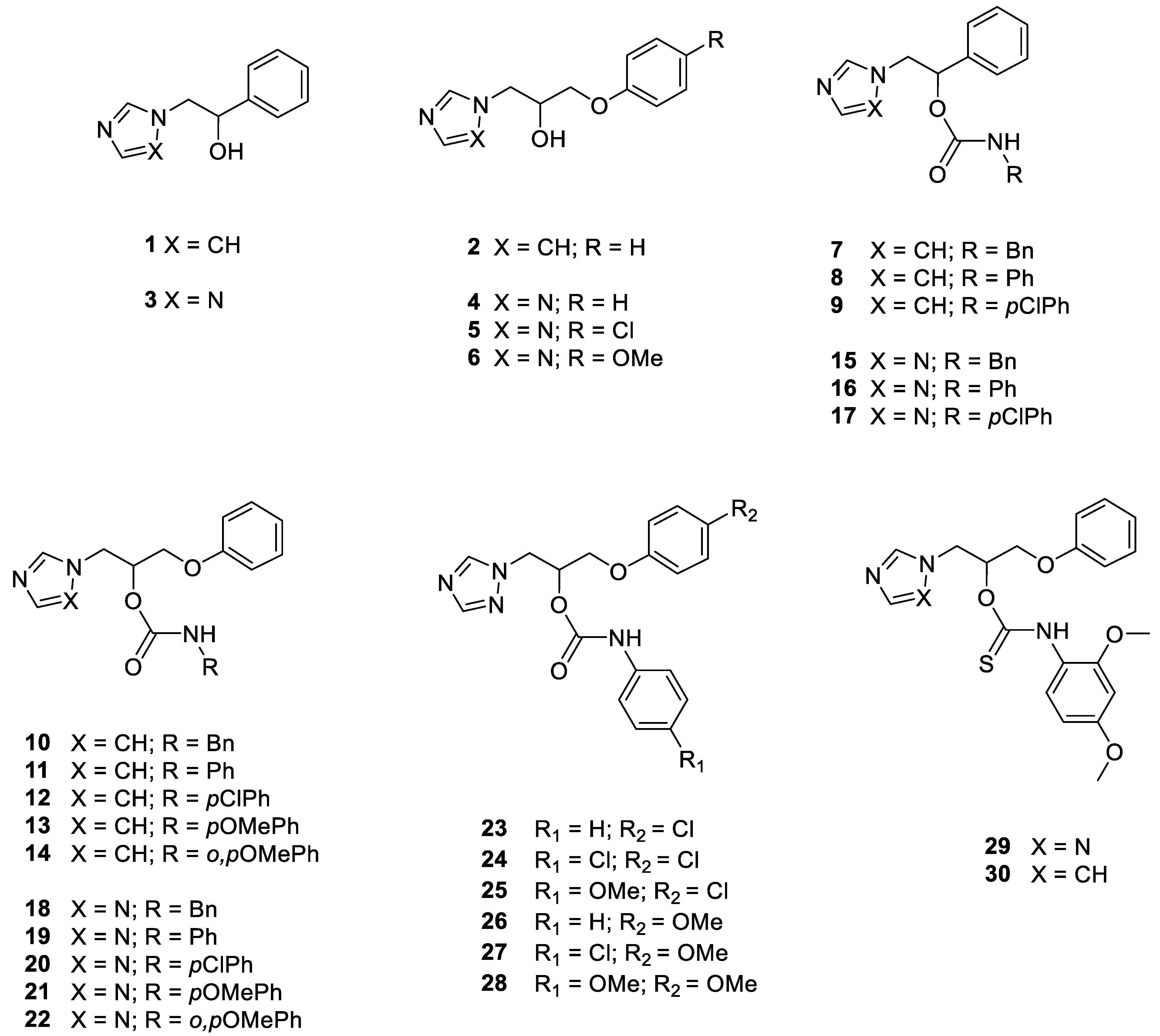
2.1. Chemistry

Melting points were determined using the Buchi Melting Point B-450 and were left uncorrected. NMR spectra were recorded using a Varian Mercury spectrometer, with 1H at 300.060 MHz and 13C at 75.475 MHz (Supplementary Materials, Figures S1–S4). Proton chemical shifts were referenced using the TMS internal standard. Chemical shifts are reported in parts per million (ppm, δ units). Coupling constants are reported in Hertz (Hz) units. Splitting patterns are designed as s, singlet; d, doublet; t, triplet; q, quartet; dd, double doublet; m, multiple; and b, broad. All commercial chemicals and solvents were of a reagent grade and were purchased from Merck (Merck, Darmstadt, Germany). They were used without further purification unless otherwise specified. The reactions were monitored by thin-layer chromatography on silica gel plates (60F-254, Merck), and the analysis of the plates was carried out using a UV lamp (254/365 nm). Flash chromatography was performed on silica gel 60 (Merck). Elemental analyses of C, H and N were recorded on a Perkin-Elmer 240 B microanalyzer (PerkinElmer, Waltham, MA, USA), obtaining analytical results within ± 0.4% of the theoretical values for all the compounds. The purity of all the compounds was over 98%. The chemical synthesis and characterization of alcohols 1–6 and carbamates 7–28 were previously reported by us [19].

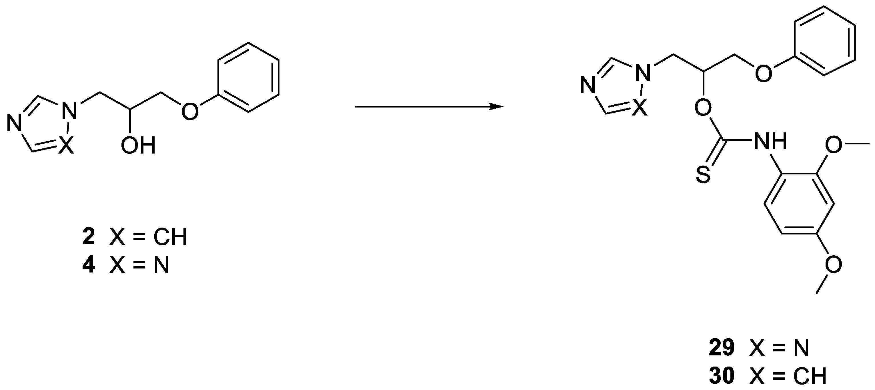
2.1.1. General Procedure for the Synthesis of Thiocarbamates 29 and 30

Thiocarbamates 29 and 30 were obtained by reacting alcohols 2 and 4 (1 mmol) with sodium hydride (1 mmol, 24 mg) in dry acetonitrile (10 mL) under nitrogen for two hours [25]. After this time, o,p-dimethoxyphenyl isothiocyanate (1.5 mmol, 293 mg) was added, and the mixtures were stirred overnight at room temperature. Then, the reaction mixtures were evaporated under reduced pressure and the residues were submitted to column chromatography on silica gel, with eluent chloroform/methanol 98:2 (v:v).

1-Phenoxy-3-(1H-1,2,4-triazol-1-yl) propan-2-yl (2,4-dimethoxyphenyl)thiocarbamate 29

Off-white solid, 62% yield; m.p. 117–119 °C; 1H NMR (CDCl3, 300 MHz) δ 3.79 and 3.81 (both s, 6H), 4.16–4.32 (m, 2H), 4.74 (d, 2H, J 6.0 Hz), 6.13–6.17 (m, 1H), 6.33–6.51 (m, 2H), 6.91–7.01 (m, 3H), 7.27–7.37 (m, 3H), 7.94–7.97 (m, 2H), 8.18–8.48 (m, 1H); 13C NMR (CDCl3, 75 MHz) δ 49.2, 49.8, 55.5, 55.8, 65.7, 74.1, 98.9, 103.5, 104.0, 114.6, 121.7, 123.6, 124.2, 129.6, 143.9, 151.2, 157.9. Calcd for C20H22N4O4S: C, 57.96; H, 5.35; N, 13.52. Found: C, 58.02; H, 5.36; N, 13.49.

1-(1H-imidazol-1-yl)-3-phenoxypropan-2-yl (2,4-dimethoxyphenyl)thiocarbamate 30

Pale yellow oil, 57% yield; 1H NMR (CDCl3, 300 MHz) δ 3.79 and 3.80 (both s, 6H), 4.00–4.15 (m, 2H), 4.49 (d, 2H, J 4.2 Hz), 6.02 (t, 1H, J 4.2 Hz), 6.36–6.52 (m, 2H), 6.85–7.05 (m, 5H), 7.27–7.33 (t, 2H, J 8.1 Hz), 7.40–7.54 (m, 1H), 8.23–8.53 (m, 1H); 13C NMR (CDCl3, 75 MHz) δ 45.8, 55.5, 55.7, 64.6, 64.8, 74.5, 98.8, 103.5, 104.0, 114.6, 119.8, 121.7, 123.6, 124.0, 129.7, 137.9, 157.8. Calcd for C21H23N3O4S: C, 61.00; H, 5.61; N, 10.16. Found: C, 60.87; H, 5.61; N, 10.19.

2.2. Antifungal Susceptibility

2.2.1. Organisms

Fungal strains were obtained from the American Type Culture Collection (ATCC, Rockville, MD, USA), the German Collection of Microorganisms (DSMZ, Braunschweig, Germany) and the Pharmaceutical Microbiology Culture Collection (PMC, Sapienza University, Rome, Italy). The fungal strains C. albicans ATCC 10231, C. albicans 3153 A, C. albicans ATCC 24422, C. glabrata PMC 0849, C. glabrata PMC 0822, C. glabrata PMC 0805, C. krusei PMC 0603, C. krusei PMC 0624, C. krusei PMC 0613, Aspergillus fumigatus DSM 790, Microsporum gypseum DSM 3824, Trichophyton rubrum PMC 6604 and Trichophyton mentagrophytes DSM 4870 were tested. All the Candida strains have MIC values of ≤4. Based on the values reported in the CLSI document and the clinical breakpoints for fungi (EUCAST), C. albicans strains are susceptible (S) or susceptible dose-dependent (SDD) and C.glabrata is susceptible dose-dependent (SDD) to fluconazole. Finally, isolates of C. krusei are not interpreted using this scale. Candida strains that were shown to form an abundant biofilm were used in the biofilm experiments.

2.2.2. Antifungal Susceptibility Testing

In vitro, the antifungal activity of the imidazole- and triazole-based compounds against C. albicans ATCC 10231, C. albicans 3153 A, C. albicans ATCC 24422, C. glabrata PMC 0849, C. glabrata PMC 0822, C. glabrata PMC 0805, C. krusei PMC 0603, C. krusei PMC 0624, C. krusei PMC 0613, A. fumigatus DSM 790, M. gypseum DSM 3824, T. rubrum PMC 6604 and T. mentagrophytes DSM 4870 was determined according to the standardized methods for yeast or filamentous fungi, using the broth microdilution method [26,27,28]. Candida strains were grown on Sabouraud dextrose agar (Sigma Aldrich, St. Louis, MO, USA) at 35 °C for 24 h. The final concentration of the inoculum was 0.5 × 10³–2.5 × 10³ cells/mL. A. fumigatus was grown on potato dextrose agar (Sigma Aldrich, St. Louis, MO, USA) at 35 °C until conidia formation was observed. The final concentration of the inoculum was 0.4 × 10⁴–5 × 10⁴ conidia/mL. Dermatophytes were grown on potato dextrose agar (Sigma Aldrich, St. Louis, MO, USA) at 30 °C until conidia formation was observed. The final concentration of the inoculum was 1 × 10³–3 × 10³ conidia/mL. The compounds and reference drug were previously dissolved in dimethyl sulfoxide at a concentration 100 times higher than the maximum tested concentration. The compounds were then serially diluted 2-fold on the 96-well plates in RPMI 1640 medium (Sigma-Aldrich, St. Louis, MO, USA). The final concentrations of the new compounds and fluconazole ranged from 128 to 0.125 µg/mL. Each experiment was performed in duplicate and was repeated at least three times on separate dates. After incubation, the minimal inhibitory concentration (MIC) was determined. After the agitation of the plates, the growth in each well was compared with that of the control (drug-free) with the aid of a reading mirror, using a microplate reader (Thermo Multiskan EX, Thermo Fisher Scientific, Monza, Italy). MIC50 was the lowest concentration that caused a prominent decrease (≥50%) in visible growth compared with the drug-free control. The results are expressed as the median.

2.2.3. In Vitro Activity of the Compounds against C. albicans, C. glabrata and C. krusei Biofilms

The Candida biofilm was formed on flat-bottomed, 96-well microtiter plates, as previously described [29]. For the evaluation of the compounds’ activity against the formation of biofilm, 100 μL aliquots of the cell suspensions (final concentration in well: 1.0 × 10⁶ cells/mL) in RPMI 1640 buffered with MOPS (4-morpholinepropanesulfonic acid) and 100 μL of the compound in a final concentration range from 128 to 8 µg/mL were added to the 96 microplate wells. Plates were then incubated for 48 h at 37 °C. After biofilm formation, the medium was aspirated, and non-adherent cells were removed by washing with physiological salt solution (0.9%).
To assess the activity against mature biofilm, 200 μL aliquots of the cell suspensions (final concentration in well: 1.0 × 10⁶ cells/mL) in RPMI 1640 buffered with MOPS were added to the 96 microplate wells. After 24 h of incubation at 37 °C, the medium was aspirated, and the compound was added. After 24 h of incubation at 37 °C, the medium was aspirated, and non-adherent cells were removed by washing with physiological salt solution (0.9%).
The metabolic activity of the Candida spp. biofilms was quantified using the XTT [2,3-bis(2-methoxy-4-nitro-5-sulfophenyl)-2H-tetrazolium-5-carboxanilide] reduction assay, as described previously [30]. In brief, the XTT solution was added to each well and incubated at 37 °C for 3 h in the dark. Finally, the colorimetric changes showing the metabolic activity of the Candida biofilm were measured at 492 nm using a microplate reader. The experiment was performed four times independently in duplicate, and the results were expressed as mean ± standard deviation (SD).

2.2.4. G. mellonella Survival Assay

G. mellonella killing assays were carried out as described previously [31]. In brief, larvae of G. mellonella of 0.3 ± 0.03 g (10 for each group), obtained from Blu Fish (Rome, Italy), were selected. Nine groups of larvae (1 for each treatment and strain) were inoculated in the last left proleg with 2 × 10⁶ cells of C. albicans ATCC 10231, C. glabrata PMC 0849 and C. krusei PMC 0603 and with 10 µL of compound 12. The concentrations of compound 12 were 36.5 mg/kg, 18.2 mg/kg and 9.1 mg/kg. Control groups (10 larvae each) were used. Three groups with 36.5 mg/kg, 18.2 mg/kg and 9.1 mg/kg, respectively, of compound 12, were defined. One group of larvae were pierced with no treatment applied, one group were treated with sterile PBS and three groups were treated with C. albicans ATCC 10231, C. glabrata PMC 0849 and C. krusei PMC 0603, respectively. The larvae were then incubated at 37 °C and monitored for 120 h and considered dead when they did not respond to physical stimulation (a slight pressure with forceps). Each experiment was repeated at least three times ad reported as a percent survival rate.

2.2.5. In Vivo Toxicity Studies

In vivo toxicity studies were conducted using G. mellonella larvae. In brief, larvae of G. mellonella of 0.3 ± 0.03 g (10 for each group), obtained from Blu Fish (Rome, Italy), were selected. Three groups of larvae (one for each concentration) were inoculated in the last left proleg with 10 µL of compound 12. The concentrations of compound 12 were 36.5 mg/kg, 18.3 mg/kg and 9.1 mg/kg. Control groups (10 larvae each) were used. A group of larvae were only pierced with no treatment applied and one group were treated with sterile PBS. The larvae were then incubated at 37 °C and monitored for 120 h and considered dead when they did not respond to physical stimulation (a slight pressure with forceps). Each experiment was repeated at least three times and reported as a percent survival rate.

2.3. Statistical Analysis

The data were expressed as mean ± S.E.M., and p < 0.05 was considered statistically significant. The statistical criteria, p and other parameters are shown for each experiment. The Kolmogorov–Smirnov test was applied to investigate the data distribution (Supplementary Materials, Figure S5). The G. mellonella survival rate was displayed via Kaplan–Meier curves. The statistical data analysis was performed using the GraphPad Prism 8.0.1.244 software (GraphPad Software Inc., San Diego, CA, USA).

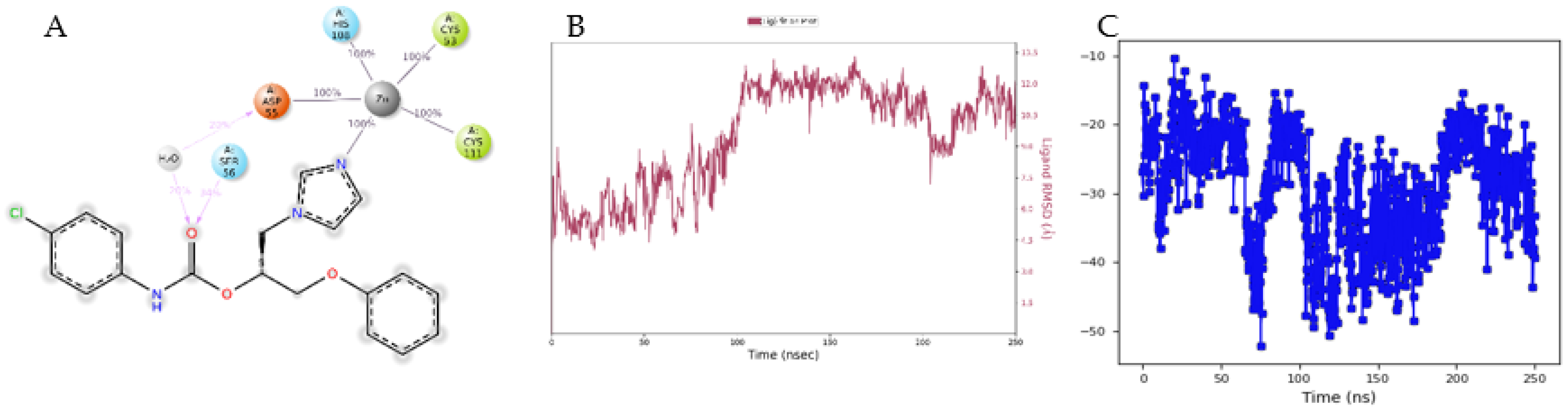
2.4. Molecular Modelling Studies

All modelling studies were performed using the tools of the Schrödinger software package (v2022-2, Schrödinger, Inc., New York, NY, USA).

2.4.1. Docking Studies

The three-dimensional structure of compound 12, in both the R and S isomers, was prepared using the LigPrep tool, and the ligand was protonated according to a pH value of 7.4, using Epik. Subsequently, the generated structures were minimized using the OPLS4 forcefield.
The crystal structure of CYP51a1 from C. glabrata (CgCYP51a1) in complex with itraconazole (PDB: 5jlc; 2.4 Å) was obtained from the RCSB Protein Data Bank. All the water and buffer molecules, as well as the ions, were deleted, and all the protein, heme and iron atoms were retained. Subsequently, hydrogen atoms were added to the system (according to pH 7.4, using Epik), an acetyl group was added to the N-terminal and a N-methyl amide group was added to the C-terminal using the Protein Preparation tool.
A homology model was constructed for the C. glabrata carbonic anhydrase enzyme (CgNce103, UniProt: Q6FTL6) using the Coccomyxa β-carbonic anhydrase in complex with acetazolamide (CmCA; 1.85 Å; 3ucj) as a template.
Subsequently, both stereoisomers of compound 12 were docked into the binding sites of the target enzymes, which were assigned as residues that are all within 5 Å of either itraconazole or acetazolamide. During the studies of the compounds’ docking to the CgCYP51a1 crystal structure, the ligand imidazole ring was superimposed onto the itraconazole triazole ring. For the studies on the docking to the CaNce103, no restraints were applied.
The docking studies were performed using the Glide tool and the SP settings. The three highest-scoring poses were obtained for both the isomers of compounds 12, and the docked ligand, as well as the active site (all residues within 5 Å of the ligand), were subsequently minimized using the Prime tool and MM–GBSA forcefield. The highest-scoring poses that underwent binding interactions (hydrogen bonds, electrostatic interactions, and hydrophobic interactions) with the active site and showed complementarity in terms of their shape and (a)polarity were selected for the molecular dynamic (MD) simulations.

2.4.2. Molecular Dynamic Simulations

All MD simulations were performed using Desmond. The ligand–protein complexes, as obtained through the docking studies, were first placed in the center of an orthorhombic box under periodic boundary conditions (minimal distance of 10 Å between the protein and boundary). Subsequently, water molecules (Tip5p) and counter ions (NaCl; 0.15 M) were added to generate a solvated and neutral system. The system was energy-minimized using the OPLS4 forcefield, while all the protein, heme and ligand heavy atoms were restrained, and only the solvent and counter ions were allowed to move. Afterwards, the system was simulated for 250 ns at a constant temperature (300 K, Nose–Hoover chain, default settings) and pressure (1 bar, Martyna–Tobias–Klein, default settings), without any position restraints. The timestep was set to 0.002 fs and the RESPA integrator was used. The ligand–protein binding interactions, as well as the protein Cα and ligand heavy atom RMSD values during the 250 ns MD run, were analyzed using Desmond.

3. Results

3.1. In Vitro Antifungal Activity Evaluation

The molecules selected for this study (Figure 1) are azole derivatives, which are differently functionalized and belong to different chemical classes. They can be classified as imidazole alcohols 12, 1,2,4-triazole alcohols 36, imidazole carbamates 714, 1,2,4-triazole carbamates 1528 and thiocarbamates 2930. The main structural modifications involved the azole ring, the alkyl linker connecting the azole to the aromatic ring, the distal aromatic ring and the carbamate portion, into which benzyl- or phenyl-substituted rings were inserted.
Carbamates 728 were previously synthesized and screened for their potential activity as aromatase inhibitors. Some of these derivatives were found to be active against aromatase, with an inhibition characterized by sub-micromolar potency [19]. With the aim to evaluate the antifungal potential of this group of compounds and expand the chemical library, the synthesis of thiocarbamates 2930 was realized by reacting intermediate alcohols 2 and 4 with sodium hydride and o,p-dimethoxyphenyl isothiocyanate in dry acetonitrile for 24 h at room temperature under nitrogen (Scheme 1).
All compounds were submitted to an in vitro evaluation of their antifungal activities against C. albicans (ATCC10231, 3153A, 24433), C. glabrata (PMC0849, PMC0822, PMC0805) and C. krusei (PMC0603, PMC0624, PMC0613) (Table 1). Their activities against A. fumigatus and selected dermatophytes, M. gypseum, T. rubrum and T. mentagrophytes, were also evaluated (Supplementary Materials, Table S1). Their antifungal activity was evaluated by the broth microdilution method, using fluconazole (FLC) as the reference compound. All Candida strains used in this experiment had fluconazole MIC values of ≤4. The results are expressed as the median MIC50 (μg/mL) value for each strain.
Imidazole and triazole alcohols 16 were found to be inactive against all the Candida strains tested, displaying median MIC50 values of >128 μg/mL for almost all the tested strains (geometric means (GM) MIC50 196–256 μg/mL). Only compounds 1 and 5 showed antifungal activity against C. glabrata PMC0849, with median MIC50 values of 48 and 64 μg/mL, respectively.
A better antifungal activity was observed when testing imidazole carbamates 714, with GMMIC50 values ranging from 15 to 123 μg/mL calculated for all the tested Candida strains. Compound 12 emerged as the most active derivative, showing a GMMIC50 value of 15 μg/mL, and the best activities were observed against C. krusei (GMMIC50 9 μg/mL) and C. glabrata (GMMIC50 10 μg/mL) compared to C. albicans (GMMIC50 40 μg/mL). A remarkable antifungal activity was exhibited by 12 against C. glabrata PMC0849 (median MIC50 1 μg/mL) and C. krusei PMC0603 (median MIC50 4 μg/mL). The C. glabrata strain PMC0849 emerged as the most sensitive to the action of carbamates 714, with median MIC50 values in the range of 1–20 μg/mL.
Only some derivatives in the group of triazole-based carbamates 1528 showed an appreciable antifungal activity. Compound 24 was active against C. glabrata and C. krusei, with GMMIC50 values of 23 and 43 μg/mL, respectively. The C. glabrata PMC0849 strain ermerged, again, the most sensitive to the actions of the tested compounds, with derivatives 24, 25, 20 and 27 displaying the lowest median MIC50 values (4 μg/mL for 24 and 25, 8 μg/mL for 20 and 27).
Thiocarbamate 29 did not show an appreciable antifungal activity, whereas 30 was moderately active against C. glabrata (GMMIC50 45 μg/mL) and C. krusei (GMMIC50 68 μg/mL). The best antifungal activity was detected against the C. glabrata strain PMC0849, with a median MIC50 value of 4 μg/mL.
When tested against A. fumigatus and the dermatophytes (M. gypseum, T. rubrum, T. mentagrophytes), all compounds were found to be essentially inactive, showing MIC50 values of >128 μg/mL or >64 μg/mL (90.51 μg/mL for 12, 90.58 μg/mL for 30 against M. gypseum) (Table S1).
These results allowed us to trace some preliminary structure–activity relationships for the assayed compounds (Figure 2). At first, alcohols were found to be markedly less active compared to carbamates, and the presence of the imidazole or triazole ring does not appear to be a critical structural feature. In the large group of carbamates, the presence of the imidazole produced the most active derivatives, compared to the triazole compounds. The p-chloro substitution of the phenyl ring linked to the carbamate portion improved the antifungal activity (9 vs. 8, 12 vs. 11) against all tested strains, whereas the p-methoxy substitution resulted in a less favorable activity (13 vs. 12). In the group of triazole carbamates, the same trend was confirmed for the p-chloro and p-methoxy substitution, with p-chloro derivative 20 showing the best activity. The further substitution of the distal phenoxy ring improved the activity, with p-chloro compound 24 displaying the best antifungal profile. The p-methoxy derivative 27 was less active than 24, confirming the status of chlorine as the best substituent in this series of compounds. Thiocarbamates did not show significant improvements in their activity compared to their corresponding carbamates (29 vs. 22, 30 vs. 14).

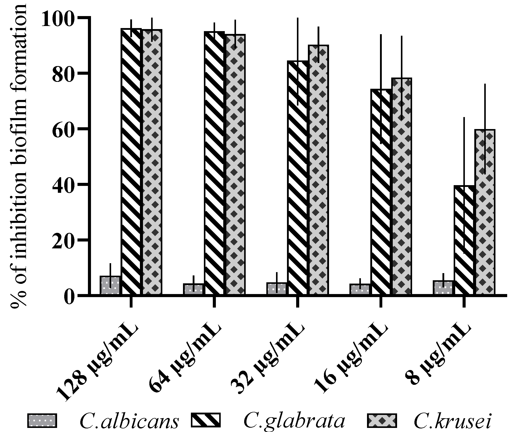
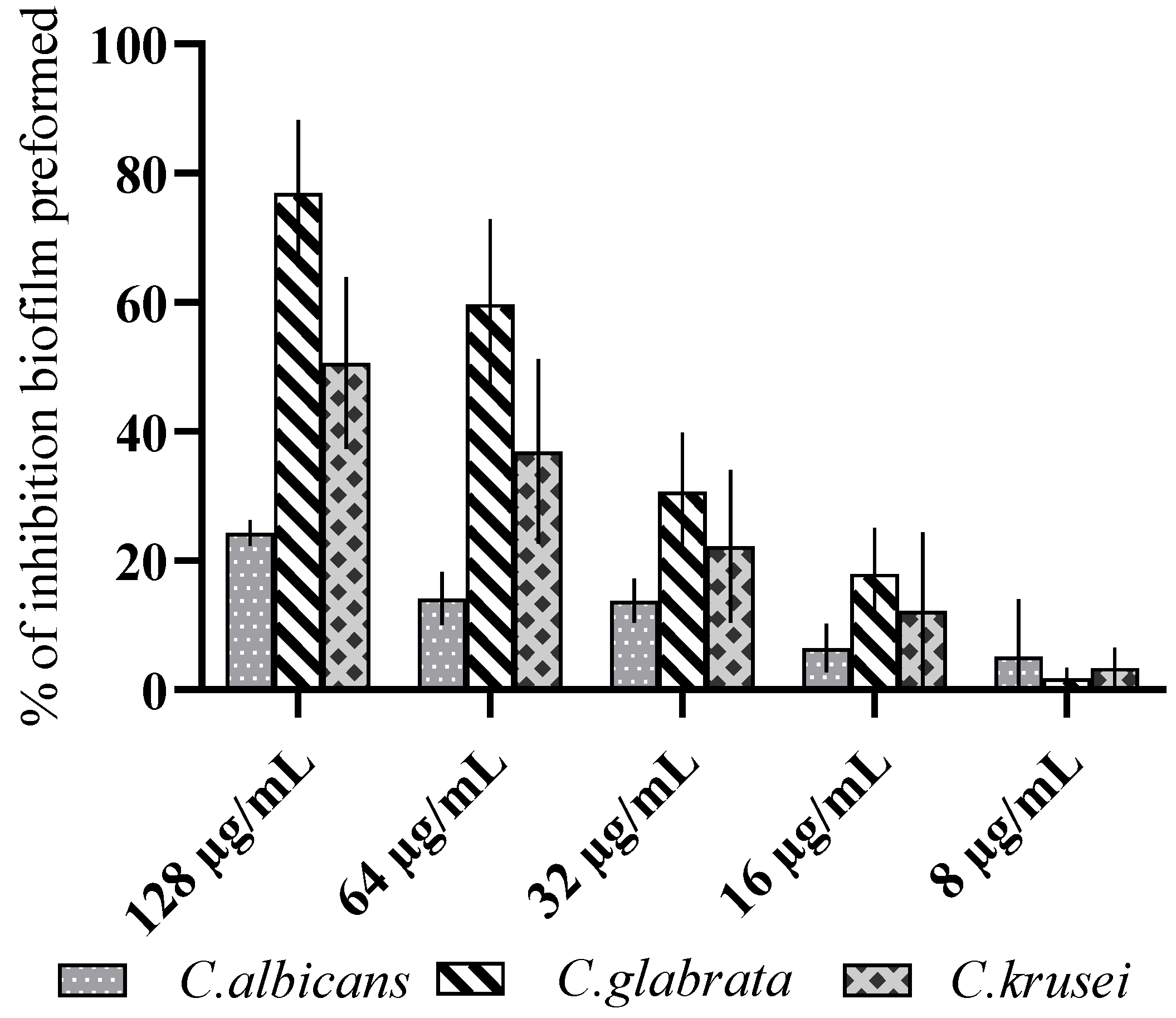
3.2. Antibiofilm Activity

Starting with these results, we subjected the most active compound 12 to further experiments using biofilm of three Candida species in a concentration range from 128 to 8 µg/mL. The results, reported in Figure 3, showed that, at 128 µg/mL, compound 12 reduced the formation of C. glabrata and C. krusei biofilm by more than 90%. Figure 4 shows the reduction in the mature biofilm of the three Candida species. Compound 12, at a concentration of 128 µg/mL, reduced the mature biofilm of C. glabrata PMC 0849 by 77% and of C. krusei PMC 0603 by 51%. The compound 12 did not show activity against C. albicans biofilms. The Kolmogorov–Smirnov test was applied to investigate the data distribution (Supplementary Materials, Figure S5).

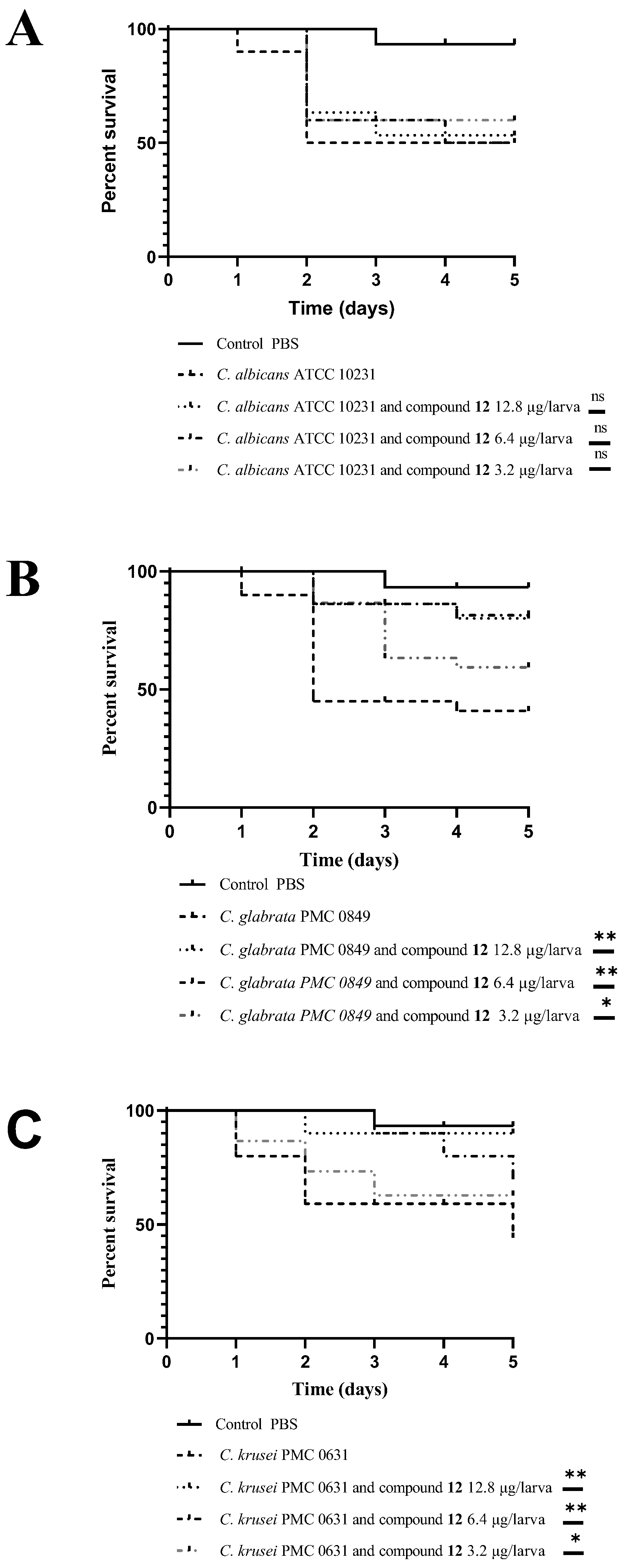
3.3. In Vivo Antifungal Activity Evaluation

Moreover, compound 12 was tested in an in vivo model using the larvae of G. mellonella. The response of the G. mellonella larvae to Candida infection was shown to have a strong correlation with the results obtained in mice. The G. mellonella were infected with C. albicans ATCC 10231, C. glabrata PMC 0849 and C. krusei PMC 0603 cells. To calculate the lethal dose, mortality curves were previously determined. After inoculation with 5 × 10⁶ cells of the Candida species/larvae, the larvae were inoculated with different concentrations of compound 12. The mortality of the larvae was reported daily for 5 days. In the groups of untouched larvae and larvae inoculated with PBS, the mortality was 0–10%. Conversely, in the group of larvae infected with Candida species cells, the mortality rate was 50–60% at 5 days post-infection. The results of tests on the larvae inoculated with compound 12, in a concentration range from 36.5 mg/kg to 9.1 mg/kg, indicated a clear relationship between the concentration of compound 12 and the mortality rate in the case of C. glabrata PMC 0849 and C. krusei PMC 0603 (Figure 5). The acquired data showed that the injection of 36.5 mg/kg of compound 12 increased the survival rate by up to 80% after 5 days post-infection in the case of C. glabrata PMC 0849 and up to 90% in the case of C. krusei PMC 0603. The LD50 detected dose was more than 36.5 mg/kg for compound 12 (Table 2).

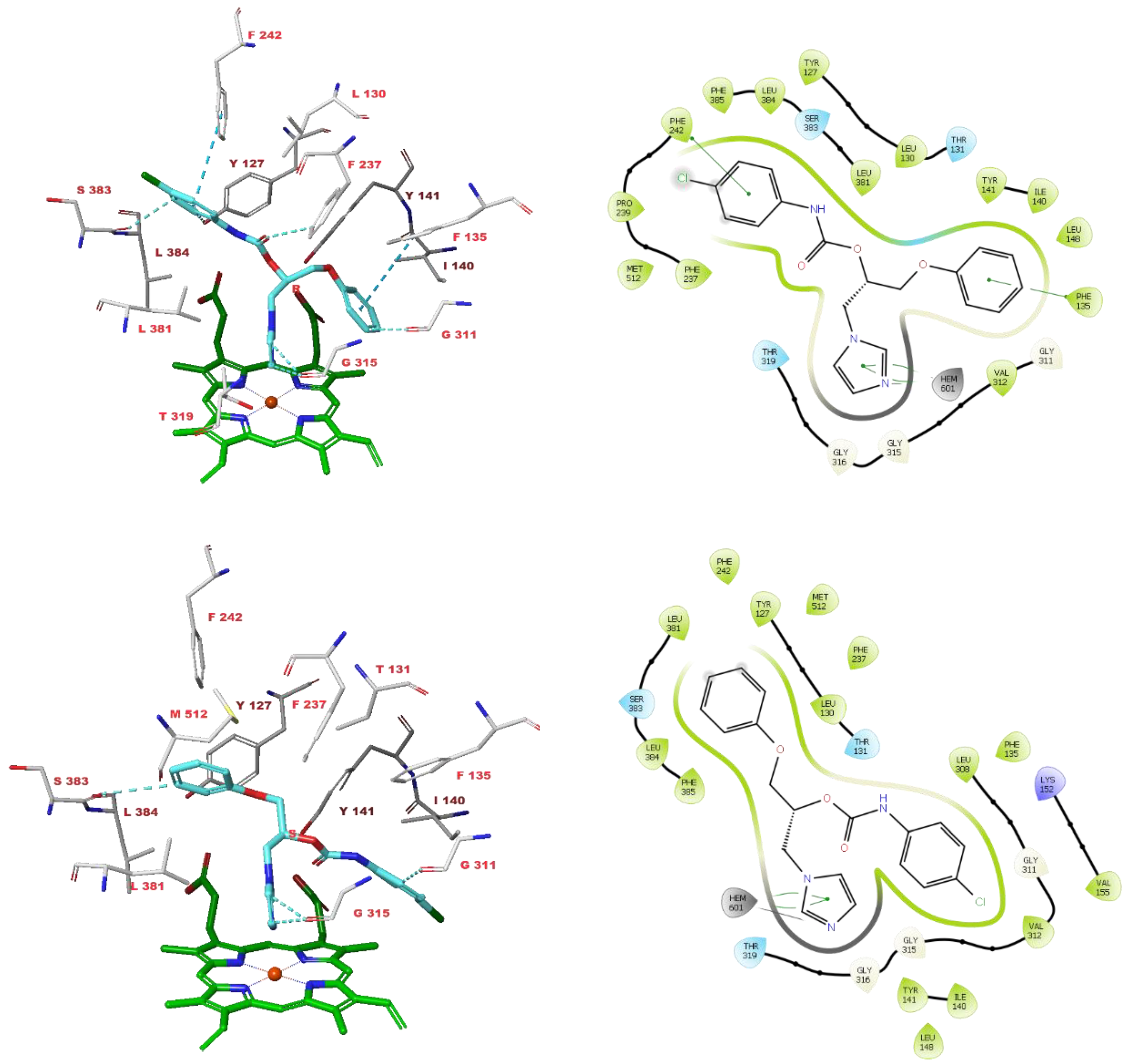
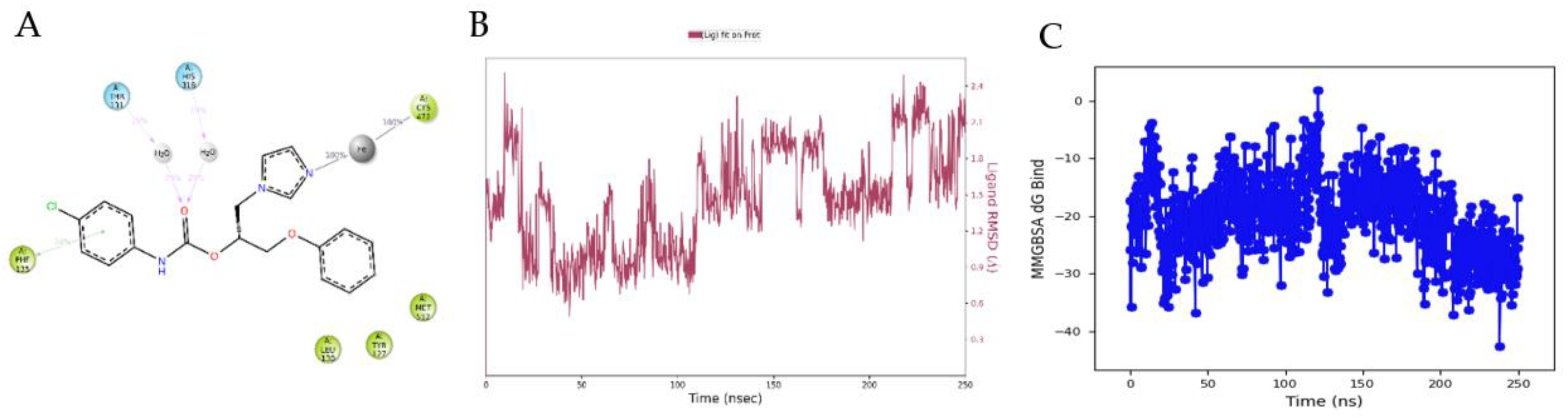
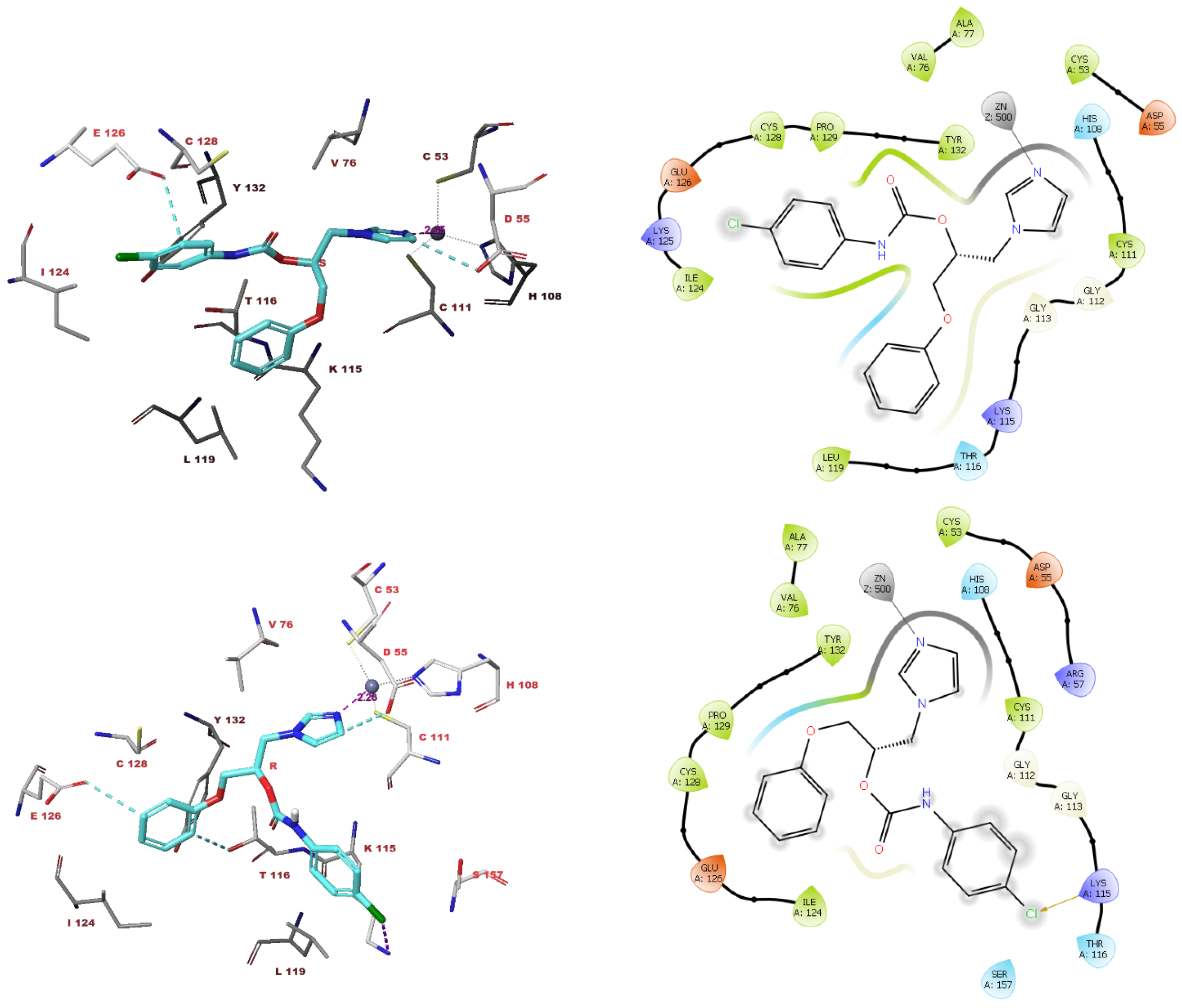
3.4. Molecular Modelling Studies

The possible binding interactions of compound 12 with the possible fungal target enzymes CgCYP51a1 (Figure 6 and Figure 7) and CgNce103 (Figure 8) were investigated through molecular modelling studies.
In the active site of CgCYP51a1, both the isomers of compound 12 were able to position their imidazole rings such that the distance between the imidazole nitrogen atom and the heme iron atom was 2.2 Å and an interaction was possible (Figure 6). The pose of compound 12(R) formed aromatic hydrogen bonds with the sidechain of Phe237 and the backbones of Gly311, Gly315 and Ser383. The ligand’s phenyl groups underwent π-π interactions with the sidechains of Phe135 and Phe242. The pose of compound 12(S) formed underwent interactions with the active site, including aromatic hydrogen bonds with the backbones of Gly311, Gly315 and Ser383.
Subsequently, 250 ns MD simulations were performed on the CgCYP51a1 ligand complexes to investigate the stability of the docked poses. Interestingly, the CgCYP51a1-12(R) pose was not stable, as the ligand’s RMSD value increased to approximately 4.5 Å, and the interactions with the heme iron were only observed for a very short period (<15% of simulation). In contrast, the CgCYP51a1-12(S) pose was stable as the ligand RMSD value was below 2.4 Å and the interaction with the heme iron was observed for the entire duration of the MD simulation (Figure 7). The ligand carbonyl group underwent water-bridged interactions with Thr131 (29% of simulation) and His318 (29% of simulation). Hydrophobic and π-π stackings were observed with Tyr127, Leu130, Phe135 and Met512. The MM–GBSA ligand–protein interaction energy during the MD simulation ranged from −42.5875 to 1.7826 kcal/mol, with an average value of −19.9918 ± 6.49 kcal/mol.
The docked pose of compound 12(S) with CgNce103 indicates the occurrence of an interaction between the ligand’s imidazole nitrogen atom and the Zn2+ ion (distance 2.25 Å) and of the aromatic hydrogen bonds with the sidechains of Asp55 and Glu126 and the backbone of Gly112 (Figure 8). The docked pose of compound 12(R) also shows an interaction with the Zn2+ ion (distance 2.26 Å). In addition, aromatic hydrogen bonds were formed with the sidechains of Asp55, Thr116 and Cys128, and an interaction took place between the ligand’s chlorine atom and the sidechain of Lys115 (Figure 8).
Again, MD simulations were performed to investigate the CgNce103-12 docked poses. For the CgNce103-12(S) complex, the interaction with the Zn2+ ion was observed throughout the simulation (Figure 9). The ligand’s carbonyl group formed a hydrogen bond with Ser56 (34% of simulation) and underwent a water-bridged interaction with Asp55 (20% of simulation). The two phenyl groups of the ligand were solvent-exposed. The MM–GBSA ligand-protein interaction energy during the MD simulation ranged from −52.1100 to −10.3882 kcal/mol, with an average value of −29.0571 ± 8.20 kcal/mol. Interestingly, the MD simulation of the CgNce103-12(R) complex indicated that the ligand only interacted with the Zn2+ ion during the entire simulation, and no additional interactions were observed for a significant amount of time (<15 % of the simulation).

4. Discussion

The widespread diffusion of fungal infections represents an important health problem, especially when the pathogens are responsible for serious diseases, with high mortality rates all over the world among immunocompromised and neonatal patients. Over one billion people suffer from severe fungal diseases [1]. Among the various human fungal pathogens, C. albicans accounts for most of the infections, followed by C. glabrata [32]. Fungal diseases induced by Candida species commonly affect the gastrointestinal and urinary systems, as well as the oral cavity. However, these infections can become systemic, reaching different organs, such as the heart, brain and eyes, as well as the blood, and producing a wide variety of symptoms. In immunocompromised or transplant patients, candidiasis represents a serious health problem. Candida spp. infections, such as candidiasis, are frequently associated with biofilm formation, and it is known that biofilm formation represents a major virulence and resistance factor [33]. In this context, new antifungal drugs that combat not only planktonic fungi but also fungal biofilms are needed. Some authors reported the activity of new compounds or plant extracts against C. albicans biofilm [30,34], but there are few studies on the C. glabrata and C. krusei biofilms [35,36]. The emergence of invasive fungal infections caused by non-albicans Candida species, and the virulence associated with the biofilm formation and drug resistance, render the search for novel antifungal drugs as a challenging field of investigation [37,38]. The extensive use of antifungal drugs, such as polyenes, azoles and echinocandins, is associated with several limitations, including systemic toxicity and cross-reactivity with other therapeutic agents [6]. In view of this, in this work, we report on the evaluation of a large library of azole derivatives used as anti-Candida agents. These molecules were previously synthesized by us and tested for the capacity as aromatase inhibitors, but the chemical similarity of our compounds with the azole antifungals prompted us to further examine their activity against fungal diseases. Candida species (C. albicans, C. glabrata, C. krusei), A. fumigatus and the dermatophytes M. gypseum, T. rubrum and T. mentagrophytes were selected to this end. Compound 12 demonstrated antibiofilm activity against C. glabrata and C. krusei.
It is well known that the immune responses of G. mellonella show similarity with the innate vertebrate immune response; thus, these larvae represent an interesting animal model that can be used to prove the virulence of fungal pathogens and the antifungal activity of new compounds [39,40,41,42]. To investigate the role of compound 12 in terms of the Candida spp. Virulence, different concentrations of compound 12 and Candida spp. were injected into G. mellonella larvae, as an excellent model for studying the virulence of Candida species. In vivo, the selected compound showed a significative antifungal activity against C. glabrata and C. krusei. Molecular modelling studies were performed on the most active compound 12 to assess its activity against the possible fungal target enzymes CgCYP51a1 and CgNce103. It was found that, in particular, the S-isomer of compound 12 may engage in stable interactions with both enzyme active sites, taking advantage of the imidazole core, the carbonyl of carbamate and the p-chlorophenyl ring. As such, CgCYP51a1 and CgNce103 may be the enzymes through which compound 12 exerts its antifungal effects. This compound also displayed a good Log Pw/o (3.12), no violations of Lipinski’s rule and a high capacity for gastrointestinal absorption and BBB permeation in silico. It is not a putative substrate of P-glycoprotein and does not behave as a scaffold, generally described by the term “PAINS” (Pan-Assay Interference Compounds). Collectively, these promising medicinal chemistry characteristics have the potential for further development in the design of more potent fungal inhibitors.

5. Conclusions

In conclusion, imidazole- and triazole-based compounds were submitted to an antifungal evaluation of the Candida species and selected dermatophytes, displaying variable degrees of activity. This evaluation allowed us to trace preliminary structure–activity relationships, with carbamates showing moderate to good anticandidal activities and with MICs comparable to that of the reference drug, fluconazole. The imidazole-based carbamate 12 was further tested against Candida species for its ability to affect the biofilm formation and growth, showing a dose-dependent inhibition profile. In vivo studies were also performed using the G. mellonella larvae, confirming the safety profile of 12 and its efficacy in the three models of Candida infections. Our molecular modelling studies suggested that imidazole 12 may exert its antifungal effects via the C. glabrata target enzymes CgCYP51a1 and CgNce103. In any case, further studies are necessary to study the experimental ADME properties of the most active compound in other in vivo models (e.g., rat, mouse) in order to corroborate its efficacy in the clinical settings. In particular, better information could be gathered regarding the impact of the stereochemistry on the pharmacodynamics and pharmacokinetics.

Supplementary Materials

The following are available online at https://www.mdpi.com/article/10.3390/antibiotics11101375/s1. Figure S1: 1H spectrum of compound 29. Figure S2: 13C spectrum of compound 29. Figure S3: 1H spectrum of compound 30. Figure S4: 13C spectrum of compound 30. Figure S5: Data distribution determined by Kolmogorov–Smirnov test. Table S1. In vitro activity of studied compounds against A. fumigatus and dermatophytes.

Author Contributions

Conceptualization, A.A. (Alessandra Ammazzalorso) and S.C.; synthesis, A.A. (Alessandra Ammazzalorso), B.D.F. and S.C.; formal analysis, A.O., G.B. and G.S.; molecular modelling, A.F.Ş., S.C. and A.A. (Atilla Akdemir); resources, A.A. (Alessandra Ammazzalorso) and S.C.; writing—original draft preparation, G.S. and A.A. (Alessandra Ammazzalorso); writing—review and editing, S.C. and A.A. (Alessandra Ammazzalorso). All authors have read and agreed to the published version of the manuscript.

Funding

This work was supported by intramural grants FAR2020 awarded to Carradori (S.C.) and Ammazzalorso (A.A.).

Institutional Review Board Statement

Not applicable.

Informed Consent Statement

Not applicable.

Data Availability Statement

Data are contained within the article.

Conflicts of Interest

The authors declare no conflict of interest.

References

  1. Almeida, F.; Rodrigues, M.L.; Coelho, C. The Still Underestimated Problem of Fungal Diseases Worldwide. Front. Microbiol. 2019, 10, 214. [Google Scholar] [CrossRef] [Green Version]
  2. Vallabhaneni, S.; Mody, R.K.; Walker, T.; Chiller, T. The Global Burden of Fungal Diseases. Infect. Dis. Clin. North Am. 2016, 30, 1–11. [Google Scholar] [CrossRef] [PubMed]
  3. Bongomin, F.; Gago, S.; Oladele, R.O.; Denning, D.W. Global and Multi-National Prevalence of Fungal Diseases—Estimate Precision. J. Fungi 2017, 3, 57. [Google Scholar] [CrossRef]
  4. Hay, R. Therapy of Skin, Hair and Nail Fungal Infections. J. Fungi 2018, 4, 99. [Google Scholar] [CrossRef] [PubMed] [Green Version]
  5. Revie, N.M.; Iyer, K.R.; Robbins, N.; Cowen, L.E. Antifungal drug resistance: Evolution, mechanisms and impact. Curr. Opin. Microbiol. 2018, 45, 70–76. [Google Scholar] [CrossRef] [PubMed]
  6. Prasad, R.; Banerjee, A.; Shah, A.H. Resistance to antifungal therapies. Essays Biochem. 2017, 61, 157–166. [Google Scholar] [CrossRef] [PubMed] [Green Version]
  7. Perlin, D.S.; Rautemaa-Richardson, R.; Alastruey-Izquierdo, A. The global problem of antifungal resistance: Prevalence, mechanisms, and management. Lancet Infect. Dis. 2017, 17, e383–e392. [Google Scholar] [CrossRef]
  8. Monk, B.C.; Sagatova, A.A.; Hosseini, P.; Ruma, Y.N.; Wilson, R.K.; Keniya, M.V. Fungal Lanosterol 14α-demethylase: A target for next-generation antifungal design. Biochim. Biophys. Acta (BBA)-Proteins Proteom. 2020, 1868, 140206. [Google Scholar] [CrossRef]
  9. Allen, D.; Wilson, D.; Drew, R.; Perfect, J. Azole antifungals: 35 years of invasive fungal infection management. Expert Rev. Anti-infective Ther. 2015, 13, 787–798. [Google Scholar] [CrossRef] [PubMed]
  10. Peyton, L.R.; Gallagher, S.; Hashemzadeh, M. Triazole antifungals: A review. Drugs Today 2015, 51, 705–718. [Google Scholar] [CrossRef]
  11. Ramage, G.; Rajendran, R.; Sherry, L.; Williams, C. Fungal Biofilm Resistance. Int. J. Microbiol. 2012, 2012, 528521. [Google Scholar] [CrossRef] [PubMed]
  12. Whaley, S.G.; Berkow, E.L.; Rybak, J.M.; Nishimoto, A.T.; Barker, K.S.; Rogers, P.D. Azole Antifungal Resistance in Candida albicans and Emerging Non-albicans Candida Species. Front. Microbiol. 2017, 7, 2173. [Google Scholar] [CrossRef] [PubMed] [Green Version]
  13. Gulati, M.; Nobile, C.J. Candida albicans biofilms: Development, regulation, and molecular mechanisms. Microbes Infect. 2016, 18, 310–321. [Google Scholar] [CrossRef] [PubMed] [Green Version]
  14. Polke, M.; Hube, B.; Jacobsen, I.D. Candida Survival Strategies. Adv. Appl. Microbiol. 2015, 91, 139–235. [Google Scholar] [CrossRef]
  15. Marichal, P.; Bossche, H.V. Mechanisms of resistance to azole antifungals. Acta Biochim. Pol. 1995, 42, 509–516. [Google Scholar] [CrossRef] [Green Version]
  16. Vanden Bossche, H.; Dromer, F.; Improvisi, I.; Lozano-Chiu, M.; Rex, J.H.; Sanglard, D. Antifungal drug resistance in pathogenic fungi. Med. Mycol. 1998, 36 (Suppl. S1), 119–128. [Google Scholar] [PubMed]
  17. Pristov, K.E.; Ghannoum, M.A. Resistance of Candida to azoles and echinocandins worldwide. Clin. Microbiol. Infect. 2019, 25, 792–798. [Google Scholar] [CrossRef] [PubMed]
  18. Nishimoto, A.T.; Sharma, C.; Rogers, P.D. Molecular and genetic basis of azole antifungal resistance in the opportunistic pathogenic fungus Candida albicans. J. Antimicrob. Chemother. 2020, 75, 257–270. [Google Scholar] [CrossRef] [PubMed]
  19. Ammazzalorso, A.; Gallorini, M.; Fantacuzzi, M.; Gambacorta, N.; De Filippis, B.; Giampietro, L.; Maccallini, C.; Nicolotti, O.; Cataldi, A.; Amoroso, R. Design, synthesis and biological evaluation of imidazole and triazole-based carbamates as novel aromatase inhibitors. Eur. J. Med. Chem. 2021, 211, 113115. [Google Scholar] [CrossRef] [PubMed]
  20. Maccallini, C.; Gallorini, M.; Sisto, F.; Akdemir, A.; Ammazzalorso, A.; De Filippis, B.; Fantacuzzi, M.; Giampietro, L.; Carradori, S.; Cataldi, A.; et al. New azolyl-derivatives as multitargeting agents against breast cancer and fungal infections: Synthesis, biological evaluation and docking study. J. Enzym. Inhib. Med. Chem. 2021, 36, 1631–1644. [Google Scholar] [CrossRef] [PubMed]
  21. Brennan, M.; Thomas, D.Y.; Whiteway, M.; Kavanagh, K. Correlation between virulence of Candida albicans mutants in mice and Galleria mellonellalarvae. FEMS Immunol. Med. Microbiol. 2002, 34, 153–157. [Google Scholar] [CrossRef] [PubMed] [Green Version]
  22. Moghimi, S.; Shafiei, M.; Foroumadi, A. Drug design strategies for the treatment azole-resistant candidiasis. Expert Opin. Drug Discov. 2022, 17, 879–895. [Google Scholar] [CrossRef]
  23. Supuran, C.; Capasso, C. A Highlight on the Inhibition of Fungal Carbonic Anhydrases as Drug Targets for the Antifungal Armamentarium. Int. J. Mol. Sci. 2021, 22, 4324. [Google Scholar] [CrossRef] [PubMed]
  24. Güzel-Akdemir, Ö.; Carradori, S.; Grande, R.; Demir-Yazıcı, K.; Angeli, A.; Supuran, C.T.; Akdemir, A. Development of Thiazolidinones as Fungal Carbonic Anhydrase Inhibitors. Int. J. Mol. Sci. 2020, 21, 2960. [Google Scholar] [CrossRef] [PubMed] [Green Version]
  25. Pan, B.-W.; Shi, Y.; Li, W.-C.; Wang, Q.; Pan, M.; Wu, Q.; Fu, H.-Z. Synthesis and biological evaluation of Vinpocetine derivatives. Bioorganic Med. Chem. Lett. 2020, 30, 126472. [Google Scholar] [CrossRef]
  26. Clinical and Laboratory Standards Institute (CLSI). Reference Method for Broth Dilution Antifungal Susceptibility Testing of Yeasts, 3rd ed.; Approved Standard. CLSI M27-A3(28); Clinical and Laboratory Standards Institute: Wayne, PA, USA, 2008. [Google Scholar]
  27. Clinical and Laboratory Standards Institute. Reference Method for Broth Dilution Antifungal Susceptibility Testing of Filamentous Fungi, 2nd ed.; Approved Standard. CLSI Document M38-A2; Clinical and Laboratory Standards Institute: Wayne, PA, USA, 2008. [Google Scholar]
  28. Clinical and Laboratory Standards Institute (CLSI). Reference Method for Broth Dilution Antifungal Susceptibility Testing of Yeasts—Fourth Informational Supplement, Document M27-A3; Clinical and Laboratory Standards Institute: Wayne, PA, USA, 2012. [Google Scholar]
  29. Ourabah, A.; Atmani-Kilani, D.; Debbache-Benaida, N.; Kolesova, O.; Azib, L.; Yous, F.; Benloukil, M.; Botta, B.; Atmani, D.; Simonetti, G. Anti-Candida albicans biofilm activity of extracts from two selected indigenous Algerian plants: Clematis flammula and Fraxinus angustifolia. J. Herb. Med. 2020, 20, 100319. [Google Scholar] [CrossRef]
  30. Simonetti, G.; Palocci, C.; Valletta, A.; Kolesova, O.; Chronopoulou, L.; Donati, L.; Di Nitto, A.; Brasili, E.; Tomai, P.; Gentili, A.; et al. Anti-Candida Biofilm Activity of Pterostilbene or Crude Extract from Non-Fermented Grape Pomace Entrapped in Biopolymeric Nanoparticles. Molecules 2019, 24, 2070. [Google Scholar] [CrossRef] [Green Version]
  31. Cairone, F.; Simonetti, G.; Orekhova, A.; Casadei, M.; Zengin, G.; Cesa, S. Health Potential of Clery Strawberries: Enzymatic Inhibition and Anti-Candida Activity Evaluation. Molecules 2021, 26, 1731. [Google Scholar] [CrossRef]
  32. Ohshima, T.; Ikawa, S.; Kitano, K.; Maeda, N. A Proposal of Remedies for Oral Diseases Caused by Candida: A Mini Review. Front. Microbiol. 2018, 9, 1522. [Google Scholar] [CrossRef] [Green Version]
  33. Wall, G.; Montelongo-Jauregui, D.; Bonifacio, B.V.; Lopez-Ribot, J.L.; Uppuluri, P. Candida albicans biofilm growth and dispersal: Contributions to pathogenesis. Curr. Opin. Microbiol. 2019, 52, 1–6. [Google Scholar] [CrossRef]
  34. De Vita, D.; Friggeri, L.; D’Auria, F.D.; Pandolfi, F.; Piccoli, F.; Panella, S.; Palamara, A.T.; Simonetti, G.; Scipione, L.; Di Santo, R.; et al. Activity of caffeic acid derivatives against Candida albicans biofilm. Bioorganic Med. Chem. Lett. 2014, 24, 1502–1505. [Google Scholar] [CrossRef] [PubMed]
  35. Gupta, P.; Gupta, H.; Poluri, K.M. Geraniol eradicates Candida glabrata biofilm by targeting multiple cellular pathways. Appl. Microbiol. Biotechnol. 2021, 105, 5589–5605. [Google Scholar] [CrossRef] [PubMed]
  36. Jamiu, A.T.; Albertyn, J.; Sebolai, O.; Gcilitshana, O.; Pohl, C.H. Inhibitory effect of polyunsaturated fatty acids alone or in combination with fluconazole on Candida krusei biofilms in vitro and in Caenorhabditis elegans. Med. Mycol. 2021, 59, 1225–1237. [Google Scholar] [CrossRef] [PubMed]
  37. Silva, L.N.; de Mello, T.P.; de Souza, R.L.; Branquinha, M.H.; dos Santos, A.L.S. New and Promising Chemotherapeutics for Emerging Infections Involving Drug-resistant Non-albicans Candida Species. Curr. Top. Med. Chem. 2019, 19, 2527–2553. [Google Scholar] [CrossRef] [PubMed]
  38. Černáková, L.; Light, C.; Salehi, B.; Rogel-Castillo, C.; Victoriano, M.; Martorell, M.; Sharifi-Rad, J.; Martins, N.; Rodrigues, C.F. Novel Therapies for Biofilm-Based Candida spp. Infections. Adv. Exp. Med. Biol. 2019, 1214, 93–123. [Google Scholar] [CrossRef]
  39. Smith, D.F.Q.; Casadevall, A. Fungal immunity and pathogenesis in mammals versus the invertebrate model organism Galleria mellonella. Pathog. Dis. 2021, 79, ftab013. [Google Scholar] [CrossRef] [PubMed]
  40. Jacobsen, I.D. Galleria mellonellaas a model host to study virulence of Candida. Virulence 2014, 5, 237–239. [Google Scholar] [CrossRef] [Green Version]
  41. Kaskatepe, B.; Erdem, S.A.; Ozturk, S.; Oz, Z.S.; Subasi, E.; Koyuncu, M.; Vlainić, J.; Kosalec, I. Antifungal and Anti-Virulent Activity of Origanum majorana L. Essential Oil on Candida albicans and In Vivo Toxicity in the Galleria mellonella Larval Model. Molecules 2022, 27, 663. [Google Scholar] [CrossRef]
  42. Piatek, M.; Sheehan, G.; Kavanagh, K. Galleria mellonella: The Versatile Host for Drug Discovery, In Vivo Toxicity Testing and Characterising Host-Pathogen Interactions. Antibiotics 2021, 10, 1545. [Google Scholar] [CrossRef]
Figure 1. Chemical structures of the azole-based alcohols, carbamates and thiocarbamates screened in this work.
Antibiotics 11 01375 g001
Scheme 1. Synthesis of thiocarbamates 2930. Reagents and conditions: 2,4-dimethoxyphenyl isothiocyanate, sodium hydride, dry ACN, N2, r.t, 24 h.
Antibiotics 11 01375 sch001
Figure 2. Schematic SARs showing the main modifications useful for promoting antifungal activity.
Antibiotics 11 01375 g002
Figure 3. The activity of compound 12 against C. albicans ATCC 10231, C. glabrata PMC 0849 and C. krusei PMC 0603 biofilm formation. The value is expressed as the median of at least three independent biological replicates.
Antibiotics 11 01375 g003
Figure 4. The activity of compound 12 against the mature biofilms of C. albicans ATCC 10231, C. glabrata PMC 0849 and C. krusei PMC 0603. The value is expressed as the median of at least three independent biological replicates.
Antibiotics 11 01375 g004
Figure 5. Compound 12 reduces the virulence of C. albicans ATCC 10231 (A), C. glabrata PMC 0849 (B) and C. krusei PMC 0603 (C) in the G. mellonella model. Survival curves of the G. mellonella larvae (n  =  30/strain) infected via injection with 106 cells of C. albicans ATCC 10231, C. glabrata PMC 0849 and C. krusei PMC 0603/larvae with or without compound 12 at different concentrations. Larvae were monitored for 5 days post-infection. The Kaplan–Meier and Mantel–Cox log-rank test was used to judge the statistical significance relative to the control. * p < 0.05 compared to the control; ** p < 0.01 compared to the control. At least three independent biological replicates were performed for each experiment.
Antibiotics 11 01375 g005
Figure 6. The docked poses of compounds 12(R) (top row) and 12(S) (bottom row) in the active site of CgCYP51a1. Aromatic hydrogen bonds are depicted as dashed turquoise lines and π-π stackings are indicated by dashed blue lines, as well as green lines, in the 2D plots.
Antibiotics 11 01375 g006
Figure 7. (A) The binding interactions during a 250 ns MD simulation of the CgCYP51a1-12(S) complex. (B) The RMSD values of the ligand atoms. (C) The MM–GBSA binding energy. Hydrogen bonds are indicated with purple dashed lines and π-π stackings are indicated by green lines.
Antibiotics 11 01375 g007
Figure 8. The docked poses of compounds 12(S) (top) and 12(R) (bottom) in the active site of CgNce103. Aromatic hydrogen bonds are depicted as dashed turquoise lines and halogen bonds, as well as distances, are indicated by purple lines.
Antibiotics 11 01375 g008
Figure 9. (A) The binding interactions during a 250 ns MD simulation of the CgNce103-12(S) complex. (B) The RMSD values of the ligand atoms. (C) The MM–GBSA binding energy. Hydrogen bonds are indicated with purple dashed lines. Interactions with the Zn2+ ion are indicated by grey lines.
Antibiotics 11 01375 g009
Table 1. In vitro antifungal activity of the studied compounds against C. albicans, C. glabrata and C. krusei.
CpdStructureCandida albicansCandida glabrataCandida krusei
MIC50 μg/mL
Median Value
MIC50 μg/mL
Median Value
MIC50 μg/mL
Median Value
ATCC
10231
3153AATCCC
24433
PMC
0849
PMC
0822
PMC
0805
PMC
0603
PMC
0624
PMC
0613
1Antibiotics 11 01375 i001>128>128>12848>128>128>128>128>128
2Antibiotics 11 01375 i002>128>128>128>128>128>128>128>128128
3Antibiotics 11 01375 i003>128>128>128>128>128>128>128>128>128
4Antibiotics 11 01375 i004>128>128>128>128>128>128>128>128>128
5Antibiotics 11 01375 i005>128>128>12864>128>128>128>128>128
6Antibiotics 11 01375 i006>128>128>128>128>128>128>128>128128
7Antibiotics 11 01375 i007>128128>12812128>1286412832
8Antibiotics 11 01375 i008>128>128>1282012812832>128>128
9Antibiotics 11 01375 i009>128128>1286128128128>12864
10Antibiotics 11 01375 i010128>128>1288128128969616
11Antibiotics 11 01375 i011>128>128>1283>128>1283296>128
12Antibiotics 11 01375 i012326432181642412
13Antibiotics 11 01375 i013128128128464641664>128
14Antibiotics 11 01375 i014128>128>1288128128128>128Nd
15Antibiotics 11 01375 i015>128>128>128>128>128>128>128>128>128
16Antibiotics 11 01375 i016>128>128>12816>128>128>128>128>128
17Antibiotics 11 01375 i0176480>1281296>128128>128>128
18Antibiotics 11 01375 i018>128>128>128>128>128128>128>128>128
19Antibiotics 11 01375 i019>128>128>12840>128>128>128>128>128
20Antibiotics 11 01375 i020>128>128>1288128128>128>128>128
21Antibiotics 11 01375 i021>128>128>12816>128>128>128>128>128
22Antibiotics 11 01375 i022>128>128>128>128>128128>128>128>128
23Antibiotics 11 01375 i023>128>128>128>128>128>128>128128>128
24Antibiotics 11 01375 i024>128>128>128432644812816
25Antibiotics 11 01375 i025>128>128>1284>128>128>128>12896
26Antibiotics 11 01375 i026>128>128>12824128>128>128>128>128
27Antibiotics 11 01375 i027>128>128>128864128>128>128>128
28Antibiotics 11 01375 i028>128>128>12832>128>128>128>128>128
29Antibiotics 11 01375 i029>128>128>128>1286464>128>12864
30Antibiotics 11 01375 i030>128>128>128412812832>12848
FLC 2420.50.51242
MIC50: minimal inhibitory concentration, resulting in a 50% reduction in growth compared to the control. FLC: fluconazole.
Table 2. Survival of G. mellonella larvae following the administration of compound 12 by intra-haemocoel injection.
CompoundLD50 (mg kg−1) *Solvent
12>36.5 mg/kg100 H2O: 1 DMSO
* At least three independent biological replicates were performed for each experiment. For each experiment, 10 larvae were used.
Publisher’s Note: MDPI stays neutral with regard to jurisdictional claims in published maps and institutional affiliations.

