Article

Long-Read Whole Genome Sequencing Elucidates the Mechanisms of Amikacin Resistance in Multidrug-Resistant Klebsiella pneumoniae Isolates Obtained from COVID-19 Patients

1
Central Research Institute of Epidemiology, Novogireevskaya Street 3a, Moscow 111123, Russia
2
National Medical and Surgical Center Named after N.I. Pirogov, Nizhnyaya Pervomayskaya Street 70, Moscow 105203, Russia
*
Author to whom correspondence should be addressed.
Antibiotics 2022, 11(10), 1364; https://doi.org/10.3390/antibiotics11101364
Submission received: 21 September 2022 / Revised: 29 September 2022 / Accepted: 4 October 2022 / Published: 6 October 2022

Abstract

:
Klebsiella pneumoniae is a Gram-negative, encapsulated, non-motile bacterium, which represents a global challenge to public health as one of the major causes of healthcare-associated infections worldwide. In the recent decade, the World Health Organization (WHO) noticed a critically increasing rate of carbapenem-resistant K. pneumoniae occurrence in hospitals. The situation with extended-spectrum beta-lactamase (ESBL) producing bacteria further worsened during the COVID-19 pandemic, due to an increasing number of patients in intensive care units (ICU) and extensive, while often inappropriate, use of antibiotics including carbapenems. In order to elucidate the ways and mechanisms of antibiotic resistance spreading within the K. pneumoniae population, whole genome sequencing (WGS) seems to be a promising approach, and long-read sequencing is especially useful for the investigation of mobile genetic elements carrying antibiotic resistance genes, such as plasmids. We have performed short- and long read sequencing of three carbapenem-resistant K. pneumoniae isolates obtained from COVID-19 patients in a dedicated ICU of a multipurpose medical center, which belonged to the same clone according to cgMLST analysis, in order to understand the differences in their resistance profiles. We have revealed the presence of a small plasmid carrying aph(3′)-VIa gene providing resistance to amikacin in one of these isolates, which corresponded perfectly to its phenotypic resistance profile. We believe that the results obtained will facilitate further elucidating of antibiotic resistance mechanisms for this important pathogen, and highlight the need for continuous genomic epidemiology surveillance of clinical K. pneumoniae isolates.

1. Introduction

The antimicrobial resistance (AMR) of pathogenic and opportunistic bacteria, especially in clinical settings, has become a major challenge that threatens the success of different protection measures in various medical applications [1]. Currently, drug-resistant infections account for about 700,000 deaths globally, and this number may increase up to several millions in the next decades [2]. According to the pathogens priority list of the World Health Organization (WHO), carbapenem-resistant Enterobacteriaceae were assigned a critical level due to increasing morbidity and mortality caused by them [3]. Within this bacterial family, Klebsiella pneumoniae possessing resistance to carbapenems is the most common and dangerous species associated with mortality rates exceeding 30% [4,5]. K. pneumoniae is one of the leading causes of healthcare-associated infections worldwide, including sepsis, pulmonary diseases, and urinary tract infections [6].
The global influence of the COVID-19 pandemic on bacterial AMR is yet to be estimated, but some reports have already confirmed the increasing number of infections caused by bacteria producing extended-spectrum beta-lactamases (ESBLs) and carbapenemases [7,8]. The optimal treatment regimen for infections caused by carbapenem-resistant K. pneumoniae is yet to be developed [9,10], and the existing options include the administration of colistin, ceftazidime/avibactam, or meropenem/vaborbactam, in high doses [10,11], which are also prone to resistance development and are burdened with substantial toxicity profile [10].
In order to address these challenges appropriately, novel prevention strategies and treatment plans are required, and their development would hardly be possible without the investigation of AMR mechanisms and routes of resistance spreading within the bacterial population. The diffusion of resistance genes is usually attributed to horizontal gene transfer mediated by plasmids, and conjugated plasmids are recognized as important vectors for AMR gene transmission in Gram-negative bacteria [12,13]. In recent years, whole genome sequencing (WGS), especially long-read sequencing, has become a powerful tool for the determination of plasmid structures and AMR gene locations [14,15,16]. WGS also allows for performing a reliable epidemiological surveillance for outbreak investigations, in which it is vitally important to determine whether particular bacterial isolates belong to an outbreak-causing strain, or not [17].
In this work, we have performed short- and long read sequencing of three carbapenem-resistant K. pneumoniae isolates obtained from COVID-19 patients in a dedicated intensive care unit (ICU). Although these isolates constituted the same strain according to cgMLST analysis, their phenotypic AMR profiles were different. Hybrid short- and long-read assembly allowed us to reveal a small plasmid carrying aph(3′)-VIa gene providing resistance to amikacin in one of these isolates, which explained the difference in resistance. We believe that the results obtained will contribute to the understanding of antibiotic resistance mechanisms, both in general, and for this important pathogen in particular. Further investigations in this field will ultimately lead to developing better prevention strategies in hospital settings.

2. Results

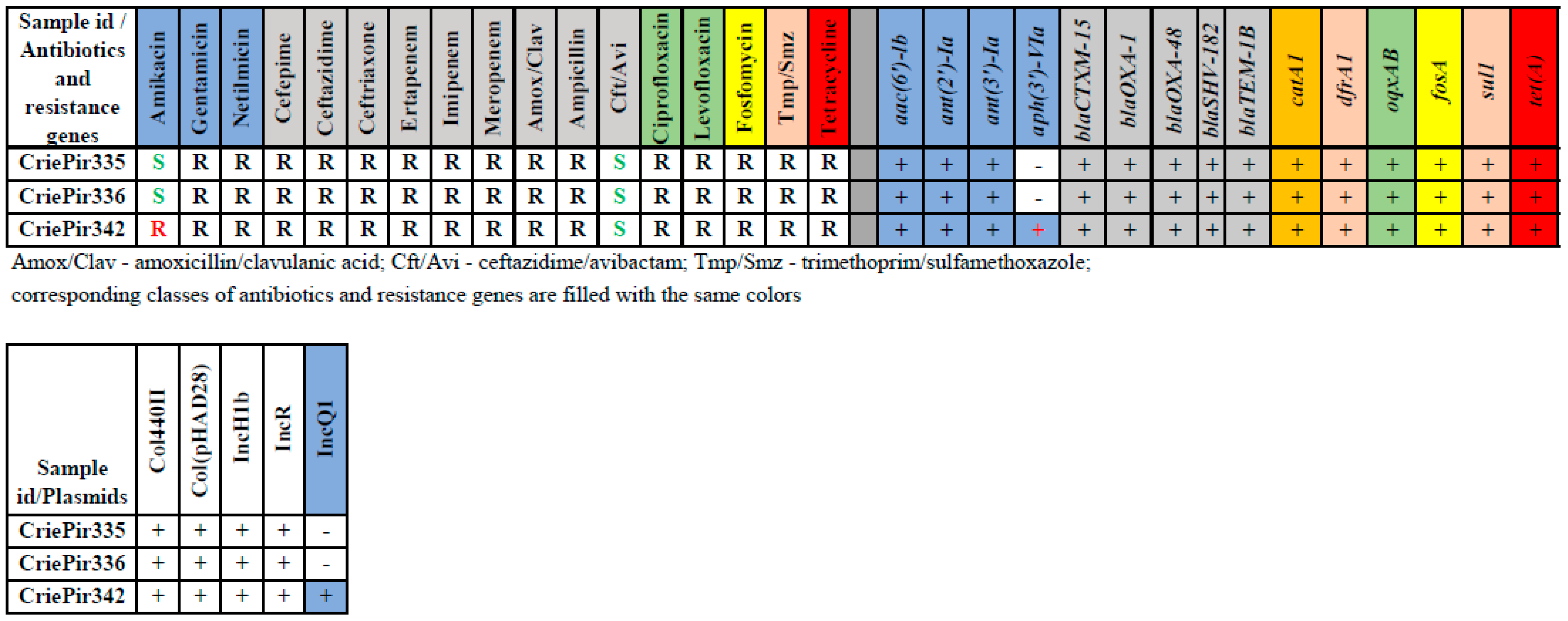
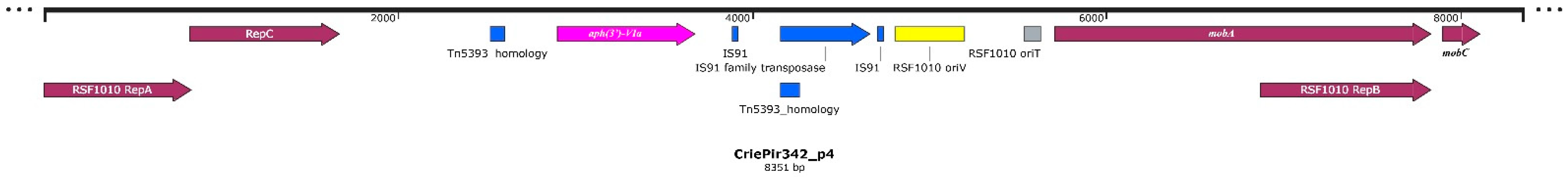
2.1. Isolate Typing, Resistance Profile, AMR Gene and Plasmid Content Determination

The typing results revealed the same profile ST395/KL39/O1/O2v1 for all three isolates. cgMLST analysis revealed that the genomic sequences of all isolates were very close (no allele differences between CriePir335 and 336 and six different alleles between either of these isolates and CriePir342). Thus, according to the criterion described previously (less than 18 different cgMLST alleles for K. pneumoniae [18]), these isolates were highly likely representing a single strain. Complete cgMLST profiles for the isolates studied are given in Table S1.
All isolates were multidrug-resistant (MDR) and susceptible only to ceftazidime/avibactam and amikacin (except CriePir342). They carried multiple antibiotic resistance determinants, including ESBL-coding genes blaCTX-M-15, blaOXA-48 and blaTEM-1B (see Figure 1). The only difference in genomic resistance was revealed for CriePir342, that carried a aph(3′)-VIa providing resistance to amikacin [19], which corresponded perfectly with phenotypic data. In general, we have not revealed any discrepancies between phenotypic and genomic resistance profiles. The susceptibility to ceftazidime/avibactam can be attributed to the synergistic effect of this drug.
The isolates CriePir335 and CriePir336 possessed the same five plasmids, while CriePir342 included an additional IncQ1 plasmid. This plasmid carried the aph(3′)-VIa gene mentioned above, which provides resistance to amikacin [19]. Plasmid data are provided in Table 1.
We investigated IncQ plasmid more thoroughly to get additional insights into the resistance mechanisms. We revealed a Tn5393 transposon homology, as well as two copies of IS91 insertion sequence, and IS91 family transposase between them, near the aph(3′)-VIa gene. The plasmid also included the genes encoding replication proteins RepA, RepB and RepC, and mobilization protein genes mobA and mobC. The plasmid structure is shown in Figure 2.

2.2. Virulence Factors

The three isolates studied included exactly the same set of virulence factors. The list and brief description of important gene clusters are presented in Table 2. The complete list of 87 virulence genes is provided in Table S2. Most genes were located on chromosomes, except for iucABCD, iutA and rmpA2, which were located on IncH1B virulence plasmid.
The virulence factor content of CriePir isolates does not allow assigning them to the hypervirulent type since the set of corresponding genes was limited and most heavy metal resistance genes like pbr (lead resistance),pco (copper),ter (tellurite) and sil (silver) were missing. However, the isolates deserve additional attention to the presence of mucoid phenotype regulator rmpA2 and their rapid spread within ICU.

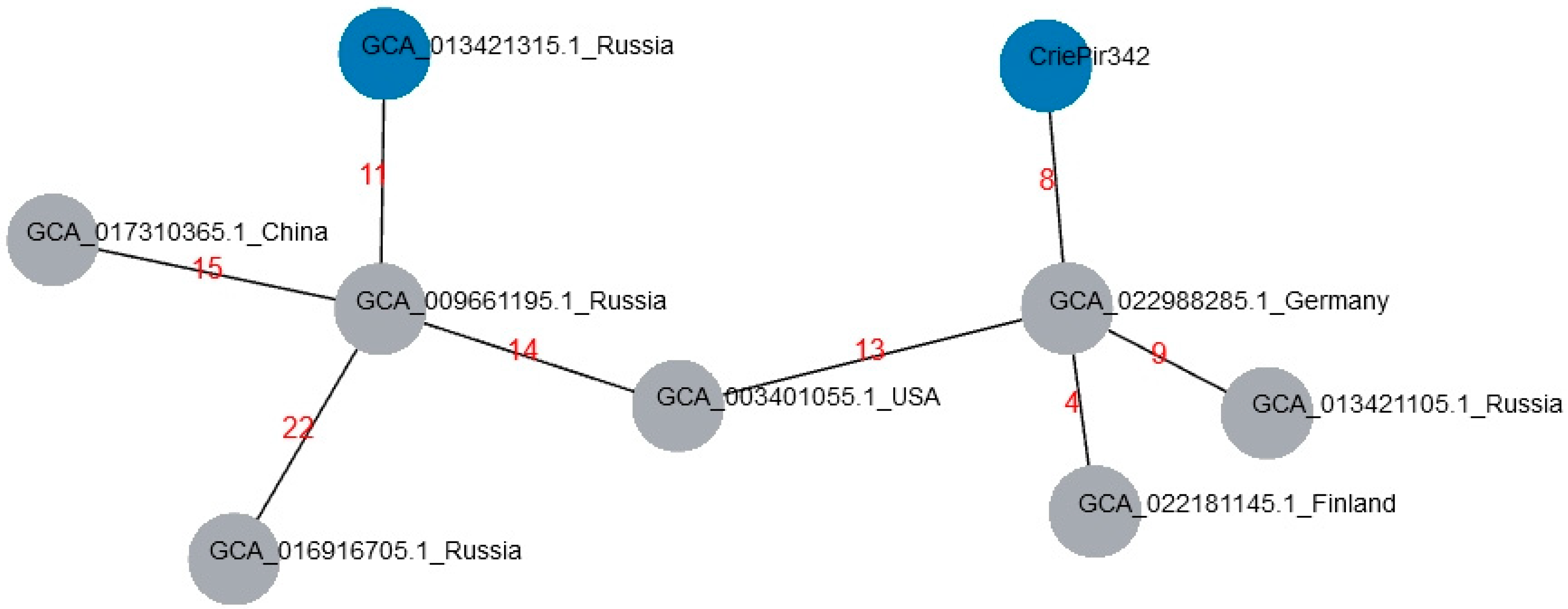
2.3. Comparison with Reference Isolates of ST395 from Genbank Database

The isolates obtained were compared to the genomes of ST395 available in Genbank, based on cgMLST. A list of closest matches and their description are provided in Table 3, and the minimum spanning tree for these isolates is shown in Figure 3 All comparisons were made based on genome sequences as the phenotype data were not available.
AMR gene content was similar for reference and CriePir isolates, except for aac(3)-IIa found in the USA isolate only, aph(3′)-VIa revealed in CriePir342 and GCA_013421315 only, and ant genes (see Table S3). An important difference was also that GCA_009661195 did not include blaCTX-M-15 and blaTEM-1B beta-lactamase genes, although it possessed IncR plasmid, on which these genes were located in CriePir342.
Virulence factor sets were also quite similar for all isolates, but several noticeable differences were revealed. GCA_003401055 (USA), GCA_009661195 (Russia) and GCA_022988285 (Germany) lack the genes from the iuc cluster and rmpA2. These genes were located on IncH1B plasmid in CriePir342, and the first two reference isolates did not have this plasmid, while the isolate from Germany did. However, this is not surprising since the plasmid structure has a high degree of plasticity.
Only the isolate GCA_013421315 included IncQ1 plasmid, which was likely to carry aph(3′)-VIa. Unfortunately, the genome record did not contain the plasmid structure, so the direct comparison of plasmid sequences was impossible. However, the contig containing the aph(3′)-VIa AMR gene was mapped to the plasmid IncQ1 of CriePir342 with 100% identity (data not shown). At the same time, GCA_013421315 included four additional plasmids in comparison to CriePir342.
The isolates provided in the Table 3 constitute a single strain, or clone, with CriePir342 based on criteria proposed by Schursch et al. [18]. The closest genomic matches to our isolates were revealed in Germany, Finland and the USA. However, the plasmid content of these isolates was different from the one of CriePir342 (see Table S3), namely, the isolates from Finland and Germany did not include IncQ1 plasmid, and the USA isolate possessed two additional plasmids, ColRNAI and IncL.

3. Discussion

In this study, a genomic epidemiology investigation of three MDR K. pneumoniae isolates from the patients of a dedicated COVID-19 ICU was conducted. Long-read sequencing allowed us to reveal that although these isolates constituted one strain, one of them (CriePir342) had a slightly different resistance profile and possessed additional plasmid encoding aph(3′)-VIa providing resistance to amikacin, which corresponded perfectly to its phenotype.
Currently K. pneumoniae is the most common cause of nosocomial infections in Russia, accounting for almost 30% of cases in 2020 according to the AMRmap database [20] (https://amrmap.ru/, accessed on 23 August 2022). The isolates studied belonged to the ST395 group, which is known as a high-risk clone with high capacity of drug resistance acquisition [21] and was revealed in different countries, for example, in France [22], Hungary [23] and China [24]. Additionally, ST395/KL39 isolates carrying the blaOXA-48 gene were recently revealed in Russia, including CriePir234 (GCA_009661195.1) described earlier by us [25], as well as in Finland in the same year, and were also obtained in 2013–2016 in such distant regions of the world as China, Germany and the USA. All these isolates belonged to the same strain as our isolates according to the proposed cgMLST allele difference threshold (≤18, [18]), and contained similar sets of AMR and virulence genes. The AMR determinants included ESBL-encoding genes blaOXA-48, blaCTX-M-15 (except GCA_009661195.1) and blaTEM-1B (except GCA_009661195.1), as well as various other AMR genes sufficient to consider these isolates as multidrug-resistant (MDR). However, only the Russian isolate NNKP343 (GCA_013421315.1) possessed the aph(3′)-VIa AMR gene mentioned above.
The virulence gene content was also similar for most isolates, except for the rmpA2 encoding mucoid phenotype regulator, which was revealed in Russian isolates only, including the CriePir ones. Most virulence genes, except the iuc cluster and rmpA2 mentioned above, were located on the chromosomes, which complies with previous data [25,26,27]. Another important determinant is a capsule surrounding the surface of K. pneumoniae, which serves as a main virulence factor associated with the viscous phenotype [28]. A capsular polysaccharide on the bacterial cell surface plays an important role in the pathogenicity of various bacteria, including Acinetobacter baumannii [29] and K. pneumoniae [30]. Although the KL39 capsule type is not generally considered as providing hypervirulence characteristics, at least one recent report described the increased virulence for the isolate having this capsule type and O1/O2v1 O-locus, which was found in all the isolates described above [31].
At the same time, the reference isolates exhibited differences in plasmid type and number, both between each other and with our set. For example, all isolates had IncR plasmid encoding, among others, the blaOXA-1 beta-lactamase gene, but IncF1B was revealed in two Russian isolates only, while large IncHI1B pNDM-MAR-like plasmid carrying both resistance and virulence (iuc cluster) traits was revealed in all but two isolates. Last but not the least, IncQ1 plasmid, which carried the amikacin resistance gene in CriePir342, was revealed only in NNKP343 (GCA_013421315.1) reference isolate, thus allowing us to suppose, together with the fact that this isolate exhibited amikacin resistance [32], that this plasmid included aph(3′)-VIa. Unfortunately, the exact plasmid structures were not reconstructed in any reference isolates, and thus the direct comparison was not possible. However, additional analysis revealed that the IncQ1 plasmid mentioned above included fragments of the Tn5393 transposon that was known to carry aminoglycoside resistance genes (in our case, aminoglycoside-O-phosphotransferase), and partial sequences of which were more common than the complete transposon in plasmids and genomic islands [33]. We also revealed that this plasmid had a MOBQ type and was mobilizable, rather than conjugative, which was reported to be a common characteristic of the IncQ1 plasmids [34]. Plasmids of this type are non-self-transmissible, but their host independent replication system allowed them to have a broader host range than any other known replicating components in bacteria [35]. Mobilizable plasmids rely on conjugative plasmids to provide the mating pair formation components, and MPFT plasmids can serve as helpers in MOBQ plasmid conjugation [36]. In CriePir342, conjugation could be assisted by large IncH plasmid, but more data are required to prove this hypothesis.
In general, the chromosomal DNA of K. pneumoniae carries only inherent resistance genes, and most acquired resistance determinants, including ESBLs, are located on plasmids [6,12,25]. Thus, it is not surprising that differences in plasmid carriage designated the dissimilarities in acquired AMR gene content for CriePir and reference isolates. It should be mentioned that although the reference and our isolates appeared to constitute a single clone, which supposedly spread across the world more than 10 years ago, the acquisition of virulence and AMR determinants through plasmid routes could significantly change the pathogenicity and morbidity of particular isolates. Meanwhile, the plasmid IncQ1 carrying the amikacin resistance gene was revealed in CriePir26 and CriePir28 isolates sequenced by us in 2017 [25]. Although these isolates belonged to a different clone with ST377, the plasmid sequence was completely the same as in CriePir342. Therefore, a continuous genomic epidemiology surveillance of clinical K. pneumoniae isolates is required to assess their threat to the patients of particular health care institutions since the determination of a sequence type and clonal lineage appears to be insufficient for this purpose.
The current study is limited to three isolates only since the main goal was to elucidate the difference in the phenotypic resistance profiles for the three K. pneumoniae isolates obtained in the same ward during this limited period. This was achieved with the help of third-generation sequencing. During recent years, the increasing use of long-read sequencing greatly facilitated the investigations of outbreaks and exploration of possible resistance transmission routes [37,38,39], and several bioinformatics protocols were developed for these purposes [15,17,40]. Another possible application of this powerful technology is the investigation of bacterial clone diversity within a particular hospital department or some other healthcare facility [41,42,43].
At the same time, various factors limit the ability to analyze the putative outbreak genomes in real-time, which, surprisingly, might include not the restrictions imposed by sequencing technologies, but rather data management and bioinformatics, for example, the lack of common outbreak repositories and delays between data collection and computational analysis [44,45]. Thus, creating more sophisticated bioinformatics tools can also advance AMR prevention strategies and the epidemiological surveillance of K. pneumoniae and other important pathogens. The development of such tools is one of the future perspectives of our investigations.

4. Materials and Methods

4.1. Sample Collection, Susceptibility Testing, DNA Isolation, and Sequencing

Three K. pneumoniae isolates (named CriePir335 (from urine), CriePir336 (from blood) and CriePir342 (from urine)) were obtained from COVID-19 patients in a dedicated ICU of a multipurpose medical center during the first half of June 2020. The patients were females of 81, 72 and 85 years of age, respectively, with a confirmed diagnosis of COVID-19.
Species identification for the isolates studied was performed using time-of-flight mass spectrometry (MALDI-TOF MS) with the VITEK MS (bioMerieux, Marcy-l’Étoile, France). Antimicrobial susceptibility/resistance was determined by the disc diffusion method using the Mueller-Hinton medium (bioMerieux, Marcy-l’Étoile, France) and disks with antibiotics (BioRad, Marnes-la-Coquette, France), and the minimum inhibitory concentration (MIC) was determined on VITEK 2 Compact 30 analyzer automated system (bioMerieux, Marcy-l’Étoile, France). The antibiotics tested included amikacin, amoxicillin/clavulanic acid, ampicillin, cefepime, ceftazidime, ceftriaxone, ceftazidime/avibactam, ciprofloxacin, ertapenem, fosfomycin, gentamicin, imipenem, levofloxacin, meropenem, netilmicin, tetracycline and trimethoprim/sulfamethoxazole. We used the EUCAST clinical breakpoints, version 11.0 (https://www.eucast.org/clinical_breakpoints/, accessed on 20 December 2020) to interpret the susceptibility/resistance results obtained.
The isolates described above represent a subset of multidrug-resistant (MDR) samples, which were subjected to WGS.
Genomic DNA was isolated using DNeasy Blood and Tissue kit (Qiagen, Hilden, Germany), and Nextera™ DNA Sample Prep Kit (Illumina®, San Diego, CA, USA) was applied for paired-end library preparation and WGS of the isolates on Illumina® Hiseq 2500 platform (Illumina®, San Diego, CA, USA). The same genomic DNA was used to prepare the libraries for the Oxford Nanopore MinION sequencing system (Oxford Nanopore Technologies, Oxford, UK) with the Rapid Barcoding Sequencing kit SQK-RBK004 (Oxford Nanopore Technologies, Oxford, UK). The amount of initial DNA was 400 ng for each sample. The libraries were prepared according to the manufacturer’s protocols, and were sequenced on R9 SpotON flow cell with a standard 24 h sequencing protocol using the MinKNOW software (Oxford Nanopore Technologies, Oxford, UK).

4.2. Data Processing and Genome Assembly

The base calling of the raw MinION data was performed using the Guppy Basecalling Software version 4.2.2 (Oxford Nanopore Technologies, Oxford, UK), and demultiplexing was performed using the Guppy barcoding software version 4.2.2 (Oxford Nanopore Technologies, Oxford, UK). Hybrid short- and long-read assemblies were obtained using the Unicycler version 0.4.9 (normal mode) [46]. Genome assemblies were submitted to the NCBI Genbank under the project PRJNA839643.
The pipeline described earlier [25] was used for the assembled genome processing and annotation. The Resfinder 4.0 database was used for antimicrobial gene detection (https://cge.cbs.dtu.dk/services/ResFinder/, accessed on 20 August 2022, using default parameters). Virulence factors were revealed by searching in VFDB (http://www.mgc.ac.cn/VFs/main.htm, accessed on 20 August 2022, using default parameters). Plasmids were detected using the PlasmidFinder (https://cge.cbs.dtu.dk/services/PlasmidFinder/, accessed on 20 August 2022, using default parameters).
Isolate typing was performed by MLST using BIGSdb (https://bigsdb.pasteur.fr/klebsiella/, accessed on 25 May 2022). In addition, the types based on capsule synthesis loci (K-loci) and lipooligosaccharide outer core loci (OCL) were also deduced using the Kaptive software v.2.0.3 with default parameters [30].
The detection of cgMLST profiles was performed using the MentaList (https://github.com/WGS-TB/MentaLiST, version 0.2.4, default parameters, accessed on 25 August 2022) [47] using the scheme obtained from cgmlst.org (https://www.cgmlst.org/ncs/schema/schema/2187931/, contained 2,358 loci, last update 25 May 2022). The minimum spanning tree was built using PHYLOViz online (http://online.phyloviz.net, accessed on 20 August 2022).
We used the TnCentral database (https://tncentral.proteininformationresource.org/tn_blast.html, accessed on 20 August 2022) to search for transposon sequences and ISEscan [48] for searching insertion sequences.

5. Conclusions

We performed WGS and obtained hybrid short- and long-read assemblies of three MDR ESBL-producing K. pneumoniae isolates, representing a single clone, obtained from COVID-19 patients in a dedicated ICU. Bioinformatics analysis allowed us to elucidate the differences in their antibiotic resistance profiles and reveal the possible mechanism of amikacin resistance found in one of the isolates. We believe that our data will facilitate the understanding of transfer mechanisms and developing new strategies for preventing resistance spreading within the clinical K. pneumoniae population.

Supplementary Materials

The following supporting information can be downloaded at: https://www.mdpi.com/article/10.3390/antibiotics11101364/s1, Table S1: cgMLST profiles for clinical K. pneumoniae isolates studied and reference isolates from Genbank.; Table S2: complete list of virulence genes revealed in clinical K. pneumoniae isolates studied; Table S3: comparison of antibiotic resistance gene, virulence factor and plasmid content for the reference and CriePir isolates.

Author Contributions

Conceptualization, A.S., M.Z. and Y.M.; formal analysis, A.S.; funding acquisition, V.A. investigation, A.S., L.P., A.M. and Y.M.; methodology, A.S., L.P. and Y.M.; project administration, M.Z. and V.A.; resources, L.P., A.M. and Y.M.; software, A.S.; supervision, M.Z. and Y.M.; validation, L.P., A.M. and Y.M.; visualization, A.S.; writing—original draft, A.S.; writing—review and editing, A.S. and Y.M. All authors have read and agreed to the published version of the manuscript.

Funding

This research received no external funding.

Institutional Review Board Statement

Ethical review and approval were waived for this study since the human samples were routinely collected and patients’ data remained anonymous.

Informed Consent Statement

Informed consent was obtained from all subjects involved in the study.

Data Availability Statement

Genome assemblies were submitted to NCBI Genbank under the project PRJNA839643. Accession numbers are as follows: JAMXKW000000000 (CriePir335), JAMXKV000000000 (CriePir336), JAMXKU000000000 (CriePir342).

Conflicts of Interest

The authors declare no conflict of interest.

References

  1. Wang, M.; Earley, M.; Chen, L.; Hanson, B.M.; Yu, Y.; Liu, Z.; Salcedo, S.; Cober, E.; Li, L.; Kanj, S.S.; et al. Clinical outcomes and bacterial characteristics of carbapenem-resistant Klebsiella pneumoniae complex among patients from different global regions (CRACKLE-2): A prospective, multicentre, cohort study. Lancet Infect. Dis. 2022, 22, 401–412. [Google Scholar] [CrossRef]
  2. Manesh, A.; Varghese, G.M. Rising antimicrobial resistance: An evolving epidemic in a pandemic. Lancet Microbe 2021, 2, e419–e420. [Google Scholar] [CrossRef]
  3. Tacconelli, E.; Carrara, E.; Savoldi, A.; Harbarth, S.; Mendelson, M.; Monnet, D.L.; Pulcini, C.; Kahlmeter, G.; Kluytmans, J.; Carmeli, Y.; et al. Discovery, research, and development of new antibiotics: The WHO priority list of antibiotic-resistant bacteria and tuberculosis. Lancet Infect. Dis. 2018, 18, 318–327. [Google Scholar] [CrossRef]
  4. Agyeman, A.A.; Bergen, P.J.; Rao, G.G.; Nation, R.L.; Landersdorfer, C.B. A systematic review and meta-analysis of treatment outcomes following antibiotic therapy among patients with carbapenem-resistant Klebsiella pneumoniae infections. Int. J. Antimicrob. Agents 2020, 55, 105833. [Google Scholar] [CrossRef]
  5. Li, Y.; Li, J.; Hu, T.; Hu, J.; Song, N.; Zhang, Y.; Chen, Y. Five-year change of prevalence and risk factors for infection and mortality of carbapenem-resistant Klebsiella pneumoniae bloodstream infection in a tertiary hospital in North China. Antimicrob. Resist. Infect. Control 2020, 9, 79. [Google Scholar] [CrossRef]
  6. Gorrie, C.L.; Mirceta, M.; Wick, R.R.; Judd, L.M.; Lam, M.M.C.; Gomi, R.; Abbott, I.J.; Thomson, N.R.; Strugnell, R.A.; Pratt, N.F.; et al. Genomic dissection of Klebsiella pneumoniae infections in hospital patients reveals insights into an opportunistic pathogen. Nat. Commun. 2022, 13, 3017. [Google Scholar] [CrossRef]
  7. Saini, V.; Jain, C.; Singh, N.P.; Alsulimani, A.; Gupta, C.; Dar, S.A.; Haque, S.; Das, S. Paradigm Shift in Antimicrobial Resistance Pattern of Bacterial Isolates during the COVID-19 Pandemic. Antibiotics 2021, 10, 954. [Google Scholar] [CrossRef]
  8. Lai, C.C.; Chen, S.Y.; Ko, W.C.; Hsueh, P.R. Increased antimicrobial resistance during the COVID-19 pandemic. Int. J. Antimicrob. Agents 2021, 57, 106324. [Google Scholar] [CrossRef]
  9. Pitout, J.D.; Nordmann, P.; Poirel, L. Carbapenemase-Producing Klebsiella pneumoniae, a Key Pathogen Set for Global Nosocomial Dominance. Antimicrob. Agents Chemother. 2015, 59, 5873–5884. [Google Scholar] [CrossRef] [Green Version]
  10. Spaziante, M.; Oliva, A.; Ceccarelli, G.; Venditti, M. What are the treatment options for resistant Klebsiella pneumoniae carbapenemase (KPC)-producing bacteria? Expert Opin. Pharmacother. 2020, 21, 1781–1787. [Google Scholar] [CrossRef]
  11. Tsuji, B.T.; Pogue, J.M.; Zavascki, A.P.; Paul, M.; Daikos, G.L.; Forrest, A.; Giacobbe, D.R.; Viscoli, C.; Giamarellou, H.; Karaiskos, I.; et al. International Consensus Guidelines for the Optimal Use of the Polymyxins: Endorsed by the American College of Clinical Pharmacy (ACCP), European Society of Clinical Microbiology and Infectious Diseases (ESCMID), Infectious Diseases Society of America (IDSA), International Society for Anti-infective Pharmacology (ISAP), Society of Critical Care Medicine (SCCM), and Society of Infectious Diseases Pharmacists (SIDP). Pharmacotherapy 2019, 39, 10–39. [Google Scholar] [CrossRef] [PubMed] [Green Version]
  12. Wang, X.; Zhao, J.; Ji, F.; Chang, H.; Qin, J.; Zhang, C.; Hu, G.; Zhu, J.; Yang, J.; Jia, Z.; et al. Multiple-Replicon Resistance Plasmids of Klebsiella Mediate Extensive Dissemination of Antimicrobial Genes. Front. Microbiol. 2021, 12, 754931. [Google Scholar] [CrossRef]
  13. Meng, M.; Li, Y.; Yao, H. Plasmid-Mediated Transfer of Antibiotic Resistance Genes in Soil. Antibiotics 2022, 11, 525. [Google Scholar] [CrossRef] [PubMed]
  14. Shaidullina, E.; Shelenkov, A.; Yanushevich, Y.; Mikhaylova, Y.; Shagin, D.; Alexandrova, I.; Ershova, O.; Akimkin, V.; Kozlov, R.; Edelstein, M. Antimicrobial Resistance and Genomic Characterization of OXA-48- and CTX-M-15-Co-Producing Hypervirulent Klebsiella pneumoniae ST23 Recovered from Nosocomial Outbreak. Antibiotics 2020, 9, 862. [Google Scholar] [CrossRef]
  15. Peter, S.; Bosio, M.; Gross, C.; Bezdan, D.; Gutierrez, J.; Oberhettinger, P.; Liese, J.; Vogel, W.; Dorfel, D.; Berger, L.; et al. Tracking of Antibiotic Resistance Transfer and Rapid Plasmid Evolution in a Hospital Setting by Nanopore Sequencing. mSphere 2020, 5, 4. [Google Scholar] [CrossRef] [PubMed]
  16. Bird, M.T.; Greig, D.R.; Nair, S.; Jenkins, C.; Godbole, G.; Gharbia, S.E. Use of Nanopore Sequencing to Characterise the Genomic Architecture of Mobile Genetic Elements Encoding blaCTX-M-15 in Escherichia coli Causing Travellers’Diarrhoea. Front. Microbiol. 2022, 13, 862234. [Google Scholar] [CrossRef]
  17. Ferreira, F.A.; Helmersen, K.; Visnovska, T.; Jorgensen, S.B.; Aamot, H.V. Rapid nanopore-based DNA sequencing protocol of antibiotic-resistant bacteria for use in surveillance and outbreak investigation. Microb. Genom. 2021, 7, 000557. [Google Scholar] [CrossRef] [PubMed]
  18. Schurch, A.C.; Arredondo-Alonso, S.; Willems, R.J.L.; Goering, R.V. Whole genome sequencing options for bacterial strain typing and epidemiologic analysis based on single nucleotide polymorphism versus gene-by-gene-based approaches. Clin. Microbiol. Infect. 2018, 24, 350–354. [Google Scholar] [CrossRef] [Green Version]
  19. Lambert, T.; Gerbaud, G.; Courvalin, P. Characterization of transposon Tn1528, which confers amikacin resistance by synthesis of aminoglycoside 3’-O-phosphotransferase type VI. Antimicrob. Agents Chemother. 1994, 38, 702–706. [Google Scholar] [CrossRef] [PubMed] [Green Version]
  20. Kuzmenkov, A.Y.; Trushin, I.V.; Vinogradova, A.G.; Avramenko, A.A.; Sukhorukova, M.V.; Malhotra-Kumar, S.; Dekhnich, A.V.; Edelstein, M.V.; Kozlov, R.S. AMRmap: An Interactive Web Platform for Analysis of Antimicrobial Resistance Surveillance Data in Russia. Front. Microbiol. 2021, 12, 620002. [Google Scholar] [CrossRef]
  21. Dogan, O.; Vatansever, C.; Atac, N.; Albayrak, O.; Karahuseyinoglu, S.; Sahin, O.E.; Kilicoglu, B.K.; Demiray, A.; Ergonul, O.; Gonen, M.; et al. Virulence Determinants of Colistin-Resistant K. pneumoniae High-Risk Clones. Biology 2021, 10, 436. [Google Scholar] [CrossRef]
  22. Muggeo, A.; Guillard, T.; Klein, F.; Reffuveille, F.; Francois, C.; Babosan, A.; Bajolet, O.; Bertrand, X.; de Champs, C.; CarbaFrEst, G. Spread of Klebsiella pneumoniae ST395 non-susceptible to carbapenems and resistant to fluoroquinolones in North-Eastern France. J. Glob. Antimicrob. Resist. 2018, 13, 98–103. [Google Scholar] [CrossRef]
  23. Kovacs, K.; Nyul, A.; Mestyan, G.; Melegh, S.; Fenyvesi, H.; Jakab, G.; Szabo, H.; Janvari, L.; Damjanova, I.; Toth, A. Emergence and interhospital spread of OXA-48-producing Klebsiella pneumoniae ST395 clone in Western Hungary. Infect. Dis. 2017, 49, 231–233. [Google Scholar] [CrossRef]
  24. Cienfuegos-Gallet, A.V.; Zhou, Y.; Ai, W.; Kreiswirth, B.N.; Yu, F.; Chen, L. Multicenter Genomic Analysis of Carbapenem-Resistant Klebsiella pneumoniae from Bacteremia in China. Microbiol. Spectr. 2022, 10, e0229021. [Google Scholar] [CrossRef] [PubMed]
  25. Shelenkov, A.; Mikhaylova, Y.; Yanushevich, Y.; Samoilov, A.; Petrova, L.; Fomina, V.; Gusarov, V.; Zamyatin, M.; Shagin, D.; Akimkin, V. Molecular Typing, Characterization of Antimicrobial Resistance, Virulence Profiling and Analysis of Whole-Genome Sequence of Clinical Klebsiella pneumoniae Isolates. Antibiotics 2020, 9, 261. [Google Scholar] [CrossRef] [PubMed]
  26. Muraya, A.; Kyany’a, C.; Kiyaga, S.; Smith, H.J.; Kibet, C.; Martin, M.J.; Kimani, J.; Musila, L. Antimicrobial Resistance and Virulence Characteristics of Klebsiella pneumoniae Isolates in Kenya by Whole-Genome Sequencing. Pathogens 2022, 11, 545. [Google Scholar] [CrossRef] [PubMed]
  27. Lery, L.M.; Frangeul, L.; Tomas, A.; Passet, V.; Almeida, A.S.; Bialek-Davenet, S.; Barbe, V.; Bengoechea, J.A.; Sansonetti, P.; Brisse, S.; et al. Comparative analysis of Klebsiella pneumoniae genomes identifies a phospholipase D family protein as a novel virulence factor. BMC Biol. 2014, 12, 41. [Google Scholar] [CrossRef] [PubMed]
  28. Zhu, J.; Wang, T.; Chen, L.; Du, H. Virulence Factors in Hypervirulent Klebsiella pneumoniae. Front. Microbiol. 2021, 12, 642484. [Google Scholar] [CrossRef] [PubMed]
  29. Arbatsky, N.P.; Shneider, M.M.; Dmitrenok, A.S.; Popova, A.V.; Shagin, D.A.; Shelenkov, A.A.; Mikhailova, Y.V.; Edelstein, M.V.; Knirel, Y.A. Structure and gene cluster of the K125 capsular polysaccharide from Acinetobacter baumannii MAR13-1452. Int. J. Biol. Macromol. 2018, 117, 1195–1199. [Google Scholar] [CrossRef] [PubMed]
  30. Wick, R.R.; Heinz, E.; Holt, K.E.; Wyres, K.L. Kaptive Web: User-Friendly Capsule and Lipopolysaccharide Serotype Prediction for Klebsiella Genomes. J. Clin. Microbiol. 2018, 56, e00197-18. [Google Scholar] [CrossRef]
  31. Arena, F.; Menchinelli, G.; Di Pilato, V.; Torelli, R.; Antonelli, A.; Henrici De Angelis, L.; Coppi, M.; Sanguinetti, M.; Rossolini, G.M. Resistance and virulence features of hypermucoviscous Klebsiella pneumoniae from bloodstream infections: Results of a nationwide Italian surveillance study. Front. Microbiol. 2022, 13, 983294. [Google Scholar] [CrossRef]
  32. Alekseeva, A.E.; Brusnigina, N.F.; Gordinskaya, N.A.; Makhova, M.A.; Kolesnikova, E.A. Molecular genetic characteristics of resistome and virulome of carbapenem-resistant Klebsiella pneumoniae clinical strains. Klin. Lab. Diagn. 2022, 67, 186–192. [Google Scholar] [CrossRef] [PubMed]
  33. Partridge, S.R.; Kwong, S.M.; Firth, N.; Jensen, S.O. Mobile Genetic Elements Associated with Antimicrobial Resistance. Clin. Microbiol. Rev. 2018, 31, e00088-17. [Google Scholar] [CrossRef] [Green Version]
  34. Orlek, A.; Phan, H.; Sheppard, A.E.; Doumith, M.; Ellington, M.; Peto, T.; Crook, D.; Walker, A.S.; Woodford, N.; Anjum, M.F.; et al. Ordering the mob: Insights into replicon and MOB typing schemes from analysis of a curated dataset of publicly available plasmids. Plasmid 2017, 91, 42–52. [Google Scholar] [CrossRef]
  35. Meyer, R. Replication and conjugative mobilization of broad host-range IncQ plasmids. Plasmid 2009, 62, 57–70. [Google Scholar] [CrossRef] [PubMed] [Green Version]
  36. Loftie-Eaton, W.; Rawlings, D.E. Diversity, biology and evolution of IncQ-family plasmids. Plasmid 2012, 67, 15–34. [Google Scholar] [CrossRef]
  37. Prussing, C.; Snavely, E.A.; Singh, N.; Lapierre, P.; Lasek-Nesselquist, E.; Mitchell, K.; Haas, W.; Owsiak, R.; Nazarian, E.; Musser, K.A. Nanopore MinION Sequencing Reveals Possible Transfer of bla KPC-2 Plasmid Across Bacterial Species in Two Healthcare Facilities. Front. Microbiol. 2020, 11, 2007. [Google Scholar] [CrossRef] [PubMed]
  38. Greig, D.R.; Jenkins, C.; Gharbia, S.E.; Dallman, T.J. Analysis of a small outbreak of Shiga toxin-producing Escherichia coli O157:H7 using long-read sequencing. Microb. Genom. 2021, 7, 000545. [Google Scholar] [CrossRef]
  39. Roberts, L.W.; Harris, P.N.A.; Forde, B.M.; Ben Zakour, N.L.; Catchpoole, E.; Stanton-Cook, M.; Phan, M.D.; Sidjabat, H.E.; Bergh, H.; Heney, C.; et al. Integrating multiple genomic technologies to investigate an outbreak of carbapenemase-producing Enterobacter hormaechei. Nat. Commun. 2020, 11, 466. [Google Scholar] [CrossRef] [Green Version]
  40. Steinig, E.; Duchene, S.; Aglua, I.; Greenhill, A.; Ford, R.; Yoannes, M.; Jaworski, J.; Drekore, J.; Urakoko, B.; Poka, H.; et al. Phylodynamic Inference of Bacterial Outbreak Parameters Using Nanopore Sequencing. Mol. Biol. Evol. 2022, 39, 3. [Google Scholar] [CrossRef]
  41. de Siqueira, G.M.V.; Pereira-Dos-Santos, F.M.; Silva-Rocha, R.; Guazzaroni, M.E. Nanopore Sequencing Provides Rapid and Reliable Insight Into Microbial Profiles of Intensive Care Units. Front. Public Health 2021, 9, 710985. [Google Scholar] [CrossRef]
  42. Shelenkov, A.; Petrova, L.; Zamyatin, M.; Mikhaylova, Y.; Akimkin, V. Diversity of International High-Risk Clones of Acinetobacter baumannii Revealed in a Russian Multidisciplinary Medical Center during 2017–2019. Antibiotics 2021, 10, 1009. [Google Scholar] [CrossRef] [PubMed]
  43. Mustapha, M.M.; Srinivasa, V.R.; Griffith, M.P.; Cho, S.T.; Evans, D.R.; Waggle, K.; Ezeonwuka, C.; Snyder, D.J.; Marsh, J.W.; Harrison, L.H.; et al. Genomic Diversity of Hospital-Acquired Infections Revealed through Prospective Whole-Genome Sequencing-Based Surveillance. mSystems 2022, 7, e0138421. [Google Scholar] [CrossRef] [PubMed]
  44. Hodcroft, E.B.; De Maio, N.; Lanfear, R.; MacCannell, D.R.; Minh, B.Q.; Schmidt, H.A.; Stamatakis, A.; Goldman, N.; Dessimoz, C. Want to track pandemic variants faster? Fix the bioinformatics bottleneck. Nature 2021, 591, 30–33. [Google Scholar] [CrossRef]
  45. Viehweger, A.; Blumenscheit, C.; Lippmann, N.; Wyres, K.L.; Brandt, C.; Hans, J.B.; Holzer, M.; Irber, L.; Gatermann, S.; Lubbert, C.; et al. Context-aware genomic surveillance reveals hidden transmission of a carbapenemase-producing Klebsiella pneumoniae. Microb. Genom. 2021, 7, 000741. [Google Scholar] [CrossRef] [PubMed]
  46. Wick, R.R.; Judd, L.M.; Gorrie, C.L.; Holt, K.E. Unicycler: Resolving bacterial genome assemblies from short and long sequencing reads. PLoSComput. Biol. 2017, 13, e1005595. [Google Scholar] [CrossRef] [PubMed] [Green Version]
  47. Feijao, P.; Yao, H.T.; Fornika, D.; Gardy, J.; Hsiao, W.; Chauve, C.; Chindelevitch, L. MentaLiST-A fast MLST caller for large MLST schemes. Microb. Genom. 2018, 4, 2. [Google Scholar] [CrossRef] [PubMed]
  48. Xie, Z.; Tang, H. ISEScan: Automated identification of insertion sequence elements in prokaryotic genomes. Bioinformatics 2017, 33, 3340–3347. [Google Scholar] [CrossRef]
Figure 1. Phenotypic and genomic antibiotic resistance profiles of clinical K. pneumoniae isolates obtained from COVID-19 patients.
Antibiotics 11 01364 g001
Figure 2. The structure of IncQ1 plasmid carrying amikacin resistance gene.
Antibiotics 11 01364 g002
Figure 3. Minimum spanning tree based on cgMLST profiles for reference K. pneumoniae isolates and CriePir342. Since all our isolates possessed the close profiles, only CriePir342 is indicated. Red numbers show the number of allele differences between the corresponding isolates. The isolates carrying IncQ1 plasmid are colored in blue.
Antibiotics 11 01364 g003
Table 1. Plasmid data for the clinical K. pneumoniae isolates studied.
IdLengthRepliconTypeRelaxaseFamilyPlasmid Type
2282,773IncHMOBH, MPFTConjugative
374,680IncR-Non-mobilizable
48351IncQMOBQMobilizable
55010ColRNAI-Non-mobilizable
64052ColRNAIMOBPMobilizable
73511ColRNAIMOBPMobilizable
Table 2. Description of important virulence gene clusters in clinical K. pneumoniae isolates studied.
Gene ClusterFunctionLocation
acrABefflux pump genesChromosome
fimABCDEFGHIKfimbria production and biofilm formationChromosome
entABCEF, fepABCDGenterobactin biosynthesis (siderophore)Chromosome
irp1,2iron acquisition systemChromosome
iucABCD, iutAaerobactin cluster-iron acquisition systemIncH1B plasmid
manBCpromoters of capsule synthesis genesChromosome
mrkABCDFHIJfimbria production and biofilm formationChromosome
rcsABexopolysaccharide biosynthesisChromosome
rmpA2regulator of mucoid phenotypeIncH1B plasmid
wbbMNOlipopolysaccharide synthesisChromosome
ybtAEPQSTUyersiniabactin cluster-iron acquisition systemChromosome
Table 3. Reference isolates from Genbank, with closest matches to the isolates studied according to cgMLST analysis.
Genbank Acc.Number of Allele DifferencesCountry of IsolationIsolate Collection Year
GCA_022988285.18Germany2016
GCA_022181145.110Finland2018
GCA_003401055.113USA2013
GCA_013421105.115Russia2018
GCA_009661195.117Russia2018
GCA_013421315.118Russia2018
GCA_017310365.118China2015
Publisher’s Note: MDPI stays neutral with regard to jurisdictional claims in published maps and institutional affiliations.

