Article

Antimicrobial Resistance, Serologic and Molecular Characterization of E. coli Isolated from Calves with Severe or Fatal Enteritis in Bavaria, Germany

1
Bavarian Health and Food Safety Authority, 91058 Erlangen, Germany
2
Bavarian Health and Food Safety Authority, 85764 Oberschleissheim, Germany
3
Chemical and Veterinary Investigation Office, 72488 Sigmaringen, Germany
4
Department of Veterinary Sciences, Faculty of Veterinary Medicine, Institute of Infectious Diseases and Zoonoses, Ludwig-Maximilians-University, 80539 Munich, Germany
*
Author to whom correspondence should be addressed.
Antibiotics 2022, 11(1), 23; https://doi.org/10.3390/antibiotics11010023
Submission received: 23 November 2021 / Revised: 17 December 2021 / Accepted: 24 December 2021 / Published: 27 December 2021

Abstract

:
Worldwide, enterotoxigenic Escherichia coli (ETEC) cause neonatal diarrhea and high mortality rates in newborn calves, leading to great economic losses. In Bavaria, Germany, no recent facts are available regarding the prevalence of virulence factors or antimicrobial resistance of ETEC in calves. Antimicrobial susceptibility of 8713 E. coli isolates obtained from 7358 samples of diseased or deceased diarrheic calves were investigated between 2015 to 2019. Considerably high rates of 84.2% multidrug-resistant and 15.8% extensively drug-resistant isolates were detected. The resistance situation of the first, second and third line antimicrobials for the treatment, here amoxicillin-clavulanate, enrofloxacin and trimethoprim-sulfamethoxazole, is currently acceptable with mean non-susceptibility rates of 28.1%, 37.9% and 50.0% over the investigated 5-year period. Furthermore, the ETEC serotypes O101:K28, O9:K35, O101:K30, O101:K32, O78:K80, O139:K82, O8:K87, O141:K85 and O147:K89, as well as the virulence factors F17, F41, F5, ST-I and stx1 were identified in a subset of samples collected in 2019 and 2020. The substantially high rates of multi- and extensively drug-resistant isolates underline the necessity of continuous monitoring regarding antimicrobial resistance to provide reliable prognoses and adjust recommendations for the treatment of bacterial infections in animals.

1. Introduction

Escherichia coli account to the major enteric and systemic pathogens of the Gram-negative rods within the family Enterobacteriaceae. Most of the E. coli colonizing the intestinal tract of animals and humans are commensal, but facultative pathogenic strains may cause intestinal disorder or even severe and life-threatening extraintestinal disease [1,2]. In calves, enterotoxigenic E. coli (ETEC) pose a leading cause of intestinal disease, especially within the first four days of life [3,4,5]. ETEC encode lipopolysaccharide structures (LPS) that may act as endotoxins, fimbrial adhesins and finally enterotoxins. The endotoxins within the blood stream cause fever, damage of endothelial cells and disseminated intravascular coagulation (DIC), that leads to acute shock and sudden death [1]. The serological LPS characterization in calves comprise the E. coli serogroups O8, O9 and O101, and respective serotypes O9:K35 and O101:K30, as these are known for endotoxin effect [6]. Further, the serotype O78:K80 plays a major role in systemic disease, septicemia and endotoxic shock of newborn calves [1,6,7]. In piglets, the serotype O141:K85 in combination with F4 fimbria is specific for the postweaning diarrhea syndrome [6]. As well, three further serotypes O139:K82, O8:K87 and O147:K89 play an important role as pathogens for swine [6,8]. Proteinaceous fimbrial adhesins precipitate the bacterial attachment to the enteric mucosa that avert the mechanical shedding of virulent strains from the gut by peristalsis [1,4,9]. Former studies showed that the fimbrial adhesins F5, F17 and F41 are associated with calf diarrhea [4]. For ETEC, two different types of enterotoxins contribute to diarrhea in calves, the heat-stable toxin (ST) and heat-labile toxin (LT), respectively [1,10,11]. On a molecular level, the toxins increase the second messengers cyclic adenosine/ guanosine monophosphate (cAMP/cGMP), that effect an active secretion of fluid and electrolytes in the small intestine leading to extreme loss of fluid within the organism [11,12]. Further, ruminants are known to be a major reservoir of human pathogenic Shiga toxin-producing E. coli (STEC) [13,14,15,16]. Shiga toxins (stx1, stx2) may lead to enterocyte damage, subsequent bloody diarrhea and endothelial damage leading to internal hemorrhages and septicemia in susceptible neonatal calves [1,17,18]. Enterohemorrhagic E. coli (EHEC), a subset of STEC, further include intimin, an adhesin coded from the enterocyte effacement pathogenicity island (eaeA) [19,20] and enterohemolysin, a toxin encoded by the ehxA gene [21]. As published in several case reports, a majority of human EHEC disease outbreaks are caused by the serotype O157:H7 originating from contaminated ground beef [13,22,23]. This serotype is responsible for the hemorrhagic colitis and the life-threatening hemolytic uremic syndrome with the occurrence of thrombocytopenia, hemolytic anemia and thrombotic microangiopathy that may lead to acute renal failure and death [23,24,25,26].
Worldwide, neonatal diarrhea is still a major economic problem on cattle farms and the therapy with antimicrobials is crucial in routine practice [27]. However, the medication with bactericide antibiotics is solely, but highly indicated exclusively in the case of life-threatening sepsis [28,29]. The Swiss antibiotic therapy guidelines for veterinarians recommend amoxicillin-clavulanate as a first line, sulfonamide-trimethoprim as a second line and fluoroquinolones as a third line choice, here enrofloxacin [29]. A study from 2014 revealed that veterinarians in Europe mainly used polymyxins (44%), (fluoro)quinolones (18%), penicillins (13%), aminoglycosides (9%) and third and fourth generation cephalosporins (8%) in calves with diarrhea emphasizing the problem of an inappropriate use of antibiotics [30]. This contributes to a higher level of antimicrobial resistant bacteria in young animals compared to adults [31,32,33]. In addition, the emergence of multidrug- and pandrug-resistant E. coli in fecal samples of diarrheic calves has been recently and repeatedly reported [33,34]. According to the expert proposal for standard definitions for acquired resistance from the European Centre for Disease Prevention and Control (ECDC), strains are classified as “multidrug-resistant” if these are non-susceptible (resistant or intermediate) to at least one antimicrobial agent in more than three categories. Isolates meet the definition “extensively drug-resistant” if these are non-susceptible in all agents but two or fewer categories. Finally, isolates non-susceptible to all agents in all antimicrobial categories are ranked as “pandrug-resistant” [35].
Previous data show that the prevalence of extended-spectrum β-lactamase (ESBL)-producing E. coli in calves increased from 7% to 29% between 2006 and 2013 in Germany [27]. ESBL-producing strains do encode for numerous resistance genes and may transduce these to other, even commensal, bacteria [36]. Animals hosting these E. coli bacteria constitute a resistance gene reservoir that may affect the health of man and animals [36,37].
Only few data are available on the identification of ETEC from calves in Bavaria. However, the discrimination between the physiological intestinal flora and pathogenic E. coli is crucial [1,6,38]. The aim of the present study was to provide recent information about the most prevalent pathotypes of E. coli. These include the investigation of the current virulence factors, serotypes and trends in antimicrobial resistance [9,39,40,41,42].

2. Results

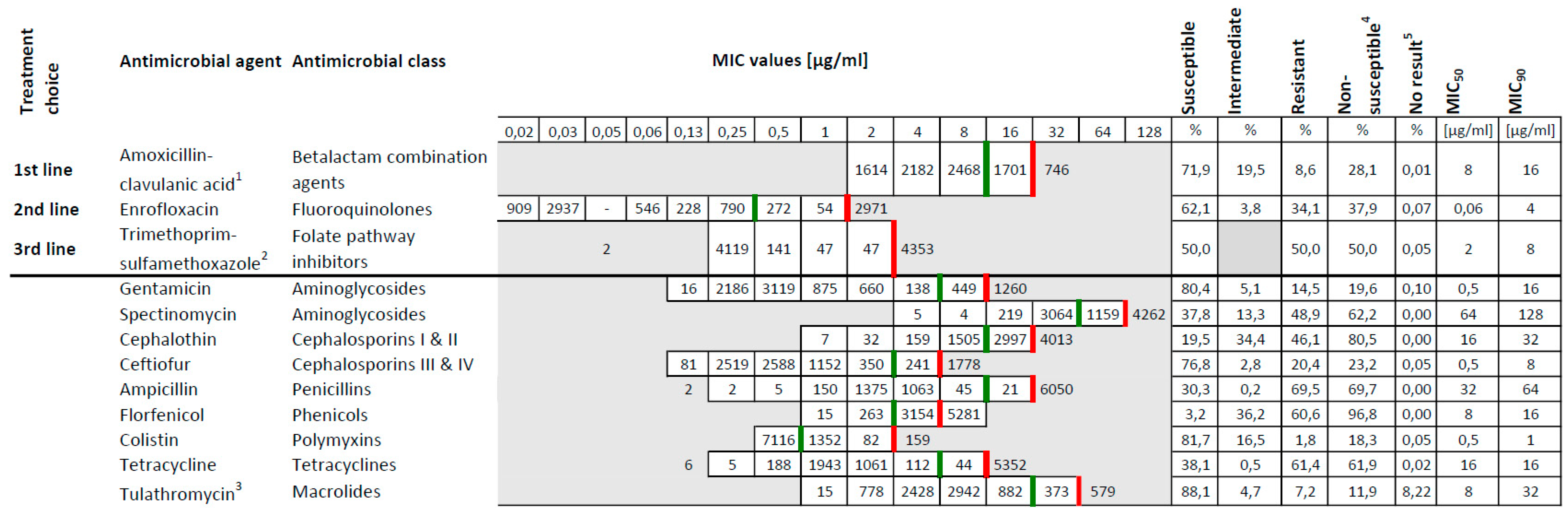
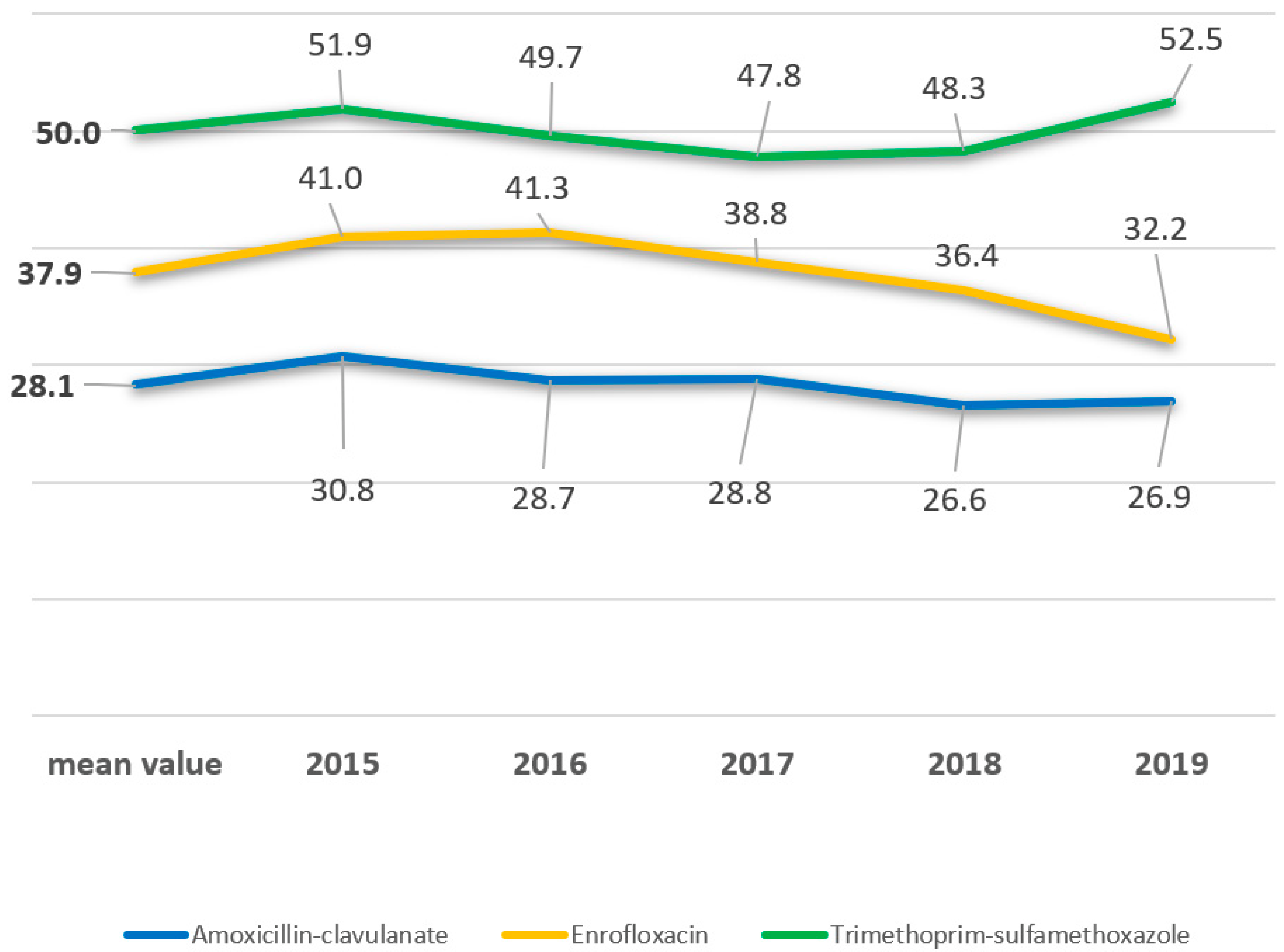
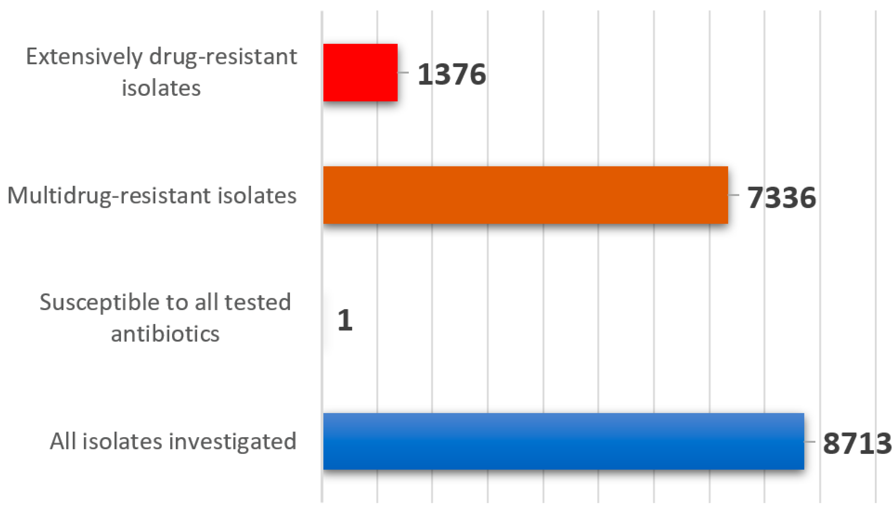
2.1. Antimicrobial Susceptibility

Within the study period 8713 E. coli were isolated from 7358 diarrheic calves at the federal state veterinary laboratory in Bavaria, Germany (Table S1). This number matches an average count of 1740 isolates per year that is in accordance with previous years (data not shown). The results on antimicrobial susceptibility testing revealed mean non-susceptibility values of 28.1% for amoxicillin-clavulanate, 37.9% for enrofloxacin and 50% for trimethoprim-sulfamethoxazole (Figure 1 and Figure 2, Table S1). The highest non-susceptibility value of a substance within each antimicrobial class revealed 11.9% for tulathromycin (macrolides), 18.3% for colistin (polymixins), 61.9% for tetracycline (tetracyclines), 62.2% for spectinomycin (aminoglycosides), 69.7% for ampicillin (penicillins), 80.5% for cephalothin (cephalosporins) and 96.8 % for florfenicol (phenicols) (Figure 1). A 5-year tendency from 2015 to 2019, evaluated for amoxicillin-clavulanate, enrofloxacin and trimethoprim-sulfamethoxazole, revealed a statistically significant decrease of the non-susceptibility rates for amoxicillin-clavulanate and enrofloxacin (p < 0.05) (Figure 2, Table 1). Regarding trimethoprim-sulfamethoxazole a significant decrease was assessed from 51.9% to 47.8% between 2015 and 2017 regarding the non-susceptible E. coli isolates (p < 0.05). A subsequent increase was further revealed from 47.8% to 52.5% in the years 2017 to 2019 (p < 0.05) (Figure 2, Table 1). Categorizing the 8713 isolates according to the ECDC expert proposal, 84.2% of the isolates (7336/8713) were multidrug-resistant, 15.7% (1368/8713) were extensively drug-resistant, eight isolates (0.1%) were pandrug-resistant and one isolate was susceptible to all antimicrobials tested. As we only tested antimicrobials licensed for the veterinary use, and none of the latest antimicrobials available on the market, we rededicated the eight presumably pandrug-resistant as extensively drug-resistant summing up to 1376 isolates in this specification (Figure 3).

2.2. Serologic Characterization

Serotyping of a randomly chosen subset of 108 E. coli isolated in 2019 and 2020 revealed 38 unequivocally typeable (35.2%), 29 untypeable (26.8%) and 41 seronegative (38%) strains (Table 2, Table S2). The most frequently detected serotypes were O101:K28 (8.3%; n = 9), O9:K35 and O139:K82 (6.5%; n = 7), O101:K30 (3.7%; n = 4), O101:K32, O78:K80 and O8:K87 (2.8%; n = 3). The serotypes O141:K85 and O147:K89 were detected once each (Table 2, Table S2). Finally, the serotypes O138:K81, O149:K91 and O157:H7 were not detected at all.
The fimbrial antigen F5 agglutinated in 6.5% of the isolates (n = 7) in combination with the serotypes O101:K30, O101:K28 and O9:K35. The fimbrial antigen F4 agglutinated in 4.6% of the isolates (n = 5), and exclusively combined with the serotype O139:K82 (Table 2, Table S2).

2.3. Molecular Characterization

Within the molecular characterization, 14 PCR assays targeted genes for the expression of fimbria, adhesin, hemolysin and toxins. A positive result was obtained for 24 isolates and 35 single assays, respectively (Table 2, Table S2). The most frequently detected genes coded for the fimbria F17 (13.9%; 15/108), F41 (3.7%; 4/108) and F5. The latter was always detected in combination with the toxin gene coding for ST-I (6.5%; 7/108). Finally, the gene coding for stx1 was detected in two of 108 isolates (1.9%). Seven of 108 isolates (6.5%) carried more than one type of virulence-associated genes (Table 2, Table S2). The fimbrial antigens F4, F6, F18, O157, adhesin eaeA, hemolysin ehxA and the toxins LT, ST-II and stx2 were not detected in any isolate. The occurrence of F4 fimbria in the serotyping assays could not be confirmed in the PCR investigation (Table 2, Table S2). In all, 84 of 108 isolates were negative in all PCR assays (Table 2, Table S2).

3. Discussion

Antibiotic treatment is the fundamental therapy regarding serious or life-threatening bacterial infections in man and animals [28,29]. Records regarding antimicrobial susceptibility on single substances are collected in many countries all over the world [43]. Worldwide this is a critical topic in line with the One Health issue [44]. Monitoring on the application and more important efficacy of antimicrobials regarding bacterial infections of farm animals is possible on principle in industrial countries. However, it is costly and difficult to standardize [36]. Published data from Canada in 2018 revealed a 51.6% susceptibility rate of 489 E. coli against trimethoprim-sulfamethoxazole, which is in consensus with our data (50%) (Figure 1 and Figure 2) [45]. Tetracycline was accounted to be effective in 36.8% and resembles our findings at 38.1% (Figure 1) [45]. Further, authors from the United States and Germany determined similar high resistance rates for tetracycline, with 71.1% and 70.9%. These data rather resemble the rate of 61.4% revealed in the present study (Figure 1) [46,47].
The antimicrobial class of fluoroquinolones includes enrofloxacin which is one of the substances of choice for the treatment of diarrhea in young cattle [29,48]. In Germany, the usage of fluoroquinolones has risen from 2011 to 2013 in human and veterinary medicine. This trend needs close monitoring to preserve the efficacy of the agent [27]. Fluoroquinolones are assessed as highest priority clinically important antimicrobials and as one of the few options for the treatment of serious Salmonella and E. coli infections in children recommended by the World Health Organization (WHO) [49]. The legislation reacted and passed a law in 2017 including obligatory antimicrobial susceptibility testing in case of the application of fluoroquinolones or third or fourth generation cephalosporines in Germany [50]. In the present study, the investigated E. coli isolated revealed a resistance rate of 34.1% regarding enrofloxacin (Figure 1). This finding correlates with published results from South America in 2017, with 36.4% [51].
Antimicrobial substances or closely related compounds may likewise be licensed for the use in man and animals. The application in an organism does trigger the development of antimicrobial resistance in present bacteria [49]. Legal restrictions regarding the use of cephalosporines, especially from the third and fourth generation, aim at a high prioritization of critically important antimicrobials in human medicine [49]. This is again in accordance with the terms of One Health [27,44]. The use of cephalosporines for the therapy of E. coli diarrhea in calves is a malpractice, as the effective therapeutic concentration is not reached within the gut [29]. Nonetheless, cephalosporin is the fifth-most commonly prescribed antimicrobial in the case of diarrhea with 8% according to a recent survey in Europe [30]. Regarding the third generation cephalosporine ceftiofur, a susceptibility rate of 86.4% could be determined in a study from Canada between 1994 and 2013 [45]. Significantly, our findings revealed 76.8% (Figure 1). Compared to data from the USA collected within the years 1960 until 2002 and in 2007, the resistance rate was at 7.4% and 11%, whereas in the present study the resistance rate of ceftiofur revealed 20.4% (Figure 1) [46,52]. This result is concerning, and the use of ceftiofur must be scrutinized critically, if not avoided completely. The resistance rates of the first generation cephalosporine, cephalothin, were lower in a comparable study regarding data within the period of 1960 to 2002, with 20.1%, in contrast to our results with an average rate at 46.1% from 2015 to 2019 (Figure 1) [46]. Currently, the standard antimicrobial therapy of mastitis in cows includes penicillins as well as first and second generation cephalosporines in the EU. Traces of antibiotics may reach the calves through the feeding of antibiotic contaminated waste milk [36]. To predict a reliable trend regarding the prevalence of ESBL-producing E. coli, PCR and sequencing methods should be applied to investigate the existence of ESBL- encoding genes as these are probably more accurate than the phenotypic characterization [53]. A study from 2013 revealed high rates (32.8%, 196 of 598 samples) of ESBL-encoding E. coli on dairy and beef cattle farms in Bavaria [54].
Completely inconsistent data are publicly available regarding the resistant rates for E. coli isolates and the substance florfenicol within the phenicol group. A 78% share of resistant isolates was determined in a study from the USA in 2006, only a 28% share from Canada in 2018, and a share of 35% from Bavaria, Germany, in 2002 [45,52,55]. In the present study, a rather higher resistance rate of 60.6% was determined for florfenicol (Figure 1). There was no information about ages of animals within the American and Canadian studies [45,52]. Since lower resistance rates were previously published in older animals for the substances ampicillin, tetracycline, streptomycin, sulfamethoxazole and chloramphenicol, this might accordingly apply for florfenicol [32]. This argument, however, still does not explain the diverse results of the Bavarian study from 2002 and the present study (Figure 1) [55].
With a 9% share of the most frequently listed antimicrobials, aminoglycosides remain at the fourth top position for the treatment of diarrhea in calves [30]. As these are almost solely used in the therapy of enterococcal endocarditis and multidrug-resistant tuberculosis in humans, they account to the high priority, clinically important antimicrobials in human medicine [49]. An application in veterinary medicine should therefore be prudent and well considered. Gentamicin belongs to the aminoglycoside antimicrobial class and has a withdrawal time for meat of more than 200 days in Germany for cattle and the indication of gastrointestinal disease. As this is economically hardly acceptable, the application of gentamicin is quite limited [48]. However, resistance to gentamicin among E. coli isolated from animals has been increasing from 0% to 40% between 1970 and 2002 within the United States [46]. Another long-term investigation from Germany revealed a further decrease of resistance rates including data from 2010 until 2013, and 2016 until 2017, respectively [47]. In the present study, the resistance rate of E. coli against Gentamicin was at 14.1% (Figure 1). Likewise, spectinomycin is an aminoglycoside antibiotic as well, and frequently used in combination with lincomycin for oral application in the treatment of simultaneous infection of the respiratory and the gastrointestinal tract in calves. The meat withdrawal time of 21 days is acceptable for farmers and practitioners and may be an explanation for the frequent prescription [48]. Within the present study and correspondingly a resistance rate of 48.9% was revealed in calves (Figure 1).
As stated by the WHO, the antimicrobial class of polymyxins accounts for the highest priority in critically important antimicrobials regarding the treatment of serious infections with Enterobacteriaceae and Pseudomonas aeruginosa in human medicine [49]. Despite rather frequent prescription of polymyxins in the treatment of diarrhea in animals, investigated E. coli isolates are still highly susceptible [30]. In the present study, the resistance rate against colistin revealed to be only 1.8% (Figure 1). Corresponding to this suggestion, another study revealed that only 3.8% of the isolates were resistant to colistin [47].
The aminopenicillin family, as well as the preparation amoxicillin-clavulanate, belong to the high priority critically important antimicrobials for the therapy of Listeria and Enterococcus spp. infections in humans according to the WHO [49]. For the aminopenicillin, ampicillin, an alarming resistance rate of 76.3% was determined in E. coli published in a most recent study from Germany [47]. Regrettably, a rate of 69.5% was determined in the present work as a similar result (Figure 1). Consequently, the recommendation on the usage of ampicillin for the treatment of calf diarrhea cannot further be continued. The amoxicillin-clavulanate susceptibility rate averaged at 57% in Germany in 2013 [27]. In the present study, the average susceptibility rate was 71.9%, and the resistance rate was 8.6% (Figure 1). Accordingly, a recently published study reported 7% of resistant E. coli isolates in Germany in 2018 [34]. Analogical to the report on the resistance monitoring study 2018 of the Federal Office of Consumer Protection and Food Safety, Germany, we determined decreasing non-susceptibility rates regarding the clinically important antimicrobial amoxicillin-clavulanate [34]. In conclusion, the resistance rates of E. coli against amoxicillin-clavulanate have decreased since 2013 and remained on a constant level within the years 2015 and 2019. This is a positive trend is beneficial for the One Health point of view [27].
Comparing data originating from other continents and collected over the last 60 years clearly reveals an increase of resistance regarding E. coli in nine out of the 12 tested drugs, namely gentamicin, cephalothin, ceftiofur, enrofloxacin, trimethoprim-sulfamethoxazole, ampicillin, amoxicillin-clavulanate, florfenicol and tetracycline [27,34,45,46,47,51,52,55]. Out of the 12 tested drugs in the present study, eight substances are similarly suitable for the treatment of human patients, namely gentamicin, spectinomycin, cephalothin, ampicillin, tetracycline, amoxicillin-clavulanate, colistin and trimethoprim-sulfamethoxazole (Figure 1) [49]. The application of these in veterinary medicine should be prudent due to the One Health aspect.
In a published study from Canada in 2018, 48.7% of multidrug-resistant E. coli were isolated from ruminants [45]. Within another study from the USA covering the years 1950 until 2002, a significantly increasing trend in resistance was observed for ampicillin, sulfonamide and tetracycline antibiotics regarding more than 1700 E. coli isolates. Two of these strains were identified as pandrug-resistant and originated from cattle in 2001 [46]. Further, multidrug resistance in E. coli increased from 7.2% to 63% between 1950 and 2002. Finally, 59.1% of the strains recovered form cattle were classified as multidrug resistant in the USA [46]. In the present study, we detected an even higher rate of 84.2% regarding multidrug resistance, 15.7% extensively drug-resistance and 0.1% pandrug-resistance (Figure 3). Furthermore, there were no exclusively susceptible isolates found amongst 108 isolates recovered in 2019 and 2020 from diarrheic calves in Bavaria (Table S2). Comparably high levels of antimicrobial resistance were published regarding the countries Brazil and Uruguay. Calves aged up to 60 days revealed a multidrug-resistance rate in E. coli at 78.7%, and at 61.6%, respectively [51]. As published, these bacteria occurred frequently in herds with high levels of diarrhea symptoms and subsequent antimicrobial therapy, as equally described in the present study [31].
Besides antimicrobial resistance, the determination of virulence regarding infectious agents is crucial in diagnostics. The discrimination from commensal E. coli was determined investigating virulence factors and evaluating the pathogenicity of isolates. As published, the E. coli serotypes O139:K82, O8:K87 and O147:K89 are pathogenic in swine [6]. However, in the present study, a fair amount of such isolates, six out of 108, were isolated from cattle, respectively (Table 2, Table S2). In laboratory diagnostics, implication of these serotypes should therefore be considered. Three isolates were identified as the serotype O78:K80, which frequently causes septicemia in calves (Table 2) [5,7,56]. However, more than one third, 38%, of the E. coli in this study revealed to be entirely seronegative (Table 3, Table S2), as it was as well published previously [57]. Preferably and in accordance with the One Health approach, the screening of E. coli isolated from diseased animals should always be of interest to identify zoonotic and human pathogenic serotypes [25]. As a matter of fact, formula associated with severe human syndromes included the serotypes O26, O103, O111, O117, O128, O145 and O146 respectively [13,22,23,58].
In recent studies, the fimbrial adhesins F17, F41 and F5 were frequently and significantly correlated with diseased calves compared to healthy animals [4,9]. These findings clearly correspond to the results of the present study (Table 2, Table S2). Other selective fimbrial antigens, F4, F6 and F18, occur frequently in isolates from diarrheic piglets [1,10,59]. As to be expected, we did not detect these amongst our strains isolated from calves (Table S2). Even five serologically F4 positive isolates were not confirmed within our molecular investigation (Table 2, Table S2). We assume that none of these isolates carry the specific primer sites, or agglutination was non-specific [9]. However, working at a federal state laboratory, we do research cross species infections especially among farm animals [60]. Furthermore, we consider the One Health approach, here especially the idea from farm to fork, and therefore continuously consider possible correlations between food-borne human pathogens and isolates from farm animals [27,44].
As published, hemolysis in E. coli isolates from piglets is a reliable diagnostic marker for virulence and pathogenicity [61,62,63]. Within the present study, only few (3/108) isolates revealed a hemolytic phenotype that was not even confirmed within the molecular analysis (Table S2). We conclude that hemolysis is not a relevant marker for virulence of E. coli isolated from calves in the present study. This statement is in accordance with prior publications [64,65].
Regarding the present study, ST-I was found in similar prevalence at a rate of 6.5% (7/108) compared to published data (Table 2, Table S2) [4,66]. The enterotoxins LT and ST-II were not detected in the present study (Table S2) and this again resembles data of relevant previous studies [4,56]. Concluding published data, ETEC isolated from calves only produced ST-I, whereas ETEC isolated from pigs may encode varying combinations of the enterotoxins LT, ST-I and ST-II [11,67]. In the present study, the detection rate of stx1 was very low and stx2 as well as intimin were not detected at all among the diarrheic calves’ isolates (Table 2, Table S2). This finding matches the results of previously published data to a high degree [9,51,68]. Obviously, the detection rate of Shiga toxins rose with the number of colonies isolated from each clinical sample, suggesting the selection of up to 35 colonies [69,70]. In the present investigation however, only up to three colonies were analyzed per clinical sample (Table S2). Other published results suggested a positive correlation between animal age and the amount of Shiga toxin, supporting our findings including animals of young age [69,70,71]. Targeted infection studies with STEC led to severe disease and bloody diarrhea in neonatal calves, but more recent studies disproved this observation revealing a still controversial discussion [4,72,73,74].

Limits of the Study

The antimicrobial susceptibility testing was carried out with a standard panel of antibiotics currently used in veterinary diagnostics in Germany. The results are therefore limited to substances only partially prescribed in human diagnostics and sometimes even in veterinary medicine regarding other countries of the world.
A thorough molecular investigation of single isolates is fairly time consuming and costly compared to the benefit that might be drawn from the results. In routine diagnostics, the molecular methods therefore can hardly be kept up.

4. Materials and Methods

4.1. Study Design and Bacterial Isolates

At the Bavarian Health and Food Safety Authority in Germany 7358 fecal samples of diseased or deceased calves with enteritis younger than six weeks of age were analyzed and included in the present study. Samples were collected between January 2015 and December 2019. Clinical symptoms ranged from low general condition, diarrhea, fever, sepsis and sudden death, respectively. A total of 8713 E. coli strains were isolated and confirmed through positive fluorescence on ECD agar (Merck Millipore, Burlington, MA, USA) and a positive Kovacs-Indole reaction (Merck Millipore, Burlington, MA, USA). All isolates were subject to antimicrobial resistance testing, further analysis and cryopreservation at the internal vaccine laboratory.

4.2. Antimicrobial Susceptibility Testing

Antimicrobial susceptibility testing was carried out according to the protocols published in CLSI VET01, 5th edition (Clinical and Laboratory Standards Institute, Wayne, PA, USA) [41]. Breakpoints were adopted from CLSI Vet01S, 5th edition, and national breakpoints for farm animals [41,42,75]. We used the microbroth dilution method on the following twelve different antimicrobial agents (antimicrobial class): Amoxicillin-clavulanic acid (betalactam combination agent), enrofloxacin (fluoroquinolone), Trimethoprim-sulfamethoxazole (folate pathway inhibitor), gentamicin and spectinomycin (aminoglycosides), cephalothin (cephalosporin I and II), ceftiofur (cephalosporin III and IV), ampicillin (penicillin), florfenicol (phenicol), colistin (polymyxin), tetracycline (tetracycline) and tulathromycin (macrolide). A commercially available set was used according to the manufacturer’s instructions (Micronaut-S, Grosstiere 4, Merlin, Bruker, Bornheim, Germany). The minimum inhibitory concentration (MIC) of each isolate and antibiotic substance was metered using a photometric plate reader system (Micronaut Scan and MCN6 software, Merlin/ Sifin, Bruker, Bornheim, Germany). Subsequently, the MIC value was reconciled with supplemented CLSI breakpoints, to categorize the respective E. coli isolate into “susceptible”, “intermediate” and “non-susceptible” for each antimicrobial substance tested [41,42,75,76]. E. coli ATCC 25922 was used as quality control strain [41].

4.3. Phenotypic Analysis and Serotyping

We deeper investigated a subset of 108 E. coli isolated in 2019 and 2020 originating from 66 diarrheic calves. The isolates were subcultured on Gassner agar (Oxoid Deutschland GmbH, Wesel, Germany) to differentiate specific colony morphology. The expression of potential virulent F5 fimbria was investigated by subculturing the isolates on pH 7.5 stabile, “minimum of casein” (Minca) agar (Sifin Diagnostics GmbH, Berlin, Germany) as previously published [76]. Finally, potential hemolytic properties of isolates were interpreted as described with subcultures on Columbia Sheep Blood Agar (Sifin Diagnostics GmbH, Berlin, Germany) [77]. Growth incubation was carried out for 18 to 24 h at 37 °C at all times. Serotyping for specific O-antigens was carried out using two polyvalent and 14 monovalent agglutination sera in a hierarchical approach according to the manufacturer’s instructions (Sifin Diagnostics GmbH Berlin, Germany) (Table 3). If an isolate showed a positive agglutination reaction with a polyvalent serum, but none with any correspondent monovalent or several reactions with various correspondent monovalent sera, it was categorized as untypeable. If an isolate showed no positive agglutination with any serum, it was categorized as seronegative.

4.4. Molecular Investigation

The molecular characterization of the E. coli isolates in the present study aimed at surface antigens, toxins and virulence factors. In all, 14 different target genes were of interest. Amongst were seven fimbrial genes F4, F5, F6, F17, F18, F41 and the outer membrane protein O157:H-. Further, two virulence genes were included, here adhesin intimin (eaeA), and enterohemolysin (ehxA). Finally, PCR targets coding for five toxins were screened, including heat-labile toxin (LT), heat-stabile toxin I (ST-I) and II (ST-II), Shiga toxin 1 (stx1) and stx2 (Table 4). Primer sequences were adopted from published protocols [9,39,40]. All 14 qPCR assays were performed applying a singleplex high resolution melting method, using AccuMelt HRM SuperMix (Quantabio, Beverly MA, USA) in 20 µL volumes according to the manufacturer’s instructions. DNA was extracted after thermolysis. The primers were added in a concentration of 0.2 µM each, and 3 µL of template DNA was used. Polymerase chain reaction assays were conducted on a Stratagene MX3000P device (Agilent Technologies, Waldbronn, Germany). The cycling protocol comprised an initial single denaturation step for 10 min at 95 °C, followed by 40 cycles of annealing and polymerization for 30 s at 60 °C and 10 s at 95 °C. After completing amplification, the melting curve analysis was performed. Specific melting temperatures were determined for each molecular target and all tested isolates. Reference strains were used as positive controls and kindly provided from Prof. R. Bauerfeind (Justus-Liebig-Universität, Gießen, Germany), and purchased from the German Collection of Microorganisms and Cell Cultures GmbH (DSMZ, Braunschweig, Germany) (Table 4).

4.5. Statistical Analysis

All statistical analyses were performed using the free software R Studio version 1.2.5033 (RStudio, Inc., Boston, MA, USA). Resistance trends of three clinically relevant antimicrobials amoxicillin-clavulanate, enrofloxacin and trimethoprim-sulfamethoxazole were evaluated by calculating a logistic regression model. The respective year was set as a continuous variable. The resulting odds ratio (OR) > 1 indicated an increased resistance trend, whereat an OR < 1 indicated a decreased antimicrobial resistance. The Wald test was used to determine the statistical significance of the year-antimicrobial trend. A value of p < 0.05 was considered significant (Table 1).

5. Conclusions

We conclude that an extensive monitoring, characterization and the analysis of antimicrobial resistance regarding enteritis causing E. coli is crucial to determine the currently raging serotypes, virulent genotypes and most important, the resistance situation. It is then possible to calculate reliable tendencies and prognoses from data collected over long terms in routine diagnostics. This is an important premise for objective and professional treatment recommendations regarding humans and animals within the scope of One Health. A further goal should be a slowdown of the increasing antimicrobial resistance situation that constitutes a global public health threat.

Supplementary Materials

The following supporting information can be downloaded at: https://www.mdpi.com/article/10.3390/antibiotics11010023/s1, Table S1: data set for all 8713 isolates from 2015–2019. Table S2: data set for a subset of 108 isolates in 2019–2020.

Author Contributions

Conceptualization, A.F., R.K.S. and J.M.R., methodology, A.F., A.H., K.B. and J.M.R.; software, A.F., A.M. and K.B.; validation, A.F., N.S., C.K., A.H., S.F. and J.M.R.; formal analysis, A.F., A.H., A.R., S.F., R.K.S. and J.M.R.; investigation, A.F., A.M., K.B., A.R. and S.F.; resources, C.K., K.B., A.R., S.F. and J.M.R.; data curation, A.F. and J.M.R.; writing—original draft preparation, A.F., N.S., A.M. and J.M.R.; writing—review and editing, N.S., C.K., K.B., A.R. and R.K.S.; visualization, A.F. and J.M.R.; supervision, R.K.S. and J.M.R.; project administration, J.M.R.; funding acquisition, K.B. and J.M.R. All authors have read and agreed to the published version of the manuscript.

Funding

This research received no external funding.

Institutional Review Board Statement

Not applicable.

Acknowledgments

The authors are grateful to the veterinary bacteriology staff members for technical support. Furthermore, the authors are grateful to the staff of the StabLab, Ludwig-Maximilians-University Munich, for a basic teaching class in statistical analyses.

Conflicts of Interest

The authors declare no conflict of interest.

References

  1. Quinn, P.J.; Markey, B.K.; Leonard, F.C.; Fitzpatrick, E.S.; Fanning, S.; Hartigan, P.J. Veterinary Microbiology and Microbial Disease; Wiley: Chichester, UK, 2011. [Google Scholar]
  2. Kaper, J.B.; Nataro, J.P.; Mobley, H.L. Pathogenic Escherichia coli. Nat. Rev. Genet. 2004, 2, 123–140. [Google Scholar] [CrossRef] [PubMed]
  3. Foster, D.M.; Smith, G.W. Pathophysiology of Diarrhea in Calves. Veter Clin. North Am. Food Anim. Pract. 2009, 25, 13–36. [Google Scholar] [CrossRef] [PubMed]
  4. Kolenda, R.; Burdukiewicz, M.; Schierack, P. A systematic review and meta-analysis of the epidemiology of pathogenic Escherichia coli of calves and the role of calves as reservoirs for human pathogenic E. coli. Front. Cell. Infect. Microbiol. 2015, 5, 23. [Google Scholar] [CrossRef] [PubMed]
  5. Cho, Y.-I.; Yoon, K.-J. An overview of calf diarrhea—Infectious etiology, diagnosis, and intervention. J. Veter Sci. 2014, 15, 1–17. [Google Scholar] [CrossRef] [Green Version]
  6. Linton, A.H.; Hinton, M.H. Enterobacteriaceae associated with animals in health and disease. Soc. Appl. Bacteriol. Symp. Ser. 1988, 65, S71–S85. [Google Scholar] [CrossRef]
  7. Ewers, C.; Schüffner, C.; Weiss, R.; Baljer, G.; Wieler, L.H. Molecular characteristics ofEscherichia coli serogroup O78 strains isolated from diarrheal cases in bovines urge further investigations on their zoonotic potential. Mol. Nutr. Food Res. 2004, 48, 504–514. [Google Scholar] [CrossRef]
  8. Nagy, B.; Fekete, P.Z. Enterotoxigenic Escherichia coli in veterinary medicine. Int. J. Med. Microbiol. 2005, 295, 443–454. [Google Scholar] [CrossRef]
  9. Sting, R.; Stermann, M. Duplex real-time PCr assays for rapid detection of virulence genes in E. coli isolated from post-weaning pigs and calves with diarrhoea Duplex Real-Time PCR-Assays für den raschen Nachweis von Virulenz-Genen in E. Coli-Isolaten durchfallerkrankter Absatzferkel und Kälber. Dtsch. Tierärztliche Wochenschr. 2008, 115, 231–238. [Google Scholar]
  10. Dubreuil, J.D.; Isaacson, R.E.; Schifferli, D.M. Animal Enterotoxigenic Escherichia coli. EcoSal Plus 2016, 7. [Google Scholar] [CrossRef] [Green Version]
  11. Gyles, C.L. Escherichia coli cytotoxins and enterotoxins. Can. J. Microbiol. 1992, 38, 734–746. [Google Scholar] [CrossRef]
  12. Field, M.; Graf, L.H., Jr.; Laird, W.J.; Smith, P.L. Heat-stable enterotoxin of Escherichia coli: In vitro effects on guanylate cyclase activity, cyclic GMP concentration, and ion transport in small intestine. Proc. Natl. Acad. Sci. USA 1978, 75, 2800–2804. [Google Scholar] [CrossRef] [Green Version]
  13. Gyles, C.L. Shiga toxin-producing Escherichia coli: An overview. J. Anim. Sci. 2007, 85, E45–E62. [Google Scholar] [CrossRef]
  14. Beutin, L.; Geier, D.; Steinrück, H.; Zimmermann, S.; Scheutz, F. Prevalence and some properties of verotoxin (Shiga-like toxin)-producing Escherichia coli in seven different species of healthy domestic animals. J. Clin. Microbiol. 1993, 31, 2483–2488. [Google Scholar] [CrossRef] [Green Version]
  15. Montenegro, M.A.; Bülte, M.; Trumpf, T.; Aleksić, S.; Reuter, G.; Bulling, E.; Helmuth, R. Detection and characterization of fecal verotoxin-producing Escherichia coli from healthy cattle. J. Clin. Microbiol. 1990, 28, 1417–1421. [Google Scholar] [CrossRef] [Green Version]
  16. Blanco, M.; Blanco, J.; Blanco, J.E.; González, E.A.; Gomes, T.; Zerbini, L.; Yano, T.; de Castro, A. Genes coding for Shiga-like toxins in bovine verotoxin-producing Escherichia coli (VTEC) strains belonging to different O:K:H serotypes. Veter Microbiol. 1994, 42, 105–110. [Google Scholar] [CrossRef]
  17. Wieler, L.H.; Bauernfeind, R. E coli: Shiga Toxin Methods and Protocols; Philpott, D., Ebel, F., Eds.; Humana Press Inc.: Totowa, NJ, USA, 2003; ISBN 978-1-59259-316-3. [Google Scholar] [CrossRef] [Green Version]
  18. Dean-Nystrom, E.A.; Bosworth, B.T.; Moon, H.W. Pathogenesis of Escherichia Coli O157:H7 in Weaned Calves. Adv. Exp. Med. Biol. 1999, 473, 173–177. [Google Scholar] [CrossRef]
  19. Dean-Nystrom, E.A.; Bosworth, B.T.; Cray, W.C., Jr.; Moon, H.W. Pathogenicity of Escherichia coli O157:H7 in the intestines of neonatal calves. Infect. Immun. 1997, 65, 1842–1848. [Google Scholar] [CrossRef] [Green Version]
  20. Dean-Nystrom, E.A.; Bosworth, B.T.; Moon, H.W.; O’Brien, A.D. Escherichia coli O157:H7 requires intimin for enteropathogenicity in calves. Infect. Immun. 1998, 66, 4560–4563. [Google Scholar] [CrossRef]
  21. Taneike, I.; Zhang, H.-M.; Wakisaka-Saito, N.; Yamamoto, T. Enterohemolysin operon of Shiga toxin-producingEscherichia coli: A virulence function of inflammatory cytokine production from human monocytes. FEBS Lett. 2002, 524, 219–224. [Google Scholar] [CrossRef] [Green Version]
  22. Beutin, L.; Aleksic’, S.; Zimmermann, S.; Gleier, K. Virulence factors and phenotypical traits of verotoxigenic strains of Escherichia coli isolated from human patients in Germany. Med. Microbiol. Immunol. 1994, 183, 13–21. [Google Scholar] [CrossRef]
  23. Boerlin, P.; McEwen, S.A.; Boerlin-Petzold, F.; Wilson, J.B.; Johnson, R.P.; Gyles, C.L. Associations between Virulence Factors of Shiga Toxin-Producing Escherichia coli and Disease in Humans. J. Clin. Microbiol. 1999, 37, 497–503. [Google Scholar] [CrossRef] [Green Version]
  24. Nataro, J.P.; Kaper, J.B. Diarrheagenic Escherichia coli. Clin. Microbiol. Rev. 1998, 11, 142–201. [Google Scholar] [CrossRef] [Green Version]
  25. Croxen, M.A.; Law, R.J.; Scholz, R.; Keeney, K.M.; Wlodarska, M.; Finlay, B.B. Recent Advances in Understanding Enteric Pathogenic Escherichia coli. Clin. Microbiol. Rev. 2013, 26, 822–880. [Google Scholar] [CrossRef] [Green Version]
  26. Mayer, C.L.; Leibowitz, C.S.; Kurosawa, S.; Stearns-Kurosawa, D.J. Shiga Toxins and the Pathophysiology of Hemolytic Uremic Syndrome in Humans and Animals. Toxins 2012, 4, 1261–1287. [Google Scholar] [CrossRef] [PubMed] [Green Version]
  27. Federal Office of Consumer Protection and Food Safety, Paul-Ehrlich-Gesellschaft für Chemotherapie e.V. Report on the Consumption of Antimicrobials and the Spread of Antimicrobial Resistance in Human and Veterinary Medicine in Germany. Available online: https://www.bvl.bund.de/SharedDocs/Downloads/05_Tierarzneimittel/germap2015_EN.pdf?__blob=publicationFile&v=5 (accessed on 23 December 2021).
  28. Constable, P.D. Treatment of Calf Diarrhea: Antimicrobial and Ancillary Treatments. Veter Clin. North Am. Food Anim. Pract. 2009, 25, 101–120. [Google Scholar] [CrossRef] [PubMed]
  29. Vetsuisse-Fakultät. Umsichtiger Einsatz von Antibiotika bei Rindern, Schweinen und kleinen Wiederkäuern, Therapieleitfaden für Tierärztinnen und Tierärzte; Gesellschaft Schweizer Tierärztinnen und Tierärzte; Bundesamt für Lebensmittelsicherheit und Veterinärwesen: Bern, Switzerland, 2019; Available online: https://www.blv.admin.ch/blv/de/home/tiere/tierarzneimittel/antibiotika/nationale-strategie-antibiotikaresistenzen--star--/sachgemaesser-antibiotikaeinsatz.html (accessed on 23 December 2021).
  30. De Briyne, N.; Atkinson, J.; Borriello, S.P.; Pokludová, L. Antibiotics used most commonly to treat animals in Europe. Vet. Rec. 2014, 175, 325. [Google Scholar] [CrossRef] [PubMed] [Green Version]
  31. De Verdier, K.; Nyman, A.; Greko, C.; Bengtsson, B. Antimicrobial resistance and virulence factors in Escherichia coli from swedish dairy calves. Acta Veter Scand. 2012, 54, 2. [Google Scholar] [CrossRef] [PubMed] [Green Version]
  32. Khachatryan, A.R.; Hancock, D.D.; Besser, T.E.; Call, D.R. Role of Calf-Adapted Escherichia coli in Maintenance of Antimicrobial Drug Resistance in Dairy Calves. Appl. Environ. Microbiol. 2004, 70, 752–757. [Google Scholar] [CrossRef] [Green Version]
  33. Cao, H.; Pradhan, A.K.; Karns, J.S.; Hovingh, E.; Wolfgang, D.R.; Vinyard, B.T.; Kim, S.W.; Salaheen, S.; Haley, B.J.; Van Kessel, J.A.S. Age-Associated Distribution of Antimicrobial-Resistant Salmonella enterica and Escherichia coli Isolated from Dairy Herds in Pennsylvania, 2013–2015. Foodborne Pathog. Dis. 2019, 16, 60–67. [Google Scholar] [CrossRef]
  34. Federal Office of Consumer Protection and Food Safety. Report on the Resistance Monitoring Study 2018. 2020. Available online: https://www.bvl.bund.de/EN/Home/home_node.html (accessed on 23 December 2021).
  35. Magiorakos, A.-P.; Srinivasan, A.; Carey, R.B.; Carmeli, Y.; Falagas, M.E.; Giske, C.G.; Harbarth, S.; Hindler, J.F.; Kahlmeter, G.; Olsson-Liljequist, B.; et al. Multidrug-resistant, extensively drug-resistant and pandrug-resistant bacteria: An international expert proposal for interim standard definitions for acquired resistance. Clin. Microbiol. Infect. 2012, 18, 268–281. [Google Scholar] [CrossRef] [Green Version]
  36. Murphy, D.; Ricci, A.; Auce, Z.; Beechinor, J.G.; Bergendahl, H.; Breathnach, R.; Bureš, J.; Duarte Da Silva, J.P.; Hederová, J.; Hekman, P.; et al. EMA and EFSA Joint Scientific Opinion on measures to reduce the need to use antimicrobial agents in animal husbandry in the European Union, and the resulting impacts on food safety (RONAFA). EFSA J. 2017, 15, 4666. [Google Scholar] [CrossRef]
  37. Astorga, F.; Navarrete-Talloni, M.J.; Miró, M.P.; Bravo, V.; Toro, M.; Blondel, C.J.; Hervé-Claude, L.P. Antimicrobial resistance in E. coli isolated from dairy calves and bedding material. Heliyon 2019, 5, e02773. [Google Scholar] [CrossRef]
  38. Ørskov, F.; Ørskov, I. Escherichia coli serotyping and disease in man and animals. Can. J. Microbiol. 1992, 38, 699–704. [Google Scholar] [CrossRef]
  39. Perelle, S.; Dilasser, F.; Grout, J.; Fach, P. Detection by 5′-nuclease PCR of Shiga-toxin producing Escherichia coli O26, O55, O91, O103, O111, O113, O145 and O157:H7, associated with the world’s most frequent clinical cases. Mol. Cell. Probes 2004, 18, 185–192. [Google Scholar] [CrossRef]
  40. Nielsen, E.M.; Andersen, M.T. Detection and Characterization of Verocytotoxin-Producing Escherichia coli by Automated 5′ Nuclease PCR Assay. J. Clin. Microbiol. 2003, 41, 2884–2893. [Google Scholar] [CrossRef] [Green Version]
  41. CLSI. Performance Standards for Antimicrobial Disk and Dilution Susceptibility Tests for Bacteria Isolated from Animals, 5th ed.; CLSI standard Vet01; Clinical and Laboratory Standards Institute: Wayne, PA, USA, 2018. [Google Scholar]
  42. CLSI. Performance Standards for Antimicrobial Disk and Dilution Susceptibility Tests for Bacteria Isolated from Animals, 4th ed.; CLSI supplement Vet08; Clinical and Laboratory Standards Institute: Wayne, PA, USA, 2018. [Google Scholar]
  43. Franklin, A.; Acar, J.; Anthony, F.; Gupta, R.; Nicholls, T.; Tamura, Y.; Thompson, S.; Threlfall, E.; Vose, D.; Van Vuuren, M.; et al. Antimicrobial resistance: Harmonisation of national antimicrobial resistance monitoring and surveillance programmes in animals and in animal-derived food. Rev. Sci. Tech. 2001, 20, 859–870. [Google Scholar] [CrossRef]
  44. German Federal Government. DART 2020—Fighting Antibiotic Resistance for the Good of Both Humans and Animals. Available online: https://www.bmel.de/SharedDocs/Downloads/EN/Publications/DART2020.html (accessed on 23 December 2021).
  45. Awosile, B.B.; Heider, L.C.; Saab, M.E.; McClure, J.T. Antimicrobial resistance in mastitis, respiratory and enteric bacteria isolated from ruminant animals from the Atlantic Provinces of Canada from 1994-2013. Can. Veter J. 2018, 59, 1099–1104. [Google Scholar]
  46. Tadesse, D.A.; Zhao, S.; Tong, E.; Ayers, S.; Singh, A.; Bartholomew, M.J.; McDermott, P.F. Antimicrobial Drug Resistance inEscherichia colifrom Humans and Food Animals, United States, 1950–2002. Emerg. Infect. Dis. 2012, 18, 741–749. [Google Scholar] [CrossRef]
  47. Tenhagen, B.-A.; Käsbohrer, A.; Grobbel, M.; Hammerl, J.; Kaspar, H. Antibiotikaresistenz von E. coli aus Rinderpopulationen in Deutschland. Tierärztliche Prax. Ausg. G Großtiere Nutztiere 2020, 48, 218–227. [Google Scholar] [CrossRef]
  48. Insitute for Pharmacology, Pharmacy and Toxicology, Veterinary Faculty, University Leipzig. Vetidata Veterinary Information Service. Available online: https://www.vetidata.de/public/index.php (accessed on 19 February 2021).
  49. World Health Organization. Critically Important Antimicrobials for Human Medicine, 5th Revision 2016. Available online: https://www.who.int/foodsafety/publications/antimicrobials-fifth/en/ (accessed on 10 October 2020).
  50. Federal Ministry of Justice and Consumer Protection. Verordnung über tierärztliche Hausapotheken; 2018. Available online: https://www.gesetze-im-internet.de/t_hav/BJNR021150975.html (accessed on 23 December 2021).
  51. Umpiérrez, A.; Bado, I.; Oliver, M.; Acquistapace, S.; Etcheverría, A.; Padola, N.L.; Vignoli, R.; Zunino, P. Zoonotic Potential and Antibiotic Resistance of Escherichia coli in Neonatal Calves in Uruguay. Microbes Environ. 2017, 32, 275–282. [Google Scholar] [CrossRef] [Green Version]
  52. Sawant, A.A.; Hegde, N.V.; Straley, B.A.; Donaldson, S.C.; Love, B.C.; Knabel, S.J.; Jayarao, B.M. Antimicrobial-Resistant Enteric Bacteria from Dairy Cattle. Appl. Environ. Microbiol. 2007, 73, 156–163. [Google Scholar] [CrossRef] [PubMed] [Green Version]
  53. Hoffmann, H.; Stürenburg, E.; Heesemann, J.; Roggenkamp, A. Prevalence of extended-spectrum β-lactamases in isolates of the Enterobacter cloacae complex from German hospitals. Clin. Microbiol. Infect. 2006, 12, 322–330. [Google Scholar] [CrossRef] [PubMed] [Green Version]
  54. Schmid, A.; Hörmansdorfer, S.; Messelhäusser, U.; Käsbohrer, A.; Sauter-Louis, C.; Mansfeld, R. Prevalence of Extended-Spectrum β-Lactamase-Producing Escherichia coli on Bavarian Dairy and Beef Cattle Farms. Appl. Environ. Microbiol. 2013, 79, 3027–3032. [Google Scholar] [CrossRef] [PubMed] [Green Version]
  55. Werckenthin, C.; Seidl, S.; Riedl, J.; Kiossis, E.; Wolf, G.; Stolla, R.; Kaaden, O.-R. Escherichia coli Isolates from Young Calves in Bavaria: In Vitro Susceptibilities to 14 Anti-microbial Agents. J. Veter Med. Ser. B 2002, 49, 61–65. [Google Scholar] [CrossRef]
  56. Blanco, J.; Gonzalez, E.; Garcia, S.; Blanco, M.; Regueiro, B.; Bernardez, I. Production of toxins by Escherichia coli strains isolated from calves with diarrhoea in Galicia (North-western Spain). Veter Microbiol. 1988, 18, 297–311. [Google Scholar] [CrossRef]
  57. Holland, R.E.; Wilson, R.A.; Holland, M.S.; Yuzbasiyan-Gurkan, V.; Mullaney, T.P.; White, D.G. Characterization of eae+ Escherichia coli isolated from healthy and diarrheic calves. Veter Microbiol. 1999, 66, 251–263. [Google Scholar] [CrossRef]
  58. Bielaszewska, M.; Mellmann, A.; Bletz, S.; Zhang, W.; Köck, R.; Kossow, A.; Prager, R.; Fruth, A.; Orth-Höller, D.; Marejková, M.; et al. Enterohemorrhagic Escherichia coli O26:H11/H−: A New Virulent Clone Emerges in Europe. Clin. Infect. Dis. 2013, 56, 1373–1381. [Google Scholar] [CrossRef]
  59. Casey, T.A.; Bosworth, B.T. Design and Evaluation of a Multiplex Polymerase Chain Reaction Assay for the Simultaneous Identification of Genes for Nine Different Virulence Factors Associated with Escherichia Coli that Cause Diarrhea and Edema Disease in Swine. J. Veter Diagn. Investig. 2009, 21, 25–30. [Google Scholar] [CrossRef] [Green Version]
  60. Bavarian Health and Food Safety Authority. Available online: https://www.lgl.bayern.de/ (accessed on 16 December 2021).
  61. Frydendahl, K. Prevalence of serogroups and virulence genes in Escherichia coli associated with postweaning diarrhoea and edema disease in pigs and a comparison of diagnostic approaches. Veter Microbiol. 2002, 85, 169–182. [Google Scholar] [CrossRef]
  62. Khac, H.V.; Holoda, E.; Pilipcinec, E.; Blanco, M.; Blanco, J.E.; Mora, A.; Dahbi, G.; López, C.; González, E.A.; Blanco, J. Serotypes, virulence genes, and PFGE profiles of Escherichia coliisolated from pigs with postweaning diarrhoea in Slovakia. BMC Veter Res. 2006, 2, 10. [Google Scholar] [CrossRef] [Green Version]
  63. Schierack, P.; Steinrück, H.; Kleta, S.; Vahjen, W. Virulence Factor Gene Profiles of Escherichia coli Isolates from Clinically Healthy Pigs. Appl. Environ. Microbiol. 2006, 72, 6680–6686. [Google Scholar] [CrossRef] [Green Version]
  64. Coura, F.M.; Diniz, S.D.A.; Mussi, J.M.S.; Silva, M.X.; Lage, A.P.; Heinemann, M.B. Characterization of virulence factors and phylogenetic group determination of Escherichia coli isolated from diarrheic and non-diarrheic calves from Brazil. Folia Microbiol. 2016, 62, 139–144. [Google Scholar] [CrossRef]
  65. Nguyen, T.D.; Vo, T.T.; Vu-Khac, H. Virulence factors inEscherichia coliisolated from calves with diarrhea in Vietnam. J. Veter Sci. 2011, 12, 159–164. [Google Scholar] [CrossRef] [Green Version]
  66. Yadegari, Z.; Brujeni, G.N.; Ghorbanpour, R.; Moosakhani, F.; Lotfollahzadeh, S. Molecular characterization of enterotoxigenic Escherichia coli isolated from neonatal calves diarrhea. Veter Res. Forum 2019, 10, 73–78. [Google Scholar] [CrossRef]
  67. World Health Organization. Escherichia coli diarrhoea. Bull. World Health Organ. 1980, 58, 23–36. [Google Scholar]
  68. Mainil, J.G.; Duchesnes, C.J.; Whipp, S.C.; Marques, L.R.; O’Brien, A.D.; Casey, T.A.; Moon, H.W. Shiga-like toxin production and attaching effacing activity of Escherichia coli associated with calf diarrhea. Am. J. Veter Res. 1987, 48, 743–748. [Google Scholar]
  69. Wieler, L.H.; Bauerfeind, R.; Baljer, G. Characterization of Shiga-like Toxin Producing Escherichia coli (SLTEC) Isolated from Calves with and without Diarrhoea. Zent. Für Bakteriol. 1992, 276, 243–253. [Google Scholar] [CrossRef]
  70. Mainil, J.G.; Jacquemin, E.R.; Kaeckenbeeck, A.E.; Pohl, P.H. Association between the effacing (eae) gene and the Shiga-like toxin-encoding genes in Escherichia coli isolates from cattle. Am. J. Veter Res. 1993, 54, 1064–1068. [Google Scholar]
  71. Wells, J.G.; Shipman, L.D.; Greene, K.D.; Sowers, E.G.; Green, J.H.; Cameron, D.N.; Downes, F.P.; Martin, M.L.; Griffin, P.M.; Ostroff, S.M. Isolation of Escherichia coli serotype O157:H7 and other Shiga-like-toxin-producing E. coli from dairy cattle. J. Clin. Microbiol. 1991, 29, 985–989. [Google Scholar] [CrossRef] [Green Version]
  72. Chanter, N.; Morgan, J.H.; Bridger, J.C.; Hall, G.A.; Reynolds, D.J. Dysentery in gnotobiotic calves caused by atypical Escherichia coli. Veter Rec. 1984, 114, 71. [Google Scholar] [CrossRef]
  73. Hall, G.A.; Reynolds, D.J.; Chanter, N.; Morgan, J.H.; Parsons, K.R.; Debney, T.G.; Bland, A.P.; Bridger, J.C. Dysentery Caused by Escherichia coli (S102-9) in Calves: Natural and Experimental Disease. Veter Pathol. 1985, 22, 156–163. [Google Scholar] [CrossRef] [Green Version]
  74. Ngeleka, M.; Godson, D.; Vanier, G.; Desmarais, G.; Wojnarowicz, C.; Sayi, S.; Huang, Y.; Movasseghi, R.; Fairbrother, J.M. Frequency of Escherichia colivirotypes in calf diarrhea and intestinal morphologic changes associated with these virotypes or other diarrheagenic pathogens. J. Veter Diagn. Investig. 2019, 31, 611–615. [Google Scholar] [CrossRef] [Green Version]
  75. Luhofer, G.; Böttner, M.; Hafez, H.; Kaske, M. Vorschläge der Arbeitsgruppe "Antibiotikaresistenz" für die Belegung von Mikrotiterplatten zur Empfindlichkeitsprüfung von Bakterien gegenüber antimikrobiellen Wirkstoffen in der Routinediagnostik—Mastitis- und Großtierlayouts. Berl. Munch. Tierarztl. Wochenschr 2004, 117, 245–251. [Google Scholar]
  76. Guinée, P.A.; Jansen, W.H.; Agterberg, C.M. Detection of the K99 antigen by means of agglutination and immunoelectrophoresis in Escherichia coli isolates from calves and its correlation with entertoxigenicity. Infect. Immun. 1976, 13, 1369–1377. [Google Scholar] [CrossRef] [PubMed] [Green Version]
  77. Pasayo, R.A.G.; Sanz, M.E.; Padola, N.L.; Moreira, A.R. Phenotypic and genotypic characterization of enterotoxigenic Escherichia coli isolated from diarrheic calves in Argentina. Open Veter J. 2019, 9, 65–73. [Google Scholar] [CrossRef] [PubMed] [Green Version]
Figure 1. Minimum inhibitory concentration (MIC) distribution of 8713 E. coli isolates on 12 antimicrobial agents from 11 antimicrobial classes. The three first lines represent the clinically relevant substances, first to third treatment choices in buiatrics. The red line demarcates the breakpoint towards resistance, the green line a breakpoint towards intermediate. Regarding the two combination compounds, only the concentration of the former substance is presented; the ratio of amoxicillin:clavulanic acid is 2:1 (1), concentration ratio of trimethoprim:sulfamethoxazole is 1:19 (2). Tulathromycin has not been tested in the first quarter of 2015 (3). The summation of intermediate and resistant isolates was named non-susceptible (4). Some results were not evaluable (5).
Antibiotics 11 00023 g001
Figure 2. The mean value (bold) and the five-year trend on non-susceptible E. coli isolated from calves revealed the highest proportion of isolates against trimethoprim-sulfamethoxazole, followed by enrofloxacin and amoxicillin-clavulanate. The trends regarding enrofloxacin and amoxicillin-clavulanate remain at a stable level and rather tend towards a decrease regarding the number of non-susceptible isolates. The graph of non-susceptible isolates regarding trimethoprim-sulfamethoxazole reveals a decrease, 2016–2017, followed by a steep increase of non-susceptible isolates in 2019. The corresponding statistic parameters are presented in Table 1.
Antibiotics 11 00023 g002
Figure 3. The classification of 8713 E. coli into extensively drug-resistant and multi drug-resistant isolates was carried out according to the expert proposal for standard definitions for acquired resistance. We categorized eight potential pandrug-resistant isolates in the category extensively drug resistant, as we only tested antimicrobials licensed for the veterinary use and did not include the latest antimicrobials available on the market.
Antibiotics 11 00023 g003
Table 1. Statistic parameters regarding the increase or decrease of resistance values within the five-year period for the three clinically relevant antimicrobials (Figure 2).
AntimicrobialYearsORCI (95%)
amoxicillin-clavulanate2015–20190.950.92–0.98 1
enrofloxacin2015–20190.910.88–0.94 1
2015–20170.920.85–1.0 1
trimethoprim-sulfamethoxazole2015–20191.00.97–1.03
2017–20191.111.03–1.19 1
OR: odds ratio, CI: confidence interval, 1 p-value (Wald test) < 0.05.
Table 2. The serologic and molecular characterization revealed 13 different serotypes known to be pathogenic for cattle and other species. Furthermore, four different genotypes were detected with five different coding sequences for fimbria and/or toxins in one or more isolates. Some of the isolates were untypeable/ seronegative and did not reveal any of the investigated virulence factors (green box).
SerotypeAdditionally Known for
Pathogenicity in
Number of
Isolates
Non-VirulentMolecular Results
F17F5ST-IF5F41ST-Istx1
O9:K35 651
O9:K35/F5 1 1
O101:K28 66
O101:K28/F5 3 3
O101:K30 1 1
O101:K30/F5 3 3
O101:K32 33
O78:K80Human/sheep33
O8:K87Swine33
O139:K82Swine22
O139:K82/F4Swine541
O141:K85Swine11
O147:K89Swine1 1
untypeable 29207 2
seronegative 41374
Total 1088415342
Table 3. In all, 16 different polyvalent and monovalent (mono) antisera were used for the agglutination and the characterization of E. coli. The listed serotypes are known for their pathogenicity in humans and farm animals.
Antiserum for
Initial Screening
Respective Follow Up
Agglutination
Specific Serotypes Occur in Cattle, but Are Found as Well/Especially in
Polyvalent anti-E. coli C
O9:K35, mono
O101:K28, mono
O101:K30, mono
O101:K32, mono
F5, mono
O78:K80, mono Human, sheep
Polyvalent anti-E. coli P Swine
O8:K87, mono
O138:K81, mono
O139:K82, mono
O141:K85, mono
O147:K89, mono
O149:K91, mono
F4, mono
O157:H7, mono Association with
food-poisoning
Table 4. Targets and primers for the molecular characterization of E. coli isolated from calves.
Target ProteinGene(s)PrimerOligo Sequence (5’ -> 3’)Size (bp)Melting Temperature (°C) ± 0.2 °CReferenceReference Isolate
F4F4_FGGTGGAACCAAACTGACCATTAC10281.0[9]7156
Fimbria/outer membrane protein F4_RTCCATCTACACCACCAGTTACTGG
F5F5_FTTGGAAGCACCTTGCTTTAACC10177.4[9]7159
F5_RTCACTTGAGGGTATATGCGATCTTT
F6F6_FGCGGATTAGCTCTTTCAGACCA10283.2[9]7155
F6_RTGACAGTACCGGCCGTAACTC
F17F17_FACTGAGGATTCTATGCRGAAAATTCAA8379.7[9]5397
F17_RCCGTCATAAGCAAGCGTAGCAG
F18F18_FCCTGCTAAGCAAGAGAATATATCCAGA8273.3[9]7160
F18_RAGAACATATACTCAGTGCCAACAGAGAT
F41F41_FCCTTTGTCATTTGGTGCGG10181.5[9]7159
F41_RTCAAATACTGTACCAGCAGAACCAC
O157 (rfbE)O157_FCGATGAGTTTATCTGCAAGGTGAT8878.3[39]DSMZ 19206
O157_RTTTCACACTTATTGGATGGTCTCAA
Adhesinintimin (eaeA)Intimin_FCCAGCTTCAGTCGCGATCTC9186.1[9]7158
Intimin_RGGCCTGCAACTGTGACGAA
Hemolysinenterohemolysin (ehxA)ehec-F2CGTTAAGGAACAGGAGGTGTCAGTA14279.5[40]DSMZ 19206
ehec-RATCATGTTTTCCGCCAATGAG
Toxinheat-labile toxin (LT)LT_FCTGCCATCGATTCCGTATATGAT8175.3[9]7157
LT_RCAGAACTATGTTCGGAATATCGCA
heat-stabile toxin (ST-I)ST-I_FTACCTCCCGTCATGTTGTTTCAC10176.1[9]7155
ST-I_RCCTCGACATATAACATGATGCAACTC
heat-stabile toxin (ST-II)St-II_FTTTTTCTATTGCTACAAATGCCTATGC10175.9[9]7156
St-II_RAACCTTTTTTACAACTTTCCTTGGC
Shiga toxin 1 (stx1)Stx1_FTCCCCAGTTCAATGTAAGATCAAC8179.0[9]7158
Stx1_RTTTCGTACAACACTGGATGATCTCA
Shiga toxin 2 (stx2)Stx2_FGAGTGACGACTGATTTGCATTCC8284.6[9]7158
Stx2_RCCATGACAACGGACAGCAGTT
Publisher’s Note: MDPI stays neutral with regard to jurisdictional claims in published maps and institutional affiliations.

