Current Trends in RNA Virus Detection via Nucleic Acid Isothermal Amplification-Based Platforms


Abstract
:1. Introduction
2. Loop-Mediated Isothermal Amplification
2.1. Principle of LAMP for RNA Virus Detection
2.2. Applications of LAMP for RNA Virus Detection
2.2.1. Fluorescence-Based Detection Methods
2.2.2. Colorimetric-Based Detection Method
2.2.3. Others Detection Methods
3. Recombinase Polymerase Amplification
3.1. Principle of RPA
3.2. Applications of RPA for RNA Virus Detection
3.2.1. Fluorescence-Based Detection Methods
3.2.2. Colorimetric-Based Detection Methods
4. Recombinase-Aided Amplification
4.1. Principle of RAA
4.2. Applications of RAA for Virus Detection
5. Other Isothermal Amplification-Based Platforms for RNA Virus Detection
5.1. Nucleic Acid Sequence-Based Amplification
5.2. Helicase-Dependent Amplification
6. Commercialized Isothermal Amplification Devices for SARS-CoV-2 Detection
7. Limitations and Future Perspectives on Isothermal Amplification Methods
7.1. Recent Challenges of Isothermal Amplification Techniques
7.2. Future Perspectives
8. Conclusions
Author Contributions
Funding
Data Availability Statement
Conflicts of Interest
References
- Domingo, E.; Escarmís, C.; Sevilla, N.; Moya, A.; Elena, S.F.; Quer, J.; Novella, I.S.; Holland, J.J. Basic concepts in RNA virus evolution. FASEB J. 1996, 10, 859–864. [Google Scholar] [CrossRef]
- Elena, S.F.; Sanjuán, R. Adaptive value of high mutation rates of RNA viruses: Separating causes from consequences. J. Virol. 2005, 79, 11555–11558. [Google Scholar] [CrossRef] [PubMed]
- Cassedy, A.; Parle-McDermott, A.; O’Kennedy, R. Virus detection: A review of the current and emerging molecular and immunological methods. Front. Mol. Biosci. 2021, 8, 637559. [Google Scholar] [CrossRef] [PubMed]
- Minchin, S.; Lodge, J. Understanding biochemistry: Structure and function of nucleic acids. Essays Biochem. 2019, 63, 433–456. [Google Scholar] [CrossRef] [PubMed]
- Fooks, A.R.; Johnson, N.; Freuling, C.M.; Wakeley, P.R.; Banyard, A.C.; McElhinney, L.M.; Marston, D.A.; Dastjerdi, A.; Wright, E.; Weiss, R.A. Emerging technologies for the detection of rabies virus: Challenges and hopes in the 21st century. PLoS Negl. Trop. Dis. 2009, 3, e530. [Google Scholar] [CrossRef] [PubMed]
- Wassenegger, M. Advantages and disadvantages of using PCR techniques to characterize transgenic plants. Mol. Biotechnol. 2001, 17, 73–82. [Google Scholar] [CrossRef] [PubMed]
- Wei, Z.; Wang, X.; Feng, H.; Ji, F.; Bai, D.; Dong, X.; Huang, W. Isothermal nucleic acid amplification technology for rapid detection of virus. Crit. Rev. Biotechnol. 2023, 43, 415–432. [Google Scholar] [CrossRef] [PubMed]
- Craw, P.; Balachandran, W. Isothermal nucleic acid amplification technologies for point-of-care diagnostics: A critical review. Lab. Chip 2012, 12, 2469–2486. [Google Scholar] [CrossRef] [PubMed]
- Chang, C.-C.; Chen, C.-C.; Wei, S.-C.; Lu, H.-H.; Liang, Y.-H.; Lin, C.-W. Diagnostic devices for isothermal nucleic acid amplification. Sensors 2012, 12, 8319–8337. [Google Scholar] [CrossRef] [PubMed]
- Obande, G.A.; Banga Singh, K.K. Current and future perspectives on isothermal nucleic acid amplification technologies for diagnosing infections. Infect. Drug Resist. 2020, 13, 455–483. [Google Scholar] [CrossRef]
- Meyers, E.; Park, J.; Coen, A.; Raman, L.; Heytens, S.; Rhee, J.; Padalko, E.; Cools, P. Evaluation of a smartphone-operated point-of-care device using loop-mediated isothermal amplification technology for rapid and remote detection of SARS-CoV-2. J. Med. Virol. 2023, 95, e29158. [Google Scholar] [CrossRef] [PubMed]
- Fukuta, S.; Iida, T.; Mizukami, Y.; Ishida, A.; Ueda, J.; Kanbe, M.; Ishimoto, Y. Detection of Japanese yam mosaic virus by RT-LAMP. Arch. Virol. 2003, 148, 1713–1720. [Google Scholar] [CrossRef]
- Lobato, I.M.; O’Sullivan, C.K. Recombinase polymerase amplification: Basics, applications and recent advances. Trac. Trends Anal. Chem. 2018, 98, 19–35. [Google Scholar] [CrossRef] [PubMed]
- Zaghloul, H.; El-Shahat, M. Recombinase polymerase amplification as a promising tool in hepatitis C virus diagnosis. World J. Hepatol. 2014, 6, 916. [Google Scholar] [CrossRef]
- Zhou, Y.; Zhang, J.; Sun, H.; Tao, D.; Xu, B.; Han, X.; Ren, R.; Ruan, J.; Steinaa, L.; Hemmink, J.D. Sensitive and specific exonuclease III-assisted recombinase-aided amplification colorimetric assay for rapid detection of nucleic acids. ACS Synth. Biol. 2023, 12, 2877–2886. [Google Scholar] [CrossRef] [PubMed]
- Chen, C.; Li, X.; Li, G.; Zhao, L.; Duan, S.; Yan, T.; Feng, Z.; Ma, X. Use of a rapid reverse-transcription recombinase aided amplification assay for respiratory syncytial virus detection. Diagn. Microbiol. Infect. Dis. 2018, 90, 90–95. [Google Scholar] [CrossRef] [PubMed]
- Li, H.; Song, W.; Li, H.; Cui, J.; Xie, Y.; Wu, B.; Chen, R. Advances in isothermal nucleic acid amplification methods for hepatitis B virus detection. Analyst 2023, 148, 3708–3718. [Google Scholar] [CrossRef] [PubMed]
- Gabrielle, M.E.; Schukkink, R.A.F.; van Gemen, B.; Schepers, Y.; Klatser, Y. Nucleic acid sequence-based amplification (NASBA) for the identification of mycobacteria. Microbiology 1993, 139, 2423–2429. [Google Scholar] [CrossRef] [PubMed]
- Zasada, A.A.; Mosiej, E.; Prygiel, M.; Polak, M.; Wdowiak, K.; Formińska, K.; Ziółkowski, R.; Żukowski, K.; Marchlewicz, K.; Nowiński, A. Detection of SARS-CoV-2 Using Reverse Transcription Helicase Dependent Amplification and Reverse Transcription Loop-Mediated Amplification Combined with Lateral Flow Assay. Biomedicines 2022, 10, 2329. [Google Scholar] [CrossRef] [PubMed]
- Goldmeyer, J.; Kong, H.; Tang, W. Development of a novel one-tube isothermal reverse transcription thermophilic helicase-dependent amplification platform for rapid RNA detection. J. Mol. Diagnostics 2007, 9, 639–644. [Google Scholar] [CrossRef] [PubMed]
- Notomi, T.; Okayama, H.; Masubuchi, H.; Yonekawa, T.; Watanabe, K.; Amino, N.; Hase, T. Loop-mediated isothermal amplification of DNA. Nucleic Acids Res. 2000, 28, e63. [Google Scholar] [CrossRef]
- Notomi, T.; Mori, Y.; Tomita, N.; Kanda, H. Loop-mediated isothermal amplification (LAMP): Principle, features, and future prospects. J. Microbiol. 2015, 53, 1–5. [Google Scholar] [CrossRef]
- Becherer, L.; Borst, N.; Bakheit, M.; Frischmann, S.; Zengerle, R.; Von Stetten, F. Loop-mediated isothermal amplification (LAMP)-review and classification of methods for sequence-specific detection. Anal. Methods 2020, 12, 717–746. [Google Scholar] [CrossRef]
- Xu, C.; Wang, H.; Jin, H.; Feng, N.; Zheng, X.; Cao, Z.; Li, L.; Wang, J.; Yan, F.; Wang, L.; et al. Visual detection of Ebola virus using reverse transcription loop-mediated isothermal amplification combined with nucleic acid strip detection. Arch. Virol. 2016, 161, 1125–1133. [Google Scholar] [CrossRef] [PubMed]
- Yang, B.; Shi, Z.; Ma, Y.; Wang, L.; Cao, L.; Luo, J.; Wan, Y.; Song, R.; Yan, Y.; Yuan, K. LAMP assay coupled with CRISPR/Cas12a system for portable detection of African swine fever virus. Transbound. Emerg. Dis. 2022, 69, e216–e223. [Google Scholar] [CrossRef] [PubMed]
- Zhang, X.; Li, H.; Liu, Z.; Zhao, Y.; Zeng, Y.; Dong, Y.; Li, L.; Zhang, C. An HFman probe-based reverse transcription loop-mediated isothermal amplification (RT-LAMP) assay for HIV-1 detection. Mol. Cell. Probes 2022, 64, 101834. [Google Scholar] [CrossRef]
- Sarkes, A.; Fu, H.; Feindel, D.; Harding, M.; Feng, J. Development and evaluation of a loop-mediated isothermal amplification (LAMP) assay for the detection of Tomato brown rugose fruit virus (ToBRFV). PLoS ONE 2020, 15, e0230403. [Google Scholar] [CrossRef] [PubMed]
- Alipanah, M.; Manzanas, C.; Hai, X.; Lednicky, J.A.; Paniz-Mondolfi, A.; Morris, J.G.; Fan, Z.H. Mayaro virus detection by integrating sample preparation with isothermal amplification in portable devices. Anal. Bioanal. Chem. 2023, 415, 5605–5617. [Google Scholar] [CrossRef] [PubMed]
- Ahn, G.; Lee, S.H.; Song, M.-S.; Han, B.-K.; Kim, Y.-H.; Ahn, J.-Y. JEV-nanobarcode and colorimetric reverse transcription loop-mediated isothermal amplification (cRT-LAMP). Microchim. Acta 2021, 188, 333. [Google Scholar] [CrossRef] [PubMed]
- Sabalza, M.; Yasmin, R.; Barber, C.A.; Castro, T.; Malamud, D.; Kim, B.J.; Zhu, H.; Montagna, R.A.; Abrams, W.R. Detection of Zika virus using reverse-transcription LAMP coupled with reverse dot blot analysis in saliva. PLoS ONE 2018, 13, e0192398. [Google Scholar] [CrossRef] [PubMed]
- Pardee, K.; Green, A.A.; Takahashi, M.K.; Braff, D.; Lambert, G.; Lee, J.W.; Ferrante, T.; Ma, D.; Donghia, N.; Fan, M.; et al. Rapid, Low-Cost Detection of Zika Virus Using Programmable Biomolecular Components. Cell 2016, 165, 1255–1266. [Google Scholar] [CrossRef] [PubMed]
- Yin, K.; Pandian, V.; Kadimisetty, K.; Ruiz, C.; Cooper, K.; You, J.; Liu, C. Synergistically enhanced colorimetric molecular detection using smart cup: A case for instrument-free HPV-associated cancer screening. Theranostics 2019, 9, 2637. [Google Scholar] [CrossRef] [PubMed]
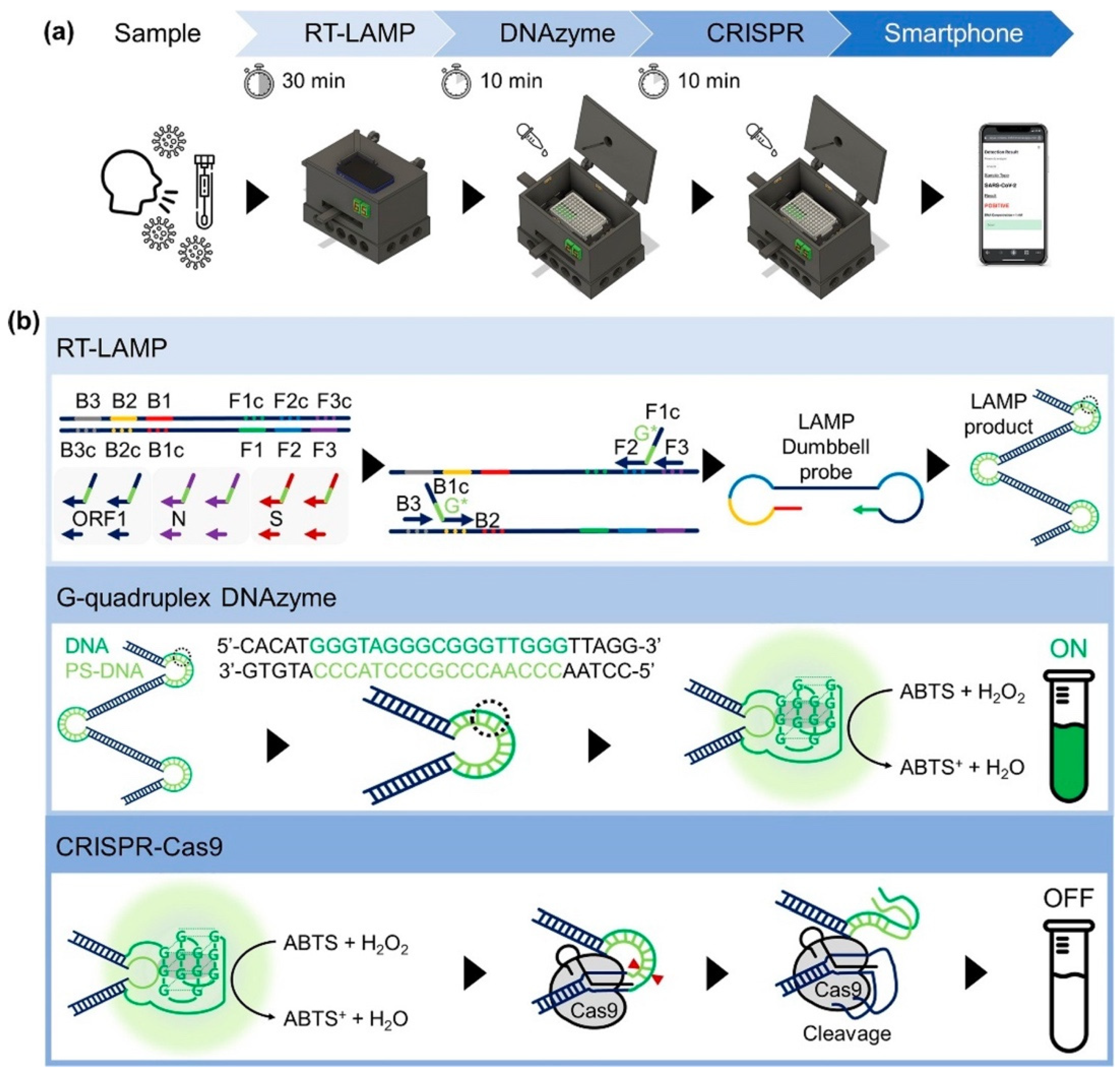
- Song, J.; Cha, B.; Moon, J.; Jang, H.; Kim, S.; Jang, J.; Yong, D.; Kwon, H.-J.; Lee, I.-C.; Lim, E.-K.; et al. Smartphone-Based SARS-CoV-2 and Variants Detection System using Colorimetric DNAzyme Reaction Triggered by Loop-Mediated Isothermal Amplification (LAMP) with Clustered Regularly Interspaced Short Palindromic Repeats (CRISPR). ACS Nano 2022, 16, 11300–11314. [Google Scholar] [CrossRef] [PubMed]
- Pang, B.; Xu, J.; Liu, Y.; Peng, H.; Feng, W.; Cao, Y.; Wu, J.; Xiao, H.; Pabbaraju, K.; Tipples, G.; et al. Isothermal Amplification and Ambient Visualization in a Single Tube for the Detection of SARS-CoV-2 Using Loop-Mediated Amplification and CRISPR Technology. Anal. Chem. 2020, 92, 16204–16212. [Google Scholar] [CrossRef] [PubMed]
- Dong, Y.; Zhao, Y.; Li, S.; Wan, Z.; Lu, R.; Yang, X.; Yu, G.; Reboud, J.; Cooper, J.M.; Tian, Z. Multiplex, real-time, point-of-care RT-LAMP for SARS-CoV-2 detection using the HFman probe. ACS Sensors 2022, 7, 730–739. [Google Scholar] [CrossRef] [PubMed]
- Ooi, K.H.; Liu, M.M.; Moo, J.R.; Nimsamer, P.; Payungporn, S.; Kaewsapsak, P.; Tan, M.H. A Sensitive and Specific Fluorescent RT-LAMP Assay for SARS-CoV-2 Detection in Clinical Samples. ACS Synth. Biol. 2022, 11, 448–463. [Google Scholar] [CrossRef]
- Rodriguez-Manzano, J.; Malpartida-Cardenas, K.; Moser, N.; Pennisi, I.; Cavuto, M.; Miglietta, L.; Moniri, A.; Penn, R.; Satta, G.; Randell, P.; et al. Handheld Point-of-Care System for Rapid Detection of SARS-CoV-2 Extracted RNA in under 20 min. ACS Cent. Sci. 2021, 7, 307–317. [Google Scholar] [CrossRef]
- Dao Thi, V.L.; Herbst, K.; Boerner, K.; Meurer, M.; Kremer, L.P.M.; Kirrmaier, D.; Freistaedter, A.; Papagiannidis, D.; Galmozzi, C.; Stanifer, M.L.; et al. A colorimetric RT-LAMP assay and LAMP-sequencing for detecting SARS-CoV-2 RNA in clinical samples. Sci. Transl. Med. 2020, 12, eabc7075. [Google Scholar] [CrossRef]
- Huang, W.E.; Lim, B.; Hsu, C.; Xiong, D.; Wu, W.; Yu, Y.; Jia, H.; Wang, Y.; Zeng, Y.; Ji, M. RT-LAMP for rapid diagnosis of coronavirus SARS-CoV-2. Microb. Biotechnol. 2020, 13, 950–961. [Google Scholar] [CrossRef]
- Diaz, L.M.; Johnson, B.E.; Jenkins, D.M. Real-time optical analysis of a colorimetric LAMP assay for SARS-CoV-2 in saliva with a handheld instrument improves accuracy compared to endpoint assessment. medRxiv 2021, preprint. [Google Scholar] [CrossRef]
- Tarim, E.A.; Oksuz, C.; Karakuzu, B.; Appak, O.; Sayiner, A.A.; Tekin, H.C. Electromechanical RT-LAMP device for portable SARS-CoV-2 detection. Talanta 2023, 254, 124190. [Google Scholar] [CrossRef] [PubMed]
- Chan, S.K.; Du, P.; Ignacio, C.; Mehta, S.; Newton, I.G.; Steinmetz, N.F. Virus-Like Particles as Positive Controls for COVID-19 RT-LAMP Diagnostic Assays. Biomacromolecules 2021, 22, 1231–1243. [Google Scholar] [CrossRef] [PubMed]
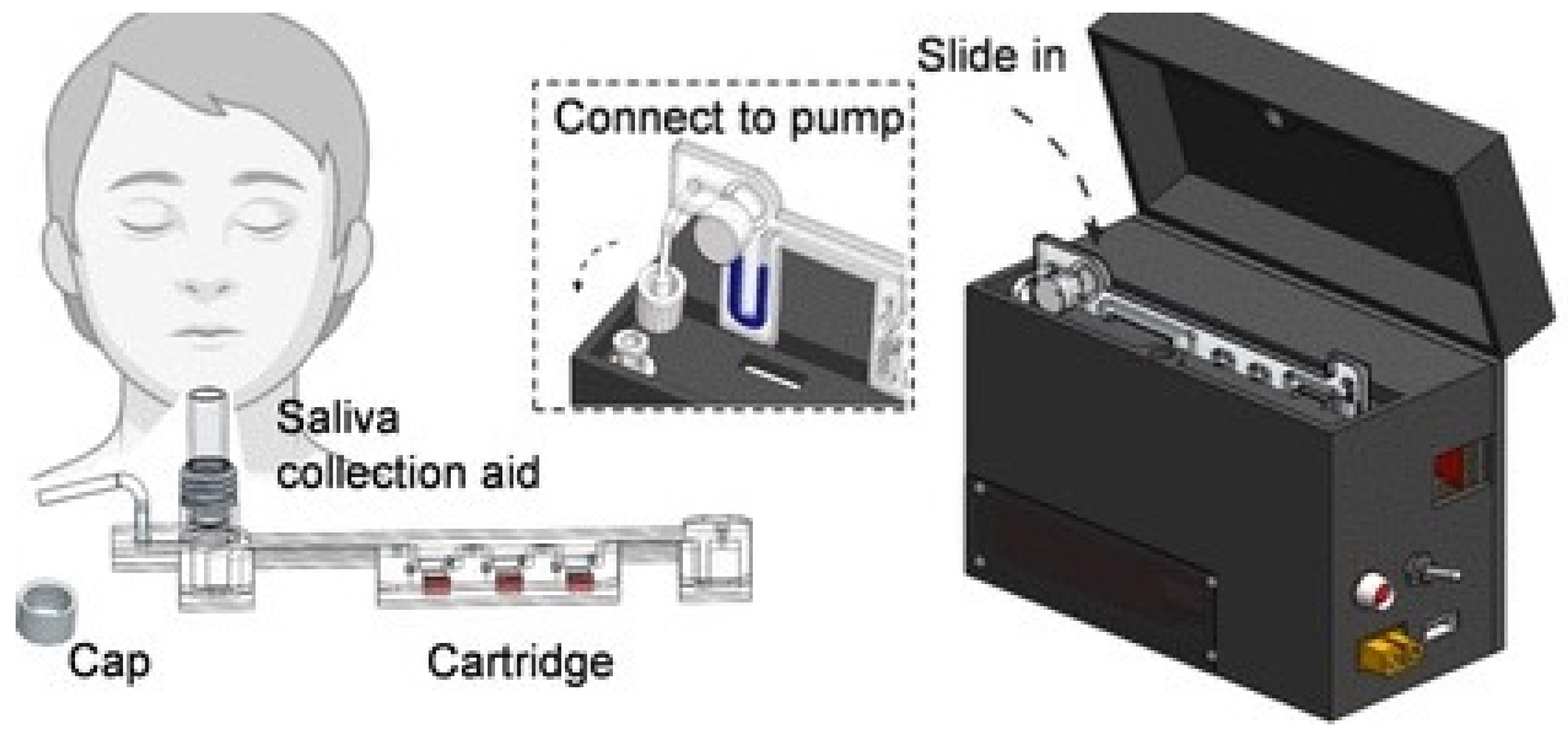
- Tang, Z.; Cui, J.; Kshirsagar, A.; Liu, T.; Yon, M.; Kuchipudi, S.V.; Guan, W. SLIDE: Saliva-Based SARS-CoV-2 Self-Testing with RT-LAMP in a Mobile Device. ACS Sens. 2022, 7, 2370–2378. [Google Scholar] [CrossRef] [PubMed]
- Ramírez-Chavarría, R.G.; Castillo-Villanueva, E.; Alvarez-Serna, B.E.; Carrillo-Reyes, J.; Ramírez-Zamora, R.M.; Buitrón, G.; Alvarez-Icaza, L. Loop-mediated isothermal amplification-based electrochemical sensor for detecting SARS-CoV-2 in wastewater samples. J. Environ. Chem. Eng. 2022, 10, 107488. [Google Scholar] [CrossRef] [PubMed]
- Lyu, W.; Zhang, J.; Yu, Y.; Xu, L.; Shen, F. Slip formation of a high-density droplet array for nucleic acid quantification by digital LAMP with a random-access system. Lab. Chip 2021, 21, 3086–3093. [Google Scholar] [CrossRef] [PubMed]
- Ganguli, A.; Mostafa, A.; Berger, J.; Aydin, M.Y.; Sun, F.; de Ramirez, S.A.S.; Valera, E.; Cunningham, B.T.; King, W.P.; Bashir, R. Rapid isothermal amplification and portable detection system for SARS-CoV-2. Proc. Natl. Acad. Sci. USA 2020, 117, 22727–22735. [Google Scholar] [CrossRef] [PubMed]
- Wang, R.; Zhao, R.; Li, Y.; Kong, W.; Guo, X.; Yang, Y.; Wu, F.; Liu, W.; Song, H.; Hao, R. Rapid detection of multiple respiratory viruses based on microfluidic isothermal amplification and a real-time colorimetric method. Lab Chip 2018, 18, 3507–3515. [Google Scholar] [CrossRef] [PubMed]
- Ma, Y.-D.; Chen, Y.-S.; Lee, G.-B. An integrated self-driven microfluidic device for rapid detection of the influenza A (H1N1) virus by reverse transcription loop-mediated isothermal amplification. Sens. Actuators B Chem. 2019, 296, 126647. [Google Scholar] [CrossRef]
- Jhou, Y.-R.; Wang, C.-H.; Tsai, H.-P.; Shan, Y.-S.; Lee, G.-B. An integrated microfluidic platform featuring real-time reverse transcription loop-mediated isothermal amplification for detection of COVID-19. Sens. Actuators B Chem. 2022, 358, 131447. [Google Scholar] [CrossRef] [PubMed]
- Kong, H.; Zhang, W.; Yao, J.; Li, C.; Lu, R.; Guo, Z.; Li, J.; Li, C.; Li, Y.; Zhang, C.; et al. A RT-LAMP based hydrogen ion selective electrode sensing for effective detection HIV-1 RNA with high-sensitivity. Sens. Actuators B Chem. 2021, 329, 129118. [Google Scholar] [CrossRef]
- Piepenburg, O.; Williams, C.H.; Stemple, D.L.; Armes, N.A. DNA detection using recombination proteins. PLoS Biol. 2006, 4, e204. [Google Scholar] [CrossRef] [PubMed]
- Crannell, Z.A.; Rohrman, B.; Richards-Kortum, R. Quantification of HIV-1 DNA using real-time recombinase polymerase amplification. Anal. Chem. 2014, 86, 5615–5619. [Google Scholar] [CrossRef] [PubMed]
- Fan, X.; Li, L.; Zhao, Y.; Liu, Y.; Liu, C.; Wang, Q.; Dong, Y.; Wang, S.; Chi, T.; Song, F. Clinical validation of two recombinase-based isothermal amplification assays (RPA/RAA) for the rapid detection of African swine fever virus. Front. Microbiol. 2020, 11, 1696. [Google Scholar] [CrossRef] [PubMed]
- Li, J.; Macdonald, J.; Von Stetten, F. A comprehensive summary of a decade development of the recombinase polymerase amplification. Analyst 2019, 144, 31–67. [Google Scholar] [CrossRef] [PubMed]
- Daher, R.K.; Stewart, G.; Boissinot, M.; Bergeron, M.G. Recombinase polymerase amplification for diagnostic applications. Clin. Chem. 2016, 62, 947–958. [Google Scholar] [CrossRef] [PubMed]
- Santiago-Felipe, S.; Tortajada-Genaro, L.A.; Puchades, R.; Maquieira, A. Recombinase polymerase and enzyme-linked immunosorbent assay as a DNA amplification-detection strategy for food analysis. Anal. Chim. Acta 2014, 811, 81–87. [Google Scholar] [CrossRef] [PubMed]
- Jiang, T.; Wang, Y.; Jiao, W.; Song, Y.; Zhao, Q.; Wang, T.; Bi, J.; Shen, A. Recombinase polymerase amplification combined with real-time fluorescent probe for Mycoplasma pneumoniae detection. J. Clin. Med. 2022, 11, 1780. [Google Scholar] [CrossRef] [PubMed]
- Choi, J.W.; Seo, W.H.; Kang, T.; Kang, T.; Chung, B.G. Droplet digital recombinase polymerase amplification for multiplexed detection of human coronavirus. Lab Chip 2023, 23, 2389–2398. [Google Scholar] [CrossRef] [PubMed]
- Dorta-Gorrín, A.; Navas-Méndez, J.; Gozalo-Margüello, M.; Miralles, L.; García-Hevia, L. Detection of SARS-CoV-2 Based on Nucleic Acid Amplification Tests (NAATs) and Its Integration into Nanomedicine and Microfluidic Devices as Point-of-Care Testing (POCT). Int. J. Mol. Sci. 2023, 24, 10233. [Google Scholar] [CrossRef] [PubMed]
- Xu, Z.; Chen, D.; Li, T.; Yan, J.; Zhu, J.; He, T.; Hu, R.; Li, Y.; Yang, Y.; Liu, M. Microfluidic space coding for multiplexed nucleic acid detection via CRISPR-Cas12a and recombinase polymerase amplification. Nat. Commun. 2022, 13, 6480. [Google Scholar] [CrossRef]
- Abd El Wahed, A.; Weidmann, M.; Hufert, F.T. Diagnostics-in-a-Suitcase: Development of a portable and rapid assay for the detection of the emerging avian influenza A (H7N9) virus. J. Clin. Virol. 2015, 69, 16–21. [Google Scholar] [CrossRef]
- Li, R.; Su, N.; Ren, X.; Sun, X.; Li, W.; Li, Y.; Li, J.; Chen, C.; Wang, H.; Lu, W. Centrifugal microfluidic-based multiplex recombinase polymerase amplification assay for rapid detection of SARS-CoV-2. Iscience 2023, 26, 106245. [Google Scholar] [CrossRef] [PubMed]
- Huang, Q.; Shan, X.; Cao, R.; Jin, X.; Lin, X.; He, Q.; Zhu, Y.; Fu, R.; Du, W.; Lv, W. Microfluidic chip with two-stage isothermal amplification method for highly sensitive parallel detection of SARS-CoV-2 and Measles Virus. Micromachines 2021, 12, 1582. [Google Scholar] [CrossRef] [PubMed]
- Qin, X.; Paul, R.; Zhou, Y.; Wu, Y.; Cheng, X.; Liu, Y. Multiplex solid-phase RPA coupled CRISPR-based visual detection of SARS-CoV-2. Biosens. Bioelectron. X 2023, 14, 100381. [Google Scholar] [CrossRef] [PubMed]
- Zhou, M.; Fan, C.; Wang, L.; Xu, T.; Zhang, X. Enhanced Isothermal Amplification for Ultrafast Sensing of SARS-CoV-2 in Microdroplets. Anal. Chem. 2022, 94, 4135–4140. [Google Scholar] [CrossRef] [PubMed]
- Kang, J.; Jang, H.; Yeom, G.; Kim, M.-G. Ultrasensitive Detection Platform of Disease Biomarkers Based on Recombinase Polymerase Amplification with H-Sandwich Aptamers. Anal. Chem. 2021, 93, 992–1000. [Google Scholar] [CrossRef] [PubMed]
- Ren, M.; Mei, H.; Zhou, J.; Zhou, M.; Han, H.; Zhao, L. Early diagnosis of rabies virus infection by RPA-CRISPR techniques in a rat model. Arch. Virol. 2021, 166, 1083–1092. [Google Scholar] [CrossRef] [PubMed]
- Park, B.J.; Park, M.S.; Lee, J.M.; Song, Y.J. Specific Detection of Influenza A and B Viruses by CRISPR-Cas12a-Based Assay. Biosensors 2021, 11, 88. [Google Scholar] [CrossRef] [PubMed]
- Lin, M.; Yue, H.; Tian, T.; Xiong, E.; Zhu, D.; Jiang, Y.; Zhou, X. Glycerol Additive Boosts 100-fold Sensitivity Enhancement for One-Pot RPA-CRISPR/Cas12a Assay. Anal. Chem. 2022, 94, 8277–8284. [Google Scholar] [CrossRef]
- Feng, W.; Peng, H.; Xu, J.; Liu, Y.; Pabbaraju, K.; Tipples, G.; Joyce, M.A.; Saffran, H.A.; Tyrrell, D.L.; Babiuk, S.; et al. Integrating Reverse Transcription Recombinase Polymerase Amplification with CRISPR Technology for the One-Tube Assay of RNA. Anal. Chem. 2021, 93, 12808–12816. [Google Scholar] [CrossRef]
- Cherkaoui, D.; Huang, D.; Miller, B.S.; Turbé, V.; McKendry, R.A. Harnessing recombinase polymerase amplification for rapid multi-gene detection of SARS-CoV-2 in resource-limited settings. Biosens. Bioelectron. 2021, 189, 113328. [Google Scholar] [CrossRef]
- Lin, C.; Chen, F.; Huang, D.; Li, W.; He, C.; Tang, Y.; Li, X.; Liu, C.; Han, L.; Yang, Y.; et al. A universal all-in-one RPA-Cas12a strategy with de novo autodesigner and its application in on-site ultrasensitive detection of DNA and RNA viruses. Biosens. Bioelectron. 2023, 239, 115609. [Google Scholar] [CrossRef]
- Kim, Y.; Yaseen, A.B.; Kishi, J.Y.; Hong, F.; Saka, S.K.; Sheng, K.; Gopalkrishnan, N.; Schaus, T.E.; Yin, P. Single-strand RPA for rapid and sensitive detection of SARS-CoV-2 RNA. MedRxiv 2020. preprint. [Google Scholar] [CrossRef]
- Ivanov, A.V.; Safenkova, I.V.; Zherdev, A.V.; Dzantiev, B.B. Nucleic acid lateral flow assay with recombinase polymerase amplification: Solutions for highly sensitive detection of RNA virus. Talanta 2020, 210, 120616. [Google Scholar] [CrossRef] [PubMed]
- Kong, M.; Li, Z.; Wu, J.; Hu, J.; Sheng, Y.; Wu, D.; Lin, Y.; Li, M.; Wang, X.; Wang, S. A wearable microfluidic device for rapid detection of HIV-1 DNA using recombinase polymerase amplification. Talanta 2019, 205, 120155. [Google Scholar] [CrossRef] [PubMed]
- Li, Z.; Uno, N.; Ding, X.; Avery, L.; Banach, D.; Liu, C. Bioinspired CRISPR-Mediated Cascade Reaction Biosensor for Molecular Detection of HIV Using a Glucose Meter. ACS Nano 2023, 17, 3966–3975. [Google Scholar] [CrossRef] [PubMed]
- Wang, H.; Dong, J.; Zhang, T.; Wang, F.; Yang, R.; Zhang, Y.; Zhao, X. A novel rapid detection of Senecavirus A using recombinase polymerase amplification (RPA) coupled with lateral flow (LF) dipstrip. Anal. Biochem. 2022, 646, 114627. [Google Scholar] [CrossRef] [PubMed]
- Wang, J.; Wang, J.; Li, R.; Liu, L.; Yuan, W. Rapid and sensitive detection of canine distemper virus by real-time reverse transcription recombinase polymerase amplification. BMC Vet. Res. 2017, 13, 241. [Google Scholar] [CrossRef] [PubMed]
- Jiao, Y.; Xu, C.; Li, J.; Gu, Y.; Xia, C.; Xie, Q.; Xie, Y.; An, M.; Xia, Z.; Wu, Y. Characterization and a RT-RPA assay for rapid detection of Chilli Veinal mottle virus (ChiVMV) in tobacco. Virol. J. 2020, 17, 33. [Google Scholar] [CrossRef] [PubMed]
- Chen, Z.; Zhang, T.; Lei, J.; Wang, Z.; Liu, P.; Zhong, K.; Chen, J.; Liu, J. Rapid, Sensitive and Simultaneous Detection of Two Wheat RNA Viruses Using Reverse Transcription Recombinase Polymerase Amplification (RT-RPA). Life 2022, 12, 1952. [Google Scholar] [CrossRef]
- Xi, Y.; Xu, C.-Z.; Xie, Z.-Z.; Zhu, D.-L.; Dong, J.-M. Rapid and visual detection of dengue virus using recombinase polymerase amplification method combined with lateral flow dipstick. Mol. Cell. Probes 2019, 46, 101413. [Google Scholar] [CrossRef]
- Song, J.; El-Tholoth, M.; Li, Y.; Graham-Wooten, J.; Liang, Y.; Li, J.; Li, W.; Weiss, S.R.; Collman, R.G.; Bau, H.H. Single-and two-stage, closed-tube, point-of-care, molecular detection of SARS-CoV-2. Anal. Chem. 2021, 93, 13063–13071. [Google Scholar] [CrossRef]
- Zhang, W.S.; Pan, J.; Li, F.; Zhu, M.; Xu, M.; Zhu, H.; Yu, Y.; Su, G. Reverse Transcription Recombinase Polymerase Amplification Coupled with CRISPR-Cas12a for Facile and Highly Sensitive Colorimetric SARS-CoV-2 Detection. Anal. Chem. 2021, 93, 4126–4133. [Google Scholar] [CrossRef] [PubMed]
- Li, X.; Zhu, S.; Zhang, X.; Ren, Y.; He, J.; Zhou, J.; Yin, L.; Wang, G.; Zhong, T.; Wang, L. Advances in the application of recombinase-aided amplification combined with CRISPR-Cas technology in quick detection of pathogenic microbes. Front. Bioeng. Biotechnol. 2023, 11, 1215466. [Google Scholar] [CrossRef] [PubMed]
- Yan, T.; Li, X.; Wang, L.; Chen, C.; Duan, S.; Qi, J.; Li, L.-X.; Ma, X. Development of a reverse transcription recombinase-aided amplification assay for the detection of coxsackievirus A10 and coxsackievirus A6 RNA. Arch. Virol. 2018, 163, 1455–1461. [Google Scholar] [CrossRef]
- Nie, M.; Zhou, Y.; Li, F.; Deng, H.; Zhao, M.; Huang, Y.; Jiang, C.; Sun, X.; Xu, Z.; Zhu, L. Epidemiological investigation of swine Japanese encephalitis virus based on RT-RAA detection method. Sci. Rep. 2022, 12, 9392. [Google Scholar] [CrossRef] [PubMed]
- Cui, H.; Tu, F.; Zhang, C.; Zhang, C.; Zhao, K.; Liu, J.; Dong, S.; Chen, L.; Liu, J.; Guo, Z. Real-time reverse transcription recombinase-aided amplification assay for rapid amplification of the N gene of SARS-CoV-2. Int. J. Mol. Sci. 2022, 23, 15269. [Google Scholar] [CrossRef] [PubMed]
- Chen, W.; Fan, J.; Li, Z.; Zhang, Y.; Qin, Y.; Wu, K.; Li, X.; Li, Y.; Fan, S.; Zhao, M. Development of recombinase aided amplification combined with disposable nucleic acid test strip for rapid detection of porcine circovirus type 2. Front. Vet. Sci. 2021, 8, 676294. [Google Scholar] [CrossRef] [PubMed]
- Tian, Y.; Fan, Z.; Zhang, X.; Xu, L.; Cao, Y.; Pan, Z.; Mo, Y.; Gao, Y.; Zheng, S.; Huang, J. CRISPR/Cas13a-Assisted Accurate and Portable Hepatitis D Virus RNA Detection: HDV RNA detection using CRISPR/Cas13a. Emerg. Microbes Infect. 2023, 12, 2276337. [Google Scholar] [CrossRef] [PubMed]
- Qin, Z.; Xiang, X.; Xue, L.; Cai, W.; Gao, J.; Yang, J.; Liang, Y.; Wang, L.; Chen, M.; Pang, R.; et al. Development of a novel RAA-based microfluidic chip for absolute quantitative detection of human norovirus. Microchem. J. 2021, 164, 106050. [Google Scholar] [CrossRef]
- Han, Y.; Li, F.; Yang, L.; Guo, X.; Dong, X.; Niu, M.; Jiang, Y.; Li, L.; Li, H.; Sun, Y. Imunocapture Magnetic Beads Enhanced and Ultrasensitive CRISPR-Cas13a-Assisted Electrochemical Biosensor for Rapid Detection of SARS-CoV-2. Biosensors 2023, 13, 597. [Google Scholar] [CrossRef]
- Qin, Z.; Xue, L.; Cai, W.; Gao, J.; Jiang, Y.; Yang, J.; Liang, Y.; Wang, L.; Zhang, J.; Hu, Y. Development of a recombinase-aided amplification assay for rapid detection of human norovirus GII. 4. BMC Infect. Dis. 2021, 21, 248. [Google Scholar] [CrossRef] [PubMed]
- Zheng, Y.-Z.; Chen, J.-T.; Li, J.; Wu, X.-J.; Wen, J.-Z.; Liu, X.-Z.; Lin, L.-Y.; Liang, X.-Y.; Huang, H.-Y.; Zha, G.-C. Reverse transcription recombinase-aided amplification assay with lateral flow dipstick assay for rapid detection of 2019 novel coronavirus. Front. Cell. Infect. Microbiol. 2021, 11, 613304. [Google Scholar] [CrossRef] [PubMed]
- Wang, H.; Zhang, Y.; Zhou, J.; Li, M.; Chen, Y.; Liu, Y.; Liu, H.; Ding, P.; Liang, C.; Zhu, X. Rapid Visual Detection of Hepatitis C Virus Using Reverse Transcription Recombinase-Aided Amplification–Lateral Flow Dipstick. Front. Cell. Infect. Microbiol. 2022, 12, 816238. [Google Scholar] [CrossRef] [PubMed]
- Xue, T.; Lu, Y.; Yang, H.; Hu, X.; Zhang, K.; Ren, Y.; Wu, C.; Xia, X.; Deng, R.; Wang, Y. Isothermal RNA Amplification for the Detection of Viable Pathogenic Bacteria to Estimate the Salmonella Virulence for Causing Enteritis. J. Agric. Food Chem. 2022, 70, 1670–1678. [Google Scholar] [CrossRef] [PubMed]
- Guoshuai, J.; Xiaomeng, X.; Zengdan, G.; Xingxing, H.; Qi, P.; Hanbing, Z.; Yi, W. A rapid and high sensitivity RNA detection based on NASBA and G4-ThT fluorescent biosensor. Sci. Rep. 2022, 12, 10076. [Google Scholar] [CrossRef] [PubMed]
- Cao, Y.; Kim, H.; Li, Y.; Kong, H.; Lemieux, B. Helicase-dependent amplification of nucleic acids. Curr. Protoc. Mol. Biol. 2013, 104, 11–15. [Google Scholar] [CrossRef] [PubMed]
- Shanmugakani, R.K.; Wu, M. An isothermal amplification-coupled dipstick for the rapid detection of COVID-19. J. Med. Microbiol. 2022, 71, 1519. [Google Scholar] [CrossRef] [PubMed]
- Lee, J.-E.; Kim, S.-A.; Park, H.-J.; Mun, H.; Ha, K.-S.; Shim, W.-B. Colorimetric detection of norovirus by helicase-dependent amplification method based on specific primers integrated with HRPzyme. Anal. Bioanal. Chem. 2022, 414, 6723–6733. [Google Scholar] [CrossRef]
- Wu, X.; Chen, C.; Xiao, X.; Deng, M.J. Development of Reverse Transcription Thermostable Helicase-Dependent DNA Amplification for the Detection of Tomato Spotted Wilt Virus. J. AOAC Int. 2016, 99, 1596–1599. [Google Scholar] [CrossRef] [PubMed]
- Li, Y.; Ma, M.; Song, Y.; Li, Y. Development of helicase-dependent amplification method for detection of Chinese sacbrood virus. Chin. J. Biol. 2014, 27, 267–271. Available online: https://www.cabidigitallibrary.org/doi/full/10.5555/20143143123. (accessed on 26 November 2023).
- Mayboroda, O.; Katakis, I.; O’Sullivan, C.K. Multiplexed isothermal nucleic acid amplification. Anal. Biochem. 2018, 545, 20–30. [Google Scholar] [CrossRef] [PubMed]
- Zhang, C.; Li, Z.; Liu, J.; Liu, C.; Zhang, H.; Lee, W.G.; Yao, C.; Guo, H.; Xu, F. Synthetic Gene Circuit-Based Assay with Multilevel Switch Enables Background-Free and Absolute Quantification of Circulating Tumor DNA. Research 2024, 6, 217. [Google Scholar] [CrossRef] [PubMed]
- Gadkar, V.J.; Goldfarb, D.M.; Gantt, S.; Tilley, P.A.G. Real-time Detection and Monitoring of Loop Mediated Amplification (LAMP) Reaction Using Self-quenching and De-quenching Fluorogenic Probes. Sci. Rep. 2018, 8, 5548. [Google Scholar] [CrossRef] [PubMed]
- Kashir, J.; Yaqinuddin, A. Loop mediated isothermal amplification (LAMP) assays as a rapid diagnostic for COVID-19. Med. Hypotheses 2020, 141, 109786. [Google Scholar] [CrossRef] [PubMed]
- Tan, M.; Liao, C.; Liang, L.; Yi, X.; Zhou, Z.; Wei, G. Recent advances in recombinase polymerase amplification: Principle, advantages, disadvantages and applications. Front. Cell. Infect. Microbiol. 2022, 12, 1744. [Google Scholar] [CrossRef] [PubMed]
- Babu, B.; Ochoa-Corona, F.M.; Paret, M.L. Recombinase polymerase amplification applied to plant virus detection and potential implications. Anal. Biochem. 2018, 546, 72–77. [Google Scholar] [CrossRef] [PubMed]
- Jaroenram, W.; Owens, L. Recombinase polymerase amplification combined with a lateral flow dipstick for discriminating between infectious Penaeus stylirostris densovirus and virus-related sequences in shrimp genome. J. Virol. Methods 2014, 208, 144–151. [Google Scholar] [CrossRef]
- Kellner, M.J.; Koob, J.G.; Gootenberg, J.S.; Abudayyeh, O.O.; Zhang, F. SHERLOCK: Nucleic acid detection with CRISPR nucleases. Nat. Protoc. 2019, 14, 2986–3012. [Google Scholar] [CrossRef]
- Barreda-García, S.; Miranda-Castro, R.; de-Los-Santos-Álvarez, N.; Miranda-Ordieres, A.J.; Lobo-Castañón, M.J. Helicase-dependent isothermal amplification: A novel tool in the development of molecular-based analytical systems for rapid pathogen detection. Anal. Bioanal. Chem. 2018, 410, 679–693. [Google Scholar] [CrossRef]
- Uyttendaele, M.R.; Debevere, J.M. LISTERIA|Listeria Monocytogenes–Detection Using NASBA (An Isothermal Nucleic Acid Amplification System); Robinson, R.K., Ed.; Elsevier: Oxford, UK, 1999; pp. 1244–1251. ISBN 978-0-12-227070-3. [Google Scholar]
- Iseki, H.; Alhassan, A.; Ohta, N.; Thekisoe, O.M.M.; Yokoyama, N.; Inoue, N.; Nambota, A.; Yasuda, J.; Igarashi, I. Development of a multiplex loop-mediated isothermal amplification (mLAMP) method for the simultaneous detection of bovine Babesia parasites. J. Microbiol. Methods 2007, 71, 281–287. [Google Scholar] [CrossRef] [PubMed]
- Fang, X.; Chen, H.; Yu, S.; Jiang, X.; Kong, J. Predicting viruses accurately by a multiplex microfluidic loop-mediated isothermal amplification chip. Anal. Chem. 2011, 83, 690–695. [Google Scholar] [CrossRef] [PubMed]
- Mahony, J.; Chong, S.; Bulir, D.; Ruyter, A.; Mwawasi, K.; Waltho, D. Multiplex loop-mediated isothermal amplification (M-LAMP) assay for the detection of influenza A/H1, A/H3 and influenza B can provide a specimen-to-result diagnosis in 40 min with single genome copy sensitivity. J. Clin. Virol. 2013, 58, 127–131. [Google Scholar] [CrossRef] [PubMed]
- Kunze, A.; Dilcher, M.; Abd El Wahed, A.; Hufert, F.; Niessner, R.; Seidel, M. On-chip isothermal nucleic acid amplification on flow-based chemiluminescence microarray analysis platform for the detection of viruses and bacteria. Anal. Chem. 2016, 88, 898–905. [Google Scholar] [CrossRef] [PubMed]
- Lau, H.Y.; Wang, Y.; Wee, E.J.H.; Botella, J.R.; Trau, M. Field demonstration of a multiplexed point-of-care diagnostic platform for plant pathogens. Anal. Chem. 2016, 88, 8074–8081. [Google Scholar] [CrossRef] [PubMed]
- Doseeva, V.; Forbes, T.; Wolff, J.; Khripin, Y.; O’Neil, D.; Rothmann, T.; Nazarenko, I. Multiplex isothermal helicase-dependent amplification assay for detection of Chlamydia trachomatis and Neisseria gonorrhoeae. Diagn. Microbiol. Infect. Dis. 2011, 71, 354–365. [Google Scholar] [CrossRef] [PubMed]
- Gines, G.; Menezes, R.; Nara, K.; Kirstetter, A.-S.; Taly, V.; Rondelez, Y. Isothermal digital detection of microRNAs using background-free molecular circuit. Sci. Adv. 2024, 6, eaay5952. [Google Scholar] [CrossRef]
- Urtel, G.; Van Der Hofstadt, M.; Galas, J.-C.; Estevez-Torres, A. rEXPAR: An isothermal amplification scheme that is robust to autocatalytic parasites. Biochemistry 2019, 58, 2675–2681. [Google Scholar] [CrossRef]
- Komiya, K.; Komori, M.; Noda, C.; Kobayashi, S.; Yoshimura, T.; Yamamura, M. Leak-free million-fold DNA amplification with locked nucleic acid and targeted hybridization in one pot. Org. Biomol. Chem. 2019, 17, 5708–5713. [Google Scholar] [CrossRef]
- Reid, M.S.; Paliwoda, R.E.; Zhang, H.; Le, X.C. Reduction of background generated from template-template hybridizations in the exponential amplification reaction. Anal. Chem. 2018, 90, 11033–11039. [Google Scholar] [CrossRef]


| Type of Virus | Target | Sample | Detection Methods | Limit of Detection (LOD) | Time (min) | Temperature (°C) | Ref. |
|---|---|---|---|---|---|---|---|
| Ebola virus | ssRNA (−) | Cell culture | Nucleic acid strip detection (NAD) | 30 copies/mL | 35 | 58 | [24] |
| African swine fever virus | dsRNA | Tissue | CRISPR-Cas12a nuclease reaction | 7 copies/μL | 30 | 37 | [25] |
| HIV | ssRNA-RT | Plasma sample | CY5 channel fluorescence | 89 copies/reaction | 50 | 64 | [26] |
| Tomato brown rugose fruit virus | ssRNA (+) | Tomato plants | Colorimetric assay | 6 copies/μL | 30 | 65 | [27] |
| Mayaro virus | ssRNA (+) | Cell culture | Fluorescence-based point-of-care device | 10 copies/reaction | 30 | 60 | [28] |
| Japanese encephalitis virus (JEV) | ssRNA (+) | Cell culture | Colorimetric assay | 1 copy/μL | 30 | 60 | [29] |
| Zika virus | ssRNA (+) | Tissue culture | Fluorescence Genie III | 6 copies/reaction | 30 | 65 | [30] |
| Zika virus | ssRNA (+) | Cell culture | Colorimetric-based toehold switch sensor | 1.7 × 10⁶ copies/mL | 30 | 37 | [31] |
| HPV | Cervical swabs | Colorimetric-based microfluidic chip | 50 copies/reaction | 60 | 68 | [32] | |
| SAR-CoV-2 virus | ssRNA (+) | Nasopharyngeal aspirates and sputum samples | Colorimetric DNAzyme reaction | 1 × 10⁵ copies/mL | 30 | 65 | [33] |
| SAR-CoV-2 virus | ssRNA (+) | Respiratory swab specimens | CRISPR-Cas12a-mediated fluorescence detection | 30 copies/μL | 40 | 60 | [34] |
| SAR-CoV-2 virus | ssRNA (+) | Nasopharyngeal swabs | High-fidelity DNA polymerase-mediated fluorescence detection | 115 copies/reaction | 30 | 60 | [35] |
| SAR-CoV-2 virus | ssRNA (+) | Nasopharyngeal and throat swabs | Cy5 channel fluorescence detection | 20 copies/reaction | 30 | 65 | [36] |
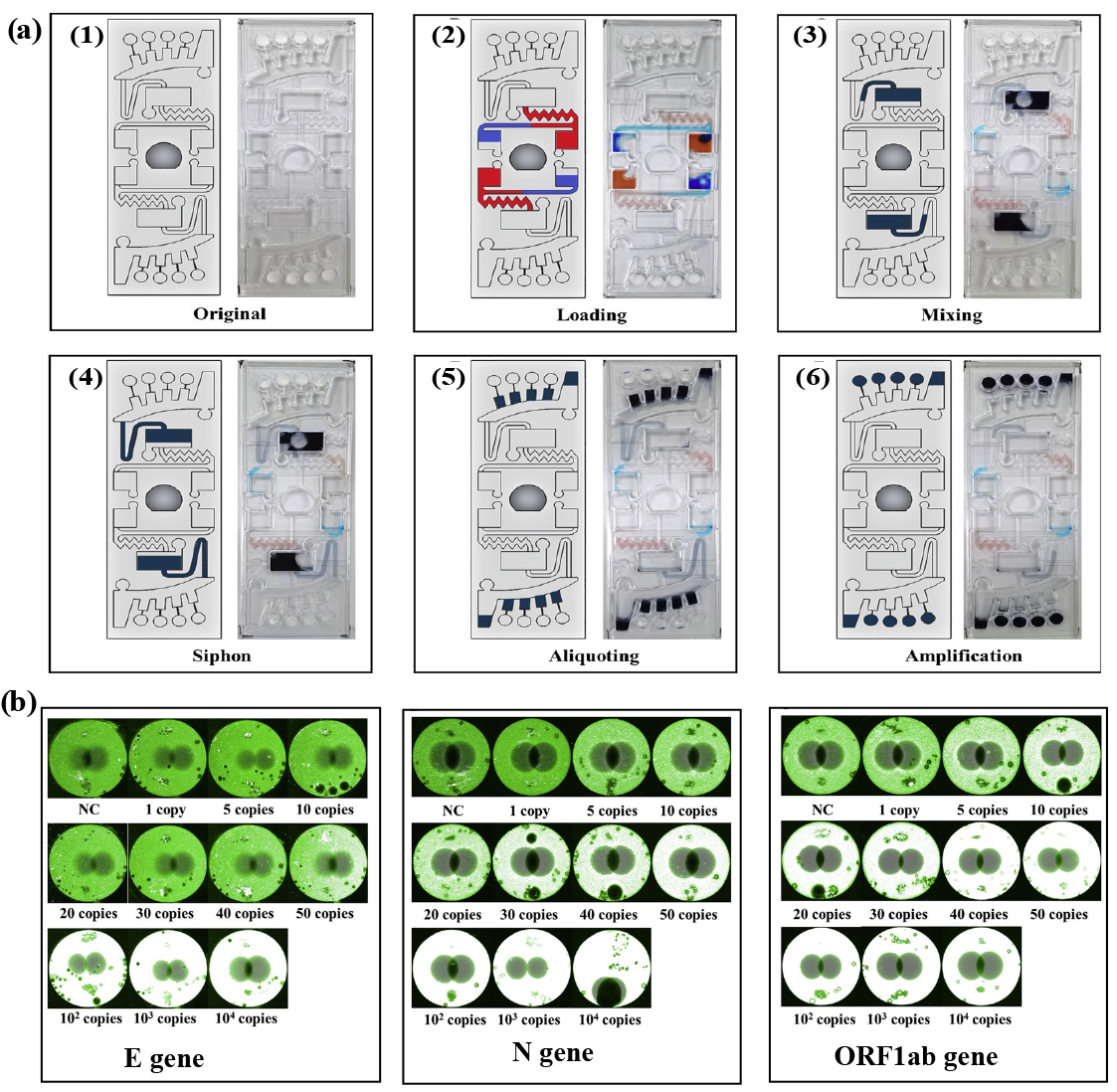
| SAR-CoV-2 virus | ssRNA (+) | Nasopharyngeal, throat, and nose swabs | Lab-on-chip platform-based fluorescence detection | 10 copies/reaction | 30 | 63 | [37] |
| SAR-CoV-2 virus | ssRNA (+) | Pharyngeal swab specimen | Colorimetric assay | 30 copies/reaction | 60 | 65 | [38] |
| SAR-CoV-2 virus | ssRNA (+) | Clinical throat swab | Colorimetric and fluorescence assay | 80 copies/mL | 30 | 65 | [39] |
| SAR-CoV-2 virus | ssRNA (+) | Nasopharyngeal swabs | Colorimetric and fluorescence assay | 50 copies/reaction | 31 | 65 | [40] |
| SAR-CoV-2 virus | ssRNA (+) | Nasopharyngeal and throat swabs | Colorimetric-based electromechanical device | 103 copies/mL | 30 | 65 | [41] |
| SAR-CoV-2 virus | ssRNA (+) | Nasopharyngeal swabs | Colorimetric assay | 0.75 copies/μL | 55 | 65 | [42] |
| SAR-CoV-2 virus | ssRNA (+) | Saliva samples | Fluorescence detection | 5 copies/μL | 45 | 65 | [43] |
| SAR-CoV-2 virus | ssRNA (+) | Wastewater samples | Colorimetric-based electrochemical sensor | 38 × 10−6 ng/μL | 30 | 60 | [44] |
| Type of Virus | Target | Sample | Detection Methods | LOD | Time (min) | Temperature (°C) | Ref. |
|---|---|---|---|---|---|---|---|
| Rabies virus | ssRNA (−) | Cell culture | IX51 Olympus fluorescence detection | 100 copies/μL | 30 | 50 | [67] |
| Influenza A (H1N1) virus | ssRNA (−) | Cell culture | CRISPR-Cas12a nuclease reaction | 10 copies/reaction | 20 | 37 | [68] |
| Influenza A (H7N9) virus | ssRNA (−) | Cell culture | Fluorescence detection | 14 copies/μL | 10 | 42 | [61] |
| Influenza A and B | ssRNA (−) | Nasal fluid samples | Fluorescence-based H-sandwich detection | 200 copies/mL | 20 | 37 | [66] |
| African swine fever virus (ASFV) | Swine sample | Glycerol-enhanced one-pot CRISP-Cas12a nuclease reaction | 10 copies/μL | 60 | 37 | [69] | |
| SAR-CoV-2 virus | ssRNA (+) | Respiratory swabs | CRISPR-Cas12a nuclease reaction | 50 copies/μL | 20 | 40 | [70] |
| SAR-CoV-2 virus | ssRNA (+) | Respiratory swabs | Real-time fluorescence and dipstick detection | 130 copies/reaction | 30 | 39 | [71] |
| SAR-CoV-2 virus | ssRNA (+) | Respiratory swabs | Cas12a nuclease reaction | 6 copies/μL | 30 | 37 | [72] |
| SAR-CoV-2 virus | ssRNA (+) | Clinical throat swab | Lateral flow | 100 copies/reaction | 5 | 42 | [73] |
| SAR-CoV-2 virus | ssRNA (+) | Clinical throat swab | CRISPR-Cas12a cleavage reaction and LED readout | 20 copies/μL | 20 | 37 | [64] |
| Potato virus Y and S | ssRNA (+) | Cell culture | Nucleic acid lateral flow | 5 × 10⁹ copies/reaction | 30 | 37 | [74] |
| HIV-1 virus | ssRNA (+) | HIV-1 integrated cells | Fluorescence-based microfluidic device | 100 copies/mL | 30 | 37 | [75] |
| HIV | ssRNA (+) | HIV clinical samples | CRISPR-mediated cascade reaction biosensor using a glucose meter | 43 copies/reaction | 60 | 40 | [76] |
| Citrus tristeza virus | ssRNA (+) | Plant samples | Lateral flow immunochromatographic assay | 3.77 × 10⁵ copies/mL | 20 | 40 | [74] |
| Senecavirus A | ssRNA (+) | Piglet blood samples | Lateral flow dipstrip | 15 copies/μL | 25 | 35 | [77] |
| Canine distemper virus | ssRNA (−) | Nasal/oropharyngeal swab | Real-time fluorescence detection | 9.4 copies/μL | 12 | 40 | [78] |
| Chilli Veinal mottle virus | ssRNA (+) | Tobacco plant | Agarose gel electrophoresis | 10 fg/μL | 20 | 38 | [79] |
| Wheat mosaic virus | ssRNA (+) | Wheat leaf | Agarose gel electrophoresis | 10−3 ng/μL | 20 | 45 | [80] |
| Dengue virus | ssRNA (+) | Clinical specimens | Lateral flow dipstick | 10 copies/μL | 15 | 37 | [81] |
| Type of Virus | Target | Sample | Detection Methods | LOD | Time (min) | Temperature (°C) | Ref. |
|---|---|---|---|---|---|---|---|
| Respiratory syncytial virus | ssRNA (−) | Nasopharyngeal aspirate | Real-time fluorescence detection | 35 copies/reaction | 30 | 39 | [16] |
| Hepatitis D virus | ssRNA (+) | Plasma sample | Fluorescence and lateral flow strip | 10 copies/μL | 30 | 39 | [89] |
| Coxsackievirus A10 and A6 | ssRNA (+) | Clinical sample | Real-time fluorescence detection | 35 copies/reaction | 30 | 39 | [85] |
| Japanese encephalitis virus | ssRNA (+) | Aborted fetuses and testicular swollen boars | Real-time fluorescence detection | 5.5 copies/μL | 30 | 39 | [86] |
| Human norovirus | ssRNA (+) | Stool clinical samples | Fluorescence-based microfluidic chip | 1.02 × 100 copies/μL | 20 | 39 | [90] |
| SAR-CoV-2 virus | ssRNA (+) | Nasopharyngeal swabs | CRISPR-Cas13a electrochemical assay | 1.66 × 101 aM | 30 | 42 | [91] |
| Platform | Manufacturer | Assay | Specimen | Analysis Time | LOD |
|---|---|---|---|---|---|
| CBDNA RT-LAMP RAPID TEST | Centrum Badan DNA, Poland | RT-LAMP | Nasopharyngeal swab | 20 | 10 copies/reaction |
| CBDNA RT-LAMP RAPID TEST | DNA Research Center Ltd., Poland | RT-LAMP | Nasal swab, nasopharyngeal swab, saliva, and throat swab | 15 | 10 copies/reaction |
| GenomtecSARS-CoV-2 EvaGreen® RT-LAMP CE-IVD Duo Kit | Genomtec SA, Poland | RT-LAMP | Nasopharyngeal swab, oropharyngeal swab, and saliva | 30 | 20 copies/reaction |
| SARS-CoV-2 EvaGreen® Direct-RT-LAMP CE-IVD Kit | Genomtec SA, Poland | RT-LAMP | Nasopharyngeal swab, oropharyngeal swab, and saliva | 40 | 2 copies/reaction (saliva)10 copies/reaction (dry swab) |
| GENEDIA W COVID-19 Colorimetric LAMP premix kit | Green Cross Medical Science Corp., South Korea | RT-LAMP | Nasopharyngeal swab and oropharyngeal swab | 32 | 100 copies/reaction |
| Hayat Rapid Colorimetric & Fluorimetric One Step LAMP SARS-CoV-2 Test Kit | Hayat Genetics Inc., Turkey | RT-LAMP | Nasal swab, nasopharyngeal swab, and saliva | 30 | 5 copies/reaction |
| COVID-19 Nucleic Acid Detection Kit (RT-LAMP) | Langfang Xinruikang Biotechnology Co. Ltd., China | RT-LAMP | Anterior nasal swab, nasopharyngeal swab, and oropharyngeal swab | 30 | 500 counts/min |
| Vivid COVID-19 LAMP Direct-G | MultiplexDX s.r.o., Slovakia | RT-LAMP | Biological fluids, sputum, throat secretion | 40 | 1000 counts/min |
| LAMPIGEN COVID19 RT-LAMP PCR KIT | Pharmaline AS, Turkey | RT-LAMP | Anterior nasal swab, nasal swab, nasopharyngeal swab, and throat swab | 30 | 3 copies/μL |
| 1copy™ COVID-19 MDx Kit Professional | 1drop Inc., South Korea | RT-LAMP | Nasopharyngeal swab | 20 | 100 copies/reaction |
| Atila iAMP COVID-19 SANO Assay | Atila BioSystems Inc., United States | RT-LAMP | Nasopharyngeal swab, oropharyngeal swab, saliva | 70 | AU 2.5 |
| BiologyWorks | BiologyWorks Inc., United States | LAMP | Anterior nasal swab, nasal swab | 45 | 30,000 counts/min |
| C4Covid-19 Human | C4Diagnostics, France | RT-LAMP | Saliva | 30 | 210,000 AU |
| DigiGENE™ COVID-19 Integrated Molecular Test | Canary Global Inc., Canada | RT-LAMP | Mid-turbinates swab, oropharyngeal swab | 20 | 600 counts/min |
| SARS-CoV-2 Nucleic Acid Detection Kit | CapitalBio Technology, China | RT-LAMP | Nasal swab, nasopharyngeal swab, throat swab | 45 | 150 counts/min |
| Dotz SARS-CoV-2 Rapid Diagnostic Kit | Dotz Nano Ltd., Israel | RT-LAMP | Nasopharyngeal swab, oropharyngeal swab, saliva | 34 | 1250 copies/reaction |
| SARS-CoV-2 POC | Enbiotech s.r.l, Italy | RT-LAMP | Nasopharyngeal swab, oropharyngeal swab | 60 | 50 copies/reaction |
| COV19-ID Kit | Genedrive, United Kingdom | RT-LAMP | Nasal swab | 17 | 500 counts/min |
| LoopDeetect COVID 19 IC | LoopDeeScience, France | RT-LAMP | Anterior nasal swab, nasal swab, nasopharyngeal swab, oropharyngeal swab | 45 | - |
| MD-Bio BCC19 Test Kit | MD-Bio Inc., United States | RT-LAMP | Nasal swab, nasopharyngeal swab | 30 | 75% |
| NaorCov19 | Rapid Diagnostic Systems Limited, Israel | RT-LAMP | Saliva | 40 | 40 copies/μL |
| Multitest COVID-19 | Selfdiagnostics Deutschland GmbH, Germany | RT-LAMP | Anterior nasal swab | 45 | - |
| Dr Vida pocket for COVID-19 | STAB VIDA, Portugal | RT-LAMP | Nasopharyngeal swab | 40 | 75 particles |
| Vicare Rapid FL | Vicare Solution GmbH, Germany | RT-LAMP | Deep (cough) sputum, nasal swab, nasopharyngeal swab, oropharyngeal swab, saliva, sputum | 30 | 150 cpu |
| SARS-CoV-2 Isothermal Amplification Detection Kit | Xiamen Jiqing Biomedical Technology Co. Ltd., China | RT-LAMP | Nasal swab, throat swab | 30 | 500 counts/min |
| ID NOWTM COVID-19 | Abbott | NEAR | nasopharyngeal swab | 15 | 125 counts/min |
| Lucira COVID-10 All-In-One Test Kit | Lucira Health | RT-LAMP | Nasal swab | 30 | 900 counts/min |
| POA-nCiVn-LFD-16 ket | BioPOA Co., Ltd. Korea | RPA | Nasopharyngeal swab and oropharyngeal swab | 20 | 20 copies/reaction |
Disclaimer/Publisher’s Note: The statements, opinions and data contained in all publications are solely those of the individual author(s) and contributor(s) and not of MDPI and/or the editor(s). MDPI and/or the editor(s) disclaim responsibility for any injury to people or property resulting from any ideas, methods, instructions or products referred to in the content. |
© 2024 by the authors. Licensee MDPI, Basel, Switzerland. This article is an open access article distributed under the terms and conditions of the Creative Commons Attribution (CC BY) license (https://creativecommons.org/licenses/by/4.0/).
Share and Cite
Ngoc, L.T.N.; Lee, Y.-C. Current Trends in RNA Virus Detection via Nucleic Acid Isothermal Amplification-Based Platforms. Biosensors 2024, 14, 97. https://doi.org/10.3390/bios14020097
Ngoc LTN, Lee Y-C. Current Trends in RNA Virus Detection via Nucleic Acid Isothermal Amplification-Based Platforms. Biosensors. 2024; 14(2):97. https://doi.org/10.3390/bios14020097
Chicago/Turabian StyleNgoc, Le Thi Nhu, and Young-Chul Lee. 2024. "Current Trends in RNA Virus Detection via Nucleic Acid Isothermal Amplification-Based Platforms" Biosensors 14, no. 2: 97. https://doi.org/10.3390/bios14020097