Recent Advances of Biosensors for Detection of Multiple Antibiotics


Abstract
:1. Introduction
2. Antibiotic Recognition Elements
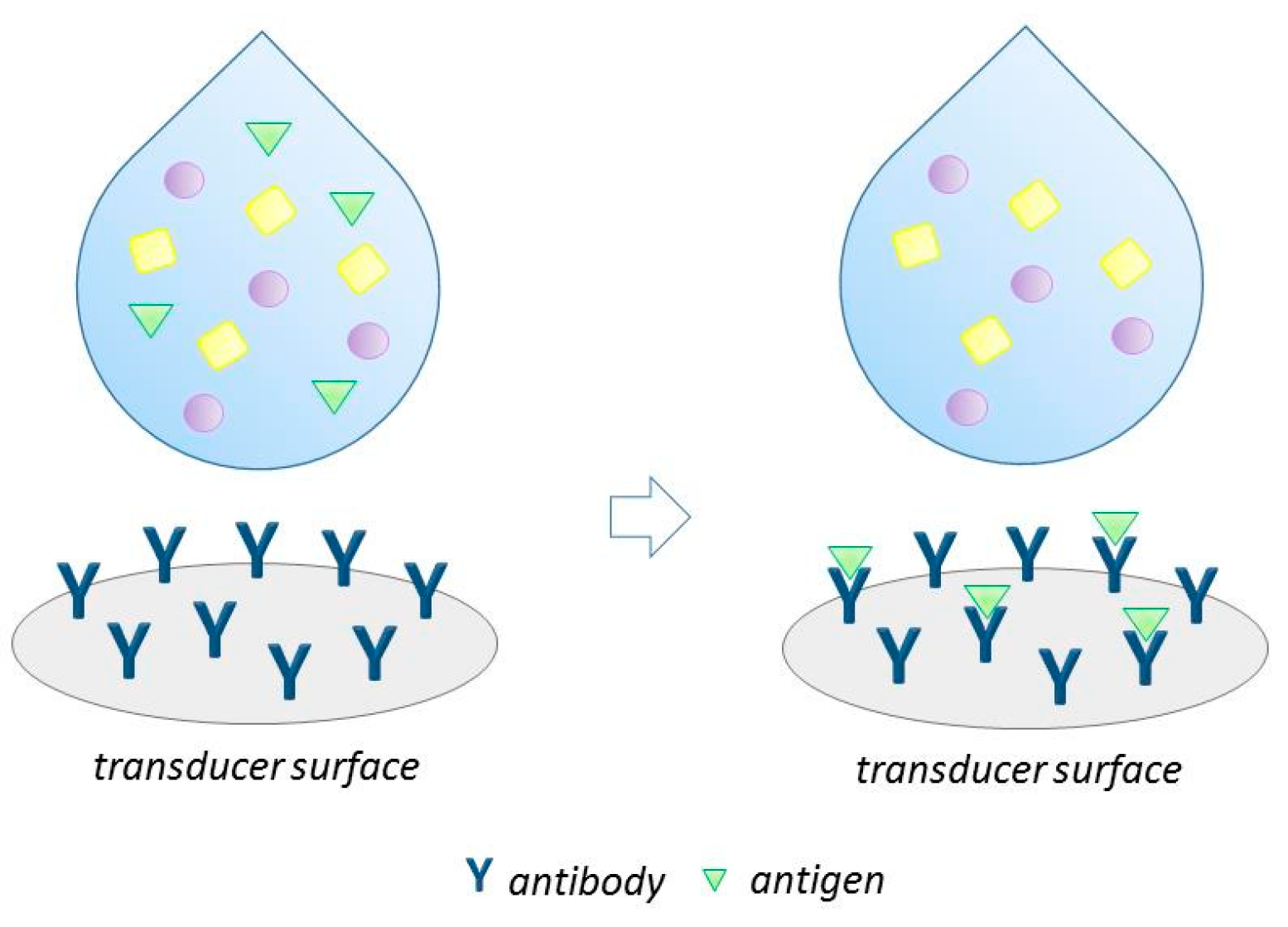
2.1. Antibody

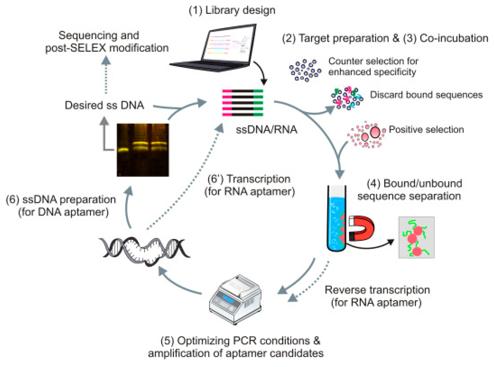
2.2. Aptamers
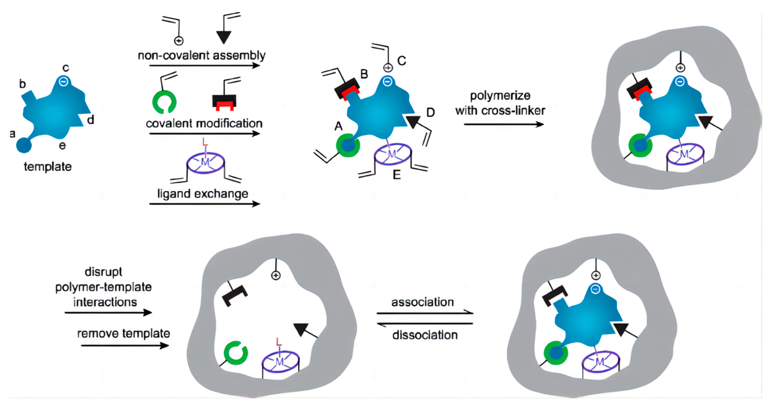
2.3. Molecularly Imprinted Polymers
3. Simultaneous Detection of Multiple Antibiotics Based on Different Methods
3.1. Fluorescence Method
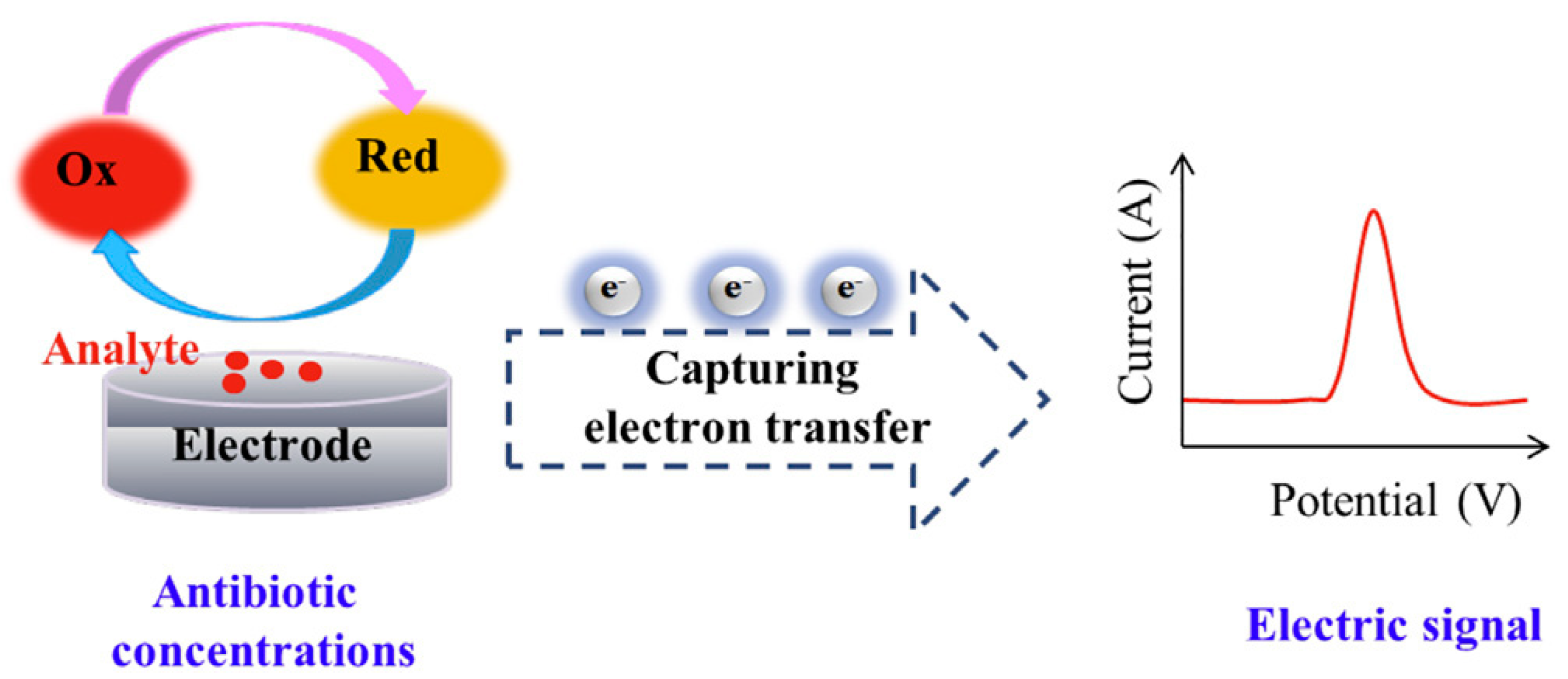
3.2. Electrochemical Method
3.3. Surface-Enhanced Raman Scattering (SERS) Method
3.4. Colorimetric Method
4. Artificial Intelligence/Machine Learning Algorithms for Antibiotic Detection
4.1. Benefits in Biosensing Antibiotics by AI/ML Algorithms
4.2. General Process and Principle of Data Analysis
4.3. Various Algorithms of AI/ML and the Application in Antibiotic Detection
5. Conclusions and Outlook
Author Contributions
Funding
Institutional Review Board Statement
Informed Consent Statement
Data Availability Statement
Conflicts of Interest
References
- Alahi, M.E.E.; Mukhopadhyay, S.C. Detection methodologies for pathogen and toxins: A review. Sensors 2017, 17, 1885. [Google Scholar] [CrossRef] [PubMed]
- Ko, E.-B.; Hwang, K.-A.; Choi, K.-C. Prenatal toxicity of the environmental pollutants on neuronal and cardiac development derived from embryonic stem cells. Reprod. Toxicol. 2019, 90, 15–23. [Google Scholar]
- Chen, C.; Shi, J.; Wang, D.; Kong, P.; Wang, Z.; Liu, Y. Antimicrobial peptides as promising antibiotic adjuvants to combat drug-resistant pathogens. Crit. Rev. Microbiol. 2023. online ahead of print. [Google Scholar] [CrossRef]
- Ridyard, K.E.; Elsawy, M.; Mattrasingh, D.; Klein, D.; Strehmel, J.; Beaulieu, C.; Wong, A.; Overhage, J. Synergy between Human Peptide LL-37 and Polymyxin B against Planktonic and Biofilm Cells of Escherichia coli and Pseudomonas aeruginosa. Antibiotics 2023, 12, 389. [Google Scholar] [CrossRef] [PubMed]
- Fungo, G.B.N.; Uy, J.C.W.; Porciuncula, K.L.J.; Candelario, C.M.A.; Chua, D.P.S.; Gutierrez, T.A.D.; Clokie, M.R.J.; Papa, D.M.D. “Two Is Better Than One”: The Multifactorial Nature of Phage-Antibiotic Combinatorial Treatments Against ESKAPE-Induced Infections. PHAGE 2023, 4, 55–67. [Google Scholar] [CrossRef] [PubMed]
- Khosla, A. Micropatternable Multifunctional Nanocomposite Polymers for Flexible Soft MEMS Applications. Ph.D. Thesis, University of Wales, Bangor, UK, 2011. [Google Scholar]
- Sharma, A.; Ahmed, A.; Singh, A.; Oruganti, S.K.; Khosla, A.; Arya, S. Recent advances in tin oxide nanomaterials as electrochemical/chemiresistive sensors. J. Electrochem. Soc. 2021, 168, 027505. [Google Scholar] [CrossRef]
- Canbaz, M.Ç.; Şimşek, Ç.S.; Sezgintürk, M.K. Electrochemical biosensor based on self-assembled monolayers modified with gold nanoparticles for detection of HER-3. Anal. Chim. Acta 2014, 814, 31–38. [Google Scholar] [CrossRef]
- Li, J.; Yang, F.; Huang, J.; Xiang, Y.; Wang, B.; Sun, X.; Liu, Y.; Kong, Q.; Chen, W.; Li, P. Novel pyramidal DNA nanostructure as a signal probe carrier platform for detection of organophosphorus pesticides. Food Anal. Methods 2022, 15, 1445–1456. [Google Scholar] [CrossRef]
- Castro, A.C.; Bezerra, Í.R.; Pascon, A.M.; Da Silva, G.H.; Philot, E.A.; De Oliveira, V.L.; Mancini, R.S.; Schleder, G.R.; Castro, C.E.; De Carvalho, L.R. Modular label-free electrochemical biosensor loading nature-inspired peptide toward the widespread use of COVID-19 antibody tests. ACS Nano 2022, 16, 14239–14253. [Google Scholar] [CrossRef]
- Srisomwat, C.; Teengam, P.; Chuaypen, N.; Tangkijvanich, P.; Vilaivan, T.; Chailapakul, O. Pop-up paper electrochemical device for label-free hepatitis B virus DNA detection. Sens. Actuators B Chem. 2020, 316, 128077. [Google Scholar] [CrossRef]
- Ahmad, R.; Mahmoudi, T.; Ahn, M.-S.; Hahn, Y.-B. Recent advances in nanowires-based field-effect transistors for biological sensor applications. Biosens. Bioelectron. 2018, 100, 312–325. [Google Scholar] [CrossRef] [PubMed]
- Hollon, T.C.; Pandian, B.; Adapa, A.R.; Urias, E.; Save, A.V.; Khalsa, S.S.S.; Eichberg, D.G.; D’Amico, R.S.; Farooq, Z.U.; Lewis, S. Near real-time intraoperative brain tumor diagnosis using stimulated Raman histology and deep neural networks. Nat. Med. 2020, 26, 52–58. [Google Scholar] [CrossRef] [PubMed]
- Cui, F.; Yue, Y.; Zhang, Y.; Zhang, Z.; Zhou, H.S. Advancing biosensors with machine learning. ACS Sens. 2020, 5, 3346–3364. [Google Scholar] [CrossRef] [PubMed]
- Vakilian, K.A.; Massah, J. A portable nitrate biosensing device using electrochemistry and spectroscopy. IEEE Sens. J. 2018, 18, 3080–3089. [Google Scholar] [CrossRef]
- Reder-Christ, K.; Bendas, G. Biosensor applications in the field of antibiotic research—A review of recent developments. Sensors 2011, 11, 9450–9466. [Google Scholar] [CrossRef]
- Wan, Y.; Deng, W.P.; Su, Y.; Zhu, X.H.; Peng, C.; Hu, H.Y.; Peng, H.Z.; Song, S.P.; Fan, C.H. Carbon nanotube-based ultrasensitive multiplexing electrochemical immunosensor for cancer biomarkers. Biosens. Bioelectron. 2011, 30, 93–99. [Google Scholar] [CrossRef]
- Song, E.; Yu, M.; Wang, Y.; Hu, W.; Cheng, D.; Swihart, M.T.; Song, Y. Multi-color quantum dot-based fluorescence immunoassay array for simultaneous visual detection of multiple antibiotic residues in milk. Biosens. Bioelectron. 2015, 72, 320–325. [Google Scholar] [CrossRef]
- Chen, Y.; Chen, Q.; Han, M.; Zhou, J.; Gong, L.; Niu, Y.; Zhang, Y.; He, L.; Zhang, L. Development and optimization of a multiplex lateral flow immunoassay for the simultaneous determination of three mycotoxins in corn, rice and peanut. Food Chem. 2016, 213, 478–484. [Google Scholar] [CrossRef]
- Zhang, X.; Yu, X.; Wen, K.; Li, C.; Mujtaba Mari, G.; Jiang, H.; Shi, W.; Shen, J.; Wang, Z. Multiplex lateral flow immunoassays based on amorphous carbon nanoparticles for detecting three fusarium mycotoxins in maize. J. Agric. Food Chem. 2017, 65, 8063–8071. [Google Scholar] [CrossRef]
- Li, Y.; Li, J.; Huang, H.; Jian, D.; Shan, Y.; Wang, S.; Liu, F. Rapid quantitative detection for multiple antibiotics in honey using a quantum dot microsphere immunochromatographic strip. Food Control 2021, 130, 108256. [Google Scholar] [CrossRef]
- Pollap, A.; Kochana, J. Electrochemical immunosensors for antibiotic detection. Biosensors 2019, 9, 61. [Google Scholar] [CrossRef] [PubMed]
- Lim, Y.; Kouzani, A.; Duan, W. Aptasensors: A review. J. Biomed. Nanotechnol. 2010, 6, 93–105. [Google Scholar] [CrossRef]
- Zhang, K.; Li, H.; Wang, W.; Cao, J.; Gan, N.; Han, H. Application of multiplexed aptasensors in food contaminants detection. ACS Sens. 2020, 5, 3721–3738. [Google Scholar] [CrossRef] [PubMed]
- Wang, T.; Chen, C.; Larcher, L.M.; Barrero, R.A.; Veedu, R.N. Three decades of nucleic acid aptamer technologies: Lessons learned, progress and opportunities on aptamer development. Biotechnol. Adv. 2019, 37, 28–50. [Google Scholar] [CrossRef] [PubMed]
- Zhu, C.; Liu, X.; Li, Y.; Yu, D.; Gao, Q.; Chen, L. Dual-ratiometric electrochemical aptasensor based on carbon nanohorns/anthraquinone-2-carboxylic acid/Au nanoparticles for simultaneous detection of malathion and omethoate. Talanta 2023, 253, 123966. [Google Scholar] [CrossRef]
- Zhu, C.; Liu, D.; Li, Y.; Ma, S.; Wang, M.; You, T. Hairpin DNA assisted dual-ratiometric electrochemical aptasensor with high reliability and anti-interference ability for simultaneous detection of aflatoxin B1 and ochratoxin A. Biosens. Bioelectron. 2021, 174, 112654. [Google Scholar] [CrossRef]
- Yang, W.; Ma, Y.; Sun, H.; Huang, C.; Shen, X. Molecularly imprinted polymers based optical fiber sensors: A review. TrAC Trends Anal. Chem. 2022, 152, 116608. [Google Scholar] [CrossRef]
- Alexander, C.; Andersson, H.S.; Andersson, L.I.; Ansell, R.J.; Kirsch, N.; Nicholls, I.A.; O’Mahony, J.; Whitcombe, M.J. Molecular imprinting science and technology: A survey of the literature for the years up to and including 2003. J. Mol. Recognit. Interdiscip. J. 2006, 19, 106–180. [Google Scholar]
- Li, W.; Zhang, Q.; Wang, Y.; Ma, Y.; Guo, Z.; Liu, Z. Controllably prepared aptamer–molecularly imprinted polymer hybrid for high-specificity and high-affinity recognition of target proteins. Anal. Chem. 2019, 91, 4831–4837. [Google Scholar] [CrossRef]
- Liu, Y.; Liu, Y.; Liu, Z.; Hill, J.P.; Alowasheeir, A.; Xu, Z.; Xu, X.; Yamauchi, Y. Ultra-durable, multi-template molecularly imprinted polymers for ultrasensitive monitoring and multicomponent quantification of trace sulfa antibiotics. J. Mater. Chem. B 2021, 9, 3192–3199. [Google Scholar] [CrossRef]
- Zhao, G.; Zhang, Y.; Sun, D.; Yan, S.; Wen, Y.; Wang, Y.; Li, G.; Liu, H.; Li, J.; Song, Z. Recent advances in molecularly imprinted polymers for antibiotic analysis. Molecules 2023, 28, 335. [Google Scholar] [CrossRef] [PubMed]
- Liao, Z.; Zhang, Y.; Li, Y.; Miao, Y.; Gao, S.; Lin, F.; Deng, Y.; Geng, L. Microfluidic chip coupled with optical biosensors for simultaneous detection of multiple analytes: A review. Biosens. Bioelectron. 2019, 126, 697–706. [Google Scholar] [CrossRef] [PubMed]
- Dincer, C.; Bruch, R.; Kling, A.; Dittrich, P.S.; Urban, G.A. Multiplexed point-of-care testing–xPOCT. Trends Biotechnol. 2017, 35, 728–742. [Google Scholar] [CrossRef] [PubMed]
- Majdinasab, M.; Hayat, A.; Marty, J.L. Aptamer-based assays and aptasensors for detection of pathogenic bacteria in food samples. TrAC Trends Anal. Chem. 2018, 107, 60–77. [Google Scholar] [CrossRef]
- Yang, Q.; Hong, J.; Wu, Y.-X.; Cao, Y.; Wu, D.; Hu, F.; Gan, N. A multicolor fluorescence nanoprobe platform using two-dimensional metal organic framework nanosheets and double stirring bar assisted target replacement for multiple bioanalytical applications. ACS Appl. Mater. Interfaces 2019, 11, 41506–41515. [Google Scholar] [CrossRef]
- Li, J.; Liu, B.; Liu, L.; Zhang, N.; Liao, Y.; Zhao, C.; Cao, M.; Zhong, Y.; Chai, D.; Chen, X. Fluorescence-based aptasensors for small molecular food contaminants: From energy transfer to optical polarization. Spectrochim. Acta Part A Mol. Biomol. Spectrosc. 2022, 285, 121872. [Google Scholar] [CrossRef]
- Stanisavljevic, M.; Krizkova, S.; Vaculovicova, M.; Kizek, R.; Adam, V. Quantum dots-fluorescence resonance energy transfer-based nanosensors and their application. Biosens. Bioelectron. 2015, 74, 562–574. [Google Scholar] [CrossRef]
- Li, Y.; Zhou, Y.; Peng, Y.; He, Y.; Shen, Y.; Wang, W.; Liu, X.; Liu, Y.; Lin, J.; Li, Y. Rapid Detection of Multiple Antibiotics in Chicken Samples via a Fluorescence Nanobiosensor Coupled with a Homemade Fluorescence Analyzer. Anal. Methods 2023, 15, 3362–3372. [Google Scholar] [CrossRef]
- Song, X.; Ding, Q.; Pu, Y.; Zhang, J.; Sun, R.; Yin, L.; Wei, W.; Liu, S. Application of the Dimeric G-Quadruplex and toehold-mediated strand displacement reaction for fluorescence biosensing of ochratoxin A. Biosens. Bioelectron. 2021, 192, 113537. [Google Scholar] [CrossRef]
- Niazi, S.; Khan, I.M.; Yu, Y.; Pasha, I.; Lv, Y.; Mohsin, A.; Mushtaq, B.S.; Wang, Z. A novel fluorescent aptasensor for aflatoxin M1 detection using rolling circle amplification and g-C3N4 as fluorescence quencher. Sens. Actuators B Chem. 2020, 315, 128049. [Google Scholar] [CrossRef]
- Wang, Q.; Xue, Q.; Chen, T.; Li, J.; Liu, Y.; Shan, X.; Liu, F.; Jia, J. Recent advances in electrochemical sensors for antibiotics and their applications. Chin. Chem. Lett. 2021, 32, 609–619. [Google Scholar] [CrossRef]
- Li, F.; Yu, Z.; Han, X.; Lai, R.Y. Electrochemical aptamer-based sensors for food and water analysis: A review. Anal. Chim. Acta 2019, 1051, 1–23. [Google Scholar] [CrossRef]
- Li, F.; Wu, Y.; Chen, D.; Guo, Y.; Wang, X.; Sun, X. Sensitive dual-labeled electrochemical aptasensor for simultaneous detection of multi-antibiotics in milk. Int. J. Hydrog. Energy 2021, 46, 23301–23309. [Google Scholar] [CrossRef]
- Vasilescu, A.; Marty, J.-L. Electrochemical aptasensors for the assessment of food quality and safety. TrAC Trends Anal. Chem. 2016, 79, 60–70. [Google Scholar] [CrossRef]
- Li, F.; Guo, Y.; Wang, X.; Sun, X. Multiplexed aptasensor based on metal ions labels for simultaneous detection of multiple antibiotic residues in milk. Biosens. Bioelectron. 2018, 115, 7–13. [Google Scholar] [CrossRef] [PubMed]
- Li, Y.; Zhang, Z.; Li, J.; Li, H.; Chen, Y.; Liu, Z. Simple, stable and sensitive electrogenerated chemiluminescence detector for high-performance liquid chromatography and its application in direct determination of multiple fluoroquinolone residues in milk. Talanta 2011, 84, 690–695. [Google Scholar] [CrossRef]
- Feng, D.; Tan, X.; Wu, Y.; Ai, C.; Luo, Y.; Chen, Q.; Han, H. Electrochemiluminecence nanogears aptasensor based on MIL-53(Fe)@CdS for multiplexed detection of kanamycin and neomycin. Biosens. Bioelectron. 2019, 129, 100–106. [Google Scholar] [CrossRef] [PubMed]
- Xue, J.; Liu, J.; Wang, C.; Tian, Y.; Zhou, N. Simultaneous electrochemical detection of multiple antibiotic residues in milk based on aptamers and quantum dots. Anal. Methods 2016, 8, 1981–1988. [Google Scholar] [CrossRef]
- Che, H.; Nie, Y.; Tian, X.; Li, Y. New method for morphological identification and simultaneous quantification of multiple tetracyclines by a white fluorescent probe. J. Hazard. Mater. 2023, 441, 129956. [Google Scholar] [CrossRef]
- Xu, X.; Gao, J.; Cao, D.; Liu, X.; Ma, M.; Zhang, L. Novel magnetic porous biochar derived from degreasing cotton as a multifunctional adsorbent for simultaneous efficient capturing and monitoring of multiple antibiotic residues. J. Environ. Chem. Eng. 2022, 10, 108377. [Google Scholar] [CrossRef]
- Reta, N.; Saint, C.P.; Michelmore, A.; Prieto-Simon, B.; Voelcker, N.H. Nanostructured electrochemical biosensors for label-free detection of water-and food-borne pathogens. ACS Appl. Mater. Interfaces 2018, 10, 6055–6072. [Google Scholar] [CrossRef] [PubMed]
- Amiri, M.; Nekoueian, K.; Saberi, R.S. Graphene-family materials in electrochemical aptasensors. Anal. Bioanal. Chem. 2021, 413, 673–699. [Google Scholar] [CrossRef] [PubMed]
- Zhao, M.; Cao, F.; Chen, J.; Hong, J.; Deng, D.; Wang, Q.; Sun, Y.; Li, Q.; Xin, H.; Wang, X. Rapid, direct, visualized and antibody-free bacterial detection with extra species identification and susceptibility evaluation capabilities. Biosens. Bioelectron. 2023, 221, 114902. [Google Scholar] [CrossRef]
- Chai, F.; Wang, D.; Zhu, L.; Zheng, W.; Jiang, X. Dual gold nanoparticle/chemiluminescent immunoassay for sensitive detection of multiple analytes. Anal. Chem. 2022, 94, 6628–6634. [Google Scholar] [CrossRef] [PubMed]
- Yang, X.; Zhao, C.; Zhang, C.; Wen, K.; Zhu, Y. Bi-directionally amplified ratiometric electrochemical aptasensor for the ultrasensitive detection of alpha-fetoprotein. Sens. Actuators B Chem. 2020, 323, 128666. [Google Scholar] [CrossRef]
- Chen, P.; Qiao, X.; Liu, J.; Xia, F.; Tian, D.; Zhou, C. Dual-signaling amplification electrochemical aptasensor based on hollow polymeric nanospheres for acetamiprid detection. ACS Appl. Mater. Interfaces 2019, 11, 14560–14566. [Google Scholar] [CrossRef]
- Li, J.; Yu, C.; Wu, Y.-n.; Zhu, Y.; Xu, J.; Wang, Y.; Wang, H.; Guo, M.; Li, F. Novel sensing platform based on gold nanoparticle-aptamer and Fe-metal-organic framework for multiple antibiotic detection and signal amplification. Environ. Int. 2019, 125, 135–141. [Google Scholar] [CrossRef]
- Chen, M.; Gan, N.; Zhou, Y.; Li, T.; Xu, Q.; Cao, Y.; Chen, Y. A novel aptamer-metal ions-nanoscale MOF based electrochemical biocodes for multiple antibiotics detection and signal amplification. Sens. Actuators B Chem. 2017, 242, 1201–1209. [Google Scholar] [CrossRef]
- Chen, M.; Gan, N.; Li, T.; Wang, Y.; Xu, Q.; Chen, Y. An electrochemical aptasensor for multiplex antibiotics detection using Y-shaped DNA-based metal ions encoded probes with NMOF substrate and CSRP target-triggered amplification strategy. Anal. Chim. Acta 2017, 968, 30–39. [Google Scholar] [CrossRef]
- Shen, Z.; He, L.; Cao, Y.; Hong, F.; Zhang, K.; Hu, F.; Lin, J.; Wu, D.; Gan, N. Multiplexed electrochemical aptasensor for antibiotics detection using metallic-encoded apoferritin probes and double stirring bars-assisted target recycling for signal amplification. Talanta 2019, 197, 491–499. [Google Scholar] [CrossRef]
- Kneipp, J.; Kneipp, H.; Kneipp, K. SERS–A single-molecule and nanoscale tool for bioanalytics. Chem. Soc. Rev. 2008, 37, 1052–1060. [Google Scholar] [CrossRef] [PubMed]
- Han, X.X.; Zhao, B.; Ozaki, Y. Surface-enhanced Raman scattering for protein detection. Anal. Bioanal. Chem. 2009, 394, 1719–1727. [Google Scholar] [CrossRef] [PubMed]
- Xu, M.-L.; Gao, Y.; Han, X.X.; Zhao, B. Detection of pesticide residues in food using surface-enhanced Raman spectroscopy: A review. J. Agric. Food Chem. 2017, 65, 6719–6726. [Google Scholar] [CrossRef] [PubMed]
- Wang, P.; Wu, L.; Lu, Z.; Li, Q.; Yin, W.; Ding, F.; Han, H. Gecko-inspired nanotentacle surface-enhanced Raman spectroscopy substrate for sampling and reliable detection of pesticide residues in fruits and vegetables. Anal. Chem. 2017, 89, 2424–2431. [Google Scholar] [CrossRef]
- Jin, M.; Wang, X.; Russel, M.; Shan, J. Towards the rapid detection of multiple antibiotics in eggs by Surface-enhanced Raman spectroscopy coupled with hollow fiber micro-extraction. Microchem. J. 2022, 181, 107743. [Google Scholar] [CrossRef]
- Majdinasab, M.; Mitsubayashi, K.; Marty, J.L. Optical and electrochemical sensors and biosensors for the detection of quinolones. Trends Biotechnol. 2019, 37, 898–915. [Google Scholar] [CrossRef]
- Zhu, D.; Liu, B.; Wei, G. Two-dimensional material-based colorimetric biosensors: A review. Biosensors 2021, 11, 259. [Google Scholar] [CrossRef]
- Yang, T.; Luo, Z.; Tian, Y.; Qian, C.; Duan, Y. Design strategies of AuNPs-based nucleic acid colorimetric biosensors. TrAC Trends Anal. Chem. 2020, 124, 115795. [Google Scholar] [CrossRef]
- Ghodake, G.; Shinde, S.; Saratale, R.G.; Kadam, A.; Saratale, G.D.; Syed, A.; Marraiki, N.; Elgorban, A.M.; Kim, D.Y. Silver nanoparticle probe for colorimetric detection of aminoglycoside antibiotics: Picomolar-level sensitivity toward streptomycin in water, serum, and milk samples. J. Sci. Food Agric. 2020, 100, 874–884. [Google Scholar] [CrossRef]
- Majdinasab, M.; Mishra, R.K.; Tang, X.; Marty, J.L. Detection of antibiotics in food: New achievements in the development of biosensors. TrAC Trends Anal. Chem. 2020, 127, 115883. [Google Scholar] [CrossRef]
- Katano, H.; Kuroda, Y.; Taira, S.; Maruyama, C.; Hamano, Y. Colorimetric Microtiter Plate Assay of Polycationic Aminoglycoside Antibiotics in Culture Broth Using Amaranth. Anal. Sci. 2017, 33, 499–503. [Google Scholar] [CrossRef] [PubMed]
- Sethu, N.; Krishnakumar, S.; Mitra, V.; Tagad, C.; Vyas, R. Design and development of a novel colorimetric assay and a portable optical system for the detection of aminoglycoside antibiotics. Sens. Actuators Rep. 2023, 5, 100151. [Google Scholar] [CrossRef]
- Kim, D.-Y.; Sharma, S.K.; Rasool, K.; Koduru, J.R.; Syed, A.; Ghodake, G. Development of Novel Peptide-Modified Silver Nanoparticle-Based Rapid Biosensors for Detecting Aminoglycoside Antibiotics. J. Agric. Food Chem. 2023. [Google Scholar] [CrossRef] [PubMed]
- Ryan, J.A. Colorimetric determination of gentamicin, kanamycin, tobramycin, and amikacin aminoglycosides with 2, 4-dinitrofluorobenzene. J. Pharm. Sci. 1984, 73, 1301–1302. [Google Scholar] [CrossRef]
- Shinde, S.K.; Kim, D.-Y.; Saratale, R.G.; Kadam, A.A.; Saratale, G.D.; Syed, A.; Bahkali, A.H.; Ghodake, G.S. Histidine Functionalized Gold Nanoparticles for Screening Aminoglycosides and Nanomolar Level Detection of Streptomycin in Water, Milk, and Whey. Chemosensors 2021, 9, 358. [Google Scholar] [CrossRef]
- Derbyshire, N.; White, S.J.; Bunka, D.H.; Song, L.; Stead, S.; Tarbin, J.; Sharman, M.; Zhou, D.; Stockley, P.G. Toggled RNA aptamers against aminoglycosides allowing facile detection of antibiotics using gold nanoparticle assays. Anal. Chem. 2012, 84, 6595–6602. [Google Scholar] [CrossRef]
- Epanchintseva, A.; Vorobjev, P.; Pyshnyi, D.; Pyshnaya, I. Fast and strong adsorption of native oligonucleotides on citrate-coated gold nanoparticles. Langmuir 2018, 34, 164–172. [Google Scholar] [CrossRef]
- Wu, Y.-Y.; Huang, P.; Wu, F.-Y. A label-free colorimetric aptasensor based on controllable aggregation of AuNPs for the detection of multiplex antibiotics. Food Chem. 2020, 304, 125377. [Google Scholar] [CrossRef]
- Kamble, K.; Sengupta, J. A comprehensive survey on emotion recognition based on electroencephalograph (EEG) signals. Multimed. Tools Appl. 2023, 82, 27269–27304. [Google Scholar] [CrossRef]
- Khare, S.K.; March, S.; Barua, P.D.; Gadre, V.M.; Acharya, U.R. Application of data fusion for automated detection of children with developmental and mental disorders: A systematic review of the last decade. Inf. Fusion 2023, 99, 101898. [Google Scholar] [CrossRef]
- Yaacob, H.; Hossain, F.; Shari, S.; Khare, S.K.; Ooi, C.P.; Acharya, U.R. Application of Artificial Intelligence Techniques for Brain-Computer Interface in Mental Fatigue Detection: A Systematic Review (2011–2022). IEEE Access 2023, 11, 74736–74758. [Google Scholar] [CrossRef]
- Rajendra Acharya, U.; Paul Joseph, K.; Kannathal, N.; Lim, C.M.; Suri, J.S. Heart rate variability: A review. Med. Biol. Eng. Comput. 2006, 44, 1031–1051. [Google Scholar] [CrossRef] [PubMed]
- Melo, M.C.; Maasch, J.R.; de la Fuente-Nunez, C. Accelerating antibiotic discovery through artificial intelligence. Commun. Biol. 2021, 4, 1050. [Google Scholar] [CrossRef] [PubMed]
- Lv, J.; Deng, S.; Zhang, L. A review of artificial intelligence applications for antimicrobial resistance. Biosaf. Health 2021, 3, 22–31. [Google Scholar] [CrossRef]
- Singh, K.P.; Gupta, S.; Singh, A.K.; Sinha, S. Experimental design and response surface modeling for optimization of Rhodamine B removal from water by magnetic nanocomposite. Chem. Eng. J. 2010, 165, 151–160. [Google Scholar] [CrossRef]
- Zhao, Y.; Zhang, H.; Li, Y.; Yu, X.; Cai, Y.; Sha, X.; Wang, S.; Zhan, Z.; Xu, J.; Liu, L. AI powered electrochemical multi-component detection of insulin and glucose in serum. Biosens. Bioelectron. 2021, 186, 113291. [Google Scholar] [CrossRef]
- Guo, Q.; Yang, X.; Chen, Z.; Wang, G.; Yao, L.; Lin, Z. Low-cost electrochemical sensor based on montmorillonite for antibiotic tetracycline hydrochloride detection. J. Mater. Sci. Mater. Electron. 2022, 33, 427–442. [Google Scholar] [CrossRef]
- Fang, G.; Lin, X.; Liang, X.; Wu, J.; Xu, W.; Hasi, W.; Dong, B. Machine Learning-Driven 3D Plasmonic Cavity-in-Cavity Surface-Enhanced Raman Scattering Platform with Triple Synergistic Enhancement Toward Label-Free Detection of Antibiotics in Milk. Small 2022, 18, 2204588. [Google Scholar] [CrossRef]
- Gutierrez, P.; Godoy, S.E.; Torres, S.; Oyarzun, P.; Sanhueza, I.; Diaz-Garcia, V.; Contreras-Trigo, B.; Coelho, P. Improved Antibiotic Detection in Raw Milk Using Machine Learning Tools over the Absorption Spectra of a Problem-Specific Nanobiosensor. Sensors 2020, 20, 4552. [Google Scholar] [CrossRef]
- Huang, Y.; Chen, J.; Duan, Q.; Feng, Y.; Luo, R.; Wang, W.; Liu, F.; Bi, S.; Lee, J. A fast antibiotic detection method for simplified pretreatment through spectra-based machine learning. Front. Environ. Sci. Eng. 2022, 16, 38. [Google Scholar] [CrossRef]
- Mandal, S.; Paul, D.; Saha, S.; Das, P. Multi-layer perceptron for detection of different class antibiotics from visual fluorescence response of a carbon nanoparticle-based multichannel array sensor. Sens. Actuators B Chem. 2022, 360, 131660. [Google Scholar] [CrossRef]
- Thrift, W.J.; Ragan, R. Quantification of analyte concentration in the single molecule regime using convolutional neural networks. Anal. Chem. 2019, 91, 13337–13342. [Google Scholar] [CrossRef] [PubMed]
- Guo, J.; Deng, H.; Liu, Q.; Chen, L.; Xiong, Z.; Shang, L. A reliable method for identification of antibiotics by terahertz spectroscopy and SVM. J. Spectrosc. 2020, 2020, 8811467. [Google Scholar] [CrossRef]
- Yang, J.; Xu, J.; Zhang, X.; Wu, C.; Lin, T.; Ying, Y. Deep learning for vibrational spectral analysis: Recent progress and a practical guide. Anal. Chim. Acta 2019, 1081, 6–17. [Google Scholar] [CrossRef]
- Hou, Y.; Aldrich, C.; Lepkova, K.; Machuca, L.; Kinsella, B. Analysis of electrochemical noise data by use of recurrence quantification analysis and machine learning methods. Electrochim. Acta 2017, 256, 337–347. [Google Scholar] [CrossRef]
- Xu, Z.; Wang, K.; Zhang, M.; Wang, T.; Du, X.; Gao, Z.; Hu, S.; Ren, X.; Feng, H. Machine learning assisted dual-emission fluorescence/colorimetric sensor array detection of multiple antibiotics under stepwise prediction strategy. Sens. Actuators B Chem. 2022, 359, 131590. [Google Scholar] [CrossRef]
- Rani, A.; Kumar, N.; Kumar, J.; Sinha, N.K. Machine learning for soil moisture assessment. In Deep Learning for Sustainable Agriculture; Elsevier: Amsterdam, The Netherlands, 2022; pp. 143–168. [Google Scholar]
- Asefpour Vakilian, K.; Massah, J. An artificial neural network approach to identify fungal diseases of cucumber (Cucumis sativus L.) plants using digital image processing. Arch. Phytopathol. Plant Prot. 2013, 46, 1580–1588. [Google Scholar] [CrossRef]
- Khayet, M.; Cojocaru, C. Artificial neural network modeling and optimization of desalination by air gap membrane distillation. Sep. Purif. Technol. 2012, 86, 171–182. [Google Scholar] [CrossRef]
- Wen, X.; Fang, J.; Diao, M.; Zhang, C. Artificial neural network modeling of dissolved oxygen in the Heihe River, Northwestern China. Environ. Monit. Assess. 2013, 185, 4361–4371. [Google Scholar] [CrossRef]
- Ali, S.; Hassan, A.; Hassan, G.; Eun, C.-H.; Bae, J.; Lee, C.H.; Kim, I.-J. Disposable all-printed electronic biosensor for instantaneous detection and classification of pathogens. Sci. Rep. 2018, 8, 5920. [Google Scholar] [CrossRef]
- Li, Z.; Li, Y. A comparative study on the prediction of the BP artificial neural network model and the ARIMA model in the incidence of AIDS. BMC Med. Inform. Decis. Mak. 2020, 20, 143. [Google Scholar] [CrossRef] [PubMed]
- Zhu, Q.; Chen, J.; Zhu, L.; Duan, X.; Liu, Y. Wind speed prediction with spatio–temporal correlation: A deep learning approach. Energies 2018, 11, 705. [Google Scholar] [CrossRef]
- Gao, M.; Qu, J.; Chen, K.; Jin, L.; Dahlgren, R.A.; Wang, H.; Tan, C.; Wang, X. Salting-out-enhanced ionic liquid microextraction with a dual-role solvent for simultaneous determination of trace pollutants with a wide polarity range in aqueous samples. Anal. Bioanal. Chem. 2017, 409, 6287–6303. [Google Scholar] [CrossRef]
- Turiel, E.; Bordin, G.; Rodríguez, A.R. Trace enrichment of (fluoro) quinolone antibiotics in surface waters by solid-phase extraction and their determination by liquid chromatography–ultraviolet detection. J. Chromatogr. A 2003, 1008, 145–155. [Google Scholar] [CrossRef] [PubMed]
- Song, X.; Zhou, T.; Li, J.; Zhang, M.; Xie, J.; He, L. Determination of ten macrolide drugs in environmental water using molecularly imprinted solid-phase extraction coupled with liquid chromatography-tandem mass spectrometry. Molecules 2018, 23, 1172. [Google Scholar] [CrossRef] [PubMed]


Disclaimer/Publisher’s Note: The statements, opinions and data contained in all publications are solely those of the individual author(s) and contributor(s) and not of MDPI and/or the editor(s). MDPI and/or the editor(s) disclaim responsibility for any injury to people or property resulting from any ideas, methods, instructions or products referred to in the content. |
© 2023 by the authors. Licensee MDPI, Basel, Switzerland. This article is an open access article distributed under the terms and conditions of the Creative Commons Attribution (CC BY) license (https://creativecommons.org/licenses/by/4.0/).
Share and Cite
Lu, N.; Chen, J.; Rao, Z.; Guo, B.; Xu, Y. Recent Advances of Biosensors for Detection of Multiple Antibiotics. Biosensors 2023, 13, 850. https://doi.org/10.3390/bios13090850
Lu N, Chen J, Rao Z, Guo B, Xu Y. Recent Advances of Biosensors for Detection of Multiple Antibiotics. Biosensors. 2023; 13(9):850. https://doi.org/10.3390/bios13090850
Chicago/Turabian StyleLu, Ning, Juntao Chen, Zhikang Rao, Boyu Guo, and Ying Xu. 2023. "Recent Advances of Biosensors for Detection of Multiple Antibiotics" Biosensors 13, no. 9: 850. https://doi.org/10.3390/bios13090850