Application of Various Optical and Electrochemical Nanobiosensors for Detecting Cancer Antigen 125 (CA-125): A Review

 , ,
, ,  and
and
Abstract
:1. Introduction
2. Optical Biosensing of CA-125
2.1. Fluorescence-Based Biosensors
Fluorescence Resonance Energy Transfer (FRET)-Based Biosensors
2.2. Chemiluminescence-Based Biosensor
2.3. Electrochemiluminescence-Based Biosensors
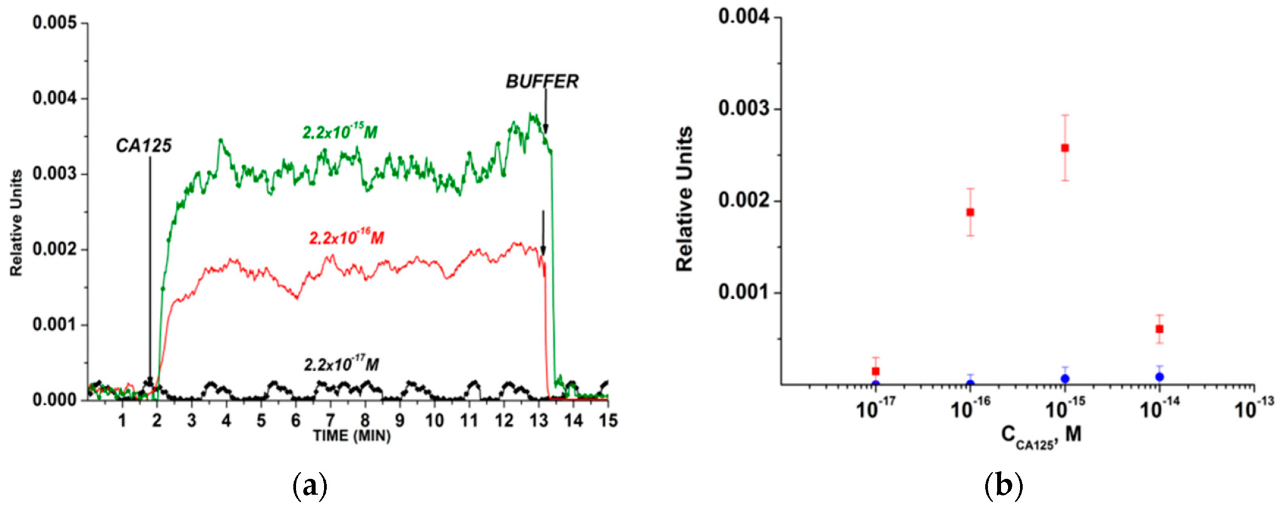
2.4. Surface Plasmon Resonance (SPR)-Based Biosensor
2.5. Surface-Enhanced Raman Scattering (SERS)-Based Biosensor
2.6. Colorimetric Biosensor
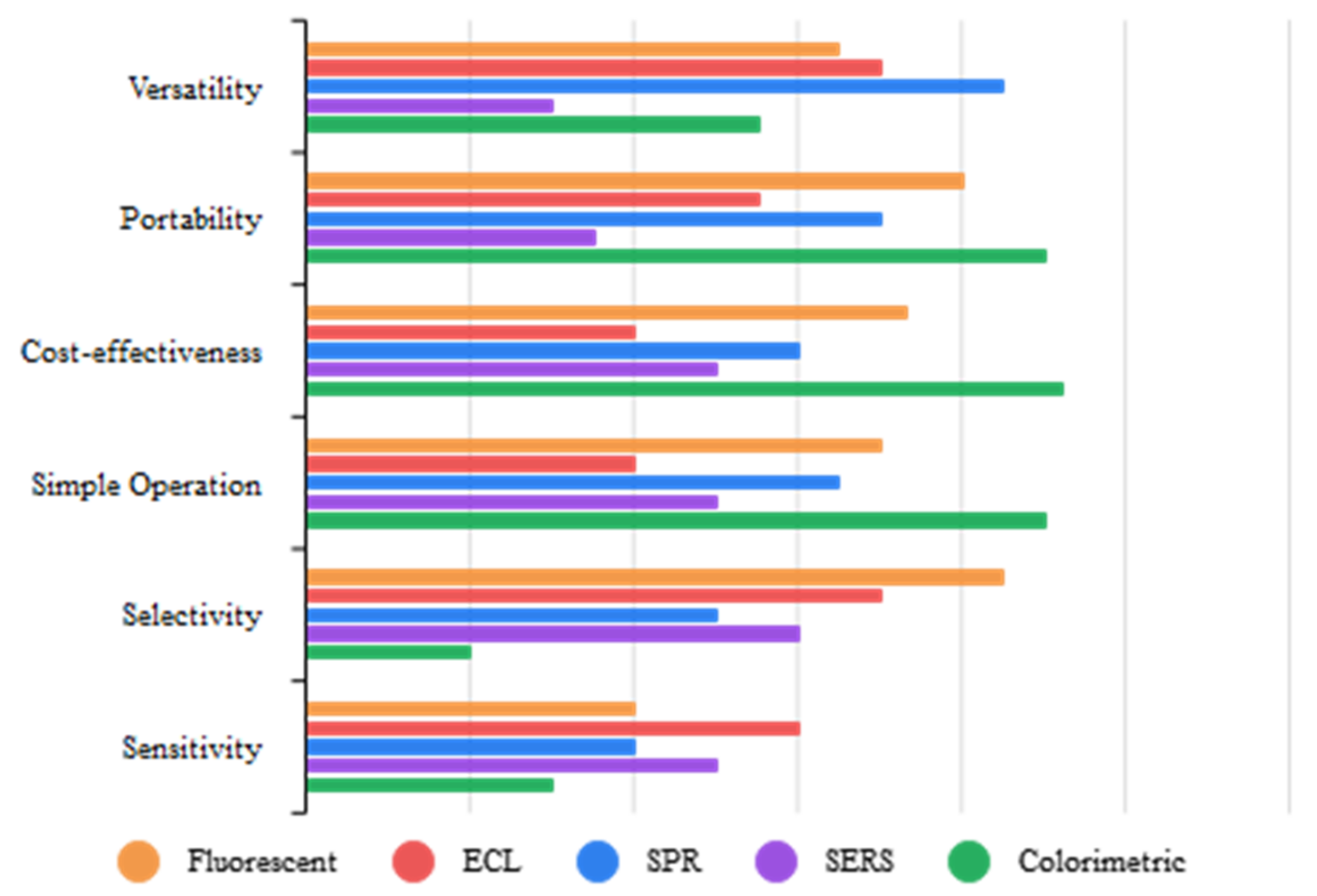
2.7. Brief Overview of Optical CA-125 Biosensors
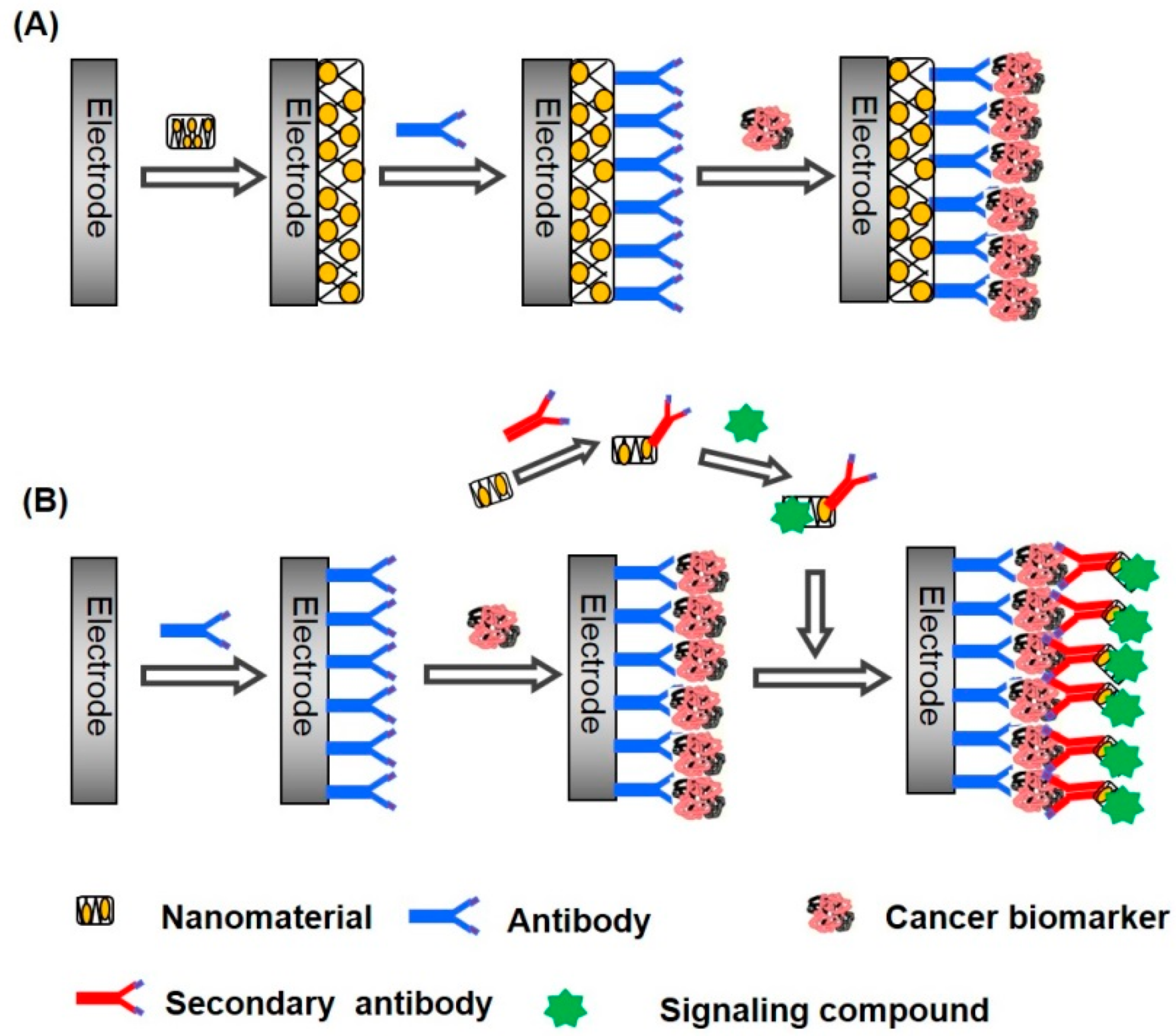
3. Electrochemical Biosensors
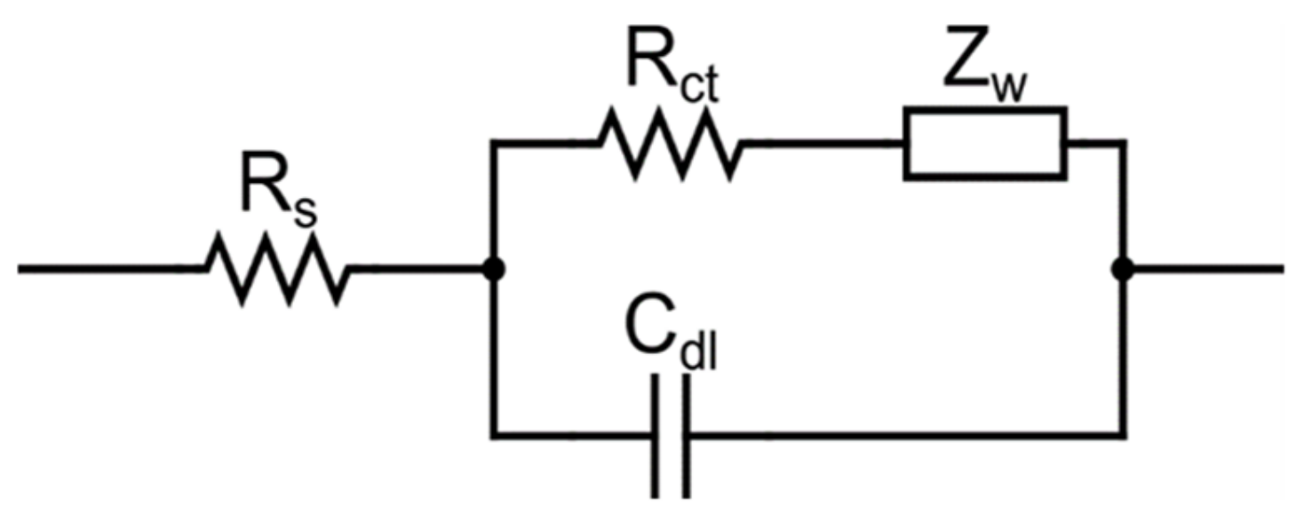
3.1. Electrical Impedance Spectroscopy-Based CA-125
3.2. Voltammetry-Based CA-125
3.3. PEC Electrochemical CA-125
3.4. Other Electrochemical CA-125
3.5. Brief Overview of Electrochemical CA-125
4. Comparison of the Performance of CA-125

| Principle | Benefits | Drawbacks | References |
|---|---|---|---|
| Optical biosensors |
|
| [18,115,116] |
| Electrochemical biosensors |
|
| [10,117,118] |

5. Comparison of CA-125 Commercial Detection Kits

6. Conclusions and Future Perspectives
Author Contributions
Funding
Conflicts of Interest
References
- Lheureux, S.; Gourley, C.; Vergote, I.; Oza, A.M. Epithelial Ovarian Cancer. Lancet 2019, 393, 1240–1253. [Google Scholar] [CrossRef] [Green Version]
- Gupta, K.K.; Gupta, V.K.; Naumann, R.W. Ovarian Cancer: Screening and Future Directions. Int. J. Gynecol. Cancer 2019, 29, 195–200. [Google Scholar] [CrossRef]
- Chandra, A.; Pius, C.; Nabeel, M.; Nair, M.; Vishwanatha, J.K.; Ahmad, S.; Basha, R. Ovarian Cancer: Current Status and Strategies for Improving Therapeutic Outcomes. Cancer Med. 2019, 8, 7018–7031. [Google Scholar] [CrossRef] [Green Version]
- Keshavarz, M.; Tan, B.; Venkatakrishnan, K. Multiplex Photoluminescent Silicon Nanoprobe for Diagnostic Bioimaging and Intracellular Analysis. Adv. Sci. 2018, 5, 1700548. [Google Scholar] [CrossRef] [Green Version]
- Charkhchi, P.; Cybulski, C.; Gronwald, J.; Wong, F.O.; Narod, S.A.; Akbari, M.R. Ca125 and Ovarian Cancer: A Comprehensive Review. Cancers 2020, 12, 3730. [Google Scholar] [CrossRef]
- Dochez, V.; Caillon, H.; Vaucel, E.; Dimet, J.; Winer, N.; Ducarme, G. Biomarkers and Algorithms for Diagnosis of Ovarian Cancer: CA125, HE4, RMI and ROMA, a Review. J. Ovarian Res. 2019, 12, 28. [Google Scholar] [CrossRef] [Green Version]
- Zhang, M.; Cheng, S.; Jin, Y.; Zhao, Y.; Wang, Y. Roles of CA125 in Diagnosis, Prediction, and Oncogenesis of Ovarian Cancer. Biochim. Biophys. Acta Rev. Cancer 2021, 1875, 188503. [Google Scholar] [CrossRef]
- Bottoni, P.; Scatena, R. The Role of CA 125 as Tumor Marker: Biochemical and Clinical Aspects. In Advances in Cancer Biomarkers; Advances in Experimental Medicine and Biology; Springer: Dordrecht, The Netherlands, 2015; Volume 867. [Google Scholar]
- De La Franier, B.; Thompson, M. Early Stage Detection and Screening of Ovarian Cancer: A Research Opportunity and Significant Challenge for Biosensor Technology. Biosens. Bioelectron. 2019, 135, 71–81. [Google Scholar] [CrossRef]
- Xiong, H.; Huang, Z.; Yang, Z.; Lin, Q.; Yang, B.; Fang, X.; Liu, B.; Chen, H.; Kong, J. Recent Progress in Detection and Profiling of Cancer Cell-Derived Exosomes. Small 2021, 17, 2007971. [Google Scholar] [CrossRef]
- Xia, L.Y.; Tang, Y.N.; Zhang, J.; Dong, T.Y.; Zhou, R.X. Advances in the DNA Nanotechnology for the Cancer Biomarkers Analysis: Attributes and Applications. Semin. Cancer Biol. 2022, 86, 1105–1119. [Google Scholar] [CrossRef] [PubMed]
- Bergua, J.F.; Álvarez-Diduk, R.; Idili, A.; Parolo, C.; Maymó, M.; Hu, L.; Merkoçi, A. Low-Cost, User-Friendly, All-Integrated Smartphone-Based Microplate Reader for Optical-Based Biological and Chemical Analyses. Anal. Chem. 2022, 94, 1271–1285. [Google Scholar] [CrossRef] [PubMed]
- Dolati, S.; Soleymani, J.; Kazem Shakouri, S.; Mobed, A. The Trends in Nanomaterial-Based Biosensors for Detecting Critical Biomarkers in Stroke. Clin. Chim. Acta 2021, 514, 107–121. [Google Scholar] [CrossRef] [PubMed]
- Hong, R.; Sun, H.; Li, D.; Yang, W.; Fan, K.; Liu, C.; Dong, L.; Wang, G. A Review of Biosensors for Detecting Tumor Markers in Breast Cancer. Life 2022, 12, 342. [Google Scholar] [CrossRef]
- Olejnik, B.; Kozioł, A.; Brzozowska, E.; Ferens-Sieczkowska, M. Application of Selected Biosensor Techniques in Clinical Diagnostics. Expert Rev. Mol. Diagn. 2021, 21, 925–937. [Google Scholar] [CrossRef]
- Huang, X.; Zhu, Y.; Kianfar, E. Nano Biosensors: Properties, Applications and Electrochemical Techniques. J. Mater. Res. Technol. 2021, 12, 1649–1672. [Google Scholar] [CrossRef]
- Sivasankarapillai, V.S.; Somakumar, A.K.; Joseph, J.; Nikazar, S.; Rahdar, A.; Kyzas, G.Z. Cancer theranostic applications of MXene nanomaterials: Recent updates. Nano-Struct. Nano-Objects 2020, 22, 100457. [Google Scholar] [CrossRef]
- Li, H. Nanomaterials-Based Biosensors for Biomarkers Detection. In Proceedings of the 2021 3rd International Academic Exchange Conference on Science and Technology Innovation, IAECST 2021, Guangzhou, China, 10–12 December 2021. [Google Scholar]
- Pourmadadi, M.; Yazdian, F.; Ghorbanian, S.; Shamsabadipour, A.; Khandel, E.; Rashedi, H.; Rahdar, A.; Díez-Pascual, A.M. Construction of Aptamer-Based Nanobiosensor for Breast Cancer Biomarkers Detection Utilizing g-C3N4/Magnetic Nano-Structure. Biosensors 2022, 12, 921. [Google Scholar] [CrossRef]
- Pourmadadi, M.; Soleimani Dinani, H.; Saeidi Tabar, F.; Khassi, K.; Janfaza, S.; Tasnim, N.; Hoorfar, M. Properties and Applications of Graphene and Its Derivatives in Biosensors for Cancer Detection: A Comprehensive Review. Biosensors 2022, 12, 269. [Google Scholar] [CrossRef]
- Dinani, H.S.; Pourmadadi, M.; Yazdian, F.; Rashedi, H.; Ebrahimi, S.A.S.; Shayeh, J.S.; Ghorbani, M. Fabrication of Au/Fe3O4/RGO Based Aptasensor for Measurement of MiRNA-128, a Biomarker for Acute Lymphoblastic Leukemia (ALL). Eng. Life Sci. 2022, 22, 519–534. [Google Scholar] [CrossRef] [PubMed]
- Triantafyllopoulos, I.K.; Papaioannou, N.A. Application of Nanotechnology in Medicine. Smart Biomaterials and Biosensors. Acta Orthop. Traumatol. Hell. 2022, 73, 3. [Google Scholar]
- Pourmadadi, M.; Rajabzadeh-Khosroshahi, M.; Saeidi Tabar, F.; Ajalli, N.; Samadi, A.; Yazdani, M.; Yazdian, F.; Rahdar, A.; Díez-Pascual, A.M. Two-Dimensional Graphitic Carbon Nitride (g-C3N4) Nanosheets and Their Derivatives for Diagnosis and Detection Applications. J. Funct. Biomater. 2022, 13, 204. [Google Scholar] [CrossRef] [PubMed]
- Cialla, D.; März, A.; Böhme, R.; Theil, F.; Weber, K.; Schmitt, M.; Popp, J. Surface-Enhanced Raman Spectroscopy (SERS): Progress and Trends. Anal. Bioanal. Chem. 2012, 403, 27–54. [Google Scholar] [CrossRef]
- Chen, Y.-T.; Lee, Y.-C.; Lai, Y.-H.; Lim, J.-C.; Huang, N.-T.; Lin, C.-T.; Huang, J.-J. Review of Integrated Optical Biosensors for Point-of-Care Applications. Biosensors 2020, 10, 209. [Google Scholar] [CrossRef] [PubMed]
- Damborský, P.; Švitel, J.; Katrlík, J. Optical Biosensors. Essays Biochem. 2016, 60, 91–100. [Google Scholar] [PubMed] [Green Version]
- Pyrak, E.; Krajczewski, J.; Kowalik, A.; Kudelski, A.; Jaworska, A. Surface Enhanced Raman Spectroscopy for DNA Biosensors—How Far Are We? Molecules 2019, 24, 4423. [Google Scholar] [CrossRef] [Green Version]
- Li, P.; Long, F.; Chen, W.; Chen, J.; Chu, P.K.; Wang, H. Fundamentals and Applications of Surface-Enhanced Raman Spectroscopy–Based Biosensors. Curr. Opin. Biomed. Eng. 2020, 13, 51–59. [Google Scholar] [CrossRef]
- Geng, Z.; Zhang, X.; Fan, Z.; Lv, X.; Su, Y.; Chen, H. Recent Progress in Optical Biosensors Based on Smartphone Platforms. Sensors 2017, 17, 2449. [Google Scholar] [CrossRef] [Green Version]
- Chen, C.; Wang, J. Optical Biosensors: An Exhaustive and Comprehensive Review. Analyst 2020, 145, 1605–1628. [Google Scholar] [CrossRef]
- Razmi, N.; Hasanzadeh, M. Current Advancement on Diagnosis of Ovarian Cancer Using Biosensing of CA 125 Biomarker: Analytical Approaches. TrAC—Trends Anal. Chem. 2018, 108, 1–12. [Google Scholar] [CrossRef]
- Sha, R.; Badhulika, S. Recent Advancements in Fabrication of Nanomaterial Based Biosensors for Diagnosis of Ovarian Cancer: A Comprehensive Review. Microchim. Acta 2020, 187, 181. [Google Scholar] [CrossRef]
- Raamanathan, A.; Simmons, G.W.; Christodoulides, N.; Floriano, P.N.; Furmaga, W.B.; Redding, S.W.; Lu, K.H.; Bast, R.C.; McDevitt, J.T. Programmable Bio-Nano-Chip Systems for Serum CA125 Quantification: Toward Ovarian Cancer Diagnostics at the Point-of-Care. Cancer Prev. Res. 2012, 5, 706–716. [Google Scholar] [CrossRef] [PubMed]
- Chakkarapani, S.K.; Zhang, P.; Ahn, S.; Kang, S.H. Total Internal Reflection Plasmonic Scattering-Based Fluorescence-Free Nanoimmunosensor Probe for Ultra-Sensitive Detection of Cancer Antigen 125. Biosens. Bioelectron. 2016, 81, 23–31. [Google Scholar] [CrossRef]
- Wang, Y.; Wang, S.; Lu, C.; Yang, X. Three Kinds of DNA-Directed Nanoclusters Cooperating with Graphene Oxide for Assaying Mucin 1, Carcinoembryonic Antigen and Cancer Antigen 125. Sens. Actuators B Chem. 2018, 262, 9–16. [Google Scholar] [CrossRef]
- Büyüktiryaki, S.; Say, R.; Denizli, A.; Ersöz, A. Phosphoserine Imprinted Nanosensor for Detection of Cancer Antigen 125. Talanta 2017, 167, 172–180. [Google Scholar] [CrossRef]
- Chen, F.; Liu, Y.; Chen, C.; Gong, H.; Cai, C.; Chen, X. Respective and Simultaneous Detection Tumor Markers CA125 and STIP1 Using Aptamer-Based Fluorescent and RLS Sensors. Sens. Actuators B Chem. 2017, 245, 470–476. [Google Scholar] [CrossRef]
- Gedi, V.; Song, C.K.; Kim, G.B.; Lee, J.O.; Oh, E.; Shin, B.S.; Jung, M.; Shim, J.; Lee, H.; Kim, Y.P. Sensitive On-Chip Detection of Cancer Antigen 125 Using a DNA Aptamer/Carbon Nanotube Network Platform. Sens. Actuators B Chem. 2018, 256, 89–97. [Google Scholar] [CrossRef]
- Pal, M.K.; Rashid, M.; Bisht, M. Multiplexed Magnetic Nanoparticle-Antibody Conjugates (MNPs-ABS) Based Prognostic Detection of Ovarian Cancer Biomarkers, CA-125, β-2M and ApoA1 Using Fluorescence Spectroscopy with Comparison of Surface Plasmon Resonance (SPR) Analysis. Biosens. Bioelectron. 2015, 73, 146–152. [Google Scholar] [CrossRef]
- Su, H.-W.; Lee, Y.-H.; Lee, M.-J.; Hsu, Y.-C.; Lee, W. Label-Free Immunodetection of the Cancer Biomarker CA125 Using High-Δn Liquid Crystals. J. Biomed. Opt. 2014, 19, 077006. [Google Scholar] [CrossRef] [PubMed] [Green Version]
- Bahari, D.; Babamiri, B.; Salimi, A. Ultrasensitive Molecularly Imprinted Fluorescence Sensor for Simultaneous Determination of CA125 and CA15–3 in Human Serum and OVCAR-3 and MCF-7 Cells Lines Using Cd and Ni Nanoclusters as New Emitters. Anal. Bioanal. Chem. 2021, 413, 4049–4061. [Google Scholar] [CrossRef]
- Liu, J.; Xu, S.; Sun, L.; Hu, S.; Sun, J.; Liu, M.; Ma, C.; Liu, H.; Wang, Z.; Yang, Y.; et al. Up-Conversion Fluorescence Biosensor for Sensitive Detection of CA-125 Tumor Markers. J. Rare Earths 2019, 37, 943–948. [Google Scholar] [CrossRef]
- Xu, X.; Ji, J.; Chen, P.; Wu, J.; Jin, Y.; Zhang, L.; Du, S. Salt-Induced Gold Nanoparticles Aggregation Lights up Fluorescence of DNA-Silver Nanoclusters to Monitor Dual Cancer Markers Carcinoembryonic Antigen and Carbohydrate Antigen 125. Anal. Chim. Acta 2020, 1125, 41–49. [Google Scholar] [CrossRef] [PubMed]
- Abou-Omar, M.N.; Attia, M.S.; Afify, H.G.; Amin, M.A.; Boukherroub, R.; Mohamed, E.H. Novel Optical Biosensor Based on a Nano-Gold Coated by Schiff Base Doped in Sol/Gel Matrix for Sensitive Screening of Oncomarker CA-125. ACS Omega 2021, 6, 20812–20821. [Google Scholar] [CrossRef] [PubMed]
- Hamd-Ghadareh, S.; Salimi, A.; Fathi, F.; Bahrami, S. An Amplified Comparative Fluorescence Resonance Energy Transfer Immunosensing of CA125 Tumor Marker and Ovarian Cancer Cells Using Green and Economic Carbon Dots for Bio-Applications in Labeling, Imaging and Sensing. Biosens. Bioelectron. 2017, 96, 308–316. [Google Scholar] [CrossRef] [PubMed]
- Ge, S.; Ge, L.; Yan, M.; Song, X.; Yu, J.; Liu, S. A Disposable Immunosensor Device for Point-of-Care Test of Tumor Marker Based on Copper-Mediated Amplification. Biosens. Bioelectron. 2013, 43, 425–431. [Google Scholar] [CrossRef] [PubMed]
- Omer, W.E.; Abdelbar, M.F.; El-Kemary, N.M.; Fukata, N.; El-Kemary, M.A. Cancer Antigen 125 Assessment Using Carbon Quantum Dots for Optical Biosensing for the Early Diagnosis of Ovarian Cancer. RSC Adv. 2021, 11, 31047–31057. [Google Scholar] [CrossRef] [PubMed]
- Jin, H.; Gui, R.; Gong, J.; Huang, W. Aptamer and 5-Fluorouracil Dual-Loading Ag2S Quantum Dots Used as a Sensitive Label-Free Probe for Near-Infrared Photoluminescence Turn-On Detection of CA125 Antigen. Biosens. Bioelectron. 2017, 92, 378–384. [Google Scholar] [CrossRef]
- Al-Ogaidi, I.; Gou, H.; Aguilar, Z.P.; Guo, S.; Melconian, A.K.; Al-Kazaz, A.K.A.; Meng, F.; Wu, N. Detection of the Ovarian Cancer Biomarker CA-125 Using Chemiluminescence Resonance Energy Transfer to Graphene Quantum Dots. Chem. Commun. 2014, 50, 1344–1346. [Google Scholar] [CrossRef]
- Yang, Z.; Xie, Z.; Liu, H.; Yan, F.; Ju, H. Streptavidin-Functionalized Three-Dimensional Ordered Nanoporous Silica Film for Highly Efficient Chemiluminescent Immunosensing. Adv. Funct. Mater. 2008, 18, 3991–3998. [Google Scholar] [CrossRef]
- Fu, Z.; Yang, Z.; Tang, J.; Liu, H.; Yan, F.; Ju, H. Channel and Substrate Zone Two-Dimensional Resolution for Chemiluminescent Multiplex Immunoassay. Anal. Chem. 2007, 79, 7376–7382. [Google Scholar] [CrossRef]
- Li, Z.; Xiao, Q.; Ying, X.; Li, Z.; Lin, J. Micro-Plate Magnetic Chemiluminescence Immunoassay of Carbohydrate Antigen 125 in Serum. Acta Chimi. Sin. 2010, 68, 162. [Google Scholar]
- Li, M.; Zhang, M.; Ge, S.; Yan, M.; Yu, J.; Huang, J.; Liu, S. Ultrasensitive Electrochemiluminescence Immunosensor Based on Nanoporous Gold Electrode and Ru-AuNPs/Graphene as Signal Labels. Sens. Actuators B Chem. 2013, 181, 50–56. [Google Scholar] [CrossRef]
- Tan, X.; Zhang, B.; Zhou, J.; Zou, G. Spectrum-Based Electrochemiluminescence Immunoassay for Selectively Determining CA125 in Greenish Waveband. ChemElectroChem 2017, 4, 1714–1718. [Google Scholar] [CrossRef]
- Babamiri, B.; Hallaj, R.; Salimi, A. Ultrasensitive Electrochemiluminescence Immunoassay for Simultaneous Determination of CA125 and CA15-3 Tumor Markers Based on PAMAM-Sulfanilic Acid-Ru(Bpy)32+ and PAMAM-CdTe@CdS Nanocomposite. Biosens. Bioelectron. 2018, 99, 353–360. [Google Scholar] [CrossRef] [PubMed]
- Gao, H.; Zhang, Z.; Zhang, Y.; Yu, H.; Rong, S.; Meng, L.; Song, S.; Mei, Y.; Pan, H.; Chang, D. Electrochemiluminescence Immunosensor for Cancer Antigen 125 Detection Based on Novel Resonance Energy Transfer between Graphitic Carbon Nitride and NIR CdTe/CdS QDs. J. Electroanal. Chem. 2021, 886, 115104. [Google Scholar] [CrossRef]
- Yin, M.; Wang, Y.; Gao, X.; Du, S.; Cheng, Y.; Yu, S.; Zou, G.; Xue, F. Electrochemiluminescence Ultrasensitive Immunoassay for Carbohydrate Antigen 125 Based on AgInS2/ZnS Nanocrystals. Anal. Bioanal. Chem. 2021, 413, 2207–2215. [Google Scholar] [CrossRef]
- Wang, S.; Ge, L.; Yan, M.; Yu, J.; Song, X.; Ge, S.; Huang, J. 3D Microfluidic Origami Electrochemiluminescence Immunodevice for Sensitive Point-of-Care Testing of Carcinoma Antigen 125. Sens. Actuators B Chem. 2013, 176, 1–8. [Google Scholar] [CrossRef]
- Liu, W.; Ma, C.; Yang, H.; Zhang, Y.; Yan, M.; Ge, S.; Yu, J.; Song, X. Electrochemiluminescence Immunoassay Using a Paper Electrode Incorporating Porous Silver and Modified with Mesoporous Silica Nanoparticles Functionalized with Blue-Luminescent Carbon Dots. Microchim. Acta 2014, 181, 1415–1422. [Google Scholar] [CrossRef]
- Xu, Q.; Li, J.; Li, S.; Pan, H. A Highly Sensitive Electrochemiluminescence Immunosensor Based on Magnetic Nanoparticles and Its Application in CA125 Determination. J. Solid State Electrochem. 2012, 16, 2891–2898. [Google Scholar] [CrossRef]
- Zhang, Y.; Li, L.; Yang, H.; Ding, Y.N.; Su, M.; Zhu, J.; Yan, M.; Yu, J.; Song, X. Gold–Silver Nanocomposite-Functionalized Graphene Sensing Platform for an Electrochemiluminescent Immunoassay of a Tumor Marker. RSC Adv. 2013, 3, 14701–14709. [Google Scholar] [CrossRef]
- Wu, L.; Sha, Y.; Li, W.; Wang, S.; Guo, Z.; Zhou, J.; Su, X.; Jiang, X. One-Step Preparation of Disposable Multi-Functionalized g-C3N4 Based Electrochemiluminescence Immunosensor for the Detection of CA125. Sens. Actuators B Chem. 2016, 226, 62–68. [Google Scholar] [CrossRef]
- Li, J.; Xu, Q.; Fu, C.; Zhang, Y. A Dramatically Enhanced Electrochemiluminescence Assay for CA125 Based on Dendrimer Multiply Labeled Luminol on Fe3O4 Nanoparticles. Sens. Actuators B Chem. 2013, 185, 146–153. [Google Scholar] [CrossRef]
- Escobedo, C.; Chou, Y.W.; Rahman, M.; Duan, X.; Gordon, R.; Sinton, D.; Brolo, A.G.; Ferreira, J. Quantification of Ovarian Cancer Markers with Integrated Microfluidic Concentration Gradient and Imaging Nanohole Surface Plasmon Resonance. Analyst 2013, 138, 1450–1458. [Google Scholar] [CrossRef]
- Szymańska, B.; Lukaszewski, Z.; Hermanowicz-Szamatowicz, K.; Gorodkiewicz, E. A Biosensor for Determination of the Circulating Biomarker CA125/MUC16 by Surface Plasmon Resonance Imaging. Talanta 2020, 206, 120187. [Google Scholar] [CrossRef] [PubMed]
- Rebelo, T.S.C.R.; Costa, R.; Brandão, A.T.S.C.; Silva, A.F.; Sales, M.G.F.; Pereira, C.M. Molecularly Imprinted Polymer SPE Sensor for Analysis of CA-125 on Serum. Anal. Chim. Acta 2019, 1082, 126–135. [Google Scholar] [CrossRef]
- Suwansa-ard, S.; Kanatharana, P.; Asawatreratanakul, P.; Wongkittisuksa, B.; Limsakul, C.; Thavarungkul, P. Comparison of Surface Plasmon Resonance and Capacitive Immunosensors for Cancer Antigen 125 Detection in Human Serum Samples. Biosens. Bioelectron. 2009, 24, 3436–3441. [Google Scholar] [CrossRef] [PubMed]
- Chang, C.C.; Chiu, N.F.; Lin, D.S.; Chu-Su, Y.; Liang, Y.H.; Lin, C.W. High-Sensitivity Detection of Carbohydrate Antigen 15-3 Using a Gold/Zinc Oxide Thin Film Surface Plasmon Resonance-Based Biosensor. Anal. Chem. 2010, 82, 1207–1212. [Google Scholar] [CrossRef] [PubMed]
- TunÇ, İ.; Susapto, H.H. Label-Free Detection of Ovarian Cancer Antigen CA125 by Surface Enhanced Raman Scattering. J. Nanosci. Nanotechnol. 2019, 20, 1358–1365. [Google Scholar] [CrossRef] [PubMed]
- Paraskevaidi, M.; Ashton, K.M.; Stringfellow, H.F.; Wood, N.J.; Keating, P.J.; Rowbottom, A.W.; Martin-Hirsch, P.L.; Martin, F.L. Raman Spectroscopic Techniques to Detect Ovarian Cancer Biomarkers in Blood Plasma. Talanta 2018, 189, 281–288. [Google Scholar] [CrossRef] [PubMed]
- Zhang, K.; Shen, X. Cancer Antigen 125 Detection Using the Plasmon Resonance Scattering Properties of Gold Nanorods. Analyst 2013, 138, 1828–1834. [Google Scholar] [CrossRef]
- Hosu, O.; Ravalli, A.; Lo Piccolo, G.M.; Cristea, C.; Sandulescu, R.; Marrazza, G. Smartphone-Based Immunosensor for CA125 Detection. Talanta 2017, 166, 234–240. [Google Scholar] [CrossRef] [PubMed]
- Zhao, Y.; Zheng, Y.; Zhao, C.; You, J.; Qu, F. Hollow PDA-Au Nanoparticles-Enabled Signal Amplification for Sensitive Nonenzymatic Colorimetric Immunodetection of Carbohydrate Antigen 125. Biosens. Bioelectron. 2015, 71, 200–206. [Google Scholar] [CrossRef]
- Ma, F.; Li, Y.; Tang, B.; Zhang, C.Y. Fluorescent Biosensors Based on Single-Molecule Counting. Acc. Chem. Res. 2016, 49, 1722–1730. [Google Scholar] [CrossRef] [PubMed]
- Kaur, B.; Kumar, S.; Kaushik, B.K. Recent Advancements in Optical Biosensors for Cancer Detection. Biosens. Bioelectron. 2022, 197, 113805. [Google Scholar] [CrossRef] [PubMed]
- Malsagova, K.A.; Pleshakova, T.O.; Galiullin, R.A.; Kozlov, A.F.; Shumov, I.D.; Popov, V.P.; Tikhonenko, F.V.; Glukhov, A.V.; Ziborov, V.S.; Petrov, O.F. Highly Sensitive Detection of CA 125 Protein with the Use of an N-Type Nanowire Biosensor. Biosensors 2020, 10, 210. [Google Scholar] [CrossRef] [PubMed]
- Sun, M.; Su, Y.; Lv, Y. Advances in Chemiluminescence and Electrogenerated Chemiluminescence Based on Silicon Nanomaterials. Luminescence 2020, 35, 978–988. [Google Scholar] [CrossRef] [PubMed]
- Sun, Y.; Lu, J. Chemiluminescence-Based Aptasensors for Various Target Analytes. Luminescence 2018, 33, 1298–1305. [Google Scholar] [CrossRef] [PubMed] [Green Version]
- Zahra, Q.U.A.; Khan, Q.A.; Luo, Z. Advances in Optical Aptasensors for Early Detection and Diagnosis of Various Cancer Types. Front. Oncol. 2021, 11, 632165. [Google Scholar] [CrossRef] [PubMed]
- Gross, E.M.; Maddipati, S.S.; Snyder, S.M. A Review of Electrogenerated Chemiluminescent Biosensors for Assays in Biological Matrices. Bioanalysis 2016, 8, 2071–2089. [Google Scholar] [CrossRef] [Green Version]
- Sina, A.A.I.; Vaidyanathan, R.; Wuethrich, A.; Carrascosa, L.G.; Trau, M. Label-Free Detection of Exosomes Using a Surface Plasmon Resonance Biosensor. Anal. Bioanal. Chem. 2019, 411, 1311–1318. [Google Scholar] [CrossRef] [Green Version]
- Unser, S.; Bruzas, I.; He, J.; Sagle, L. Localized Surface Plasmon Resonance Biosensing: Current Challenges and Approaches. Sensors 2015, 15, 15684–15716. [Google Scholar] [CrossRef]
- Choi, N.; Dang, H.; Das, A.; Sim, M.S.; Chung, I.Y.; Choo, J. SERS Biosensors for Ultrasensitive Detection of Multiple Biomarkers Expressed in Cancer Cells. Biosens. Bioelectron. 2020, 164, 112326. [Google Scholar] [CrossRef]
- Muhammad, M.; Huang, Q. A Review of Aptamer-Based SERS Biosensors: Design Strategies and Applications. Talanta 2021, 227, 122188. [Google Scholar] [CrossRef] [PubMed]
- Keshavarz, M.; Chowdhury, A.K.M.R.H.; Kassanos, P.; Tan, B.; Venkatakrishnan, K. Self-Assembled N-Doped Q-Dot Carbon Nanostructures as a SERS-Active Biosensor with Selective Therapeutic Functionality. Sens. Actuators B Chem. 2020, 323, 128703. [Google Scholar] [CrossRef]
- Zhao, V.X.T.; Wong, T.I.; Zheng, X.T.; Tan, Y.N.; Zhou, X. Colorimetric Biosensors for Point-of-Care Virus Detections. Mater. Sci. Energy Technol. 2020, 3, 237–249. [Google Scholar] [CrossRef]
- Aldewachi, H.; Chalati, T.; Woodroofe, M.N.; Bricklebank, N.; Sharrack, B.; Gardiner, P. Gold Nanoparticle-Based Colorimetric Biosensors. Nanoscale 2018, 10, 18–33. [Google Scholar] [CrossRef] [Green Version]
- Cho, I.H.; Kim, D.H.; Park, S. Electrochemical Biosensors: Perspective on Functional Nanomaterials for On-Site Analysis. Biomater. Res. 2020, 24, 6. [Google Scholar] [CrossRef] [Green Version]
- Pourmadadi, M.; Shayeh, J.S.; Omidi, M.; Yazdian, F.; Alebouyeh, M.; Tayebi, L. A Glassy Carbon Electrode Modified with Reduced Graphene Oxide and Gold Nanoparticles for Electrochemical Aptasensing of Lipopolysaccharides from Escherichia Coli Bacteria. Microchim. Acta 2019, 186, 787. [Google Scholar] [CrossRef] [PubMed]
- Kumar, S.; Kalkal, A. Electrochemical Detection: Cyclic Voltammetry/Differential Pulse Voltammetry/Impedance Spectroscopy. Nanotechnol. Cancer Manag. 2021, 43–71. [Google Scholar] [CrossRef]
- Sawhney, M.A.; Microdevices, R.S.C.-B. POISED-5, a Portable On-Board Electrochemical Impedance Spectroscopy Biomarker Analysis Device. Biomed. Microdevices 2019, 21, 70. [Google Scholar] [CrossRef] [Green Version]
- Mehdipour, G.; Shabani Shayeh, J.; Omidi, M.; Pourmadadi, M.; Yazdian, F.; Tayebi, L. An Electrochemical Aptasensor for Detection of Prostate-Specific Antigen Using Reduced Graphene Gold Nanocomposite and Cu/Carbon Quantum Dots. Biotechnol. Appl. Biochem. 2022, 69, 2102–2111. [Google Scholar] [CrossRef]
- Hu, D.; Liang, H.; Wang, X.; Luo, F.; Qiu, B.; Lin, Z.; Wang, J. Highly Sensitive and Selective Photoelectrochemical Aptasensor for Cancer Biomarker CA125 Based on AuNPs/GaN Schottky Junction. Anal. Chem. 2020, 92, 10114–10120. [Google Scholar] [CrossRef] [PubMed]
- Xue, H.; Zhao, J.; Zhou, Q.; Pan, D.; Zhang, Y.; Zhang, Y.; Shen, Y. Boosting the Sensitivity of a Photoelectrochemical Immunoassay by Using SiO2@polydopamine Core-Shell Nanoparticles as a Highly Efficient Quencher. ACS Appl. Nano Mater. 2019, 2, 1579–1588. [Google Scholar] [CrossRef]
- Zou, K.; Fu, Y.; Yang, R.; Zhang, X.; Du, C.; Chen, J. CuO–ZnO Heterojunction Derived from Cu2+-Doped ZIF-8: A New Photoelectric Material for Ultrasensitive PEC Immunoassay of CA125 with Near-Zero Background Noise. Anal. Chim. Acta 2020, 1099, 75–84. [Google Scholar] [CrossRef]
- Pourmadadi, M.; Shayeh, J.S.; Arjmand, S.; Omidi, M.; Fatemi, F. An Electrochemical Sandwich Immunosensor of Vascular Endothelial Growth Factor Based on Reduced Graphene Oxide/Gold Nanoparticle Composites. Microchem. J. 2020, 159, 105476. [Google Scholar] [CrossRef]
- Saadati, A.; Hassanpour, S.; Bahavarnia, F.; Hasanzadeh, M. A Novel Biosensor for the Monitoring of Ovarian Cancer Tumor Protein CA 125 in Untreated Human Plasma Samples Using a Novel Nano-Ink: A New Platform for Efficient Diagnosis of Cancer Using Paper Based Microfluidic Technology. Anal. Methods 2020, 12, 1639–1649. [Google Scholar] [CrossRef]
- Nunna, B.B.; Mandal, D.; Lee, J.U.; Zhuang, S.; Lee, E.S. Sensitivity Study of Cancer Antigens (CA-125) Detection Using Interdigitated Electrodes Under Microfluidic Flow Condition. BioNanoScience 2019, 9, 203–214. [Google Scholar] [CrossRef]
- Fatima, B.; Hussain, D.; Bashir, S.; Hussain, H.T.; Aslam, R.; Nawaz, R.; Rashid, H.N.; Bashir, N.; Majeed, S.; Ashiq, M.N.; et al. Catalase Immobilized Antimonene Quantum Dots Used as an Electrochemical Biosensor for Quantitative Determination of H2O2 from CA-125 Diagnosed Ovarian Cancer Samples. Mater. Sci. Eng. C 2020, 117, 111296. [Google Scholar] [CrossRef]
- Kivrak, H.; Er, O.F.; Ozok, O.; Celik, S.; Kivrak, A. Synthesis and Characterization of 4-(2-(4-Methoxyphenyl)Benzo[b]Thiophen-3-Yl)Benzaldehyde for Carbohydrate Antigen 125 Electrochemical Detection and Molecular Docking Modeling. Mater. Chem. Phys. 2022, 281, 125951. [Google Scholar] [CrossRef]
- Pourmadadi, M.; Eshaghi, M.M.; Ostovar, S.; Shamsabadipour, A.; Safakhah, S.; Mousavi, M.S.; Rahdar, A.; Pandey, S. UiO-66 Metal-Organic Framework Nanoparticles as Gifted MOFs to the Biomedical Application: A Comprehensive Review. J. Drug Deliv. Sci. Technol. 2022, 76, 103758. [Google Scholar] [CrossRef]
- Sangili, A.; Kalyani, T.; Chen, S.M.; Nanda, A.; Jana, S.K. Label-Free Electrochemical Immunosensor Based on One-Step Electrochemical Deposition of AuNP-RGO Nanocomposites for Detection of Endometriosis Marker CA 125. ACS Appl. Bio Mater. 2020, 3, 7620–7630. [Google Scholar] [CrossRef]
- Rafique, S.; Tabassum, S.; Akram, R. Sensitive Competitive Label-Free Electrochemical Immunosensor for Primal Detection of Ovarian Cancer. Chem. Pap. 2020, 74, 2591–2603. [Google Scholar] [CrossRef]
- Biswas, S.; Lan, Q.; Xie, Y.; Sun, X.; Wang, Y. Label-Free Electrochemical Immunosensor for Ultrasensitive Detection of Carbohydrate Antigen 125 Based on Antibody-Immobilized Biocompatible MOF-808/CNT. ACS Appl. Mater. Interfaces 2021, 13, 3295–3302. [Google Scholar] [CrossRef]
- Nunna, B.B.; Mandal, D.; Lee, J.U.; Singh, H.; Zhuang, S.; Misra, D.; Bhuyian, M.N.U.; Lee, E.S. Detection of Cancer Antigens (CA-125) Using Gold Nano Particles on Interdigitated Electrode-Based Microfluidic Biosensor. Nano Converg. 2019, 6, 3. [Google Scholar] [CrossRef] [PubMed]
- Bordbar, M.M.; Samadinia, H.; Sheini, A.; Halabian, R.; Parvin, S.; Ghanei, M.; Bagheri, H. A Colorimetric Electronic Tongue Based on Bi-Functionalized AuNPs for Fingerprint Detection of Cancer Markers. Sens. Actuators B Chem. 2022, 368, 132170. [Google Scholar] [CrossRef]
- Gasparotto, G.; Costa, J.P.C.; Costa, P.I.; Zaghete, M.A.; Mazon, T. Electrochemical Immunosensor Based on ZnO Nanorods-Au Nanoparticles Nanohybrids for Ovarian Cancer Antigen CA-125 Detection. Mater. Sci. Eng. C 2017, 76, 1240–1247. [Google Scholar] [CrossRef] [Green Version]
- Shen, R.; Zhang, J.; Huang, W.; Wu, S.; Li, G.; Zou, S.; Ling, L. Dynamic Light Scattering and Fluorescence Dual-Signal Sensing of Cancer Antigen-125 via Recognition of the Polymerase Chain Reaction Product with Gold Nanoparticle Probe. Anal. Chim. Acta 2021, 1145, 87–94. [Google Scholar] [CrossRef]
- Heidari, F.; Mohajeri, N.; Zarghami, N. Targeted Design of Green Carbon Dot-CA-125 Aptamer Conjugate for the Fluorescence Imaging of Ovarian Cancer Cell. Cell Biochem. Biophys. 2022, 80, 75–88. [Google Scholar] [CrossRef] [PubMed]
- Zhang, S.; Rong, F.; Guo, C.; Duan, F.; He, L.; Wang, M.; Zhang, Z.; Kang, M.; Du, M. Metal–Organic Frameworks (MOFs) Based Electrochemical Biosensors for Early Cancer Diagnosis In Vitro. Coord. Chem. Rev. 2021, 439, 213948. [Google Scholar] [CrossRef]
- Kang, S.-W.; Rainczuk, A.; Oehler, M.K.; Jobling, T.W.; Plebanski, M.; Stephens, A.N. Active Ratio Test (Art) as a Novel Diagnostic for Ovarian Cancer. Diagnostics 2021, 11, 1048. [Google Scholar] [CrossRef]
- Stabile, G.; Zinicola, G.; Romano, F.; Laganà, A.S.; Dal Pozzolo, C.; Ricci, G. Pelvic Mass, Ascites, Hydrothorax: A Malignant or Benign Condition? Meigs Syndrome with High Levels of CA 125. Menopause Rev./Przegląd Menopauzalny 2021, 20, 103–107. [Google Scholar] [CrossRef]
- Schobert, I.T.; Savic, L.J. Current Trends in Non-Invasive Imaging of Interactions in the Liver Tumor Microenvironment Mediated by Tumor Metabolism. Cancers 2021, 13, 3645. [Google Scholar] [CrossRef] [PubMed]
- Pansare, K.; Krishna, C.M. Monitoring Therapeutic Response in Cancers: A Raman Spectroscopy Approach. Recent Adv. Anal. Tech. 2022, 5, 192–275. [Google Scholar]
- Carneiro, M.C.C.G.; Rodrigues, L.R.; Moreira, F.T.C.; Sales, M.G.F. Colorimetric Paper-Based Sensors against Cancer Biomarkers. Sensors 2022, 22, 3221. [Google Scholar] [CrossRef] [PubMed]
- Valerievich Yumashev, A.; Rudiansyah, M.; Chupradit, S.; Kadhim, M.M.; Turki Jalil, A.; Abdelbasset, W.K.; Suksatan, W.; Mireya Romero Parra, R.; Fakri Mustafa, Y.; Abdullaev, B.; et al. Optical-Based Biosensor for Detection of Oncomarker CA 125, Recent Progress and Current Status. Anal. Biochem. 2022, 655, 114750. [Google Scholar] [CrossRef] [PubMed]
- Kant, T.; Shrivas, K.; Dewangan, K.; Kumar, A.; Jaiswal, N.K.; Deb, M.K.; Pervez, S. Design and Development of Conductive Nanomaterials for Electrochemical Sensors: A Modern Approach. Mater. Today Chem. 2022, 24, 100769. [Google Scholar] [CrossRef]
- Hussain, S.H.; Huertas, C.S.; Mitchell, A.; Deman, A.-L.; Laurenceau, E. Biosensors for Circulating Tumor Cells (CTCs)-Biomarker Detection in Lung and Prostate Cancer: Trends and Prospects. Biosens. Bioelectron. 2022, 197, 113770. [Google Scholar] [CrossRef] [PubMed]
- Wang, B.; Akiba, U.; Anzai, J. Recent Progress in Nanomaterial-Based Electrochemical Biosensors for Cancer Biomarkers: A Review. Molecules 2017, 22, 1048. [Google Scholar] [CrossRef] [Green Version]
- Nawaz, S.; Ahmad, B.; Ahmad, S.; Khan, S.A.; Ali, S. A Comparative Study of the IMMULITE and Enzyme Linked Immuno Sorbent Assay for Measuring Tumor Marker CA-125 in Ovarian Cancer Patients in Khyber Pakhtunkhwa. Int. J. Biosci. 2020, 16, 242–252. [Google Scholar]
- Metkar, S.K.; Girigoswami, K. Diagnostic Biosensors in Medicine—A Review. Biocatal. Agric. Biotechnol. 2019, 17, 271–283. [Google Scholar] [CrossRef]
- Novodchuk, I.; Bajcsy, M.; Yavuz, M. Graphene-Based Field Effect Transistor Biosensors for Breast Cancer Detection: A Review on Biosensing Strategies. Carbon 2021, 172, 431–453. [Google Scholar] [CrossRef]
- Sadighbayan, D.; Sadighbayan, K.; Tohid-Kia, M.R.; Khosroushahi, A.Y.; Hasanzadeh, M. Development of Electrochemical Biosensors for Tumor Marker Determination towards Cancer Diagnosis: Recent Progress. TrAC Trends Anal. Chem. 2019, 118, 73–88. [Google Scholar] [CrossRef]
- Ouyang, N.; Hong, L.; Zhou, Y.; Zhang, J.; Shafi, S.; Pan, J.; Zhao, R.; Yang, Y.; Hou, W. Application of Fluorescent Nano-Biosensor for the Detection of Cancer Bio-Macromolecular Markers. Polym. Test. 2022, 115, 107746. [Google Scholar] [CrossRef]
- Sok, D.; Clarizia, L.-J.A.; Farris, L.R.; McDonald, M.J. Novel Fluoroimmunoassay for Ovarian Cancer Biomarker CA-125. Anal. Bioanal. Chem. 2009, 393, 1521–1523. [Google Scholar] [CrossRef] [PubMed]
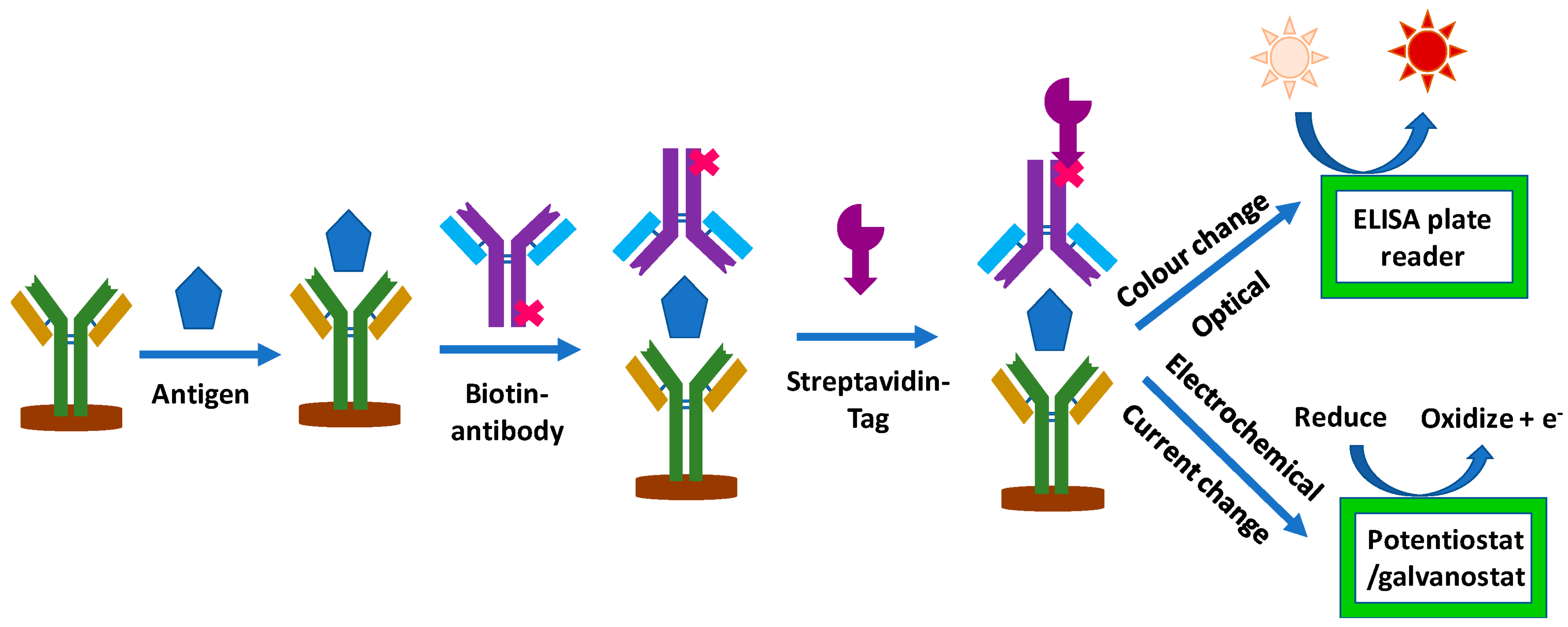
- Arya, S.K.; Estrela, P. Recent Advances in Enhancement Strategies for Electrochemical ELISA-Based Immunoassays for Cancer Biomarker Detection. Sensors 2018, 18, 2010. [Google Scholar] [CrossRef] [PubMed]


| Method | Nanoplatform | Limit of Detection (LOD) | Linear Range | Ref |
|---|---|---|---|---|
| Fluorescent | Agarose nano-net | 1.0 U/mL | 0.05–1.45 U/mL | [36] |
| Ag NPs | 0.0018 U/mL | 0.01–80 U/mL | [37] | |
| Ag NCs/GO or Ag/Au NCs/GO | 1.26 ng/mL | 2 ng/mL–6.7 µg/mL | [38] | |
| SPN/MIP or CNT/MIP | 0.49 U/mL | 3.12–150 U/mL | [39] | |
| rGO | 50 mU/mL | 50–2000 mU/mL | [40] | |
| 3D CNT | 10 pg/mL | 10 pg/mL–1 μg/mL | [41] | |
| Magnetic NPs | 0.26 U/mL | 0–500 U/Ml | [39] | |
| NA | 4 μg/mL | 4–250 μg/mL | [40] | |
| Magnetic graphene oxide (GO/Fe3O4) | 50 mU/mL | 0.0005–40 U/mL | [41] | |
| Combination of NaYF4: Yb, Tm, and Ag NPs | 120 U/mL | 5–100 U/mL | [42] | |
| Ag NCs | 0.015 U/mL | 0.01–2 U/mL | [43] | |
| Au doped sol–gel matrix | 1.45 U/mL | 2–127 U/mL | [44] | |
| FRET | PAMAM-dendrimer/Au NPs | 0.5 fg/mL | 1 fg/mL–1 ng/mL | [45] |
| CuO NPs | 3 × 10⁻⁴ ng/mL | 2 × 10⁻⁴ ng/mL–100 U/mL | [46] | |
| CQDs | 0.66 U/mL | 0.01–129 U/mL | [47] | |
| Photoluminescent | Ag2S QDs | 0.07 ng/mL | 0.1–106 ng/mL | [48] |
| CL | Graphene QDs | 0.05 U/mL | 0.1–600 U/mL | [49] |
| SiO2 NPs | 0.17 U/mL | 0.5–400 U/mL | [50] | |
| NA | 0.15 U/mL | 0.50–80 U/mL | [51] | |
| MPs | 2 U/mL | 0–400 U/mL | [52] | |
| ECL | Ru-Au NPs/GR | 0.005 U/mL | 0.01–100 U/mL | [53] |
| Cd/Se NCs | 5 × 10⁻⁵ U/mL | 10−4–1 U/mL | [54] | |
| Dendrimer-sulfanilic acid-Ru(bpy)32+ and Dendrimer-CdTe@CdS nanocomposite | 1.1 µU/mL | 1 µU/mL–1 U/mL | [55] | |
| CdTe/CdS QDs | 0.034 mU/mL | 0.0001 U/mL–10 U/mL | [56] | |
| AgInS2/ZnS nanocrystals | 1 × 10⁻⁶ U/mL | 5 × 10⁻⁶–5 × 10⁻³ U/mL | [57] | |
| Au NPs | 0.0074 U/mL | 0.01–100 U/mL | [58] | |
| Amino-functionalized mesoporous silica NPs | 4.3 mU/mL | 0.01–50 U/mL | [59] | |
| Fe3O4 | 8.0 μU/mL | 0–10 mU/mL | [60] | |
| Au-Ag nanocomposite-functionalized graphene | 2.5 mU/mL | 0.008–50 U/mL | [61] | |
| Fe3O4 | 0.4 mU/mL | 0.001–5 U/mL | [62] | |
| Fe3O4 | 0.032 μU/mL | 0.2–100 μU/mL | [63] | |
| SPR | Au NPs | 5 nM | 0.25–9.0 μg/mL | [64] |
| Au NPs | 0.66 U/mL | 2.2–150 U/mL | [65] | |
| Au-SPE film | 0.1 U/mL | 0.1–300 U/mL | [66] | |
| Au NPs | 0.1 U/mL | 0.1–40 U/mL | [67] | |
| Au/ZnO nanocomposite | 0.025 U/mL | 1–40 U/mL | [68] | |
| SERS | Au NPs | NA | NA | [69] |
| Ag NPs | NA | NA | [70] | |
| Plasmon Resonance Scattering (PRS) | Au nanorods | 0.4 U/mL | 1–80 U/mL | [71] |
| Colorimetric | Ag/Au NPs | 30 U/mL | 0–1000 U/mL | [72] |
| Hollow polydopamine-Au and Fe3O4 NPs | 0.1 U/mL | 0.1–100 U/mL | [73] |
| Electrode Material | Coating Material | Advantages | Disadvantages | Features | Ref. |
|---|---|---|---|---|---|
| GCE | AuNPs | High sensitivity, low cost, short test time | Narrow linear range, detects lower-than-average biomarker values (35 U/mL) | Stabilizer: cellulose acetate membrane, cysteamine (CysA) sulfur-containing biomolecule | [97] |
| GCE | (Silver nanoparticles) Ag NPs | High electrical conductivity and biocompatibility, and low toxicity. Optical and thermal attributes, support for electrocatalytic activity | Aggregation of Ag with solvent evaporation causes gaps and leads to low conductivity | [97] | |
| GCE | * Ag NPs with graphene quantum dot (Ag-DPA-GQDs ink) | Measures different concentrations of CA-125 biomarker | Conductivity: 290 mS [86] The linear range is 0.01–400. Descriptions: Ag-DPA-GQDs nano-ink deposition on GCE electrode | [97,98] | |
| GCE | Antimonene quantum dots (AMQDs) | Reduces the cost of analysis | LOD is 4.4 μM. | Catalase for H2O2 reduction is immobilized on AMQDs for cyclic voltammetry and amperometry detection. | [99] |
| GCE | Nafion + MPBB antibody | Detects at low concentration, detects OC early and can be used to screen at-risk individuals. | The linear range is 5–50 ng/mL and 100–500 ng/mL | [100] | |
| Three-dimensional gold electrode(Au/GNS/Ab-modified electrode) | Silicon nanoparticles (SiNPs) | Linked to the immunosensor CA-125, improved electrochemical performance. | The linear range is 1 fg/mL–1 μg/mL | [100,103] | |
| GCE | Zinc oxide (ZnO)-based NP | High repeatability, specificity, and durability | Acceptable stability | Linear range is 2.5 ng/μL–1 ng/μL | [100] |
| Graphene-polyaniline-based | Improves early-stage diagnosis | The linear range is 0.92 pg/L to 15.20 ng/L. | [100] | ||
| GCE | MOF-808/CNT | Biocompatible surface, high stability, electrochemically enhanced | The linear range is 0.001–30 ng/mL | [104] | |
| Biotin-modified carbon paste electrodes | Au NPs | Stability, biological adaptability | Narrow linear range Detects lower-than-average biomarker levels (35 U/mL) | [97] |
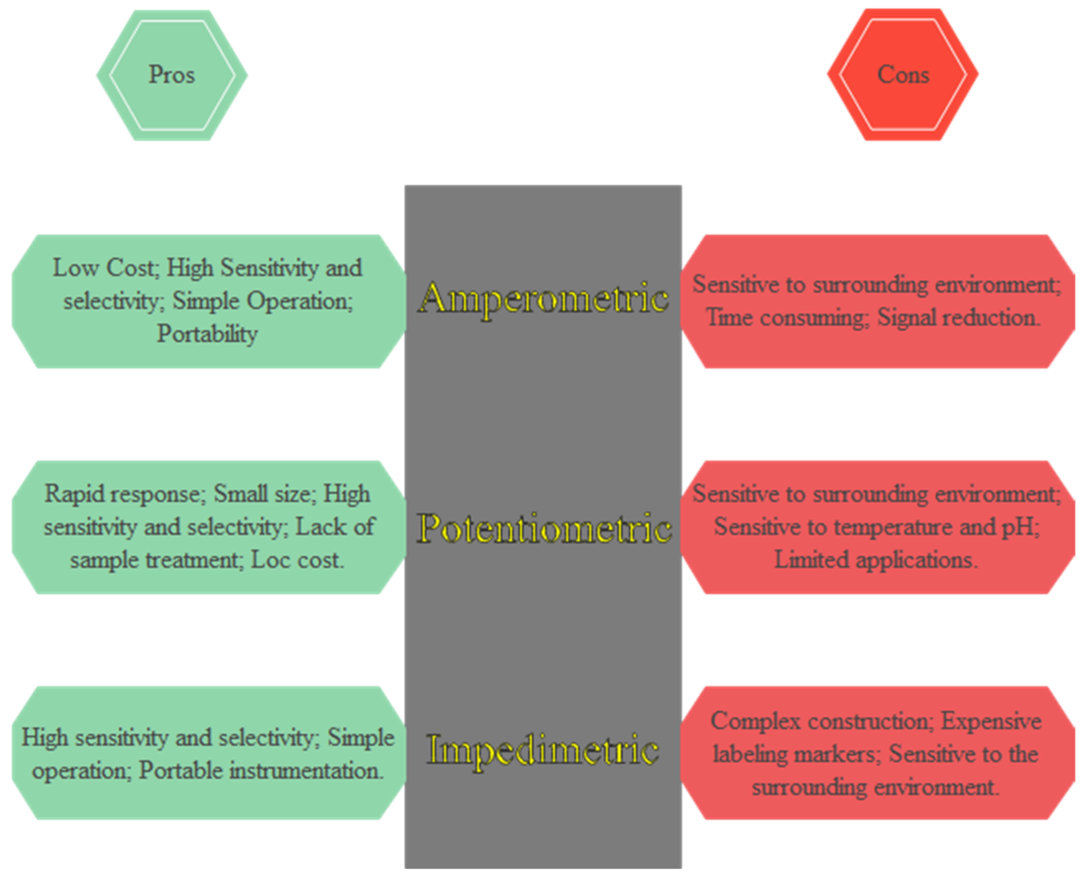
| Electrochemical Detection Methodology | Assay Strategy |
|---|---|
| Amperometric [98] | Nanostructured colloidal gold immunosensor Immunosensor based on multiwalled carbon nanotubes Tagging technique for redox probes Biosensor with a molecular imprint Magnetic bead immunosensor Immunosensor nanoparticle |
| A field-effect transistor (FET) [98] | Nanotube-based immunosensor Label-free immunosensor |
| Potentiometric [98] | Immunosensor-arrayed microfluidic device Magnetic bead immunosensor |
| Commercial CA-125 Kits | Assay Sensitivity | Assay Range | Sample Type | Assay Time (h) |
|---|---|---|---|---|
| LifeSpan | - | 1.563–100 U/mL | Plasma, serum | 3.5 |
| RayBiotech | 0.6 U/mL | 0.6–400 U/mL | Cell culture supernatants, plasma, serum | - |
| Aviva Systems | 6.5 pg/mL | 15.6–1000 pg/mL | Serum, plasma, tissue homogenates, and other biological fluids | 3 |
| Wuhan Fine | 1.875 IU/mL | 3.125–200 IU/mL | Serum, plasma, and other biological fluids | - |
| (DEMEDITEC Diagnostics GmbH) | 0.25 U/mL | 25–600 U/mL | Serum, plasma | 1 h and 15 min |
| (Thermo Fisher Scientific) | - | 0.55–400 U/mL | Plasma, 50 µL; serum, 50 µL; supernatant, 100 µL | 4 h and 45 min |
| Novus Biologicals | 3.8 U/mL | 3.8 U/mL | Serum, plasma | - |
Disclaimer/Publisher’s Note: The statements, opinions and data contained in all publications are solely those of the individual author(s) and contributor(s) and not of MDPI and/or the editor(s). MDPI and/or the editor(s) disclaim responsibility for any injury to people or property resulting from any ideas, methods, instructions or products referred to in the content. |
© 2023 by the authors. Licensee MDPI, Basel, Switzerland. This article is an open access article distributed under the terms and conditions of the Creative Commons Attribution (CC BY) license (https://creativecommons.org/licenses/by/4.0/).
Share and Cite
Pourmadadi, M.; Moammeri, A.; Shamsabadipour, A.; Moghaddam, Y.F.; Rahdar, A.; Pandey, S. Application of Various Optical and Electrochemical Nanobiosensors for Detecting Cancer Antigen 125 (CA-125): A Review. Biosensors 2023, 13, 99. https://doi.org/10.3390/bios13010099
Pourmadadi M, Moammeri A, Shamsabadipour A, Moghaddam YF, Rahdar A, Pandey S. Application of Various Optical and Electrochemical Nanobiosensors for Detecting Cancer Antigen 125 (CA-125): A Review. Biosensors. 2023; 13(1):99. https://doi.org/10.3390/bios13010099
Chicago/Turabian StylePourmadadi, Mehrab, Ali Moammeri, Amin Shamsabadipour, Yasamin Farahanian Moghaddam, Abbas Rahdar, and Sadanand Pandey. 2023. "Application of Various Optical and Electrochemical Nanobiosensors for Detecting Cancer Antigen 125 (CA-125): A Review" Biosensors 13, no. 1: 99. https://doi.org/10.3390/bios13010099