Chitosan-Based Hydrogels for Bioelectronic Sensing: Recent Advances and Applications in Biomedicine and Food Safety


Abstract
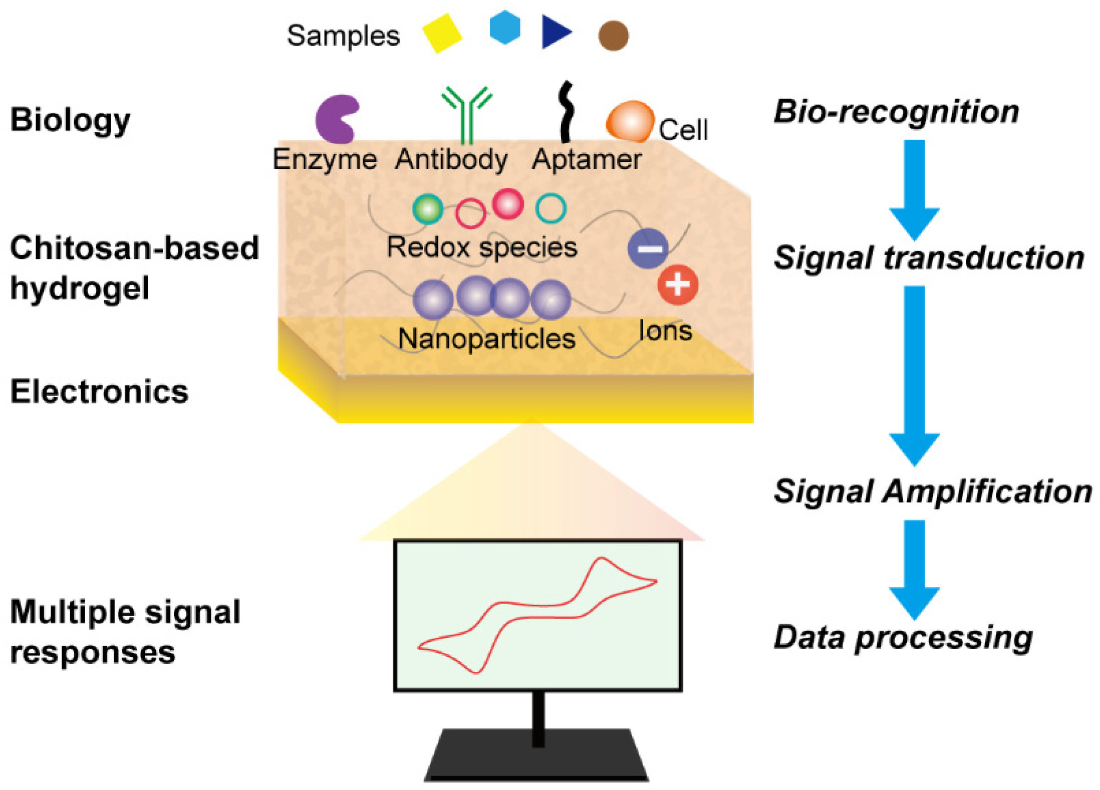
:1. Introduction
2. Fabrication and Interaction Mechanism of Chitosan-Based Hydrogels
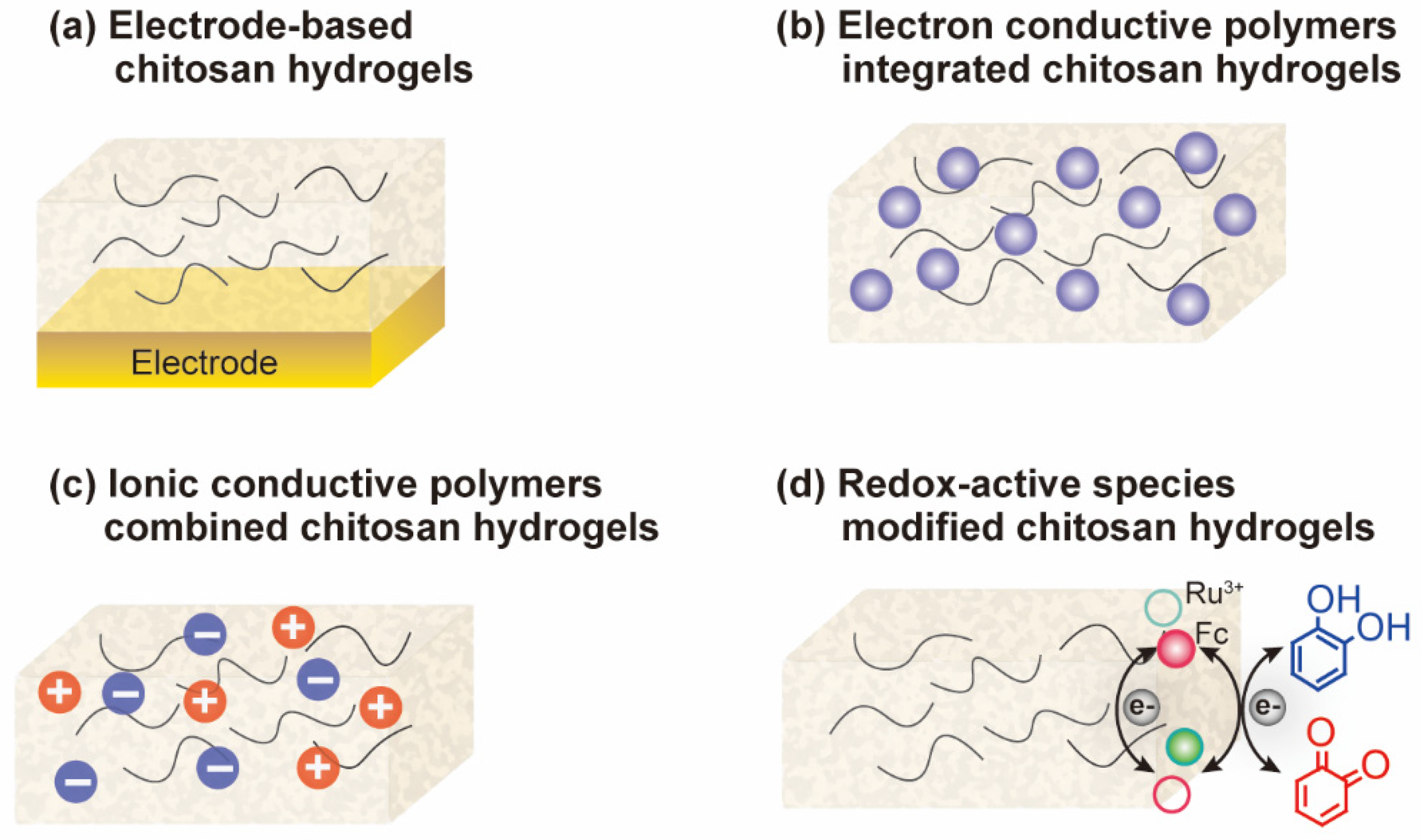
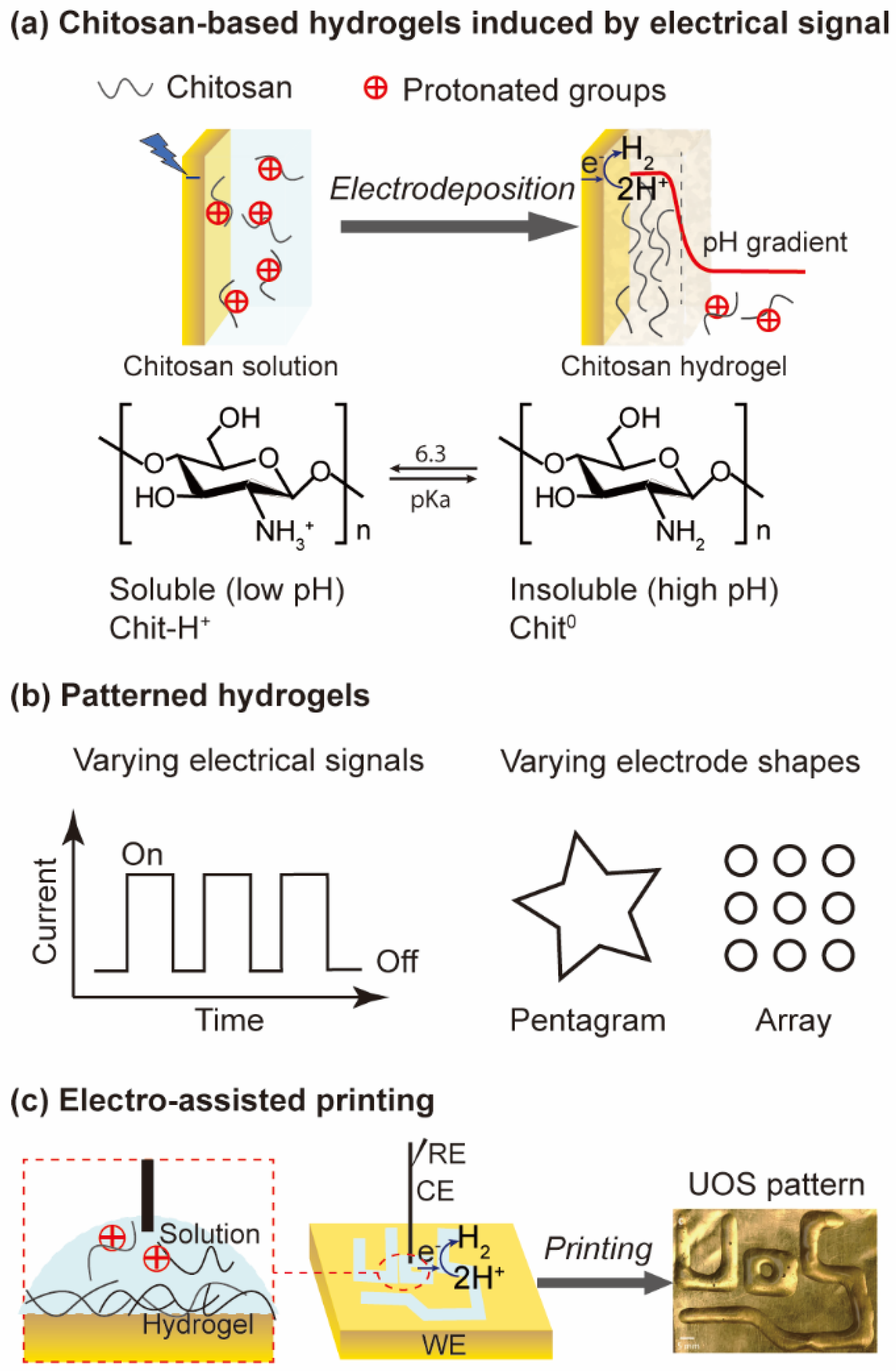
2.1. Electrode-Based Hydrogels
2.2. Conductive Materials Conjugated Hydrogels
2.3. Ionically Conductive Hydrogels
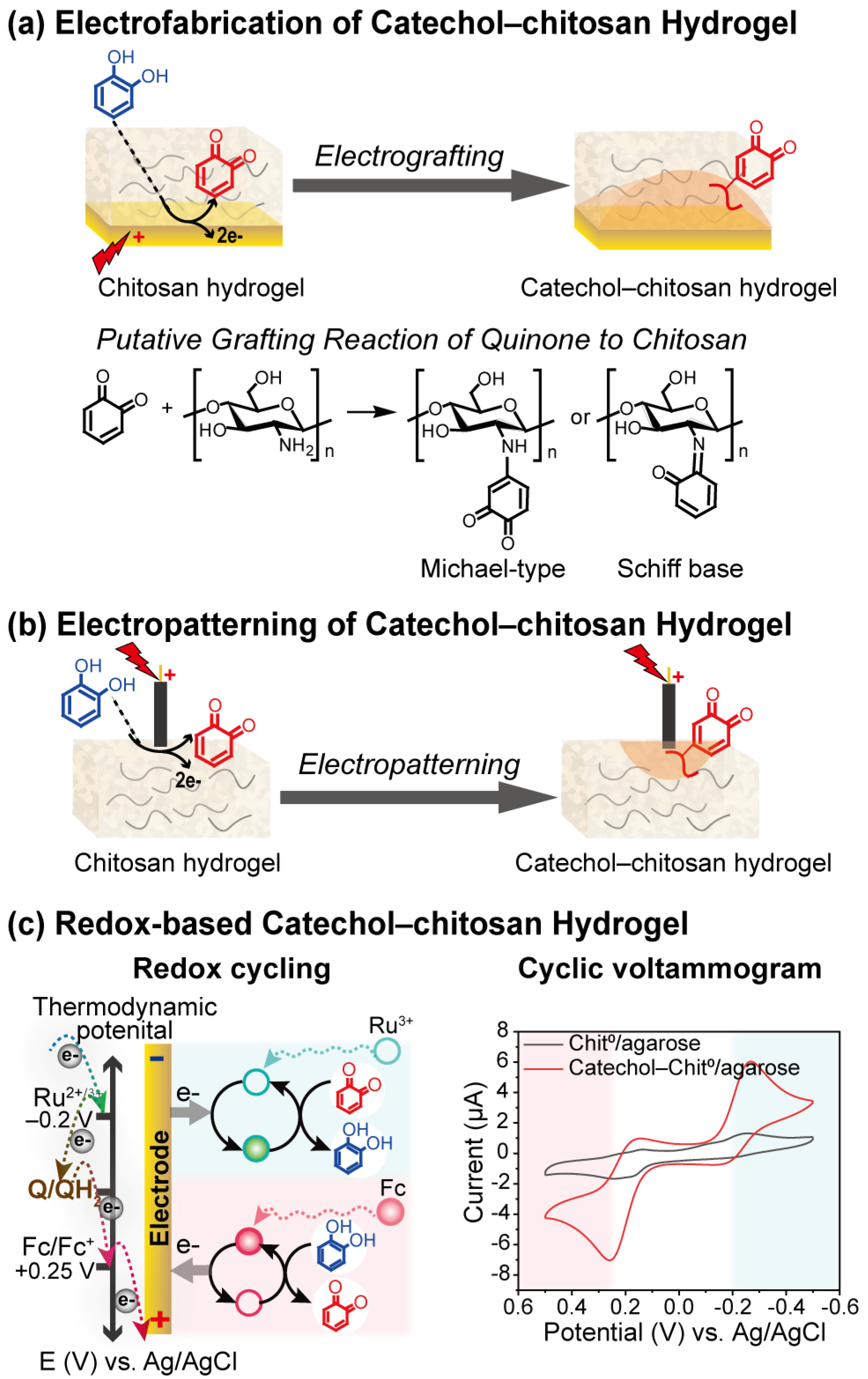
2.4. Redox-Based Hydrogels
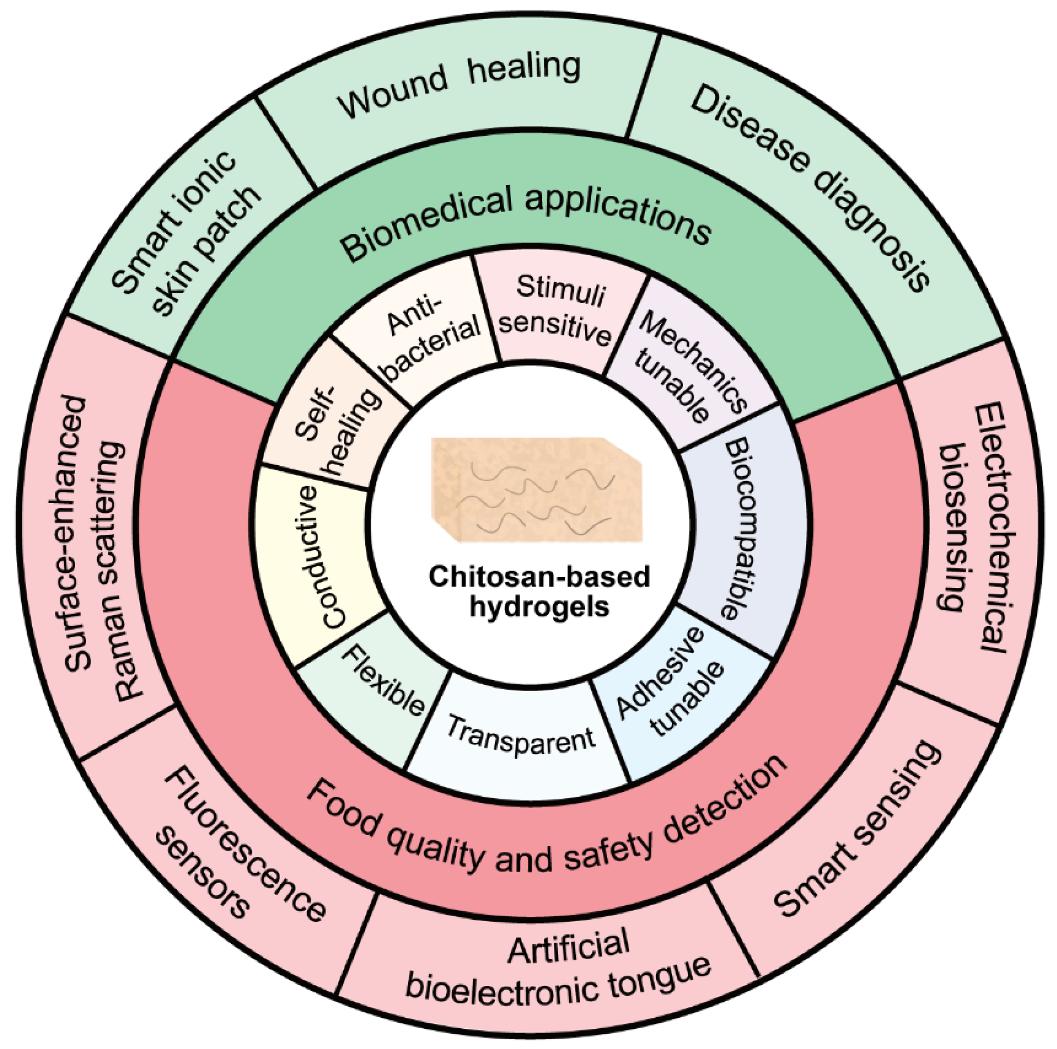
3. Functional Properties and Applications of Chitosan-Based Hydrogels
4. Biomedical Applications
4.1. Smart Ionic Skin Patch
4.2. Wood Healing
4.3. Disease Diagnosis
5. Food Quality and Safety Detection
5.1. Electrochemical Biosensing
5.2. Smart Sensing
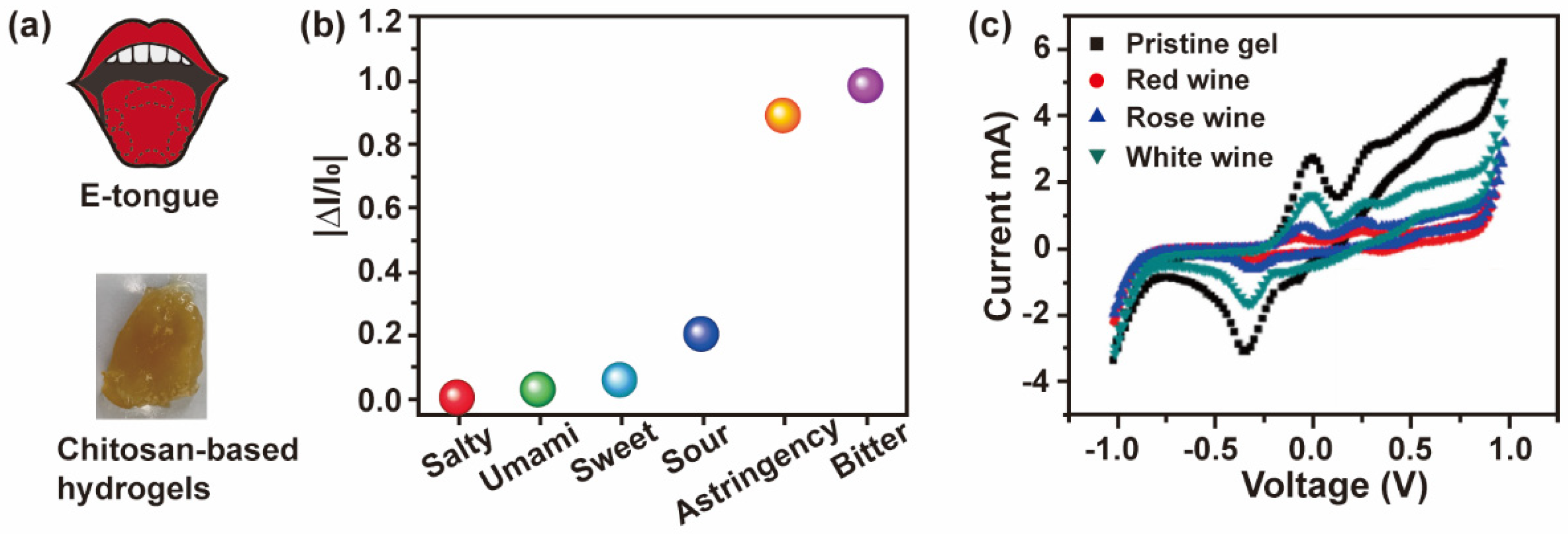
5.3. Artificial Bioelectronic Tongue
5.4. Fluorescence Sensors
5.5. Surface-Enhanced Raman Scattering
6. Conclusions and Future Prospects
Author Contributions
Funding
Institutional Review Board Statement
Informed Consent Statement
Data Availability Statement
Conflicts of Interest
References
- Lee, S.W.; Park, J.J.; Park, B.H.; Mun, S.C.; Park, Y.T.; Liao, K.; Seo, T.S.; Hyun, W.J.; Park, O.O. Enhanced Sensitivity of Patterned Graphene Strain Sensors Used for Monitoring Subtle Human Body Motions. ACS Appl. Mater. Interfaces 2017, 9, 11176–11183. [Google Scholar] [CrossRef] [PubMed]
- Wang, Y.; Niu, W.; Lo, C.Y.; Zhao, Y.; He, X.; Zhang, G.; Wu, S.; Ju, B.; Zhang, S. Interactively Full-Color Changeable Electronic Fiber Sensor with High Stretchability and Rapid Response. Adv. Funct. Mater. 2020, 30, 2000356. [Google Scholar] [CrossRef]
- Webb, R.C.; Bonifas, A.P.; Behnaz, A.; Zhang, Y.; Yu, K.J.; Cheng, H.; Shi, M.; Bian, Z.; Liu, Z.; Kim, Y.S.; et al. Ultrathin Conformal Devices for Precise and Continuous Thermal Characterization of Human Skin. Nat. Mater. 2013, 12, 938–944. [Google Scholar] [CrossRef] [PubMed] [Green Version]
- Luo, N.; Dai, W.; Li, C.; Zhou, Z.; Lu, L.; Poon, C.C.Y.; Chen, S.C.; Zhang, Y.; Zhao, N. Flexible Piezoresistive Sensor Patch Enabling Ultralow Power Cuffless Blood Pressure Measurement. Adv. Funct. Mater. 2016, 26, 1178–1187. [Google Scholar] [CrossRef]
- You, I.; Kim, B.; Park, J.; Koh, K.; Shin, S.; Jung, S.; Jeong, U. Stretchable E-Skin Apexcardiogram Sensor. Adv. Mater. 2016, 28, 6359–6364. [Google Scholar] [CrossRef]
- Khan, A.; Ahmed, S.; Sun, B.Y.; Chen, Y.C.; Chuang, W.T.; Chan, Y.H.; Gupta, D.; Wu, P.W.; Lin, H.C. Self-Healable and Anti-Freezing Ion Conducting Hydrogel-Based Artificial Bioelectronic Tongue Sensing toward Astringent and Bitter Tastes. Biosens. Bioelectron. 2022, 198, 113811. [Google Scholar] [CrossRef]
- Liu, X.; Liu, J.; Lin, S.; Zhao, X. Hydrogel Machines. Mater. Today 2020, 36, 102–124. [Google Scholar] [CrossRef]
- Yin, X.Y.; Zhang, Y.; Xiao, J.; Moorlag, C.; Yang, J. Monolithic Dual-Material 3D Printing of Ionic Skins with Long-Term Performance Stability. Adv. Funct. Mater. 2019, 29, 1904716. [Google Scholar] [CrossRef]
- Zhu, Z.; Park, H.S.; McAlpine, M.C. 3D Printed Deformable Sensors. Sci. Adv. 2020, 6, eaba5575. [Google Scholar] [CrossRef]
- Zhang, Y.S.; Khademhosseini, A. Advances in Engineering Hydrogels. Science 2017, 356, eaaf3627. [Google Scholar] [CrossRef]
- Thambi, T.; Phan, V.H.G.; Lee, D.S. Stimuli-Sensitive Injectable Hydrogels Based on Polysaccharides and Their Biomedical Applications. Macromol. Rapid Commun. 2016, 37, 1881–1896. [Google Scholar] [CrossRef] [PubMed]
- Nordin, N.; Bordonali, L.; Badilita, V.; MacKinnon, N. Spatial and Temporal Control Over Multilayer Bio-Polymer Film Assembly and Composition. Macromol. Biosci. 2019, 19, 1800372. [Google Scholar] [CrossRef]
- Vivcharenko, V.; Wojcik, M.; Przekora, A. Cellular Response to Vitamin C-Enriched Chitosan/Agarose Film with Potential Application as Artificial Skin Substitute for Chronic Wound Treatment. Cells 2020, 9, 1185. [Google Scholar] [CrossRef] [PubMed]
- Blacklow, S.O.; Li, J.; Freedman, B.R.; Zeidi, M.; Chen, C.; Mooney, D.J. Bioinspired Mechanically Active Adhesive Dressings to Accelerate Wound Closure. Sci. Adv. 2019, 5, aaw3963. [Google Scholar] [CrossRef] [Green Version]
- Shariatinia, Z.; Jalali, A.M. Chitosan-Based Hydrogels: Preparation, Properties and Applications. Int. J. Biol. Macromol. 2018, 115, 194–220. [Google Scholar] [CrossRef]
- Lee, K.Y.; Mooney, D.J. Hydrogels for Tissue Engineering. Chem. Rev. 2001, 101, 1869–1879. [Google Scholar] [CrossRef]
- Li, J.; Yu, X.; Martinez, E.E.; Zhu, J.; Wang, T.; Shi, S.; Shin, S.R.; Hassan, S.; Guo, C. Emerging Biopolymer-Based Bioadhesives. Macromol. Biosci. 2022, 22, 2100340. [Google Scholar] [CrossRef]
- Yuk, H.; Lu, B.; Zhao, X. Hydrogel Bioelectronics. Chem. Soc. Rev. 2019, 48, 1642–1667. [Google Scholar] [CrossRef] [PubMed] [Green Version]
- Yan, K.; Xiong, Y.; Wu, S.; Bentley, W.E.; Deng, H.; Du, Y.; Payne, G.F.; Shi, X.W. Electro-Molecular Assembly: Electrical Writing of Information into an Erasable Polysaccharide Medium. ACS Appl. Mater. Interfaces 2016, 8, 19780–19786. [Google Scholar] [CrossRef] [PubMed]
- Song, P.; Qin, H.; Gao, H.L.; Cong, H.P.; Yu, S.H. Self-Healing and Superstretchable Conductors from Hierarchical Nanowire Assemblies. Nat. Commun. 2018, 9, 2786. [Google Scholar] [CrossRef]
- Gao, H.L.; Xu, L.; Long, F.; Pan, Z.; Du, Y.X.; Lu, Y.; Ge, J.; Yu, S.H. Macroscopic Free-Standing Hierarchical 3D Architectures Assembled from Silver Nanowires by Ice Templating. Angew. Chemie-Int. Ed. 2014, 53, 4561–4566. [Google Scholar] [CrossRef] [PubMed]
- Ahn, Y.; Lee, H.; Lee, D.; Lee, Y. Highly Conductive and Flexible Silver Nanowire-Based Microelectrodes on Biocompatible Hydrogel. ACS Appl. Mater. Interfaces 2014, 6, 18401–18407. [Google Scholar] [CrossRef]
- Zhang, Y.; He, P.; Luo, M.; Xu, X.; Dai, G.; Yang, J. Highly Stretchable Polymer/Silver Nanowires Composite Sensor for Human Health Monitoring. Nano Res. 2020, 13, 919–926. [Google Scholar] [CrossRef]
- Tang, P.; Yan, H.; Chen, L.; Wu, Q.; Zhao, T.; Li, S.; Gao, H.; Liu, M. Anisotropic Nanocomposite Hydrogels with Enhanced Actuating Performance through Aligned Polymer Networks. Sci. China Mater. 2020, 63, 832–841. [Google Scholar] [CrossRef] [Green Version]
- Deng, J.; Yuk, H.; Wu, J.; Varela, C.E.; Chen, X.; Roche, E.T.; Guo, C.F.; Zhao, X. Electrical Bioadhesive Interface for Bioelectronics. Nat. Mater. 2021, 20, 229–236. [Google Scholar] [CrossRef] [PubMed]
- Liao, H.; Guo, X.; Wan, P.; Yu, G. Conductive MXene Nanocomposite Organohydrogel for Flexible, Healable, Low-Temperature Tolerant Strain Sensors. Adv. Funct. Mater. 2019, 29, 1904507. [Google Scholar] [CrossRef]
- Fu, S.; Zhu, Y.; Zhang, Y.; Zhang, M.; Wang, D. Recent Advances in Carbon Nanomaterials-Based Electrochemical Sensors for Phenolic Compounds Detection. Microchem. J. 2021, 171, 106776. [Google Scholar] [CrossRef]
- Liang, S.; Zhang, Y.; Wang, H.; Xu, Z.; Chen, J.; Bao, R.; Tan, B.; Cui, Y.; Fan, G.; Wang, W.; et al. Paintable and Rapidly Bondable Conductive Hydrogels as Therapeutic Cardiac Patches. Adv. Mater. 2018, 30, 1704235. [Google Scholar] [CrossRef]
- Duan, J.; Liang, X.; Guo, J.; Zhu, K.; Zhang, L. Ultra-Stretchable and Force-Sensitive Hydrogels Reinforced with Chitosan Microspheres Embedded in Polymer Networks. Adv. Mater. 2016, 28, 8037–8044. [Google Scholar] [CrossRef]
- Go, Y.M.; Jones, D.P. The Redox Proteome. J. Biol. Chem. 2013, 288, 26512–26520. [Google Scholar] [CrossRef]
- Wu, S.; Kim, E.; Li, J.; Bentley, W.E.; Shi, X.-W.; Payne, G.F. Catechol-Based Capacitor for Redox-Linked Bioelectronics. ACS Appl. Electron. Mater. 2019, 1, 1337–1347. [Google Scholar] [CrossRef] [PubMed]
- Yan, K.; Liu, Y.; Zhang, J.; Correa, S.O.; Shang, W.; Tsai, C.C.; Bentley, W.E.; Shen, J.; Scarcelli, G.; Raub, C.B.; et al. Electrical Programming of Soft Matter: Using Temporally Varying Electrical Inputs to Spatially Control Self Assembly. Biomacromolecules 2018, 19, 364–373. [Google Scholar] [CrossRef] [PubMed]
- Munoz-Robles, B.G.; Kopyeva, I.; DeForest, C.A. Surface Patterning of Hydrogel Biomaterials to Probe and Direct Cell–Matrix Interactions. Adv. Mater. Interfaces 2020, 7, 1–25. [Google Scholar] [CrossRef]
- Da Silva, A.C.; Wang, J.; Minev, I.R. Electro-Assisted Printing of Soft Hydrogels via Controlled Electrochemical Reactions. Nat. Commun. 2022, 13, 1353. [Google Scholar] [CrossRef]
- Liao, M.; Wan, P.; Wen, J.; Gong, M.; Wu, X.; Wang, Y.; Shi, R.; Zhang, L. Wearable, Healable, and Adhesive Epidermal Sensors Assembled from Mussel-Inspired Conductive Hybrid Hydrogel Framework. Adv. Funct. Mater. 2017, 27, 1703852. [Google Scholar] [CrossRef]
- Liang, Y.; Zhao, X.; Hu, T.; Chen, B.; Yin, Z.; Ma, P.X.; Guo, B. Adhesive Hemostatic Conducting Injectable Composite Hydrogels with Sustained Drug Release and Photothermal Antibacterial Activity to Promote Full-Thickness Skin Regeneration During Wound Healing. Small 2019, 15, e1900046. [Google Scholar] [CrossRef]
- Pan, Z.; Wang, Z.Y.; Wang, M.H.; Yang, L.; Yu, S.H. Adhesive Aero-Hydrogel Hybrid Conductor Assembled from Silver Nanowire Architectures. Sci. China Mater. 2021, 64, 2868–2876. [Google Scholar] [CrossRef]
- Lin, L.; Wu, Q. Improved Conductivity of Polysaccharide-Co-Polyacrylate/Polyaniline Conducting Hydrogels. Polym. Polym. Compos. 2012, 20, 377–386. [Google Scholar] [CrossRef]
- Zhang, J.; Wu, C.; Xu, Y.; Chen, J.; Ning, N.; Yang, Z.; Guo, Y.; Hu, X.; Wang, Y. Highly Stretchable and Conductive Self-Healing Hydrogels for Temperature and Strain Sensing and Chronic Wound Treatment. ACS Appl. Mater. Interfaces 2020, 12, 40990–40999. [Google Scholar] [CrossRef]
- Kim, J.H.; Kim, S.R.; Kil, H.J.; Kim, Y.C.; Park, J.W. Highly Conformable, Transparent Electrodes for Epidermal Electronics. Nano Lett. 2018, 18, 4531–4540. [Google Scholar] [CrossRef]
- Liu, Q.; Yang, S.; Ren, J.; Ling, S. Flame-Retardant and Sustainable Silk Ionotronic Skin for Fire Alarm Systems. ACS Mater. Lett. 2020, 2, 712–720. [Google Scholar] [CrossRef]
- Yang, C.; Suo, Z. Hydrogel Ionotronics. Nat. Rev. Mater. 2018, 3, 125–142. [Google Scholar] [CrossRef]
- Leger, J.; Berggren, M.; Carter, S. Iontronics: Ionic Carriers in Organic Electronic Materials and Devices; CRC Press: Boca Raton, FL, USA, 2011; p. 247. [Google Scholar]
- Gao, Z.; Li, Y.; Shang, X.; Hu, W.; Gao, G.; Duan, L. Bio-Inspired Adhesive and Self-Healing Hydrogels as Flexible Strain Sensors for Monitoring Human Activities. Mater. Sci. Eng. C 2020, 106, 110168. [Google Scholar] [CrossRef] [PubMed]
- Han, X.; Jiang, D.; Qu, X.; Bai, Y.; Cao, Y.; Luo, R.; Li, Z. A Stretchable, Self-Healable Triboelectric Nanogenerator as Electronic Skin for Energy Harvesting and Tactile Sensing. Materials 2021, 14, 1689. [Google Scholar] [CrossRef]
- Sarwar, M.S.; Dobashi, Y.; Preston, C.; Wyss, J.K.M.; Mirabbasi, S.; David, J.; Madden, W. Bend, Stretch, and Touch: Locating a Finger on an Actively Deformed Transparent Sensor Array. Sci. Adv. 2017, 3, e1602200. [Google Scholar] [CrossRef] [PubMed] [Green Version]
- Lei, Z.; Wang, Q.; Sun, S.; Zhu, W.; Wu, P. A Bioinspired Mineral Hydrogel as a Self-Healable, Mechanically Adaptable Ionic Skin for Highly Sensitive Pressure Sensing. Adv. Mater. 2017, 29, 1700321. [Google Scholar] [CrossRef] [PubMed]
- Yang, N.; Qi, P.; Ren, J.; Yu, H.; Liu, S.; Li, J.; Chen, W.; Kaplan, D.L.; Ling, S. Polyvinyl Alcohol/Silk Fibroin/Borax Hydrogel Ionotronics: A Highly Stretchable, Self-Healable, and Biocompatible Sensing Platform. ACS Appl. Mater. Interfaces 2019, 11, 23632–23638. [Google Scholar] [CrossRef] [PubMed]
- Lei, Z.; Wang, Q.; Wu, P. A Multifunctional Skin-like Sensor Based on a 3D Printed Thermo-Responsive Hydrogel. Mater. Horizons 2017, 4, 694–700. [Google Scholar] [CrossRef]
- Zhang, C.; Sun, W.; Chen, H.; Liu, L.; Li, B.; Li, D. Electromechanical Deformation of Conical Dielectric Elastomer Actuator with Hydrogel Electrodes. J. Appl. Phys. 2016, 119, 1223. [Google Scholar] [CrossRef]
- Pei, X.; Zhang, H.; Zhou, Y.; Zhou, L.; Fu, J. Stretchable, Self-Healing and Tissue-Adhesive Zwitterionic Hydrogels as Strain Sensors for Wireless Monitoring of Organ Motions. Mater. Horizons 2020, 7, 1872–1882. [Google Scholar] [CrossRef]
- Zhang, J.; Zhang, Q.; Liu, X.; Xia, S.; Gao, Y.; Gao, G. Flexible and Wearable Strain Sensors Based on Conductive Hydrogels. J. Polym. Sci. 2022, 60, 2663–2678. [Google Scholar] [CrossRef]
- Parvez, S.; Long, M.J.C.; Poganik, J.R.; Aye, Y. Correction to Redox Signaling by Reactive Electrophiles and Oxidants. Chem. Rev. 2019, 119, 4464–4469. [Google Scholar] [CrossRef] [PubMed]
- Petra, A.I.; Panagiotidou, S.; Hatziagelaki, E.; Stewart, J.M.; Conti, P.; Theoharides, T.C. Gut-Microbiota-Brain Axis and Its Effect on Neuropsychiatric Disorders with Suspected Immune Dysregulation. Clin. Ther. 2015, 37, 984–995. [Google Scholar] [CrossRef] [PubMed] [Green Version]
- Valko, M.; Leibfritz, D.; Moncol, J.; Cronin, M.T.D.; Mazur, M.; Telser, J. Free Radicals and Antioxidants in Normal Physiological Functions and Human Disease. Int. J. Biochem. Cell Biol. 2007, 39, 44–84. [Google Scholar] [CrossRef] [PubMed]
- Wu, L.-Q.; McDermott, M.K.; Zhu, C.; Ghodssi, R.; Payne, G.F. Mimicking Biological Phenol Reaction Cascades to Confer Mechanical Function. Adv. Funct. Mater. 2006, 16, 1967–1974. [Google Scholar] [CrossRef]
- Wu, S.; Yan, K.; Zhao, Y.; Tsai, C.C.; Shen, J.; Bentley, W.E.; Chen, Y.; Deng, H.; Du, Y.; Payne, G.F.; et al. Electrical Writing onto a Dynamically Responsive Polysaccharide Medium: Patterning Structure and Function into a Reconfigurable Medium. Adv. Funct. Mater. 2018, 28, 1803139. [Google Scholar] [CrossRef]
- Wu, S.; Zhao, Z.; Rzasa, J.R.; Kim, E.; Li, J.; VanArsdale, E.; Bentley, W.E.; Shi, X.; Payne, G.F. Hydrogel Patterning with Catechol Enables Networked Electron Flow. Adv. Funct. Mater. 2021, 31, 2007709. [Google Scholar] [CrossRef]
- Wu, S.; Kim, E.; Chen, C.Y.; Li, J.; VanArsdale, E.; Grieco, C.; Kohler, B.; Bentley, W.E.; Shi, X.; Payne, G.F. Catechol-Based Molecular Memory Film for Redox Linked Bioelectronics. Adv. Electron. Mater. 2020, 6, 2000452. [Google Scholar] [CrossRef]
- Kim, E.; Li, J.; Kang, M.; Kelly, D.L.; Chen, S.; Napolitano, A.; Panzella, L.; Shi, X.; Yan, K.; Wu, S.; et al. Redox Is a Global Biodevice Information Processing Modality. Proc. IEEE 2019, 107, 1402–1424. [Google Scholar] [CrossRef]
- Kim, E.; Liu, Z.; Liu, Y.; Bentley, W.E.; Payne, G.F. Catechol-Based Hydrogel for Chemical Information Processing. Biomimetics 2017, 2, 11. [Google Scholar] [CrossRef]
- Vanarsdale, E.; Hörnström, D.; Sjöberg, G.; Järbur, I.; Pitzer, J.; Payne, G.F.; Van Maris, A.J.A.; Bentley, W.E. A Coculture Based Tyrosine-Tyrosinase Electrochemical Gene Circuit for Connecting Cellular Communication with Electronic Networks. ACS Synth. Biol. 2020, 9, 1117–1128. [Google Scholar] [CrossRef] [PubMed]
- Kim, E.; Liu, Y.; Bentley, W.E.; Payne, G.F. Redox Capacitor to Establish Bio-Device Redox-Connectivity. Adv. Funct. Mater. 2012, 22, 1409–1416. [Google Scholar] [CrossRef]
- Liu, H.; Qu, X.; Kim, E.; Lei, M.; Dai, K.; Tan, X.; Xu, M.; Li, J.; Liu, Y.; Shi, X.; et al. Bio-Inspired Redox-Cycling Antimicrobial Film for Sustained Generation of Reactive Oxygen Species. Biomaterials 2018, 162, 109–122. [Google Scholar] [CrossRef]
- Kim, E.; Liu, Y.; Baker, C.J.; Owens, R.; Xiao, S.; Bentley, W.E.; Payne, G.F. Redox-Cycling and H2O2 Generation by Fabricated Catecholic Films in the Absence of Enzymes. Biomacromolecules 2011, 12, 880–888. [Google Scholar] [CrossRef] [PubMed]
- Shang, W.; Chen, C.Y.; Lo, K.; Payne, G.F.; Bentley, W.E. Chip Modularity Enables Molecular Information Access from Organ-on-Chip Devices with Quality Control. Sens. Actuators B Chem. 2019, 295, 30–39. [Google Scholar] [CrossRef]
- Yan, K.; Liu, Y.; Guan, Y.; Bhokisham, N.; Tsao, C.Y.; Kim, E.; Shi, X.W.; Wang, Q.; Bentley, W.E.; Payne, G.F. Catechol–chitosan Redox Capacitor for Added Amplification in Electrochemical Immunoanalysis. Colloids Surf. B Biointerfaces 2018, 169, 470–477. [Google Scholar] [CrossRef]
- Annu; Raja, A.N. Recent Development in Chitosan-Based Electrochemical Sensors and Its Sensing Application. Int. J. Biol. Macromol. 2020, 164, 4231–4244. [Google Scholar] [CrossRef]
- Ou, Y.; Tian, M. Advances in Multifunctional Chitosan-Based Self-Healing Hydrogels for Biomedical Applications. J. Mater. Chem. B 2021, 9, 7955–7971. [Google Scholar] [CrossRef]
- Shi, X.; Wu, P. A Smart Patch with On-Demand Detachable Adhesion for Bioelectronics. Small 2021, 17, 2101220. [Google Scholar] [CrossRef]
- Si, R.; Gao, C.; Guo, R.; Lin, C.; Li, J.; Guo, W. Human Mesenchymal Stem Cells Encapsulated-Coacervated Photoluminescent Nanodots Layered Bioactive Chitosan/Collagen Hydrogel Matrices to Indorse Cardiac Healing after Acute Myocardial Infarction. J. Photochem. Photobiol. B Biol. 2020, 206, 111789. [Google Scholar] [CrossRef]
- Shin, M.; Park, S.G.; Oh, B.C.; Kim, K.; Jo, S.; Lee, M.S.; Oh, S.S.; Hong, S.H.; Shin, E.C.; Kim, K.S.; et al. Complete Prevention of Blood Loss with Self-Sealing Haemostatic Needles. Nat. Mater. 2017, 16, 147–152. [Google Scholar] [CrossRef] [PubMed]
- Nguyen, N.; Lin, Z.H.; Barman, S.R.; Korupalli, C.; Cheng, J.Y.; Song, N.X.; Chang, Y.; Mi, F.L.; Song, H.L.; Sung, H.W.; et al. Engineering an Integrated Electroactive Dressing to Accelerate Wound Healing and Monitor Noninvasively Progress of Healing. Nano Energy 2022, 99, 107393. [Google Scholar] [CrossRef]
- Luo, X.; Liu, Y.; Qin, R.; Ao, F.; Wang, X.; Zhang, H.; Yang, M.; Liu, X. Tissue-Nanoengineered Hyperbranched Polymer Based Multifunctional Hydrogels as Flexible “Wounped Treatment-Health Monitoring” Bioelectronic Implant. Appl. Mater. Today 2022, 29, 101576. [Google Scholar] [CrossRef]
- Huang, Y.; Mu, L.; Zhao, X.; Han, Y.; Guo, B. Bacterial Growth-Induced Tobramycin Smart Release Self-Healing Hydrogel for Pseudomonas Aeruginosa-Infected Burn Wound Healing. ACS Nano 2022, 16, 13022–13036. [Google Scholar] [CrossRef]
- Chen, F.; Wu, M.; Dong, Q.; Ke, M.; Liang, X.; Ai, J.; Cheng, Q.; Cai, L.; Tong, Z.; Chen, Y. Arbitrarily Shapeable and Conductive Hydrogel with “Magic Cube” like Structure for Real-Time Monitoring and Promoting Wound Healing. Compos. Part B Eng. 2022, 238, 109903. [Google Scholar] [CrossRef]
- Shukla, R.P.; Aroosh, M.; Matzafi, A.; Ben-Yoav, H. Partially Functional Electrode Modifications for Rapid Detection of Dopamine in Urine. Adv. Funct. Mater. 2021, 31, 2004146. [Google Scholar] [CrossRef]
- Kim, J.N.; Lee, J.; Lee, H.; Oh, I.K. Stretchable and Self-Healable Catechol–chitosan-Diatom Hydrogel for Triboelectric Generator and Self-Powered Tremor Sensor Targeting at Parkinson Disease. Nano Energy 2021, 82, 105705. [Google Scholar] [CrossRef]
- Wen, J.; Tang, J.; Ning, H.; Hu, N.; Zhu, Y.; Gong, Y.; Xu, C.; Zhao, Q.; Jiang, X.; Hu, X.; et al. Multifunctional Ionic Skin with Sensing, UV-Filtering, Water-Retaining, and Anti-Freezing Capabilities. Adv. Funct. Mater. 2021, 31, 2011176. [Google Scholar] [CrossRef]
- Vakalopoulos, K.A.; Wu, Z.; Kroese, L.; Kleinrensink, G.J.; Jeekel, J.; Vendamme, R.; Dodou, D.; Lange, J.F. Mechanical Strength and Rheological Properties of Tissue Adhesives with Regard to Colorectal Anastomosis an Ex Vivo Study. Ann. Surg. 2015, 261, 323–331. [Google Scholar] [CrossRef]
- Liu, H.; Wang, C.; Li, C.; Qin, Y.; Wang, Z.; Yang, F.; Li, Z.; Wang, J. A Functional Chitosan-Based Hydrogel as a Wound Dressing and Drug Delivery System in the Treatment of Wound Healing. RSC Adv. 2018, 8, 7533–7549. [Google Scholar] [CrossRef]
- Duarte, A.P.; Coelho, J.F.; Bordado, J.C.; Cidade, M.T.; Gil, M.H. Surgical Adhesives: Systematic Review of the Main Types and Development Forecast. Prog. Polym. Sci. 2012, 37, 1031–1050. [Google Scholar] [CrossRef]
- Sedõ, J.; Saiz-Poseu, J.; Busqué, F.; Ruiz-Molina, D. Catechol-Based Biomimetic Functional Materials. Adv. Mater. 2013, 25, 653–701. [Google Scholar] [CrossRef] [Green Version]
- Faure, E.; Falentin-Daudré, C.; Jérôme, C.; Lyskawa, J.; Fournier, D.; Woisel, P.; Detrembleur, C. Catechols as Versatile Platforms in Polymer Chemistry. Prog. Polym. Sci. 2013, 38, 236–270. [Google Scholar] [CrossRef]
- Li, J.; Celiz, A.D.; Yang, J.; Yang, Q.; Wamala, I.; Whyte, W.; Seo, B.R.; Vasilyev, N.V.; Vlassak, J.J.; Suo, Z.; et al. Tough Adhesives for Diverse Wet Surfaces. Science 2017, 357, 378–381. [Google Scholar] [CrossRef] [Green Version]
- Yang, J.; Bai, R.; Chen, B.; Suo, Z. Hydrogel Adhesion: A Supramolecular Synergy of Chemistry, Topology, and Mechanics. Adv. Funct. Mater. 2020, 30, 1901693. [Google Scholar] [CrossRef]
- Jenkins, P.O.; Mehta, M.A.; Sharp, D.J. Catecholamines and Cognition after Traumatic Brain Injury. Brain 2016, 139, 2345–2371. [Google Scholar] [CrossRef] [PubMed] [Green Version]
- Puscas, A.; Hosu, A.; Cimpoiu, C. Application of a Newly Developed and Validated High-Performance Thin-Layer Chromatographic Method to Control Honey Adulteration. J. Chromatogr. A 2013, 1272, 132–135. [Google Scholar] [CrossRef] [PubMed]
- Yeganeh-Zare, S.; Farhadi, K.; Amiri, S. Rapid Detection of Apple Juice Concentrate Adulteration with Date Concentrate, Fructose and Glucose Syrup Using HPLC-RID Incorporated with Chemometric Tools. Food Chem. 2022, 370, 131015. [Google Scholar] [CrossRef]
- Li, Y.; Sun, J.; Mao, W.; Tang, S.; Liu, K.; Qi, T.; Deng, H.; Shen, W.; Chen, L.; Peng, L. Antimony-Doped Tin Oxide Nanoparticles as Peroxidase Mimics for Paper-Based Colorimetric Detection of Glucose Using Smartphone Read-Out. Microchim. Acta 2019, 186, 403. [Google Scholar] [CrossRef]
- Yang, J.; Shen, M.; Luo, Y.; Wu, T.; Chen, X.; Wang, Y.; Xie, J. Advanced Applications of Chitosan-Based Hydrogels: From Biosensors to Intelligent Food Packaging System. Trends Food Sci. Technol. 2021, 110, 822–832. [Google Scholar] [CrossRef]
- Ye, Y.; Guo, H.; Sun, X. Recent Progress on Cell-Based Biosensors for Analysis of Food Safety and Quality Control. Biosens. Bioelectron. 2019, 126, 389–404. [Google Scholar] [CrossRef]
- Artigues, M.; Gilabert-Porres, J.; Texidó, R.; Borrós, S.; Abellà, J.; Colominas, S. Analytical Parameters of a Novel Glucose Biosensor Based on Grafted Pfm as a Covalent Immobilization Technique. Sensors 2021, 21, 4185. [Google Scholar] [CrossRef]
- Li, X.; Falcone, N.; Hossain, M.N.; Kraatz, H.B.; Chen, X.; Huang, H. Development of a Novel Label-Free Impedimetric Electrochemical Sensor Based on Hydrogel/Chitosan for the Detection of Ochratoxin A. Talanta 2021, 226, 122183. [Google Scholar] [CrossRef]
- Wu, S.; Rzasa, J.R.; Kim, E.; Zhao, Z.; Li, J.; Bentley, W.E.; Payne, N.N.; Shi, X.; Payne, G.F. Catechol Patterned Film Enables the Enzymatic Detection of Glucose with Cell Phone Imaging. ACS Sustain. Chem. Eng. 2021, 9, 14836–14845. [Google Scholar] [CrossRef]
- Fang Wong, S.; Mei Khor, S. Differential Colorimetric Nanobiosensor Array as Bioelectronic Tongue for Discrimination and Quantitation of Multiple Foodborne Carcinogens. Food Chem. 2021, 357, 129801. [Google Scholar] [CrossRef] [PubMed]
- Liu, J.; Zhang, N.; Li, J.; Li, M.; Wang, G.; Wang, W.; Fan, Y.; Jiang, S.; Chen, G.; Zhang, Y.; et al. A Novel Umami Electrochemical Biosensor Based on AuNPs@ZIF-8/Ti3C2 MXene Immobilized T1R1-VFT. Food Chem. 2022, 397, 133838. [Google Scholar] [CrossRef] [PubMed]
- Motsuo, R. Role of Saliva in the Maintenance of Taste Sensitivity. Crit. Rev. Oral Biol. Med. 2000, 11, 216–229. [Google Scholar] [CrossRef] [Green Version]
- Liu, Q.; Zhang, D.; Zhang, F.; Zhao, Y.; Jimmy Hsia, K.; Wang, P. Biosensor Recording of Extracellular Potentials in the Taste Epithelium for Bitter Detection. Sensors Actuators B Chem. 2013, 176, 497–504. [Google Scholar] [CrossRef]
- Salvo-Comino, C.; García-Hernández, C.; García-Cabezón, C.; Rodríguez-Méndez, M.L. Discrimination of Milks with a Multisensor System Based on Layer-by-Layer Films. Sensors 2018, 18, 2716. [Google Scholar] [CrossRef] [Green Version]
- Li, C.; Duan, L.; Cheng, X. Facile Method to Synthesize Fluorescent Chitosan Hydrogels for Selective Detection and Adsorption of Hg2+/Hg+. Carbohydr. Polym. 2022, 288, 119417. [Google Scholar] [CrossRef]
- Fu, F.; Yang, B.; Hu, X.; Tang, H.; Zhang, Y.; Xu, X.; Zhang, Y.; Touhid, S.S.B.; Liu, X.; Zhu, Y.; et al. Biomimetic Synthesis of 3D Au-Decorated Chitosan Nanocomposite for Sensitive and Reliable SERS Detection. Chem. Eng. J. 2020, 392, 123693. [Google Scholar] [CrossRef]
- Ye, Y.; Qi, X.; Wang, H.; Zhao, B.; Xu, L.; Zhang, Y.; Wang, X.; Zhou, N. A Surface-Enhanced Raman Scattering Aptasensor for Escherichia Coli Detection Based on High-Performance 3D Substrate and Hot Spot Effect. Anal. Chim. Acta 2022, 1221, 340141. [Google Scholar] [CrossRef] [PubMed]
- Chen, Y.; Zhang, Y.; Liang, Z.; Cao, Y.; Han, Z.; Feng, X. Flexible Inorganic Bioelectronics. npj Flex. Electron. 2020, 4, 2. [Google Scholar] [CrossRef] [Green Version]
- Dixon, T.A.; Williams, T.C.; Pretorius, I.S. Sensing the Future of Bio-Informational Engineering. Nat. Commun. 2021, 12, 388. [Google Scholar] [CrossRef] [PubMed]


| Materials | Hydrogel Types | Interaction Mechanisms | Properties | Applications | Ref. |
|---|---|---|---|---|---|
| Quaternized chitosan/polyacrylic acid hydrogel | Conductive polymers combined hydrogel | Polymerization, physical cross-linking | Mechanic tunable, adhesion reversible, pH sensitive, thermosensitive, biosafe, self-healing, conductive | Smart ionic skin patch | [70] |
| Chitosan/collagen-graphene oxide quantum dots hydrogel | Combined biopolymeric conductive hydrogel | Blending, condensation reaction | Biocompatible, injectable, thermally stable, promotes gene expression | Cardiac healing | [71] |
| Catechol–chitosan hydrogel | Redox-active hydrogel | Chemical cross-linking | Adhesive, self-sealing, hemostatic | Hemostatic needle coating | [72] |
| Chitosan-polypyrrole/Zn-functionalized chitosan/poly(vinyl alcohol) hydrogel | Conductive polymers combined hydrogel | Polymerization, chemical and physical cross-linking | Stretchable, flexible, self-healing, biocompatible, antibacterial | Chronic Wound Treatment | [39] |
| Polydopamine- carboxymethyl chitosan hydrogel | Gold electrode-based hydrogel | Polymerization, chemical cross-linking | Biocompatible, non-immunogenic, flexible, conductive, antioxidant, adhesive | Real-time wound monitoring | [73] |
| Hyperbranched Polyglycidyl ether /chitosan/ human-like collagen/MXene sheets/graphene hydrogel | Conductive polymers combined hydrogel | Polymerization, chemical and physical cross-linking | Flexible, antibacterial, electroactive, bio-adhesive, self-healing, hemostatic | Wound treatment, health monitoring | [74] |
| Quaternized chitosan/oxidized dextran/tobramycin/polydopamine@polypyrrole hydrogel | Conductive polymers combined hydrogel | Polymerization, chemical and physical cross-linking | Transparent, antioxidant, antibacterial, conductive, self-healing | Drug Release, wound healing | [75] |
| Chitosan quaternary ammonium salt/ sodium alginate hydrogel | Polyelectrolyte composite hydrogel | Physical cross-linking | Flexible, conductive, biocompatible, adhesive, hemostatic | Wound healing | [76] |
| Chitosan/carbon nanotubes hydrogel | Gold electrode-based hydrogel | Physical cross-linking | Conductive, redox active | Point-of-care testing for tumors | [77] |
| Catechol–chitosan-diatom hydrogel | Ionically conductive hydrogel | Chemical and physical cross-linking | Stretchable, skin-attachable, biocompatible, self-healing, self-powered | Real time health monitoring | [78] |
Disclaimer/Publisher’s Note: The statements, opinions and data contained in all publications are solely those of the individual author(s) and contributor(s) and not of MDPI and/or the editor(s). MDPI and/or the editor(s) disclaim responsibility for any injury to people or property resulting from any ideas, methods, instructions or products referred to in the content. |
© 2023 by the authors. Licensee MDPI, Basel, Switzerland. This article is an open access article distributed under the terms and conditions of the Creative Commons Attribution (CC BY) license (https://creativecommons.org/licenses/by/4.0/).
Share and Cite
Wu, S.; Wu, S.; Zhang, X.; Feng, T.; Wu, L. Chitosan-Based Hydrogels for Bioelectronic Sensing: Recent Advances and Applications in Biomedicine and Food Safety. Biosensors 2023, 13, 93. https://doi.org/10.3390/bios13010093
Wu S, Wu S, Zhang X, Feng T, Wu L. Chitosan-Based Hydrogels for Bioelectronic Sensing: Recent Advances and Applications in Biomedicine and Food Safety. Biosensors. 2023; 13(1):93. https://doi.org/10.3390/bios13010093
Chicago/Turabian StyleWu, Si, Shijing Wu, Xinyue Zhang, Tao Feng, and Long Wu. 2023. "Chitosan-Based Hydrogels for Bioelectronic Sensing: Recent Advances and Applications in Biomedicine and Food Safety" Biosensors 13, no. 1: 93. https://doi.org/10.3390/bios13010093