Optically Active Nanomaterials and Its Biosensing Applications—A Review

 ,
,  ,
,
Abstract
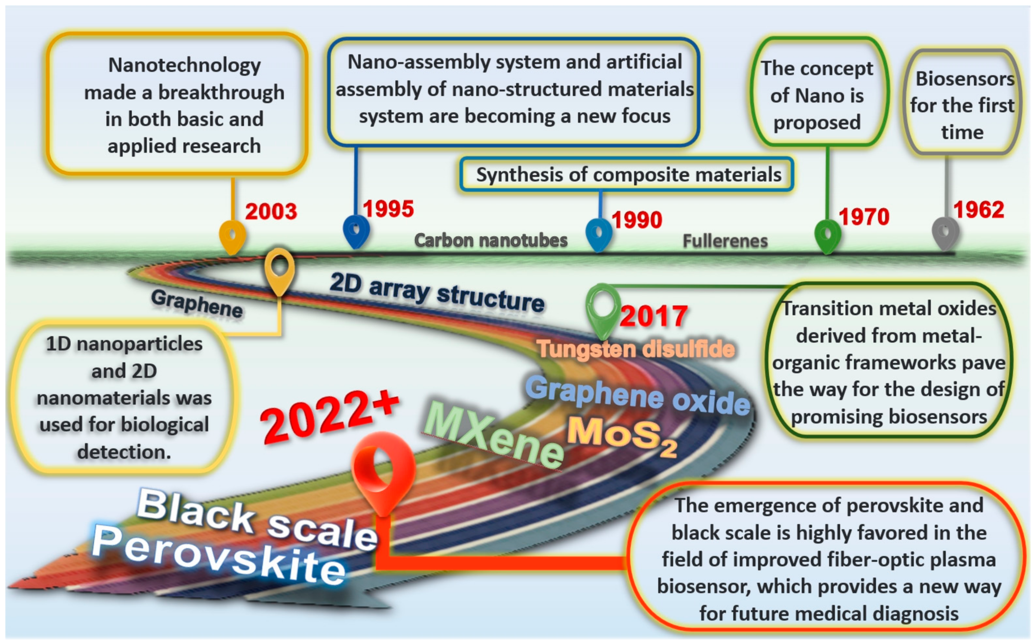
:1. Introduction
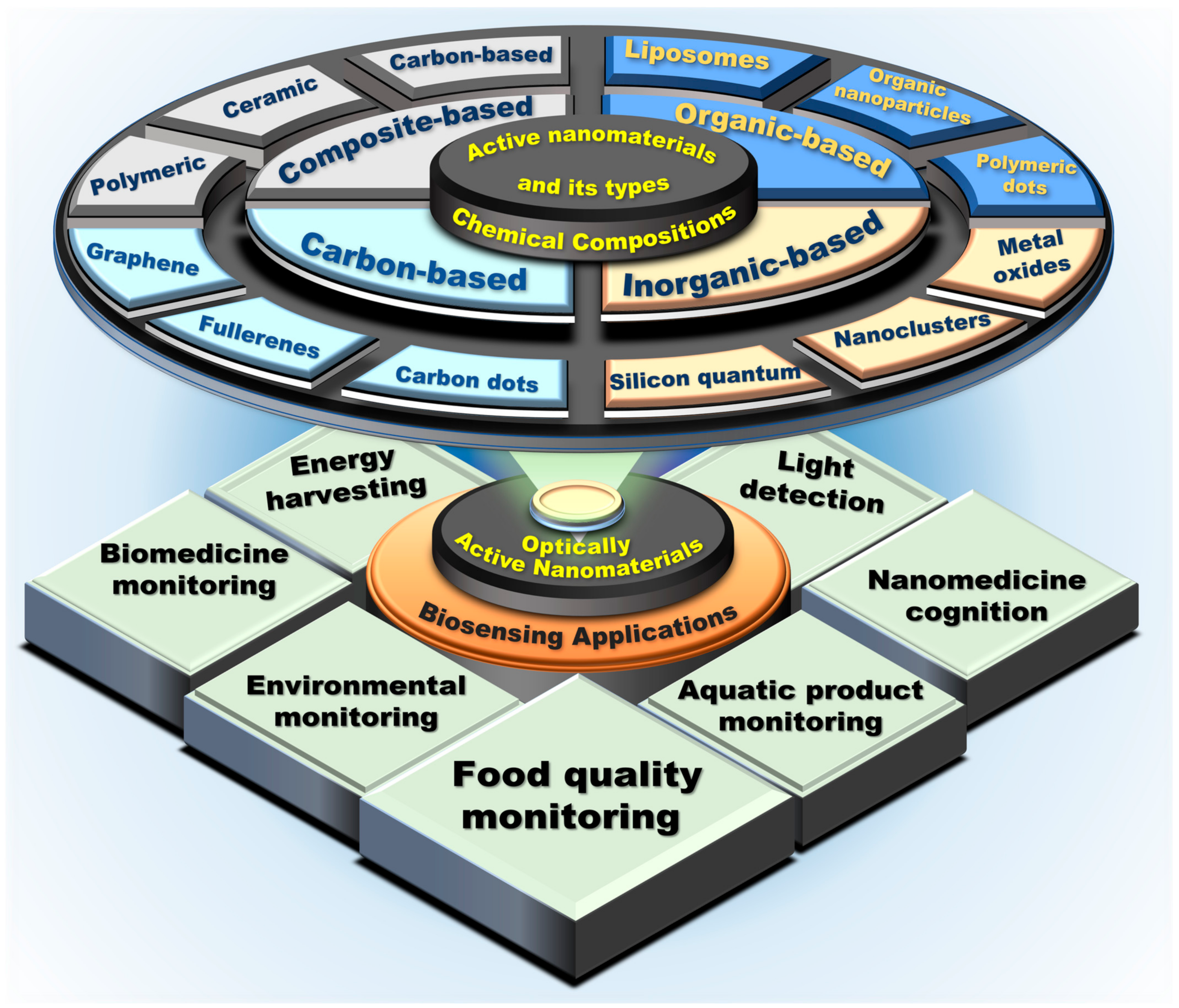
2. Optically Active Nanomaterials and Its Types: Based on Their Chemical Compositions
2.1. Carbon-Based Nanomaterials
2.2. Inorganic-Based Nanomaterials
2.3. Organic-Based Nanomaterials
2.4. Composite-Based Nanomaterials
3. Biosensing Applications
3.1. Disease Detection
3.2. Biomolecules Detection
3.3. Microorganism Detection
4. Challenges and Future Prospects
5. Conclusions
Author Contributions
Funding
Institutional Review Board Statement
Informed Consent Statement
Data Availability Statement
Acknowledgments
Conflicts of Interest
References
- Clark, L.C., Jr.; Lyons, C. Electrode systems for continuous monitoring in cardiovascular surgery. Ann. N. Y. Acad. Sci. 1962, 102, 29–45. [Google Scholar] [CrossRef] [PubMed]
- Cass, A.; Davis, G.; Francis, G.D.; Hill, H.; Aston, W.J.; Higgins, I.J.; Plotkin, E.V.; Scott, L.; Turner, A. Ferrocene-mediated enzyme electrode for amperometric determination of glucose. Anal. Chem. 1984, 56, 667–671. [Google Scholar] [CrossRef] [PubMed]
- Chen, A.C.; Chatterjee, S. Nanomaterials based electrochemical sensors for biomedical applications. Chem. Soc. Rev. 2013, 42, 5425–5438. [Google Scholar] [CrossRef] [PubMed]
- Li, T.; Shang, D.W.; Gao, S.W.; Wang, B.; Kong, H.; Yang, G.Z.; Shu, W.D.; Xu, P.L.; Wei, G. Two-Dimensional Material-Based Electrochemical Sensors/Biosensors for Food Safety and Biomolecular Detection. Biosensors 2022, 12, 314. [Google Scholar] [CrossRef]
- Kotsiri, Z.; Vidic, J.; Vantarakis, A. Applications of biosensors for bacteria and virus detection in food and water—A systematic review. J. Environ. Sci. 2022, 111, 367–379. [Google Scholar] [CrossRef] [PubMed]
- Vallabani, N.V.S.; Singh, S. Recent advances and future prospects of iron oxide nanoparticles in biomedicine and diagnostics. 3 Biotech. 2018, 8, 279. [Google Scholar] [CrossRef] [Green Version]
- Ainla, A.; Mousavi, M.P.S.; Tsaloglou, M.N.; Redston, J.; Bell, J.G.; Fernandez-Abedul, M.T.; Whitesides, G.M. Open-Source Potentiostat for Wireless Electrochemical Detection with Smartphones. Anal. Chem. 2018, 90, 6240–6246. [Google Scholar] [CrossRef] [Green Version]
- Das Mahapatra, S.; Mohapatra, P.C.; Aria, A.I.; Christie, G.; Mishra, Y.K.; Hofmann, S.; Thakur, V.K. Piezoelectric Materials for Energy Harvesting and Sensing Applications: Roadmap for Future Smart Materials. Adv. Sci. 2021, 8, 2100864. [Google Scholar] [CrossRef]
- Park, H.L.; Kim, H.; Lim, D.; Zhou, H.; Kim, Y.H.; Lee, Y.; Park, S.; Lee, T.W. Retina-Inspired Carbon Nitride-Based Photonic Synapses for Selective Detection of UV Light. Adv. Mater. 2020, 32, 1906899. [Google Scholar] [CrossRef]
- Ren, Z.G.; Sun, S.C.; Sun, R.R.; Cui, G.Y.; Hong, L.J.; Rao, B.C.; Li, A.; Yu, Z.J.; Kan, Q.C.; Mao, Z.W. A Metal-Polyphenol-Coordinated Nanomedicine for Synergistic Cascade Cancer Chemotherapy and Chemodynamic Therapy. Adv. Mater. 2020, 32, 1906024. [Google Scholar] [CrossRef]
- Li, M.; Singh, R.; Marques, C.; Zhang, B.; Kumar, S. 2D material assisted SMF-MCF-MMF-SMF based LSPR sensor for creatinine detection. Opt. Express 2021, 29, 38150–38167. [Google Scholar] [CrossRef] [PubMed]
- Du, J.H.; Cheng, H.M. The Fabrication, Properties, and Uses of Graphene/Polymer Composites. Macromol. Chem. Phys. 2012, 213, 1060–1077. [Google Scholar] [CrossRef]
- Shaw, J.C.; Zhou, H.L.; Chen, Y.; Weiss, N.O.; Liu, Y.; Huang, Y.; Duan, X.F. Chemical vapor deposition growth of monolayer MoSe2 nanosheets. Nano Res. 2014, 7, 511–517. [Google Scholar] [CrossRef]
- Huang, Q.Q.; Chen, Y.; Yu, H.Q.; Yan, L.G.; Zhang, J.H.; Wang, B.; Du, B.; Xing, L.T. Magnetic graphene oxide/MgAl-layered double hydroxide nanocomposite: One-pot solvothermal synthesis, adsorption performance and mechanisms for Pb2+, Cd2+, and Cu2+. Chem. Eng. J. 2018, 341, 1–9. [Google Scholar] [CrossRef]
- Naresh, V.; Lee, N. A Review on Biosensors and Recent Development of Nanostructured Materials-Enabled Biosensors. Sensors 2021, 21, 1109. [Google Scholar] [CrossRef] [PubMed]
- Bollella, P.; Fusco, G.; Tortolini, C.; Sanzo, G.; Favero, G.; Gorton, L.; Antiochia, R. Beyond graphene: Electrochemical sensors and biosensors for biomarkers detection. Biosens. Bioelectron. 2017, 89, 152–166. [Google Scholar] [CrossRef] [PubMed]
- Zhang, S.; Rong, F.L.; Guo, C.A.P.; Duan, F.H.; He, L.H.; Wang, M.H.; Zhang, Z.H.; Kang, M.M.; Du, M. Metal-organic frameworks (MOFs) based electrochemical biosensors for early cancer diagnosis in vitro. Coord. Chem. Rev. 2021, 439, 213948. [Google Scholar] [CrossRef]
- Ackermann, J.; Metternich, J.T.; Herbertz, S.; Kruss, S. Biosensing with Fluorescent Carbon Nanotubes. Angew. Chem.-Int. Ed. 2022, 61, 2112372. [Google Scholar] [CrossRef]
- Aldewachi, H.; Chalati, T.; Woodroofe, M.N.; Bricklebank, N.; Sharrack, B.; Gardiner, P. Gold nanoparticle-based colorimetric biosensors. Nanoscale 2018, 10, 18–33. [Google Scholar] [CrossRef] [Green Version]
- Peng, F.; Su, Y.Y.; Zhong, Y.L.; Fan, C.H.; Lee, S.T.; He, Y. Silicon Nanomaterials Platform for Bioimaging, Biosensing, and Cancer Therapy. Acc. Chem. Res. 2014, 47, 612–623. [Google Scholar] [CrossRef]
- Liang, X.; Li, N.; Zhang, R.H.; Yin, P.G.; Zhang, C.M.; Yang, N.; Liang, K.; Kong, B. Carbon-based SERS biosensor: From substrate design to sensing and bioapplication. Npg Asia Mater. 2021, 13, 8. [Google Scholar] [CrossRef]
- Loiseau, A.; Asila, V.; Boitel-Aullen, G.; Lam, M.; Salmain, M.; Boujday, S. Silver-Based Plasmonic Nanoparticles for and Their Use in Biosensing. Biosensors 2019, 9, 78. [Google Scholar] [CrossRef] [PubMed] [Green Version]
- Kumar, V.; Raghuwanshi, S.K.; Kumar, S. Recent Advances in Carbon Nanomaterials Based SPR Sensor for Biomolecules and Gas Detection—A Review. IEEE Sens. J. 2022, 22, 15661–15672. [Google Scholar] [CrossRef]
- Raghuwanshi, S.K.; Kumar, S.; Singh, Y. 2D Materials for Surface Plasmon Resonance-Based Sensors; CRC Press: Boca Raton, FL, USA, 2021. [Google Scholar]
- Kaur, B.; Kumar, S.; Kaushik, B.K. Antimonene, CNT and MoS2 Based SPR-Fiber-Optic Probe for Tuberculosis Detection. IEEE Sens. J. 2022, 22, 14903–14910. [Google Scholar] [CrossRef]
- Kim, D.S.; Yang, X.; Lee, J.H.; Yoo, H.Y.; Park, C.; Kim, S.W.; Lee, J. Development of GO/Co/Chitosan-Based Nano-Biosensor for Real-Time Detection of D-Glucose. Biosensors 2022, 12, 464. [Google Scholar] [CrossRef]
- Baraty, F.; Hamedi, S. Design of a 2D photonic crystal biosensor using X-shape ring resonator based on Graphene Oxide (GO) for detection of blood components. J. Electromagn. Waves Appl. 2022, 36, 2401–2418. [Google Scholar] [CrossRef]
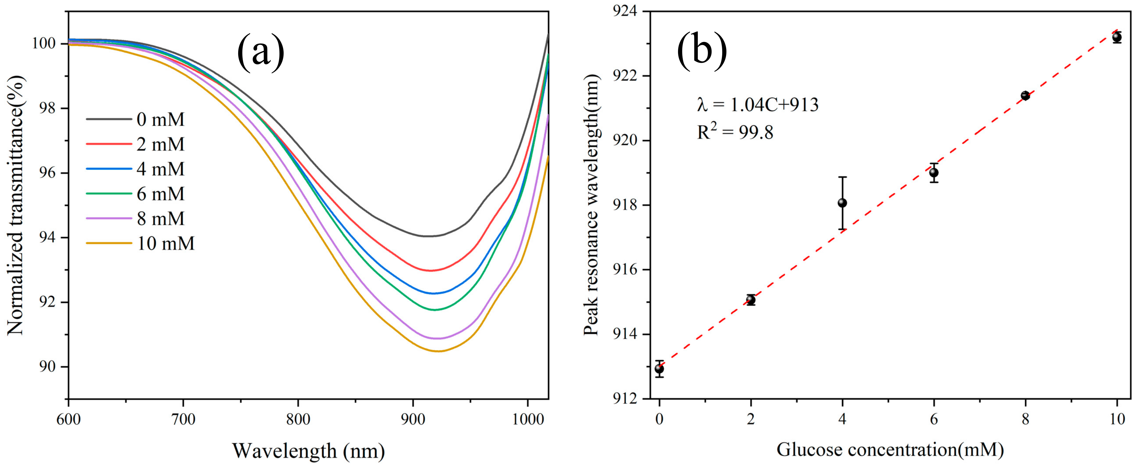
- Yang, Q.; Zhang, X.; Kumar, S.; Singh, R.; Zhang, B.; Bai, C.; Pu, X. Development of Glucose Sensor Using Gold Nanoparticles and Glucose-Oxidase Functionalized Tapered Fiber Structure. Plasmonics 2020, 15, 841–848. [Google Scholar] [CrossRef]
- Yang, Q.; Zhu, G.; Singh, L.; Wang, Y.; Singh, R.; Zhang, B.; Zhang, X.; Kumar, S. Highly sensitive and selective sensor probe using glucose oxidase/gold nanoparticles/graphene oxide functionalized tapered optical fiber structure for detection of glucose. Optik 2020, 208, 164536. [Google Scholar] [CrossRef]
- Pandey, P.S.; Raghuwanshi, S.K.; Kumar, S. Recent Advances in Two-Dimensional Materials-Based Kretschmann Configuration for SPR Sensors: A Review. IEEE Sens. J. 2022, 22, 1069–1080. [Google Scholar] [CrossRef]
- Kaur, B.; Kumar, S.; Kaushik, B.K. 2D Materials-Based Fiber Optic SPR Biosensor for Cancer Detection at 1550 nm. IEEE Sens. J. 2021, 21, 23957–23964. [Google Scholar] [CrossRef]
- Singh, R.; Kumar, S. Cancer Targeting and Diagnosis: Recent Trends with Carbon Nanotubes. Nanomaterials 2022, 12, 2283. [Google Scholar] [CrossRef]
- Li, G.; Xu, Q.; Singh, R.; Zhang, W.; Marques, C.; Xie, Y.; Zhang, B.; Kumar, S. Graphene Oxide/Multiwalled Carbon Nanotubes Assisted Serial Quadruple Tapered Structure-Based LSPR Sensor for Glucose Detection. IEEE Sens. J. 2022, 22, 16904–16911. [Google Scholar] [CrossRef]
- Wang, Y.; Singh, R.; Li, M.; Min, R.; Wu, Q.; Kaushik, B.K.; Jha, R.; Zhang, B.; Kumar, S. Cardiac Troponin I Detection using Gold/Cerium-Oxide Nanoparticles assisted Hetro-Core Fiber Structure. IEEE Trans. NanoBioscience 2022, 1. [Google Scholar] [CrossRef] [PubMed]
- Barrientos, K.; Arango, J.P.; Moncada, M.S.; Placido, J.; Patiño, J.; Macías, S.L.; Maldonado, C.; Torijano, S.; Bustamante, S.; Londoño, M.E.; et al. Carbon dot-based biosensors for the detection of communicable and non-communicable diseases. Talanta 2023, 251, 123791. [Google Scholar] [CrossRef] [PubMed]
- Mo, G.; He, X.; Zhou, C.; Ya, D.; Feng, J.; Yu, C.; Deng, B. A novel ECL sensor based on a boronate affinity molecular imprinting technique and functionalized SiO2@CQDs/AuNPs/MPBA nanocomposites for sensitive determination of alpha-fetoprotein. Biosens. Bioelectron. 2019, 126, 558–564. [Google Scholar] [CrossRef] [PubMed]
- Elugoke, S.E.; Adekunle, A.S.; Fayemi, O.E.; Mamba, B.B.; Sherif, E.-S.M.; Ebenso, E.E. Carbon-Based Quantum Dots for Electrochemical Detection of Monoamine Neurotransmitters—Review. Biosensors 2020, 10, 162. [Google Scholar] [CrossRef] [PubMed]
- Iannazzo, D.; Espro, C.; Celesti, C.; Ferlazzo, A.; Neri, G. Smart Biosensors for Cancer Diagnosis Based on Graphene Quantum Dots. Cancers 2021, 13, 3194. [Google Scholar] [CrossRef]
- Ratlam, C.; Phanichphant, S.; Sriwichai, S. Development of dopamine biosensor based on polyaniline/carbon quantum dots composite. J. Polym. Res. 2020, 27, 183. [Google Scholar] [CrossRef]
- Wei, Q.; Zhang, P.; Liu, T.; Pu, H.; Sun, D.-W. A fluorescence biosensor based on single-stranded DNA and carbon quantum dots for acrylamide detection. Food Chem. 2021, 356, 129668. [Google Scholar] [CrossRef]
- Wu, S.; Jiang, M.; Mao, H.; Zhao, N.; He, D.; Chen, Q.; Liu, D.; Zhang, W.; Song, X.-M. A sensitive cholesterol electrochemical biosensor based on biomimetic cerasome and graphene quantum dots. Anal. Bioanal. Chem. 2022, 414, 3593–3603. [Google Scholar] [CrossRef]
- Jiang, W.; Zhao, Y.; Zhu, X.; Liu, H.; Sun, B. Carbon Dot-Based Biosensors. Adv. NanoBiomed Res. 2021, 1, 2000042. [Google Scholar] [CrossRef]
- Cho, M.-J.; Park, S.-Y. Carbon-dot-based ratiometric fluorescence glucose biosensor. Sens. Actuators B Chem. 2019, 282, 719–729. [Google Scholar] [CrossRef]
- Xie, H.; Luo, G.; Niu, Y.; Weng, W.; Zhao, Y.; Ling, Z.; Ruan, C.; Li, G.; Sun, W. Synthesis and utilization of Co3O4 doped carbon nanofiber for fabrication of hemoglobin-based electrochemical sensor. Mater. Sci. Eng. C 2020, 107, 110209. [Google Scholar] [CrossRef] [PubMed]
- Kaçar, C.; Erden, P.E. An amperometric biosensor based on poly(l-aspartic acid), nanodiamond particles, carbon nanofiber, and ascorbate oxidase–modified glassy carbon electrode for the determination of l-ascorbic acid. Anal. Bioanal. Chem. 2020, 412, 5315–5327. [Google Scholar] [CrossRef] [PubMed]
- Niri, A.D.; Faridi-Majidi, R.; Saber, R.; Khosravani, M.; Adabi, M. Electrospun carbon nanofiber-based electrochemical biosensor for the detection of hepatitis B virus. Biointerface Res. Appl. Chem. 2019, 9, 4022–4026. [Google Scholar]
- Erden, P.E.; Kaçar Selvi, C.; Kılıç, E. A novel tyramine biosensor based on carbon nanofibers, 1-butyl-3-methylimidazolium tetrafluoroborate and gold nanoparticles. Microchem. J. 2021, 170, 106729. [Google Scholar] [CrossRef]
- Li, J.-x.; Zhang, W.-h.; Tong, Z.-r.; Liu, J.-w. Fiber optic sensor modified by graphene oxide–glucose oxidase for glucose detection. Opt. Commun. 2021, 492, 126983. [Google Scholar] [CrossRef]
- Esposito, F.; Sansone, L.; Srivastava, A.; Cusano, A.M.; Campopiano, S.; Giordano, M.; Iadicicco, A. Label-free detection of vitamin D by optical biosensing based on long period fiber grating. Sens. Actuators B Chem. 2021, 347, 130637. [Google Scholar] [CrossRef]
- Xu, B.; Huang, J.; Ding, L.; Cai, J. Graphene oxide-functionalized long period fiber grating for ultrafast label-free glucose biosensor. Mater. Sci. Eng. C 2020, 107, 110329. [Google Scholar] [CrossRef]
- Tong, Z.; Zhao, Y.; Wang, X.; Li, P.; Zhang, W.; Zhang, J. Research on dual-parameter biosensor based on no-core fiber coated by composite film. Optik 2022, 259, 169027. [Google Scholar] [CrossRef]
- Cao, Z.; Yao, B.; Qin, C.; Yang, R.; Guo, Y.; Zhang, Y.; Wu, Y.; Bi, L.; Chen, Y.; Xie, Z.; et al. Biochemical sensing in graphene-enhanced microfiber resonators with individual molecule sensitivity and selectivity. Light Sci. Appl. 2019, 8, 107. [Google Scholar] [CrossRef]
- Yu, S.; Ding, L.; Lin, H.; Wu, W.; Huang, J. A novel optical fiber glucose biosensor based on carbon quantum dots-glucose oxidase/cellulose acetate complex sensitive film. Biosens. Bioelectron. 2019, 146, 111760. [Google Scholar] [CrossRef]
- Wu, W.; Huang, J.; Ding, L.; Lin, H.; Yu, S.; Yuan, F.; Liang, B. A real-time and highly sensitive fiber optic biosensor based on the carbon quantum dots for nitric oxide detection. J. Photochem. Photobiol. A Chem. 2021, 405, 112963. [Google Scholar] [CrossRef]
- Sangubotla, R.; Kim, J. Fiber-optic biosensor based on the laccase immobilization on silica-functionalized fluorescent carbon dots for the detection of dopamine and multi-color imaging applications in neuroblastoma cells. Mater. Sci. Eng. C 2021, 122, 111916. [Google Scholar] [CrossRef]
- Li, Q.; Ding, L.; Zhang, Y.; Wu, T. A Cholesterol Optical Fiber Sensor Based on CQDs-COD/CA Composite. IEEE Sens. J. 2022, 22, 6247–6255. [Google Scholar] [CrossRef]
- Fukuda, T.; Muguruma, H.; Iwasa, H.; Tanaka, T.; Hiratsuka, A.; Shimizu, T.; Tsuji, K.; Kishimoto, T. Electrochemical determination of uric acid in urine and serum with uricase/carbon nanotube/carboxymethylcellulose electrode. Anal. Biochem. 2020, 590, 113533. [Google Scholar] [CrossRef]
- Pathak, A.; Gupta, B.D. Ultra-selective fiber optic SPR platform for the sensing of dopamine in synthetic cerebrospinal fluid incorporating permselective nafion membrane and surface imprinted MWCNTs-PPy matrix. Biosens. Bioelectron. 2019, 133, 205–214. [Google Scholar] [CrossRef]
- Zhao, B.; Zhou, Y.; Qu, J.; Yin, F.; Yin, S.; Chang, Y.; Zhang, W. Preparation and performance of CNTs-Pt formaldehyde sensor and CNTs-Au glucose sensor. Sens. Rev. 2022, 42, 544–553. [Google Scholar] [CrossRef]
- Homola, J.; Yee, S.S.; Gauglitz, G. Surface plasmon resonance sensors: Review. Sens. Actuators B Chem. 1999, 54, 3–15. [Google Scholar] [CrossRef]
- Li, L.; Liu, S.; Chen, Z.; Dai, Z.; Chen, N.; Pang, F.; Shang, Y.; Lu, B.; Wang, T. Remote detection of the surface-enhanced Raman spectrum with the optical fiber nanoprobe. Opt. Spectrosc. 2014, 116, 575–578. [Google Scholar] [CrossRef]
- Zhang, J.; Chen, S.; Gong, T.; Zhang, X.; Zhu, Y. Tapered Fiber Probe Modified by Ag Nanoparticles for SERS Detection. Plasmonics 2015, 11, 743–751. [Google Scholar] [CrossRef]
- Kumar, S.; Agrawal, N.; Saha, C.; Jha, R. Optical Fiber-Based Plasmonic Biosensors: Trends, Techniques, and Applications; CRC Press: Boca Raton, FL, USA, 2022. [Google Scholar]
- Kumari, A.; Vyas, V.; Kumar, S. Synthesis, characterization, and applications of gold nanoparticles in development of plasmonic optical fiber-based sensors. Nanotechnology 2023, 34, 042001. [Google Scholar] [CrossRef]
- Uniyal, S.; Choudhary, K.; Sachdev, S.; Kumar, S. Synthesis and characterization of gold nanoparticles and graphene oxide for the development of optical fiber biosensors. Appl. Opt. 2022, 61, 7618–7624. [Google Scholar] [CrossRef]
- Kumar, S.; Wang, Y.; Li, M.; Wang, Q.; Malathi, S.; Marques, C.; Singh, R.; Zhang, B. Plasmon-Based Tapered-in-Tapered Fiber Structure for p-Cresol Detection: From Human Healthcare to Aquaculture Application. IEEE Sens. J. 2022, 22, 18493–18500. [Google Scholar] [CrossRef]
- Sharma, P.; Semwal, V.; Gupta, B.D. A highly selective LSPR biosensor for the detection of taurine realized on optical fiber substrate and gold nanoparticles. Opt. Fiber Technol. 2019, 52, 101962. [Google Scholar] [CrossRef]
- Yuan, H.; Ji, W.; Chu, S.; Qian, S.; Wang, F.; Masson, J.F.; Han, X.; Peng, W. Fiber-optic surface plasmon resonance glucose sensor enhanced with phenylboronic acid modified Au nanoparticles. Biosens. Bioelectron. 2018, 117, 637–643. [Google Scholar] [CrossRef]
- Potdar, R.P.; Khollam, Y.B.; Shaikh, S.F.; More, P.S.; Rana, A. Polyvinylpyrrolidone-Capped Silver Nanoparticles for Highly Sensitive and Selective Optical Fiber-Based Ammonium Sensor. Nanomaterials 2022, 12, 3373. [Google Scholar] [CrossRef]
- Kapoor, V.; Sharma, N.K.; Gupta, S.; Kumar, P. Fiber optic SPR sensing of liquids using copper and zinc oxide. Optik 2021, 238, 166727. [Google Scholar] [CrossRef]
- Phan, L.M.T.; Baek, S.H.; Nguyen, T.P.; Park, K.Y.; Ha, S.; Rafique, R.; Kailasa, S.K.; Park, T.J. Synthesis of fluorescent silicon quantum dots for ultra-rapid and selective sensing of Cr(VI) ion and biomonitoring of cancer cells. Mater. Sci. Eng. C Mater. Biol. Appl. 2018, 93, 429–436. [Google Scholar] [CrossRef]
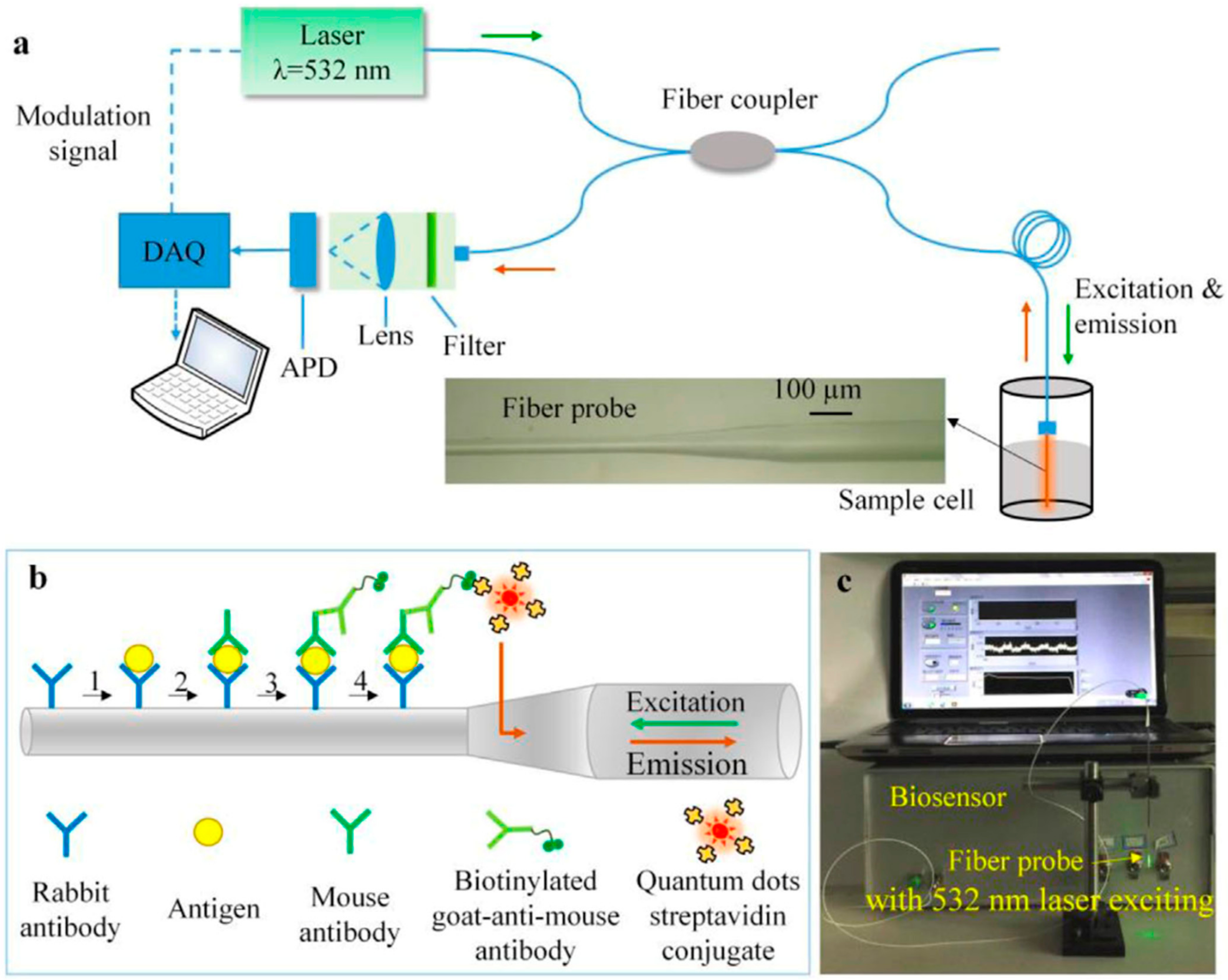
- Cui, J.; Zhou, M.; Li, Y.; Liang, Z.; Li, Y.; Yu, L.; Liu, Y.; Liang, Y.; Chen, L.; Yang, C. A New Optical Fiber Probe-Based Quantum Dots Immunofluorescence Biosensors in the Detection of Staphylococcus aureus. Front. Cell Infect. Microbiol. 2021, 11, 665241. [Google Scholar] [CrossRef]
- Liu, L.; Zhu, G.; Zeng, W.; Yi, Y.; Lv, B.; Qian, J.; Zhang, D. Silicon quantum dot-coated onto gold nanoparticles as an optical probe for colorimetric and fluorometric determination of cysteine. Mikrochim. Acta 2019, 186, 98. [Google Scholar] [CrossRef]
- Huang, B.H.; Shen, S.S.; Wei, N.; Guo, X.F.; Wang, H. Fluorescence biosensor based on silicon quantum dots and 5,5′-dithiobis-(2-nitrobenzoic acid) for thiols in living cells. Spectrochim. Acta A Mol. Biomol. Spectrosc. 2020, 229, 117972. [Google Scholar] [CrossRef]
- Du, L.; Li, Z.; Yao, J.; Wen, G.; Dong, C.; Li, H.W. Enzyme free glucose sensing by amino-functionalized silicon quantum dot. Spectrochim. Acta A Mol. Biomol. Spectrosc. 2019, 216, 303–309. [Google Scholar] [CrossRef]
- Deniz, S.A.; Goker, S.; Toppare, L.; Soylemez, S. Fabrication of D–A–D type conducting polymer, carbon nanotubes and silica nanoparticle-based laccase biosensor for catechol detection. New J. Chem. 2022, 46, 15521–15529. [Google Scholar] [CrossRef]
- Mathelié-Guinlet, M.; Cohen-Bouhacina, T.; Gammoudi, I.; Martin, A.; Béven, L.; Delville, M.-H.; Grauby-Heywang, C. Silica nanoparticles-assisted electrochemical biosensor for the rapid, sensitive and specific detection of Escherichia coli. Sens. Actuators B Chem. 2019, 292, 314–320. [Google Scholar] [CrossRef]
- Ananda, P.D.K.P.; Tillekaratne, A.; Hettiarachchi, C.; Lalichchandran, N. Sensitive detection of E. coli using bioconjugated fluorescent silica nanoparticles. Appl. Surf. Sci. Adv. 2021, 6, 100159. [Google Scholar] [CrossRef]
- Cheraghi Shahi, S.; Dadmehr, M.; Korouzhdehi, B.; Tavassoli, A. A novel colorimetric biosensor for sensitive detection of aflatoxin mediated by bacterial enzymatic reaction in saffron samples. Nanotechnology 2021, 32, 505503. [Google Scholar] [CrossRef]
- Nycz, M.; Arkusz, K.; Pijanowska, D.G. Fabrication of Electrochemical Biosensor Based on Titanium Dioxide Nanotubes and Silver Nanoparticles for Heat Shock Protein 70 Detection. Materials 2021, 14, 3767. [Google Scholar] [CrossRef]
- Yang, K.; Li, J.; Lamy de la Chapelle, M.; Huang, G.; Wang, Y.; Zhang, J.; Xu, D.; Yao, J.; Yang, X.; Fu, W. A terahertz metamaterial biosensor for sensitive detection of microRNAs based on gold-nanoparticles and strand displacement amplification. Biosens. Bioelectron. 2021, 175, 112874. [Google Scholar] [CrossRef]
- Xu, Y.; Xiong, M.; Yan, H. A portable optical fiber biosensor for the detection of zearalenone based on the localized surface plasmon resonance. Sens. Actuators B Chem. 2021, 336, 129752. [Google Scholar] [CrossRef]
- Sadeghi, S.; Rahaie, M. Design and Fabrication of a DNA-copper Nanocluster-based Biosensor for Multiple Detections of Circulating miRNAs in Early Screening of Breast Cancer. J. Fluoresc. 2022, 32, 2297–2307. [Google Scholar] [CrossRef]
- Khalef, W.K.; Aljubour, A.A.; Faisal, A.D. Glucose biosensor electrode fabrication based on CuO/ZnO nanostructures. J. Phys. Conf. Ser. 2021, 1795, 012038. [Google Scholar] [CrossRef]
- Ahmad, R.; Khan, M.; Mishra, P.; Jahan, N.; Ahsan, M.A.; Ahmad, I.; Khan, M.R.; Watanabe, Y.; Syed, M.A.; Furukawa, H.; et al. Engineered Hierarchical CuO Nanoleaves Based Electrochemical Nonenzymatic Biosensor for Glucose Detection. J. Electrochem. Soc. 2021, 168, 017501. [Google Scholar] [CrossRef]
- Sinha, K.; Chakraborty, B.; Chaudhury, S.S.; Chaudhuri, C.R.; Chattopadhyay, S.K.; Mukhopadhyay, C.D. Selective, Ultra-Sensitive, and Rapid Detection of Serotonin by Optimized ZnO Nanorod FET Biosensor. IEEE Trans. Nano Biosci. 2022, 21, 65–74. [Google Scholar] [CrossRef]
- Kamaci, U.D.; Kamaci, M. Selective and Sensitive ZnO Quantum Dots Based Fluorescent Biosensor for Detection of Cysteine. J. Fluoresc. 2021, 31, 401–414. [Google Scholar] [CrossRef] [PubMed]
- Zhang, J.; Hu, H.; Yang, L. Ultra-highly sensitive and stable acetylcholinesterase biosensor based on TiO2-NRs and rGO. Microchem. J. 2021, 168, 106435. [Google Scholar] [CrossRef]
- Wu, Z.; Sun, D.-W.; Pu, H.; Wei, Q. A dual signal-on biosensor based on dual-gated locked mesoporous silica nanoparticles for the detection of Aflatoxin B1. Talanta 2023, 253, 124027. [Google Scholar] [CrossRef]
- Jimenez-Falcao, S.; Parra-Nieto, J.; Pérez-Cuadrado, H.; Martínez-Máñez, R.; Martínez-Ruiz, P.; Villalonga, R. Avidin-gated mesoporous silica nanoparticles for signal amplification in electrochemical biosensor. Electrochem. Commun. 2019, 108, 106556. [Google Scholar] [CrossRef]
- Sahu, S.; Karuppusamy, M.; Easwaramoorthi, S. Water-dispersible polymer coated silica nanoparticle for turn-on fluorometric detection of Cephalexin. Biosens. Bioelectron. X 2022, 12, 100231. [Google Scholar] [CrossRef]
- Yuan, Y.; Hou, W.; Qin, W.; Wu, C. Recent advances in semiconducting polymer dots as optical probes for biosensing. Biomater. Sci. 2021, 9, 328–346. [Google Scholar] [CrossRef] [PubMed]
- Jo, H.J.; Robby, A.I.; Kim, S.G.; Lee, G.; Lee, B.C.; Park, S.Y. Reusable biosensor-based polymer dot-coated electrode surface for wireless detection of bacterial contamination. Sens. Actuators B Chem. 2021, 346, 130503. [Google Scholar] [CrossRef]
- Won, H.J.; Robby, A.I.; Jhon, H.S.; In, I.; Ryu, J.H.; Park, S.Y. Wireless label-free electrochemical detection of cancer cells by MnO2-Decorated polymer dots. Sens. Actuators B Chem. 2020, 320, 128391. [Google Scholar] [CrossRef]
- Qi, H.; Zhang, C. Organic nanoparticles for electrogenerated chemiluminescence assay. Curr. Opin. Electrochem. 2022, 34, 101023. [Google Scholar] [CrossRef]
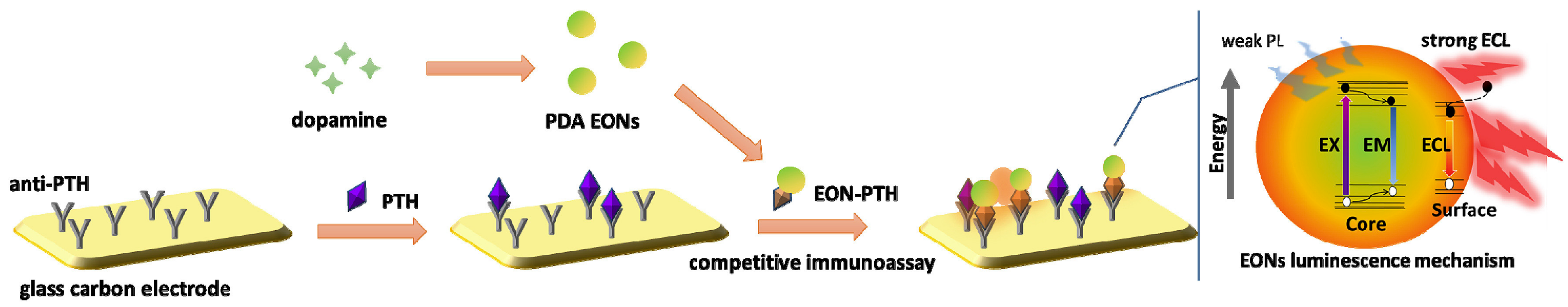
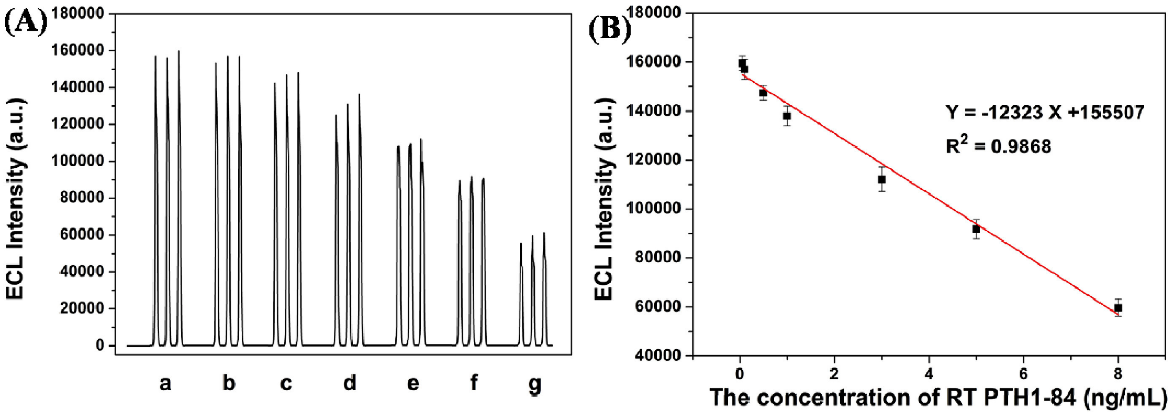
- Li, S.; Liu, Y.; Ma, Q. A novel polydopamine electrochemiluminescence organic nanoparticle-based biosensor for parathyroid hormone detection. Talanta 2019, 202, 540–545. [Google Scholar] [CrossRef] [PubMed]
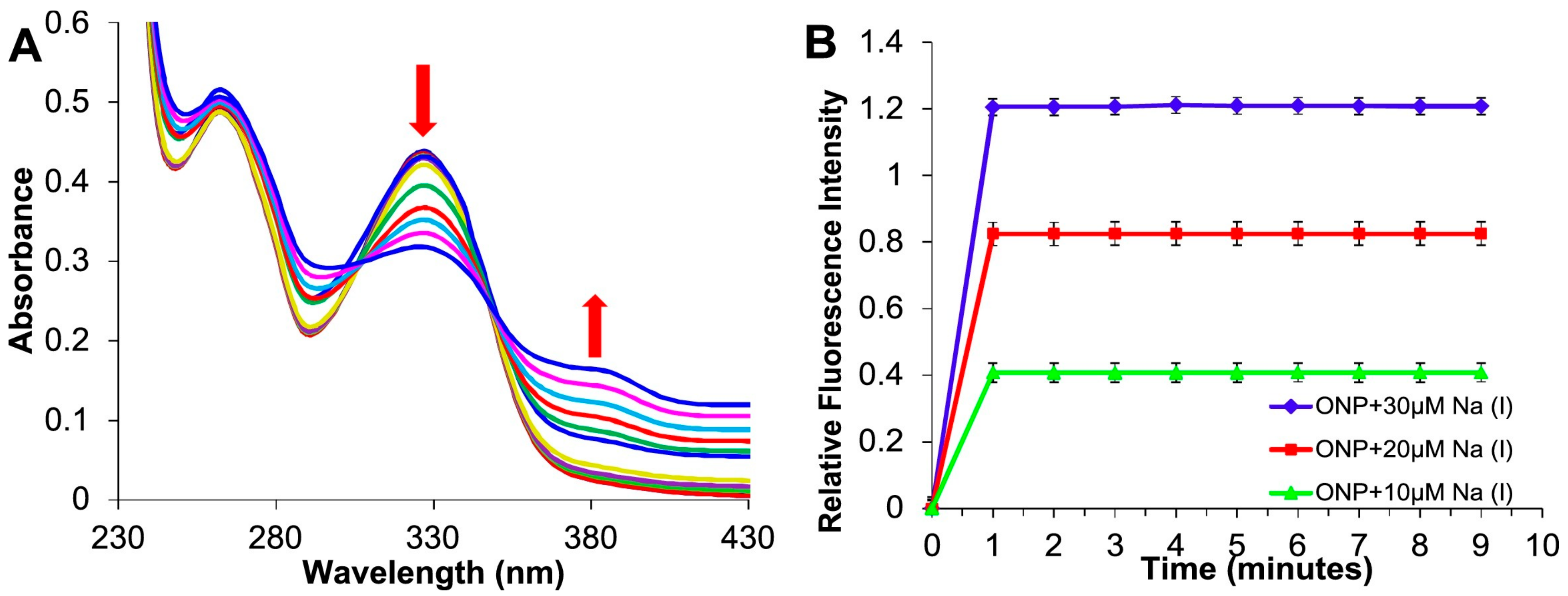
- Kaur, G.; Kaur, N. Estimation of sodium ions using easily engineered organic nanoparticles-based turn-on fluorescent sensor: Application in biological and environmental samples. Sens. Actuators B Chem. 2018, 265, 134–141. [Google Scholar] [CrossRef]
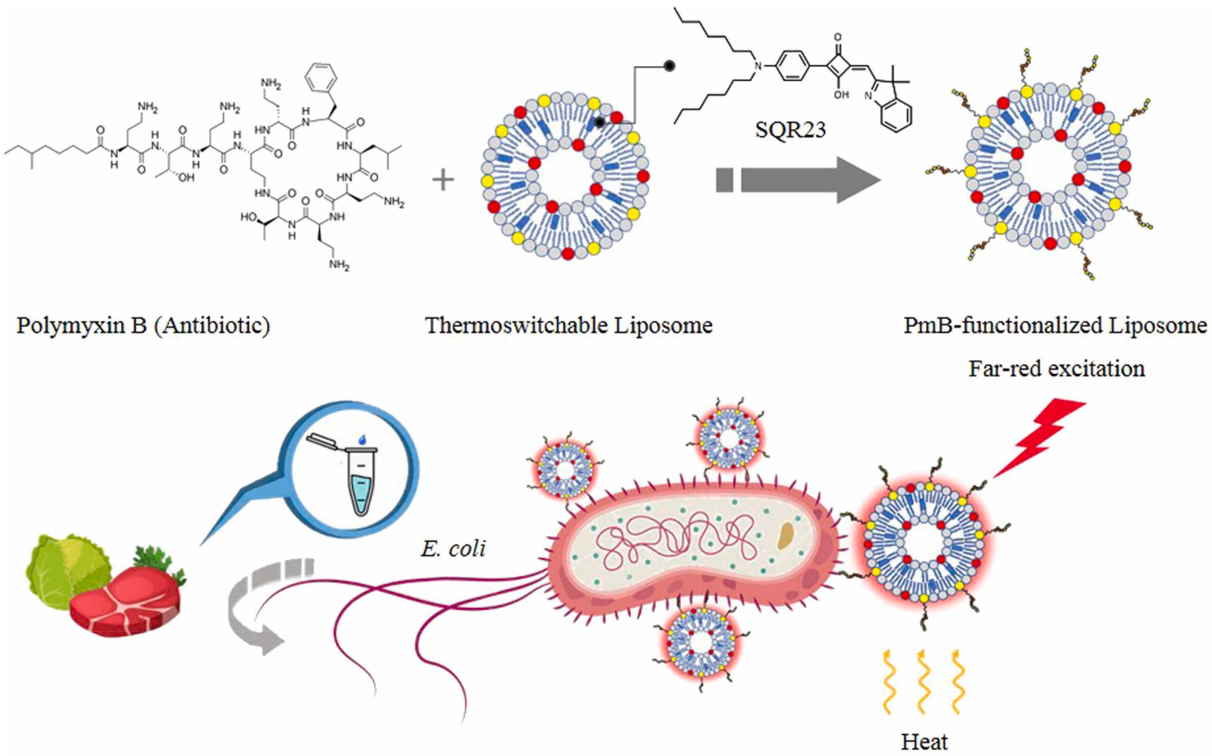
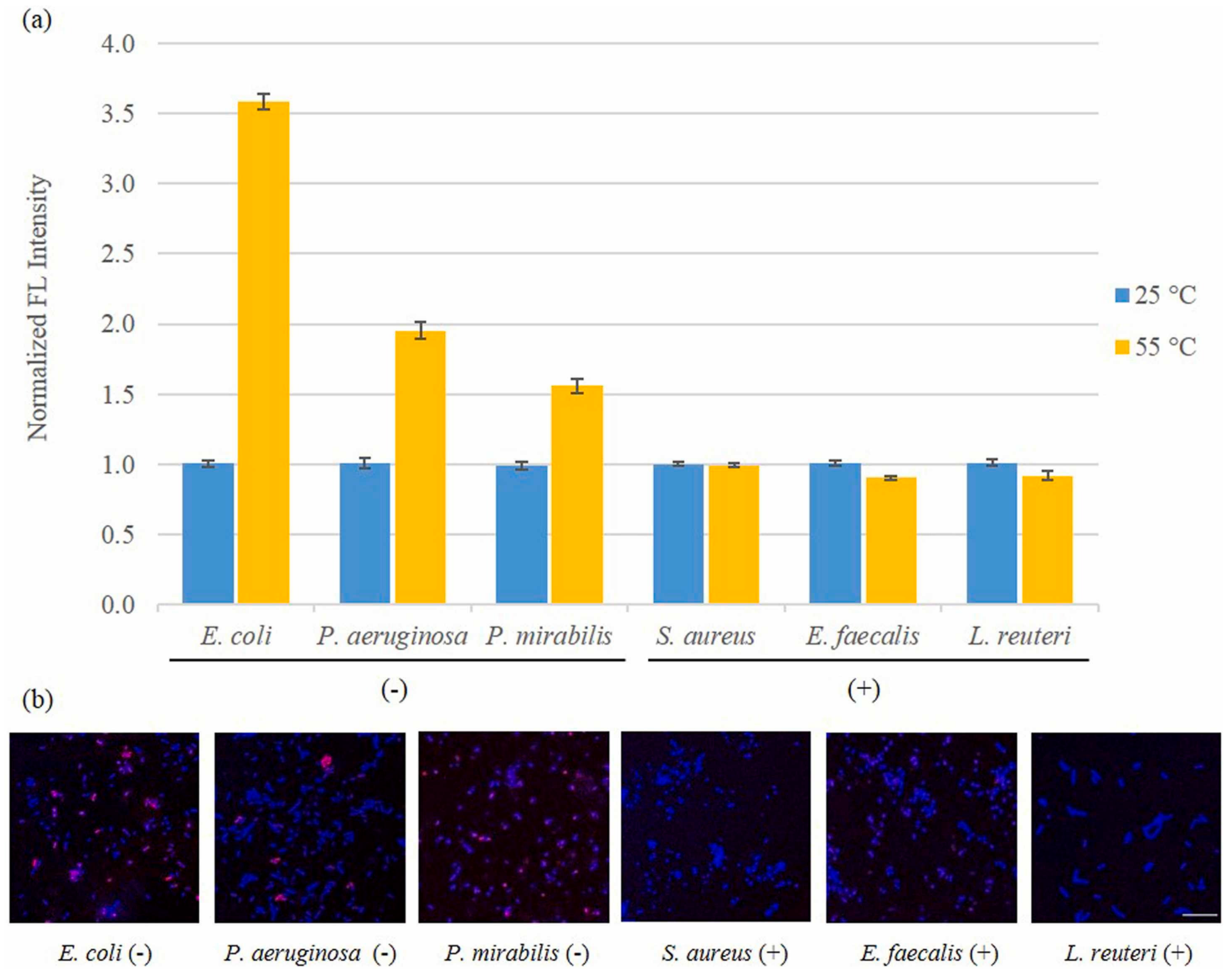
- Tan, J.R.; Ferdinandus; Xing, B.; Lee, C.-L.K. Functionalization of thermoswitchable liposomes for rapid detection of Gram-negative bacteria. Sens. Actuators B Chem. 2022, 367, 132123. [Google Scholar] [CrossRef]
- Kim, C.; Han, B.H.; Kim, D.; Lee, G.; Hong, C.; Kang, J.Y.; Lee, K. Multi-target polydiacetylene liposome-based biosensor for improved exosome detection. Sens. Actuators B Chem. 2022, 355, 131286. [Google Scholar] [CrossRef]
- Zhou, J.; Duan, M.; Huang, D.; Shao, H.; Zhou, Y.; Fan, Y. Label-free visible colorimetric biosensor for detection of multiple pathogenic bacteria based on engineered polydiacetylene liposomes. J. Colloid. Interface Sci. 2022, 606, 1684–1694. [Google Scholar] [CrossRef] [PubMed]
- Chen, H.; Yu, J.; Zhang, J.; Sun, K.; Ding, Z.; Jiang, Y.; Hu, Q.; Wu, C.; Chiu, D.T. Monitoring Metabolites Using an NAD(P)H-sensitive Polymer Dot and a Metabolite-Specific Enzyme. Angew. Chem. Int. Ed. Engl. 2021, 60, 19331–19336. [Google Scholar] [CrossRef] [PubMed]
- Jo, H.J.; Ryu, J.S.; Robby, A.I.; Kim, Y.S.; Chung, H.J.; Park, S.Y. Rapid and selective electrochemical sensing of bacterial pneumonia in human sputum based on conductive polymer dot electrodes. Sens. Actuators B Chem. 2022, 368, 132084. [Google Scholar] [CrossRef]
- Zhang, N.; Gao, H.; Jia, Y.L.; Pan, J.B.; Luo, X.L.; Chen, H.Y.; Xu, J.J. Ultrasensitive Nucleic Acid Assay Based on AIE-Active Polymer Dots with Excellent Electrochemiluminescence Stability. Anal. Chem. 2021, 93, 6857–6864. [Google Scholar] [CrossRef] [PubMed]
- Jamei, H.R.; Rezaei, B.; Ensafi, A.A. Ultra-sensitive and selective electrochemical biosensor with aptamer recognition surface based on polymer quantum dots and C(60)/MWCNTs- polyethylenimine nanocomposites for analysis of thrombin protein. Bioelectrochemistry 2021, 138, 107701. [Google Scholar] [CrossRef] [PubMed]
- Zhang, N.; Zhao, Z.-Y.; Gao, H.; Yu, Y.; Pan, J.-B.; Chen, H.-Y.; Xu, J.-J. An ultrasensitive electrochemiluminescence assay for nucleic acid detection based on carboxyl functionalized polymer dots. J. Electroanal. Chem. 2021, 900, 115743. [Google Scholar] [CrossRef]
- Ding, L.; Qin, Z.; Xiang, C.; Zhou, G. Novel fluorescent organic nanoparticles as a label-free biosensor for dopamine in serum. J. Mater. Chem. B 2017, 5, 2750–2756. [Google Scholar] [CrossRef]
- Liu, X.; Song, J.; Wang, C.; Yang, R.; Sun, P.; Huang, Y.; Zhang, L.; Fan, Q. An amplified fluorescence biosensor for intracellular telomerase determination and in situ imaging based on thioflavin T and conjugated polymer nanoparticles. Sens. Actuators B Chem. 2022, 371, 132485. [Google Scholar] [CrossRef]
- Xiao, S.Y.; Zhen, S.J.; Huang, C.Z.; Li, Y.F. Ultrasensitive ratiometric electrochemiluminescence for detecting atxA mRNA using luminol-encapsulated liposome as effectively amplified signal labels. Biosens. Bioelectron. 2021, 186, 113263. [Google Scholar] [CrossRef]
- Gong, H.; Wu, Y.; Zeng, R.; Zeng, Y.; Liu, X.; Tang, D. CRISPR/Cas12a-mediated liposome-amplified strategy for the photoelectrochemical detection of nucleic acid. Chem. Commun. 2021, 57, 8977–8980. [Google Scholar] [CrossRef] [PubMed]
- Yagati, A.K.; Behrent, A.; Tomanek, V.; Chavan, S.G.; Go, A.; Park, S.R.; Jin, Z.; Baeumner, A.J.; Lee, M.H. Polypyrrole-palladium nanocomposite as a high-efficiency transducer for thrombin detection with liposomes as a label. Anal. Bioanal. Chem. 2022, 414, 3205–3217. [Google Scholar] [CrossRef]
- Kim, J.; Moon, B.S.; Hwang, E.; Shaban, S.; Lee, W.; Pyun, D.G.; Lee, D.H.; Kim, D.H. Solid-state colorimetric polydiacetylene liposome biosensor sensitized by gold nanoparticles. Analyst 2021, 146, 1682–1688. [Google Scholar] [CrossRef]
- Breuer, O.; Sundararaj, U. Big returns from small fibers: A review of polymer/carbon nanotube composites. Polym. Compos. 2004, 25, 630–645. [Google Scholar] [CrossRef]
- Akgöl, S.; Ulucan-Karnak, F.; Kuru, C.İ.; Kuşat, K. The usage of composite nanomaterials in biomedical engineering applications. Biotechnol. Bioeng. 2021, 118, 2906–2922. [Google Scholar] [CrossRef] [PubMed]
- Kumar, V.; Raghuwanshi, S.K.; Kumar, S. Advances in Nanocomposite Thin-Film-Based Optical Fiber Sensors for Environmental Health Monitoring—A Review. IEEE Sens. J. 2022, 22, 14696–14707. [Google Scholar] [CrossRef]
- Mousumi, S. Nanocomposite Materials. In Nanotechnology and the Environment; Mousumi, S., Ed.; IntechOpen: Rijeka, Croatia, 2020; Chapter 6. [Google Scholar]
- Hule, R.A.; Pochan, D.J. Polymer Nanocomposites for Biomedical Applications. MRS Bull. 2007, 32, 354–358. [Google Scholar] [CrossRef] [Green Version]
- De Leon, A.C.; Chen, Q.; Palaganas, N.B.; Palaganas, J.O.; Manapat, J.; Advincula, R.C. High performance polymer nanocomposites for additive manufacturing applications. React. Funct. Polym. 2016, 103, 141–155. [Google Scholar] [CrossRef]
- Razavi, M.; Fathi, M.H.; Meratian, M. Fabrication and characterization of magnesium–fluorapatite nanocomposite for biomedical applications. Mater. Charact. 2010, 61, 1363–1370. [Google Scholar] [CrossRef]
- Rajamehala, M.; Pandian, A.M.; Rajasimman, M.; Gopalakrishnan, B. Synthesis of metal-based functional nanocomposite material and its application for the elimination of paracetamol from synthetic wastewater. Chemosphere 2022, 308, 136530. [Google Scholar] [CrossRef]
- Wang, H.; Rogach, A.L. Hierarchical SnO2 Nanostructures: Recent Advances in Design, Synthesis, and Applications. Chem. Mater. 2014, 26, 123–133. [Google Scholar] [CrossRef]
- Wang, L.; Kang, Y.; Liu, X.; Zhang, S.; Huang, W.; Wang, S. ZnO nanorod gas sensor for ethanol detection. Sens. Actuators B Chem. 2012, 162, 237–243. [Google Scholar] [CrossRef]
- Park, S.; An, S.; Mun, Y.; Lee, C. UV-Enhanced NO2 Gas Sensing Properties of SnO2-Core/ZnO-Shell Nanowires at Room Temperature. ACS Appl. Mater. Interfaces 2013, 5, 4285–4292. [Google Scholar] [CrossRef]
- Huang, H.; Gong, H.; Chow, C.L.; Guo, J.; White, T.J.; Tse, M.S.; Tan, O.K. Low-Temperature Growth of SnO2 Nanorod Arrays and Tunable n–p–n Sensing Response of a ZnO/SnO2 Heterojunction for Exclusive Hydrogen Sensors. Adv. Funct. Mater. 2011, 21, 2680–2686. [Google Scholar] [CrossRef]
- Yan, S.H.; Ma, S.Y.; Xu, X.L.; Li, W.Q.; Luo, J.; Jin, W.X.; Wang, T.T.; Jiang, X.H.; Lu, Y.; Song, H.S. Preparation of SnO2–ZnO hetero-nanofibers and their application in acetone sensing performance. Mater. Lett. 2015, 159, 447–450. [Google Scholar] [CrossRef]
- Liu, J.; Wang, T.; Wang, B.; Sun, P.; Yang, Q.; Liang, X.; Song, H.; Lu, G. Highly sensitive and low detection limit of ethanol gas sensor based on hollow ZnO/SnO2 spheres composite material. Sens. Actuators B: Chem. 2017, 245, 551–559. [Google Scholar] [CrossRef]
- Mikani, M.; Talaei, S.; Rahmanian, R.; Ahmadi, P.; Mahmoudi, A. Sensitive electrochemical sensor for urea determination based on F-doped SnO2 electrode modified with ZnO-Fe3O4 nanoparticles transducer: Application in biological fluids. J. Electroanal. Chem. 2019, 840, 285–294. [Google Scholar] [CrossRef]
- Ghanavati, M.; Tadayon, F.; Basiryanmahabadi, A.; Torabi Fard, N.; Smiley, E. Design of new sensing layer based on ZnO/NiO/Fe3O4/MWCNTs nanocomposite for simultaneous electrochemical determination of Naproxen and Sumatriptan. J. Pharm. Biomed. Anal. 2023, 223, 115091. [Google Scholar] [CrossRef] [PubMed]
- Alula, M.T.; Lemmens, P.; Bo, L.; Wulferding, D.; Yang, J.; Spende, H. Preparation of silver nanoparticles coated ZnO/Fe3O4 composites using chemical reduction method for sensitive detection of uric acid via surface-enhanced Raman spectroscopy. Anal. Chim. Acta 2019, 1073, 62–71. [Google Scholar] [CrossRef]
- Fayemi, O.E.; Adekunle, A.S.; Kumara Swamy, B.E.; Ebenso, E.E. Electrochemical sensor for the detection of dopamine in real samples using polyaniline/NiO, ZnO, and Fe3O4 nanocomposites on glassy carbon electrode. J. Electroanal. Chem. 2018, 818, 236–249. [Google Scholar] [CrossRef]
- Qambrani, N.; Buledi, J.A.; Khand, N.H.; Solangi, A.R.; Ameen, S.; Jalbani, N.S.; Khatoon, A.; Taher, M.A.; Moghadam, F.H.; Shojaei, M.; et al. Facile Synthesis of NiO/ZnO nanocomposite as an effective platform for electrochemical determination of carbamazepine. Chemosphere 2022, 303, 135270. [Google Scholar] [CrossRef]
- Salarizadeh, N.; Habibi-Rezaei, M.; Zargar, S.J. NiO–MoO3 nanocomposite: A sensitive non-enzymatic sensor for glucose and urea monitoring. Mater. Chem. Phys. 2022, 281, 125870. [Google Scholar] [CrossRef]
- Wang, Z.; Singh, R.; Marques, C.; Jha, R.; Zhang, B.; Kumar, S. Taper-in-taper fiber structure-based LSPR sensor for alanine aminotransferase detection. Opt. Express 2021, 29, 43793–43810. [Google Scholar] [CrossRef]
- Ramya, M.; Kumar, P.S.; Rangasamy, G.; Shankar, V.U.; Rajesh, G.; Nirmala, K. Experimental investigation of the electrochemical detection of sulfamethoxazole using copper oxide-MoS2 modified glassy carbon electrodes. Environ. Res. 2023, 216, 114463. [Google Scholar] [CrossRef]
- Lin, Y.; Shen, R.; Liu, N.; Yi, H.; Dai, H.; Lin, J. A highly sensitive peptide-based biosensor using NiCo2O4 nanosheets and g-C3N4 nanocomposite to construct amplified strategy for trypsin detection. Anal. Chim. Acta 2018, 1035, 175–183. [Google Scholar] [CrossRef] [PubMed]
- Jang, H.D.; Kim, S.K.; Chang, H.; Roh, K.-M.; Choi, J.-W.; Huang, J. A glucose biosensor based on TiO2–Graphene composite. Biosens. Bioelectron. 2012, 38, 184–188. [Google Scholar] [CrossRef] [PubMed]
- Kumar, S.; Guo, Z.; Singh, R.; Wang, Q.; Zhang, B.; Cheng, S.; Liu, F.-Z.; Marques, C.; Kaushik, B.K.; Jha, R. MoS2 Functionalized Multicore Fiber Probes for Selective Detection of Shigella Bacteria Based on Localized Plasmon. J. Light Technol. 2021, 39, 4069–4081. [Google Scholar] [CrossRef]
- Su, L.; Zou, L.; Fong, C.-C.; Wong, W.-L.; Wei, F.; Wong, K.-Y.; Wu, R.S.S.; Yang, M. Detection of cancer biomarkers by piezoelectric biosensor using PZT ceramic resonator as the transducer. Biosens. Bioelectron. 2013, 46, 155–161. [Google Scholar] [CrossRef] [PubMed]
- De Cicco, G.; Morten, B. New approach to the excitation of plate waves for piezoelectric thick-film devices. Ultrasonics 2008, 48, 697–706. [Google Scholar] [CrossRef]
- Lee, G.-M.; Kim, B.-H. Effects of thermal aging on temperature stability of Pb(ZryTi1−y)O3 + x(wt.%)Cr2O3 ceramics. Mater. Chem. Phys. 2005, 91, 233–236. [Google Scholar] [CrossRef]
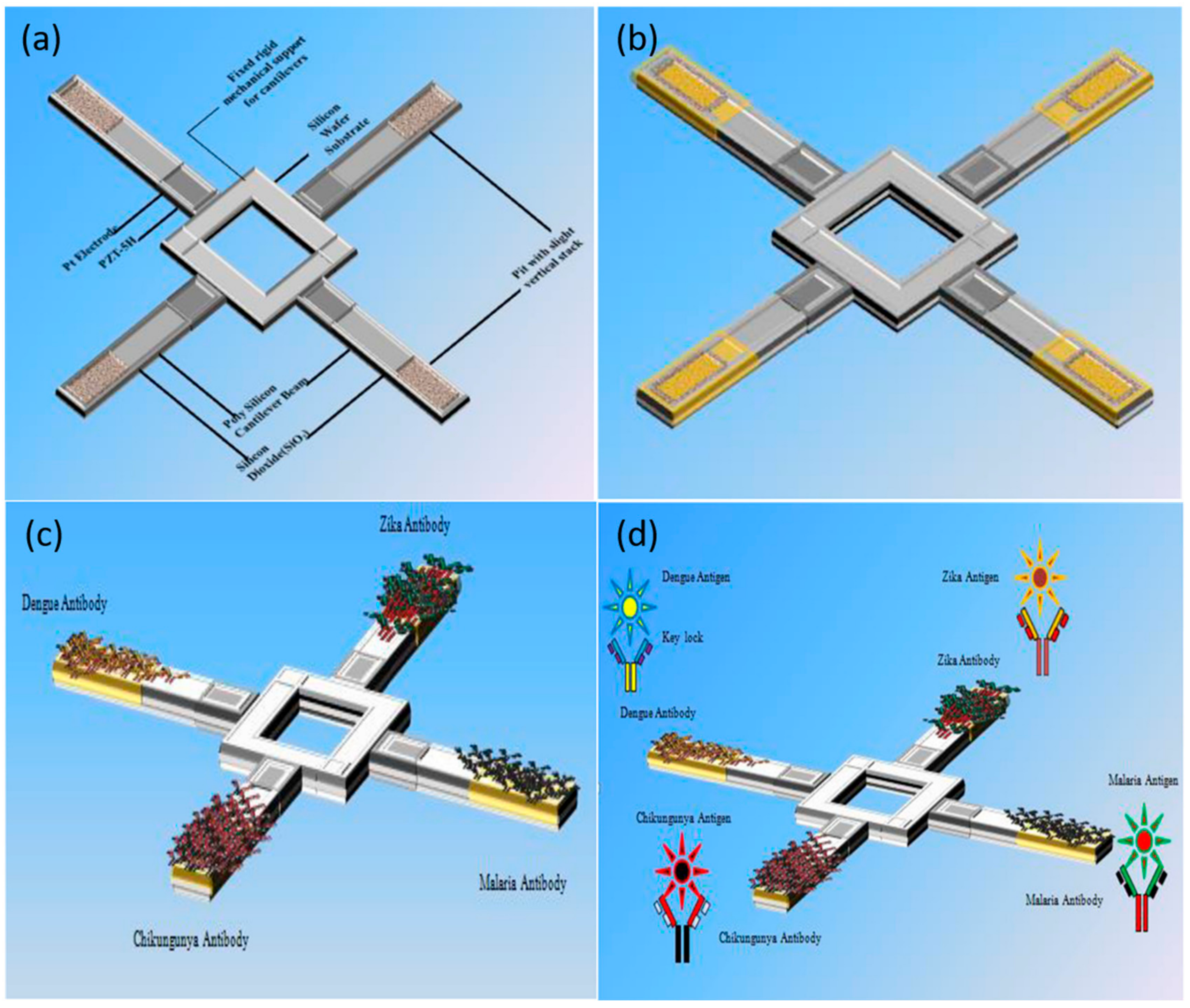
- Katta, M.; Sandanalakshmi, R. Simultaneous tropical disease identification with PZT-5H piezoelectric material including molecular mass biosensor microcantilever collection. Sens. Bio-Sens. Res. 2021, 32, 100413. [Google Scholar] [CrossRef]
- Ciriminna, R.; Fidalgo, A.; Pandarus, V.; Béland, F.; Ilharco, L.M.; Pagliaro, M. The Sol–Gel Route to Advanced Silica-Based Materials and Recent Applications. Chem. Rev. 2013, 113, 6592–6620. [Google Scholar] [CrossRef]
- Talou, M.H.; Bolaños Rivera, J.O.; Camerucci, M.A. Polymer-derived porous ceramics from novel silicon-based preceramic/sucrose systems. Ceram. Int. 2022, in press. [Google Scholar] [CrossRef]
- Su, D.; Li, Y.-L.; An, H.-J.; Liu, X.; Hou, F.; Li, J.-Y.; Fu, X. Pyrolytic transformation of liquid precursors to shaped bulk ceramics. J. Eur. Ceram. Soc. 2010, 30, 1503–1511. [Google Scholar] [CrossRef]
- Klermund, L.; Poschenrieder, S.T.; Castiglione, K. Simple surface functionalization of polymersomes using non-antibacterial peptide anchors. J. Nanobiotechnol. 2016, 14, 48. [Google Scholar] [CrossRef] [PubMed]
- Rikken, R.S.; Engelkamp, H.; Nolte, R.J.; Maan, J.C.; van Hest, J.C.; Wilson, D.A.; Christianen, P.C. Shaping polymersomes into predictable morphologies via out-of-equilibrium self-assembly. Nat. Commun. 2016, 7, 12606. [Google Scholar] [CrossRef] [Green Version]
- Einfalt, T.; Witzigmann, D.; Edlinger, C.; Sieber, S.; Goers, R.; Najer, A.; Spulber, M.; Onaca-Fischer, O.; Huwyler, J.; Palivan, C.G. Biomimetic artificial organelles with in vitro and in vivo activity triggered by reduction in microenvironment. Nat. Commun. 2018, 9, 1127. [Google Scholar] [CrossRef] [PubMed] [Green Version]
- Idrissi, M.E.; Meyer, C.E.; Zartner, L.; Meier, W. Nanosensors based on polymer vesicles and planar membranes: A short review. J. Nanobiotechnol. 2018, 16, 63. [Google Scholar] [CrossRef] [Green Version]
- Zhang, X.; Sun, J.; Liu, J.; Xu, H.; Dong, B.; Sun, X.; Zhang, T.; Xu, S.; Xu, L.; Bai, X.; et al. Label-free electrochemical immunosensor based on conductive Ag contained EMT-style nano-zeolites and the application for α-fetoprotein detection. Sens. Actuators B Chem. 2018, 255, 2919–2926. [Google Scholar] [CrossRef]
- Chen, B.; Yang, Z.; Zhu, Y.; Xia, Y. Zeolitic imidazolate framework materials: Recent progress in synthesis and applications. J. Mater. Chem. A 2014, 2, 16811–16831. [Google Scholar] [CrossRef]
- Denoual, M.; Robbes, D.; Inoue, S.; Mita, Y.; Grand, J.; Awala, H.; Mintova, S. Thermal resonant zeolite-based gas sensor. Sens. Actuators B Chem. 2017, 245, 179–182. [Google Scholar] [CrossRef]
- Wang, B.; Mu, Y.; Zhang, C.; Li, J. Blue photoluminescent carbon nanodots prepared from zeolite as efficient sensors for picric acid detection. Sens. Actuators B Chem. 2017, 253, 911–917. [Google Scholar] [CrossRef]
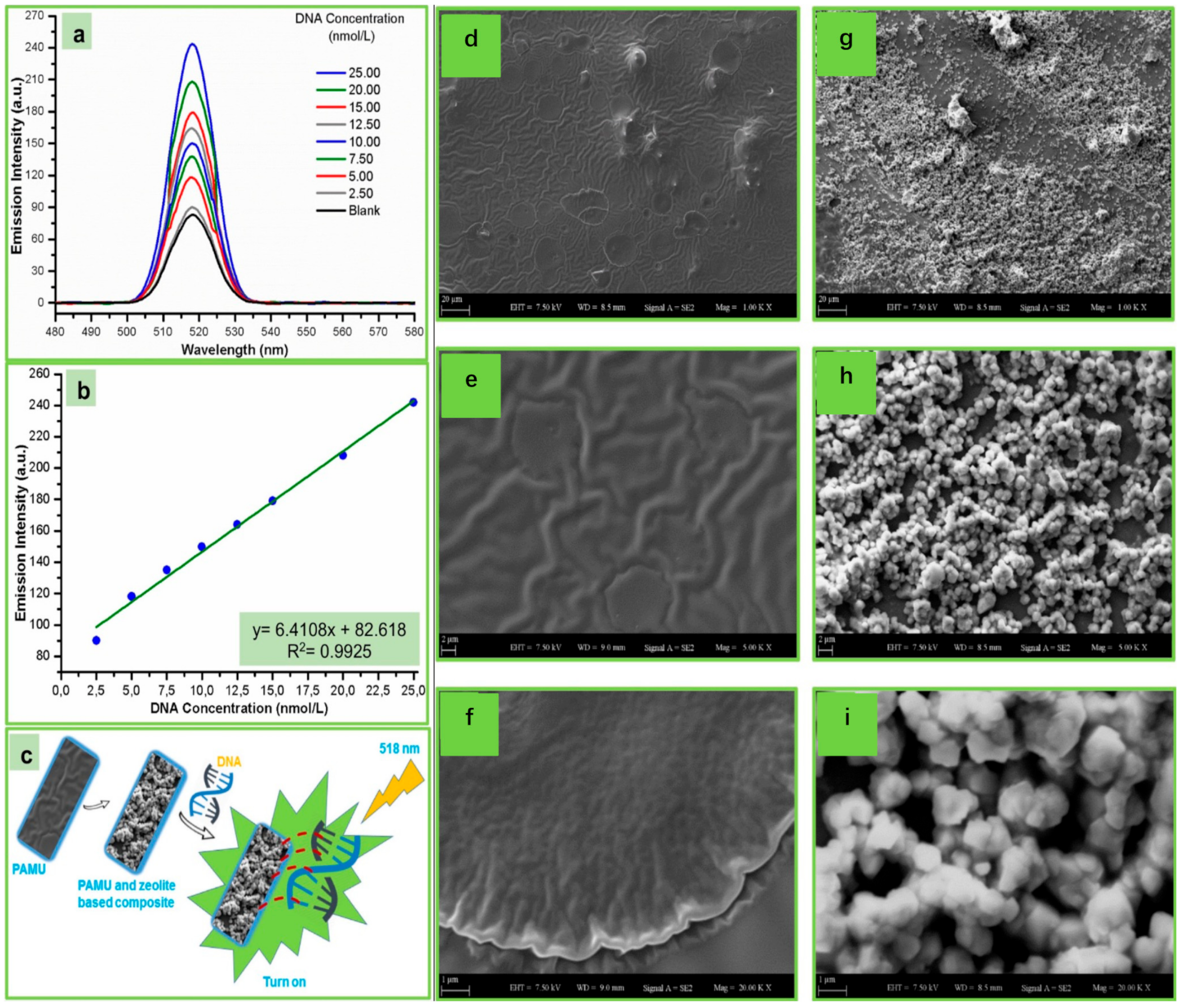
- Kamaci, U.D.; Kamaci, M.; Peksel, A. Poly(azomethine-urethane) and zeolite-based composite: Fluorescent biosensor for DNA detection. Spectrochim. Acta Part A Mol. Biomol. Spectrosc. 2019, 212, 232–239. [Google Scholar] [CrossRef]
- Braeckmans, K.; De Smedt, S.C.; Roelant, C.; Leblans, M.; Pauwels, R.; Demeester, J. Encoding microcarriers by spatial selective photobleaching. Nat. Mater. 2003, 2, 169–173. [Google Scholar] [CrossRef]
- Hanada, T. Basic Properties of ZnO, GaN, and Related Materials. In Oxide and Nitride Semiconductors: Processing, Properties, and Applications; Yao, T., Hong, S.-K., Eds.; Springer: Berlin/Heidelberg, Germany, 2009; pp. 1–19. [Google Scholar]
- Low, I. Ceramic-Matrix Composites: Microstructure, Properties & Applications; Woodhead Publishing: Sawston, UK, 2006. [Google Scholar]
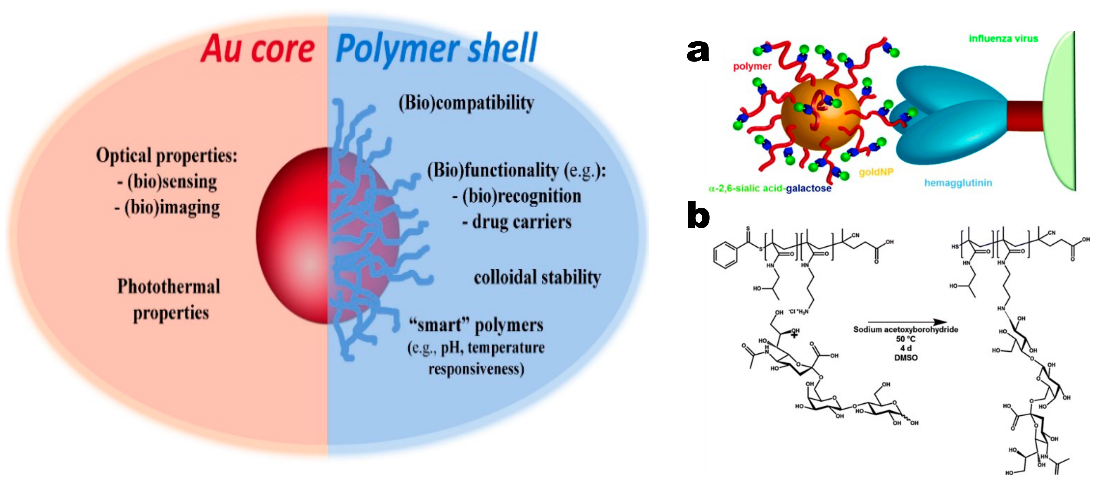
- Pereira, S.O.; Barros-Timmons, A.; Trindade, T. Polymer@gold Nanoparticles Prepared via RAFT Polymerization for Opto-Biodetection. Polymers 2018, 10, 189. [Google Scholar] [CrossRef] [PubMed]
- Zhang, Z.; Schepens, B.; Nuhn, L.; Saelens, X.; Schotsaert, M.; Callewaert, N.; De Rycke, R.; Zhang, Q.; Moins, S.; Benali, S.; et al. Influenza-binding sialylated polymer coated gold nanoparticles prepared via RAFT polymerization and reductive amination. Chem. Commun. 2016, 52, 3352–3355. [Google Scholar] [CrossRef] [PubMed]
- Takara, M.; Toyoshima, M.; Seto, H.; Hoshino, Y.; Miura, Y. Polymer-modified gold nanoparticles via RAFT polymerization: A detailed study for a biosensing application. Polym. Chem. 2014, 5, 931–939. [Google Scholar] [CrossRef]
- Parry, A.L.; Clemson, N.A.; Ellis, J.; Bernhard, S.S.R.; Davis, B.G.; Cameron, N.R. ‘Multicopy Multivalent’ Glycopolymer-Stabilized Gold Nanoparticles as Potential Synthetic Cancer Vaccines. J. Am. Chem. Soc. 2013, 135, 9362–9365. [Google Scholar] [CrossRef] [PubMed] [Green Version]
- Taylor-Pashow, K.M.L.; Della Rocca, J.; Huxford, R.C.; Lin, W. Hybrid nanomaterials for biomedical applications. Chem. Commun. 2010, 46, 5832–5849. [Google Scholar] [CrossRef] [PubMed]
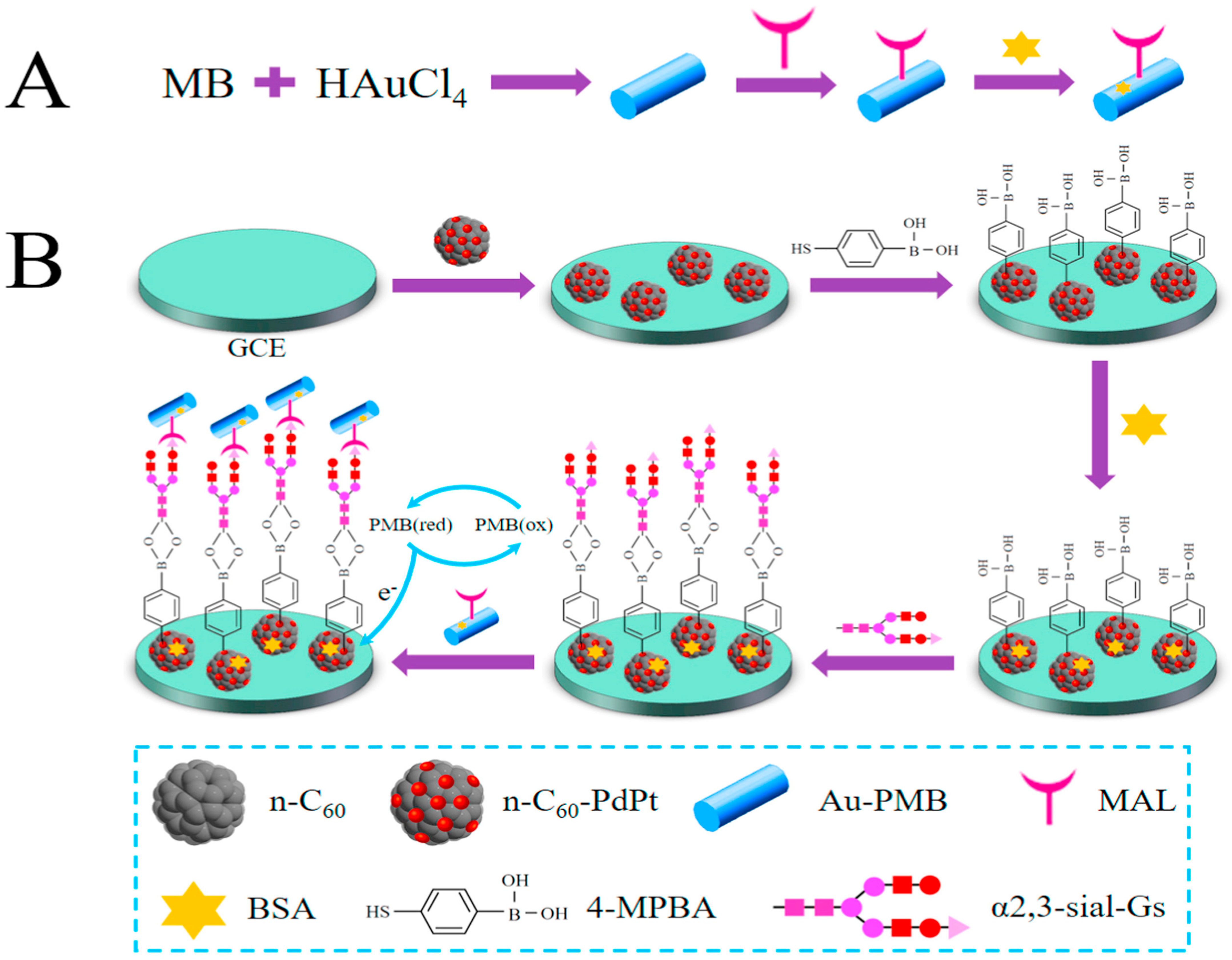
- Yuan, Q.; He, J.; Niu, Y.; Chen, J.; Zhao, Y.; Zhang, Y.; Yu, C. Sandwich-type biosensor for the detection of α2,3-sialylated glycans based on fullerene-palladium-platinum alloy and 4-mercaptophenylboronic acid nanoparticle hybrids coupled with Au-methylene blue-MAL signal amplification. Biosens. Bioelectron. 2018, 102, 321–327. [Google Scholar] [CrossRef] [PubMed]
- Singh, R.; Kumar, S.; Liu, F.-Z.; Shuang, C.; Zhang, B.; Jha, R.; Kaushik, B.K. Etched multicore fiber sensor using copper oxide and gold nanoparticles decorated graphene oxide structure for cancer cells detection. Biosens. Bioelectron. 2020, 168, 112557. [Google Scholar] [CrossRef]
- Guo, J.; Ma, X. Simultaneous monitoring of glucose and uric acid on a single test strip with dual channels. Biosens. Bioelectron. 2017, 94, 415–419. [Google Scholar] [CrossRef]
- Hatada, M.; Tran, T.-T.; Tsugawa, W.; Sode, K.; Mulchandani, A. Affinity sensor for haemoglobin A1c based on single-walled carbon nanotube field-effect transistor and fructosyl amino acid binding protein. Biosens. Bioelectron. 2019, 129, 254–259. [Google Scholar] [CrossRef]
- Comba, F.N.; Romero, M.R.; Garay, F.S.; Baruzzi, A.M. Mucin and carbon nanotube-based biosensor for detection of glucose in human plasma. Anal. Biochem. 2018, 550, 34–40. [Google Scholar] [CrossRef] [Green Version]
- Li, X.; Ren, K.; Zhang, M.; Sang, W.; Sun, D.; Hu, T.; Ni, Z. Cobalt functionalized MoS2/carbon nanotubes scaffold for enzyme-free glucose detection with extremely low detection limit. Sens. Actuators B Chem. 2019, 293, 122–128. [Google Scholar] [CrossRef]
- Lee, C.-Y.; Wu, L.-P.; Chou, T.-T.; Hsieh, Y.-Z. Functional magnetic nanoparticles–assisted electrochemical biosensor for eosinophil cationic protein in cell culture. Sens. Actuators B Chem. 2018, 257, 672–677. [Google Scholar] [CrossRef]
- Meisner, M. Update on procalcitonin measurements. Ann. Lab. Med. 2014, 34, 263. [Google Scholar] [CrossRef] [PubMed] [Green Version]
- Chiang, C.-Y.; Huang, T.-T.; Wang, C.-H.; Huang, C.-J.; Tsai, T.-H.; Yu, S.-N.; Chen, Y.-T.; Hong, S.-W.; Hsu, C.-W.; Chang, T.-C.; et al. Fiber optic nanogold-linked immunosorbent assay for rapid detection of procalcitonin at femtomolar concentration level. Biosens. Bioelectron. 2020, 151, 111871. [Google Scholar] [CrossRef]
- Sundar, S.; Kwon, S.J.; Venkatachalam, G. Magneto-Biosensor for the Detection of Uric Acid Using Citric Acid-Capped Iron Oxide Nanoparticles. J. Nanosci. Nanotechnol. 2020, 20, 2144–2153. [Google Scholar] [CrossRef]
- Baliyan, A.; Usha, S.P.; Gupta, B.D.; Gupta, R.; Sharma, E.K. Localized surface plasmon resonance-based fiber-optic sensor for the detection of triacylglycerides using silver nanoparticles. J. Biomed. Opt. 2017, 22, 107001. [Google Scholar] [CrossRef] [PubMed] [Green Version]
- Buehler, M.J. Nature designs tough collagen: Explaining the nanostructure of collagen fibrils. Proc. Natl. Acad. Sci. USA 2006, 103, 12285–12290. [Google Scholar] [CrossRef] [Green Version]
- Kaushik, B.K.; Singh, L.; Singh, R.; Zhu, G.; Zhang, B.; Wang, Q.; Kumar, S. Detection of Collagen-IV Using Highly Reflective Metal Nanoparticles-Immobilized Photosensitive Optical Fiber-Based MZI Structure. IEEE Trans. Nanobiosci. 2020, 19, 477–484. [Google Scholar] [CrossRef]
- Kaushik, S.; Tiwari, U.; Nilima; Prashar, S.; Das, B.; Sinha, R.K. Label-free detection of Escherichia coli bacteria by cascaded chirped long period gratings immunosensor. Rev. Sci. Instrum. 2019, 90, 025003. [Google Scholar] [CrossRef]
- Gahlaut, S.K.; Pathak, A.; Gupta, B.D.; Singh, J.P. Portable fiber-optic SPR platform for the detection of NS1-antigen for dengue diagnosis. Biosens. Bioelectron. 2022, 196, 113720. [Google Scholar] [CrossRef]
- Luo, B.; Liu, Z.; Wang, X.; Shi, S.; Zhong, N.; Ma, P.; Wu, S.; Wu, D.; Zhao, M.; Liang, W. Dual-peak long period fiber grating coated with graphene oxide for label-free and specific assays of H5N1 virus. J. Biophotonics 2021, 14, e202000279. [Google Scholar] [CrossRef] [PubMed]
- Friedrich, R.; Ghorbani-Asl, M.; Curtarolo, S.; Krasheninnikov, A.V. Data-Driven Quest for Two-Dimensional Non-van der Waals Materials. Nano Lett. 2022, 22, 989–997. [Google Scholar] [CrossRef] [PubMed]
- Ahmad, S.; Husain, A.; Khan, M.M.A.; Khan, I.; Khan, A.; Asiri, A.M. 5—Perovskite-based material for sensor applications. In Hybrid Perovskite Composite Materials; Khan, I., Khan, A., Khan, M.M.A., Khan, S., Verpoort, F., Umar, A., Eds.; Woodhead Publishing: Sawston, UK, 2021; pp. 135–145. [Google Scholar] [CrossRef]
- Jia, D.; Xu, M.; Mu, S.; Ren, W.; Liu, C. Recent Progress of Perovskite Nanocrystals in Chem/Bio Sensing. Biosensors 2022, 12, 754. [Google Scholar] [CrossRef] [PubMed]


| Mechanism | Materials Used | Analyte | Linear Range | LOD | Ref. |
|---|---|---|---|---|---|
| LSPR | GO | Glucose | 0–11 mM | n.r. a | [29] |
| SPR | GO | Glucose | 56–390 mM | n.r. a | [48] |
| SPR | GO | 25-hydroxyvitamin D3 | 2.39 fM–2.4 pM | 2.39 fM | [49] |
| SPR | GO | Glucose | 0–6.7 nM | n.r. a | [50] |
| SPR | GO | Glucose | 56–390 mM | n.r. a | [51] |
| Fluorescence resonance energy transfer (FRET) | GO | Dopamine | 0–10 mM | 200 μM | [52] |
| Nicotine | 0–0.7 mM | ~10 nM | |||
| ssDNA | 0–0.2 mM | ~10 nM | |||
| FRET | CODs | Glucose | 10–200 μM | 6.43 μM | [53] |
| 10–100 nM | 25.79 nM | ||||
| FRET | CODs | Nitric oxide | 10–100 nM | 9.12 nM | [54] |
| FRET | CODs | Dopamine | 0–10 μM | 46.4 nM | [55] |
| FRET | CODs | Cholesterol | 0.01–6 mM | 1 μM | [56] |
| Electrochemistry | CNTs | Uric acid | 0.02–2.7 mM | 2.8 μM | [57] |
| SPR | CNTs | Dopamine | 0−10 μM | 18.9 pM | [58] |
| Electrochemistry | CNTs | Glucose | 0.2–10.6 mM | 0.4 μM | [59] |
| Mechanism | Materials Used | Analyte | Linear Range | LOD | Ref. |
|---|---|---|---|---|---|
| Colorimetric | AuNPs@gelatin | Aflatoxin B1 | 32.02–448.33 pM | 12.81 pM | [79] |
| Electrochemical | TiO2 nanotubes/AgNPs | Heat shock protein 70 | 1.43 mM–1.43 fM | n.r. a | [80] |
| Terahertz spectroscopy | AuNPs | MicroRNAs | 1 fM–10 pM | 14.54 aM | [81] |
| LSPR | AuNPs | Zearalenone | 3.12–149.81 fM | 0.32 fM | [82] |
| Fluorescence | Cu nanocluster | Breast Cancer | 500 nM–3 µM | 1.7 pM | [83] |
| Electrochemical | CuONPs/ ZnO nanowires | Glucose | 50–500 µM | n.r. a | [84] |
| Electrochemical | CuO nanoleaves | Glucose | 0.005–5.89 mM | 12 nM | [85] |
| Field effect transistors | ZnO nanorods | Serotonin | 0.1 fM–1 nM | 0.1 fM | [86] |
| Fluorescence | ZnO-QDs | Cysteine | 0.1–600 μM | 0.642 μM | [87] |
| Electrochemical | TiO2-NRs/rGO | Dichlorvos | 2.26–565 nM | 2.23 nM | [88] |
| Fluorescence | SiQDs | Glucose | 1–90 μM | 30 μM | [75] |
| Raman/fluorescence spectroscopic | SiO2-NPs | Aflatoxin B1 | n.r. a | 0.426 aM | [89] |
| Electrochemical | SiO2-NPs | Carcinoembryonic antigen | 5 mM–0.8 aM | n.r. a | [90] |
| Turn-on fluorometric | SiO2-NPs | Cephalexin | n.r. a | 1.6 μM | [91] |
| Mechanism | Materials Used | Analyte | Linear Range | LOD | Ref. |
|---|---|---|---|---|---|
| Fluorescence | NAD(P)H-sensitive Polymer Dot | Phenylalanine | 0–2400 μM | 3.5 μM | [101] |
| Electrochemical | Polymer dot | Gram-negative and Gram-positive bacteria | n.r. a | Gram-negative 3.0 CFU/mL and Gram-positive 3.1 CFU/mL | [102] |
| Electro-chemiluminescence | AIE-Active Polymer Dots | Nucleic acid | n.r. a | 32 aM | [103] |
| Electrochemical | Polymer QDs and C60/MWCNTs- polyethyleneimine nanocomposites | Thrombin | 50 fM–20 nM | 6 fM | [104] |
| Electro-chemiluminescence | Carboxyl functionalized polymer dots | Nucleic acid | 0.1 fM–100 pM | 36 aM | [105] |
| Fluorescence | Organic nanoparticles | Dopamine | 0–10 μM | n.r. a | [106] |
| Fluorescence | Conjugated polymer nanoparticles | Intracellular telomerase | n.r. a | 3 HeLa cells in 400 μL | [107] |
| Electro-chemiluminescence | Luminol-encapsulated liposome | atxA mRNA | 10–300 fM | 8.13 fM | [108] |
| Photo-electrochemical | CRISPR/Cas12 a-mediated liposome | Nucleic acid | 0–100 nM | 1.6 pM | [109] |
| Electrochemical | Liposomes | Thrombin | 0.1–1000 nM | 0.3 pM | [110] |
| Electrochemical | Biomimetic cerasome/graphene QDs | Cholesterol | 16–6186 μM | 5 μM | [41] |
| Colorimetric | polydiacetylene liposome/AuNPs | Thrombin | 0–27.03 fM | n.r. a | [111] |
Disclaimer/Publisher’s Note: The statements, opinions and data contained in all publications are solely those of the individual author(s) and contributor(s) and not of MDPI and/or the editor(s). MDPI and/or the editor(s) disclaim responsibility for any injury to people or property resulting from any ideas, methods, instructions or products referred to in the content. |
© 2023 by the authors. Licensee MDPI, Basel, Switzerland. This article is an open access article distributed under the terms and conditions of the Creative Commons Attribution (CC BY) license (https://creativecommons.org/licenses/by/4.0/).
Share and Cite
Kumar, S.; Wang, Z.; Zhang, W.; Liu, X.; Li, M.; Li, G.; Zhang, B.; Singh, R. Optically Active Nanomaterials and Its Biosensing Applications—A Review. Biosensors 2023, 13, 85. https://doi.org/10.3390/bios13010085
Kumar S, Wang Z, Zhang W, Liu X, Li M, Li G, Zhang B, Singh R. Optically Active Nanomaterials and Its Biosensing Applications—A Review. Biosensors. 2023; 13(1):85. https://doi.org/10.3390/bios13010085
Chicago/Turabian StyleKumar, Santosh, Zhi Wang, Wen Zhang, Xuecheng Liu, Muyang Li, Guoru Li, Bingyuan Zhang, and Ragini Singh. 2023. "Optically Active Nanomaterials and Its Biosensing Applications—A Review" Biosensors 13, no. 1: 85. https://doi.org/10.3390/bios13010085