Conducting Polymers as Versatile Tools for the Electrochemical Detection of Cancer Biomarkers


Abstract
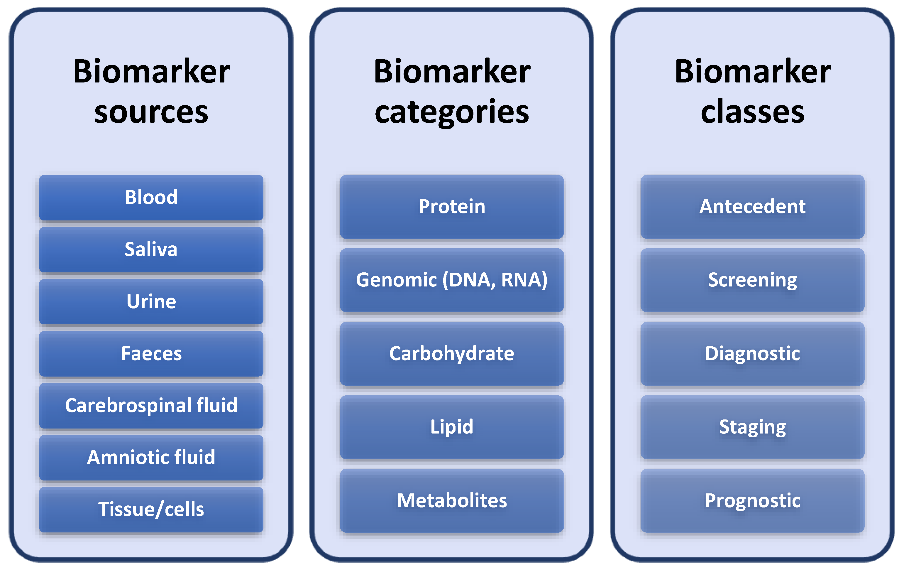
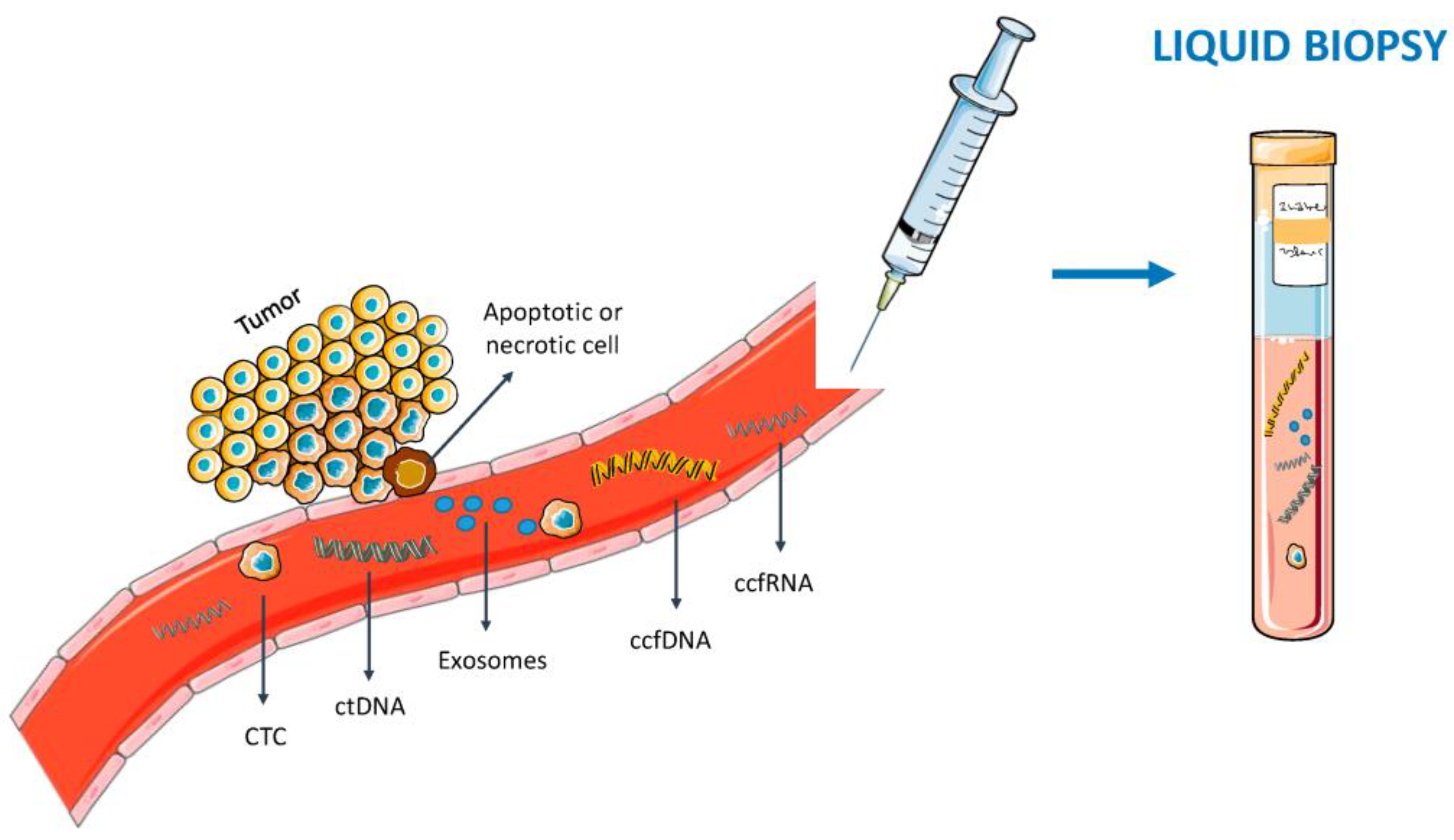
:1. Introduction
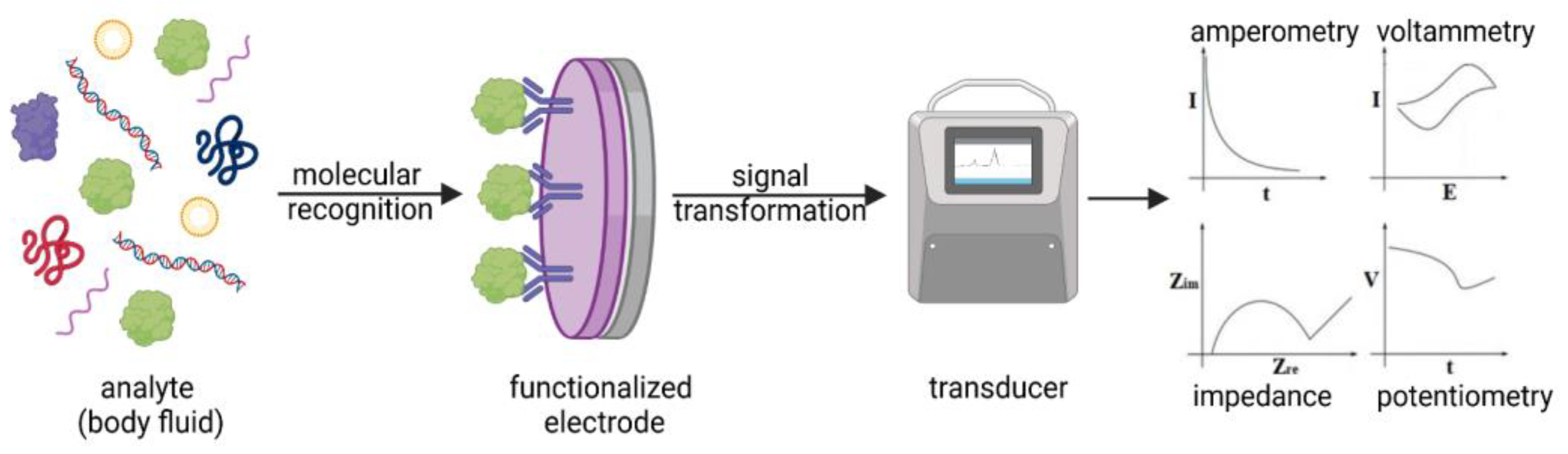
2. Electrochemical Biosensors
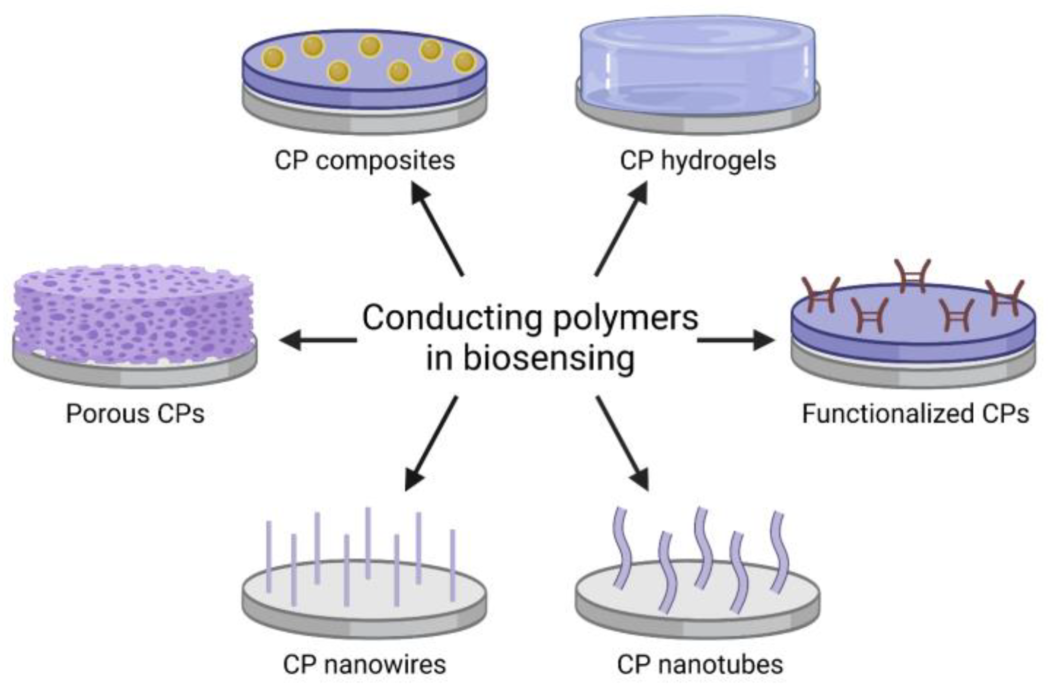
3. Conducting Polymers
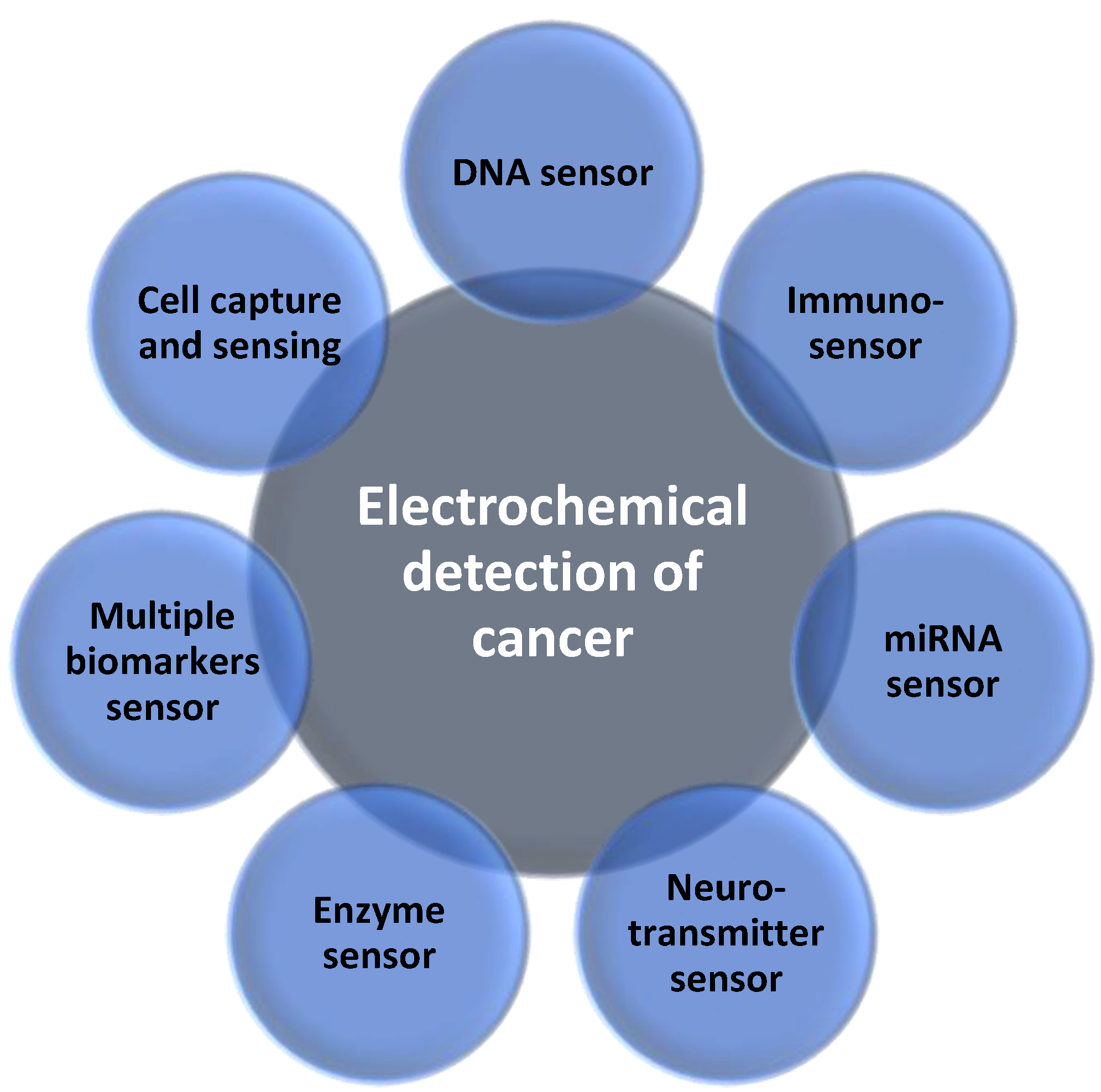
4. Conducting Polymer-Based Electrochemical Sensors for Cancer Diagnosis
4.1. DNA Sensors
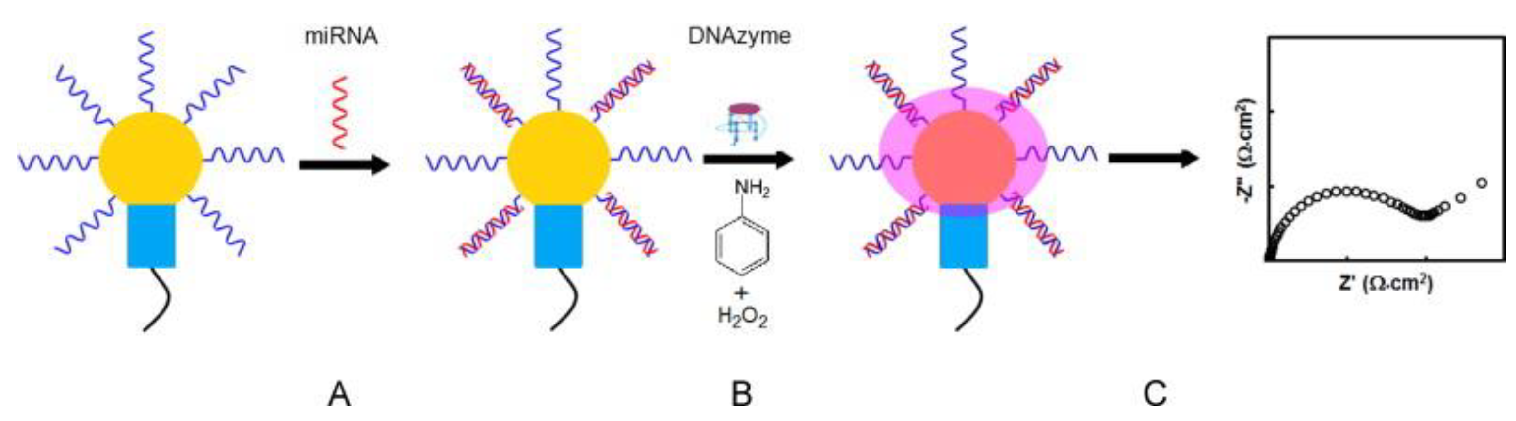
4.2. miRNA Sensors
4.3. Immunosensors
4.4. Detection of Enzymes
4.5. Detection of Neurotransmitters
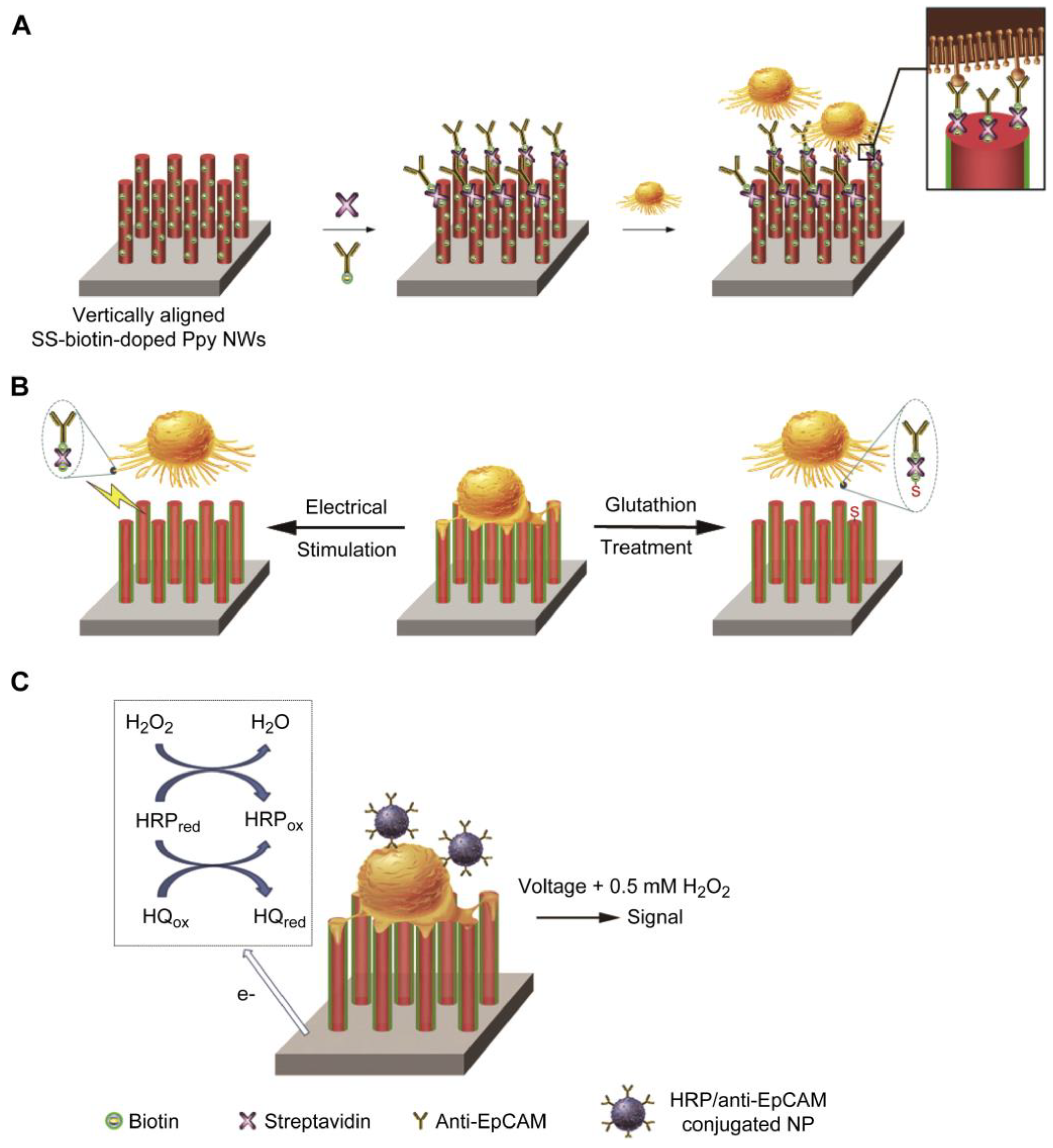
4.6. Cell Capture
4.7. Multiplex Biomarkers Detection
5. CP-Based Electrochemical Sensors for Cancer Diagnosis: Comparison
6. Summary and Outlooks
Author Contributions
Funding
Data Availability Statement
Conflicts of Interest
References
- Dyba, T.; Randi, G.; Bray, F.; Martos, C.; Giusti, F.; Nicholson, N.; Gavin, A.; Flego, M.; Neamtiu, L.; Dimitrova, N.; et al. The European cancer burden in 2020: Incidence and mortality estimates for 40 countries and 25 major cancers. Eur. J. Cancer 2021, 157, 308–347. [Google Scholar] [CrossRef] [PubMed]
- Davies, P.C.W.; Lineweaver, C.H. Cancer tumors as Metazoa 1.0: Tapping genes of ancient ancestors. Phys. Biol. 2011, 8, 015001. [Google Scholar] [CrossRef] [PubMed]
- Topkaya, S.N.; Azimzadeh, M.; Ozsoz, M. Electrochemical Biosensors for Cancer Biomarkers Detection: Recent Advances and Challenges. Electroanalysis 2016, 28, 1402–1419. [Google Scholar] [CrossRef]
- Bensalah, K.; Montorsi, F.; Shariat, S.F. Challenges of Cancer Biomarker Profiling. Eur. Urol. 2007, 52, 1601–1609. [Google Scholar] [CrossRef] [PubMed]
- Tang, Y.; Qiao, G.; Xu, E.; Xuan, Y.; Liao, M.; Yin, G. Biomarkers for early diagnosis, prognosis, prediction, and recurrence monitoring of non-small cell lung cancer. Onco. Targets. Ther. 2017, 10, 4527–4534. [Google Scholar] [CrossRef] [Green Version]
- Prajapati, D.G.; Kandasubramanian, B. Progress in the Development of Intrinsically Conducting Polymer Composites as Biosensors. Macromol. Chem. Phys. 2019, 220, 1800561. [Google Scholar] [CrossRef]
- Luong, J.H.T.; Narayan, T.; Solanki, S.; Malhotra, B.D. Recent Advances of Conducting Polymers and Their Composites for Electrochemical Biosensing Applications. J. Funct. Biomater. 2020, 11, 71. [Google Scholar] [CrossRef]
- Peng, H.; Zhang, L.; Soeller, C.; Travas-Sejdic, J. Conducting polymers for electrochemical DNA sensing. Biomaterials 2009, 30, 2132–2148. [Google Scholar] [CrossRef]
- Aydemir, N.; Malmström, J.; Travas-Sejdic, J. Conducting polymer based electrochemical biosensors. Phys. Chem. Chem. Phys. 2016, 18, 8264–8277. [Google Scholar] [CrossRef]
- Cho, I.H.; Kim, D.H.; Park, S. Electrochemical biosensors: Perspective on functional nanomaterials for on-site analysis. Biomater. Res. 2020, 24, 6. [Google Scholar] [CrossRef]
- Singh, A.; Sharma, A.; Ahmed, A.; Sundramoorthy, A.K.; Furukawa, H.; Arya, S.; Khosla, A. Recent Advances in Electrochemical Biosensors: Applications, Challenges, and Future Scope. Biosensors 2021, 11, 336. [Google Scholar] [CrossRef] [PubMed]
- Cui, F.; Zhou, Z.; Zhou, H.S. Review—Measurement and Analysis of Cancer Biomarkers Based on Electrochemical Biosensors. J. Electrochem. Soc. 2020, 167, 037525. [Google Scholar] [CrossRef]
- Sadeghi, S.J. Amperometric Biosensors. Encycl. Biophys. 2013, 61–67. [Google Scholar] [CrossRef]
- Ouyang, J. Recent Advances of Intrinsically Conductive Polymers. Acta Phys.-Chim. Sin. 2018, 34, 1211–1220. [Google Scholar] [CrossRef]
- Boehler, C.; Aqrawe, Z.; Asplund, M. Applications of PEDOT in bioelectronic medicine. Bioelectron. Med. 2019, 2, 89–99. [Google Scholar] [CrossRef] [Green Version]
- Balint, R.; Cassidy, N.J.; Cartmell, S.H. Conductive polymers: Towards a smart biomaterial for tissue engineering. Acta Biomater. 2014, 10, 2341–2353. [Google Scholar] [CrossRef]
- Poole-Warren, L.; Martens, P.; Green, R. Biosynthetic Polymers for Medical Applications; Elsevier: Amsterdam, The Netherlands, 2016; ISBN 9781782421054. [Google Scholar]
- Skorupa, M.; Więcławska, D.; Czerwińska-Główka, D.; Skonieczna, M.; Krukiewicz, K. Dopant-dependent electrical and biological functionality of pedot in bioelectronics. Polymers 2021, 13, 1948. [Google Scholar] [CrossRef]
- Le, T.-H.H.; Kim, Y.; Yoon, H. Electrical and electrochemical properties of conducting polymers. Polymers 2017, 9, 150. [Google Scholar] [CrossRef] [Green Version]
- Lakard, B. Electrochemical Biosensors Based on Conducting Polymers: A Review. Appl. Sci. 2020, 10, 6614. [Google Scholar] [CrossRef]
- Das, T.K.; Prusty, S. Review on Conducting Polymers and Their Applications. Polym. Plast. Technol. Eng. 2012, 51, 1487–1500. [Google Scholar] [CrossRef]
- Vahdatiyekta, P.; Zniber, M.; Bobacka, J.; Huynh, T.P. A review on conjugated polymer-based electronic tongues. Anal. Chim. Acta 2022, 1221, 340114. [Google Scholar] [CrossRef] [PubMed]
- Soylemez, S.; Kanik, F.E.; Uzun, S.D.; Hacioglu, S.O.; Toppare, L. Development of an efficient immobilization matrix based on a conducting polymer and functionalized multiwall carbon nanotubes: Synthesis and its application to ethanol biosensors. J. Mater. Chem. B 2014, 2, 511–521. [Google Scholar] [CrossRef] [PubMed]
- Uzun, S.D.; Unlu, N.A.; Sendur, M.; Kanik, F.E.; Timur, S.; Toppare, L. A novel promising biomolecule immobilization matrix: Synthesis of functional benzimidazole containing conducting polymer and its biosensor applications. Colloids Surf. B Biointerfaces 2013, 112, 74–80. [Google Scholar] [CrossRef] [PubMed]
- Ramanavicius, S.; Ramanavicius, A. Conducting Polymers in the Design of Biosensors and Biofuel Cells. Polymers 2020, 13, 49. [Google Scholar] [CrossRef]
- Gerard, M.; Chaubey, A.; Malhotra, B.D. Application of conducting polymers to biosensors. Biosens. Bioelectron. 2002, 17, 345–359. [Google Scholar] [CrossRef]
- Maziz, A.; Özgür, E.; Bergaud, C.; Uzun, L. Progress in conducting polymers for biointerfacing and biorecognition applications. Sens. Actuators Rep. 2021, 3, 100035. [Google Scholar] [CrossRef]
- Ma, Z.; Shi, W.; Yan, K.; Pan, L.; Yu, G. Doping engineering of conductive polymer hydrogels and their application in advanced sensor technologies. Chem. Sci. 2019, 10, 6232–6244. [Google Scholar] [CrossRef] [Green Version]
- Li, L.; Pan, L.; Ma, Z.; Yan, K.; Cheng, W.; Shi, Y.; Yu, G. All Inkjet-Printed Amperometric Multiplexed Biosensors Based on Nanostructured Conductive Hydrogel Electrodes. Nano Lett. 2018, 18, 3322–3327. [Google Scholar] [CrossRef]
- Wang, M.; Baek, P.; Akbarinejad, A.; Barker, D.; Travas-Sejdic, J. Conjugated polymers and composites for stretchable organic electronics. J. Mater. Chem. C 2019, 7, 5534–5552. [Google Scholar] [CrossRef]
- Constâncio, V.; Nunes, S.P.; Henrique, R.; Jerónimo, C. DNA Methylation-Based Testing in Liquid Biopsies as Detection and Prognostic Biomarkers for the Four Major Cancer Types. Cells 2020, 9, 624. [Google Scholar] [CrossRef]
- Lee, H.J.; Jeon, S.H.; Seo, J.S.; Goh, S.H.; Han, J.Y.; Cho, Y. A novel strategy for highly efficient isolation and analysis of circulating tumor-specific cell-free DNA from lung cancer patients using a reusable conducting polymer nanostructure. Biomaterials 2016, 101, 251–257. [Google Scholar] [CrossRef] [PubMed]
- Wang, J.; Wang, D.; Hui, N. A low fouling electrochemical biosensor based on the zwitterionic polypeptide doped conducting polymer PEDOT for breast cancer marker BRCA1 detection. Bioelectrochemistry 2020, 136, 107595. [Google Scholar] [CrossRef] [PubMed]
- Shahrokhian, S.; Salimian, R. Ultrasensitive detection of cancer biomarkers using conducting polymer/electrochemically reduced graphene oxide-based biosensor: Application toward BRCA1 sensing. Sens. Actuators B Chem. 2018, 266, 160–169. [Google Scholar] [CrossRef]
- Calin, G.A.; Dumitru, C.D.; Shimizu, M.; Bichi, R.; Zupo, S.; Noch, E.; Aldler, H.; Rattan, S.; Keating, M.; Rai, K.; et al. Frequent deletions and down-regulation of micro-RNA genes miR15 and miR16 at 13q14 in chronic lymphocytic leukemia. Proc. Natl. Acad. Sci. USA 2002, 99, 15524–15529. [Google Scholar] [CrossRef] [Green Version]
- Deng, H.; Shen, W.; Ren, Y.; Gao, Z. A highly sensitive microRNA biosensor based on hybridized microRNA-guided deposition of polyaniline. Biosens. Bioelectron. 2014, 60, 195–200. [Google Scholar] [CrossRef]
- Peng, Y.; Yi, G.; Gao, Z. A highly sensitive microRNA biosensor based on ruthenium oxide nanoparticle-initiated polymerization of aniline. Chem. Commun. 2010, 46, 9131–9133. [Google Scholar] [CrossRef]
- Tran, H.V.; Piro, B.; Reisberg, S.; Tran, L.D.; Duc, H.T.; Pham, M.C. Label-free and reagentless electrochemical detection of microRNAs using a conducting polymer nanostructured by carbon nanotubes: Application to prostate cancer biomarker miR-141. Biosens. Bioelectron. 2013, 49, 164–169. [Google Scholar] [CrossRef]
- Fan, Y.; Chen, X.; Trigg, A.D.; Tung, C.H.; Kong, J.; Gao, Z. Detection of microRNAs using target-guided formation of conducting polymer nanowires in nanogaps. J. Am. Chem. Soc. 2007, 129, 5437–5443. [Google Scholar] [CrossRef]
- Felix, F.S.; Angnes, L. Electrochemical immunosensors—A powerful tool for analytical applications. Biosens. Bioelectron. 2018, 102, 470–478. [Google Scholar] [CrossRef]
- ul Haq Zia, T.; ul Haq Ali Shah, A. Understanding the adsorption of 1 NLB antibody on polyaniline nanotubes as a function of zeta potential and surface charge density for detection of hepatitis C core antigen: A label-free impedimetric immunosensor. Colloids Surfaces A Physicochem. Eng. Asp. 2021, 626, 127076. [Google Scholar] [CrossRef]
- Liu, S.; Ma, Y.; Cui, M.; Luo, X. Enhanced electrochemical biosensing of alpha-fetoprotein based on three-dimensional macroporous conducting polymer polyaniline. Sens. Actuators B Chem. 2018, 255, 2568–2574. [Google Scholar] [CrossRef]
- Liu, Z.; Ma, Z. Fabrication of an ultrasensitive electrochemical immunosensor for CEA based on conducting long-chain polythiols. Biosens. Bioelectron. 2013, 46, 1–7. [Google Scholar] [CrossRef] [PubMed]
- Hui, N.; Sun, X.; Song, Z.; Niu, S.; Luo, X. Gold nanoparticles and polyethylene glycols functionalized conducting polyaniline nanowires for ultrasensitive and low fouling immunosensing of alpha-fetoprotein. Biosens. Bioelectron. 2016, 86, 143–149. [Google Scholar] [CrossRef] [PubMed]
- Li, Z.; Yin, J.; Gao, C.; Qiu, G.; Meng, A.; Li, Q. The construction of electrochemical aptasensor based on coral-like poly-aniline and Au nano-particles for the sensitive detection of prostate specific antigen. Sens. Actuators B Chem. 2019, 295, 93–100. [Google Scholar] [CrossRef]
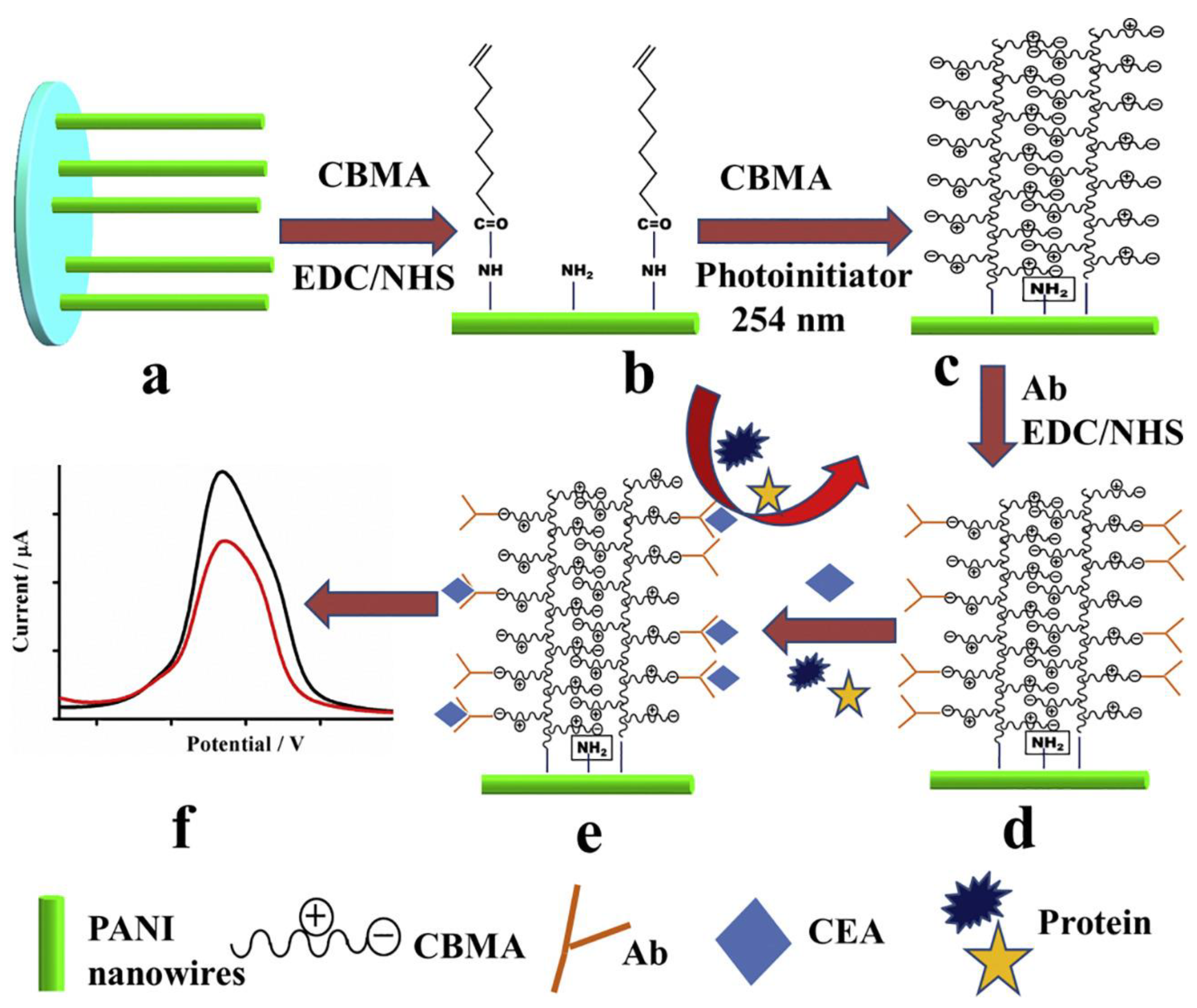
- Wang, J.; Hui, N. Zwitterionic poly(carboxybetaine) functionalized conducting polymer polyaniline nanowires for the electrochemical detection of carcinoembryonic antigen in undiluted blood serum. Bioelectrochemistry 2019, 125, 90–96. [Google Scholar] [CrossRef]
- Han, R.; Wang, G.; Xu, Z.; Zhang, L.; Li, Q.; Han, Y.; Luo, X. Designed antifouling peptides planted in conducting polymers through controlled partial doping for electrochemical detection of biomarkers in human serum. Biosens. Bioelectron. 2020, 164, 112317. [Google Scholar] [CrossRef]
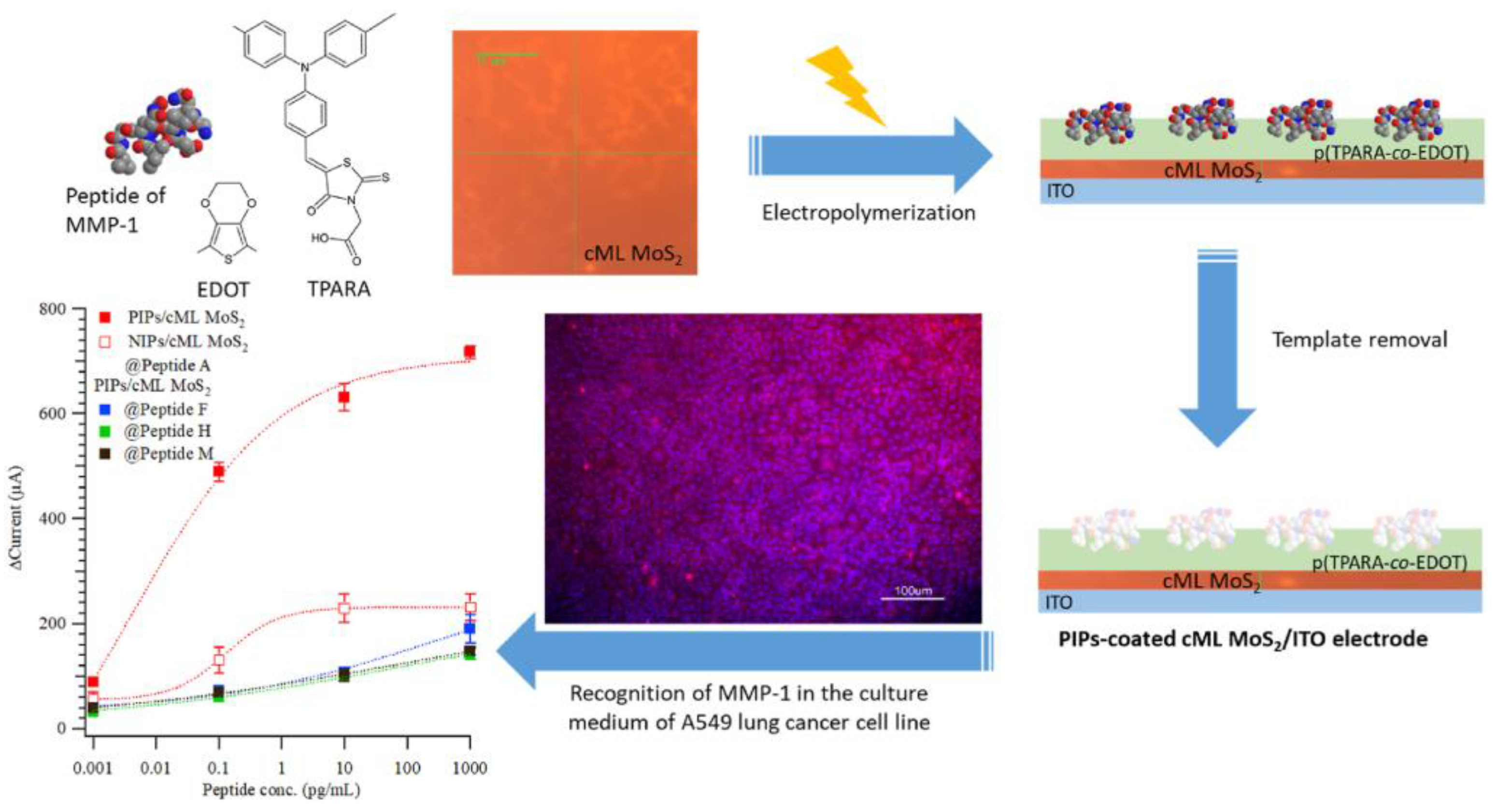
- Lee, M.-H.; Lin, C.-C.; Kutner, W.; Thomas, J.L.; Lin, C.-Y.; Iskierko, Z.; Ku, Y.-S.; Lin, C.-Y.; Borowicz, P.; Sharma, P.; et al. Peptide-imprinted conductive polymer on continuous monolayer molybdenum disulfide transferred electrodes for electrochemical sensing of Matrix Metalloproteinase-1 in lung cancer culture medium. Biosens. Bioelectron. X 2022, 13, 100258. [Google Scholar] [CrossRef]
- Ahmad, L.; Salmon, L.; Korri-Youssoufi, H. Electrochemical detection of the human cancer biomarker’ autocrine motility factor-phosphoglucose isomerase’ based on a biosensor formed with a monosaccharidic inhibitor. Sens. Actuators B Chem. 2019, 299, 126933. [Google Scholar] [CrossRef]
- Jiang, S.H.; Hu, L.P.; Wang, X.; Li, J.; Zhang, Z.G. Neurotransmitters: Emerging targets in cancer. Oncogene 2020, 39, 503–515. [Google Scholar] [CrossRef]
- Chung, S.; Akhtar, M.H.; Benboudiaf, A.; Park, D.S.; Shim, Y.B. A Sensor for Serotonin and Dopamine Detection in Cancer Cells Line Based on the Conducting Polymer−Pd Complex Composite. Electroanalysis 2020, 32, 520–527. [Google Scholar] [CrossRef]
- Moon, J.M.; Thapliyal, N.; Hussain, K.K.; Goyal, R.N.; Shim, Y.B. Conducting polymer-based electrochemical biosensors for neurotransmitters: A review. Biosens. Bioelectron. 2018, 102, 540–552. [Google Scholar] [CrossRef] [PubMed]
- Sekine, J.; Luo, S.C.; Wang, S.; Zhu, B.; Tseng, H.R.; Yu, H.H. Functionalized conducting polymer nanodots for enhanced cell capturing: The synergistic effect of capture agents and nanostructures. Adv. Mater. 2011, 23, 4788–4792. [Google Scholar] [CrossRef] [PubMed]
- Hong, W.Y.; Jeon, S.H.; Lee, E.S.; Cho, Y. An integrated multifunctional platform based on biotin-doped conducting polymer nanowires for cell capture, release, and electrochemical sensing. Biomaterials 2014, 35, 9573–9580. [Google Scholar] [CrossRef]
- Han, R.; Li, Y.; Chen, M.; Li, W.; Ding, C.; Luo, X. Antifouling Electrochemical Biosensor Based on the Designed Functional Peptide and the Electrodeposited Conducting Polymer for CTC Analysis in Human Blood. Anal. Chem. 2022, 94, 2204–2211. [Google Scholar] [CrossRef] [PubMed]
- Jeon, S.H.; Hong, W.Y.; Lee, E.S.; Cho, Y. High-purity isolation and recovery of circulating tumor cells using conducting polymer-deposited microfluidic device. Theranostics 2014, 4, 1123–1132. [Google Scholar] [CrossRef] [PubMed] [Green Version]
- Jeon, S.H.; Lee, H.J.; Bae, K.; Yoon, K.A.; Lee, E.S.; Cho, Y. Efficient capture and isolation of tumor-related circulating cell-free dna from cancer patients using electroactive conducting polymer nanowire platforms. Theranostics 2016, 6, 828–836. [Google Scholar] [CrossRef] [PubMed]
- Hsiao, Y.S.; Ho, B.C.; Yan, H.X.; Kuo, C.W.; Chueh, D.Y.; Yu, H.H.; Chen, P. Integrated 3D conducting polymer-based bioelectronics for capture and release of circulating tumor cells. J. Mater. Chem. B 2015, 3, 5103–5110. [Google Scholar] [CrossRef] [PubMed]
- An, L.; Wang, G.; Han, Y.; Li, T.; Jin, P.; Liu, S. Electrochemical biosensor for cancer cell detection based on a surface 3D micro-array. Lab Chip 2018, 18, 335–342. [Google Scholar] [CrossRef]
- Ashraf, J.; Akbarinejad, A.; Hisey, C.L.; Bryant, D.T.; Wang, J.; Zhu, B.; Evans, C.W.; Williams, D.E.; Chamley, L.W.; Barker, D.; et al. Conducting Polymer-Coated Carbon Cloth Captures and Releases Extracellular Vesicles by a Rapid and Controlled Redox Process. ACS Appl. Mater. Interfaces 2021, 14, 32880–32889. [Google Scholar] [CrossRef]
- Wei, F.; Patel, P.; Liao, W.; Chaudhry, K.; Zhang, L.; Arellano-Garcia, M.; Hu, S.; Elashoff, D.; Zhou, H.; Shukla, S.; et al. Electrochemical sensor for multiplex biomarkers detection. Clin. Cancer Res. 2009, 15, 4446–4452. [Google Scholar] [CrossRef]
- Liu, Z.; Rong, Q.; Ma, Z.; Han, H. One-step synthesis of redox-active polymer/AU nanocomposites for electrochemical immunoassay of multiplexed tumor markers. Biosens. Bioelectron. 2015, 65, 307–313. [Google Scholar] [CrossRef] [PubMed]


| Electrode Material | Biomarker | Sensing Technique | Linear Range | Limit of Detection | Real Sample | Ref. |
|---|---|---|---|---|---|---|
| GCE/PEDOT/polypeptide | BRCA1 | DPV | 1 × 10⁻⁵–1 nM | 3.4 × 10⁻⁶ nM | Serum | [33] |
| GCE/GO/PPY | BRCA1 | DPV | 1 × 10⁻⁷–100 nM | 3 × 10⁻⁶ nM | Serum | [34] |
| AuE/PAn | miRNA | EIS | 1 × 10⁻⁶–5 × 10⁻³ nM | 5 × 10⁻⁷ nM | Cancer cells & blood | [36] |
| RuO2-templated electropolymerized PAn | miRNA | SWV | 5 × 10⁻⁷–2 × 10⁻³ nM | 2 × 10⁻⁶ nM | Lung cancer cells | [37] |
| GCE/o-MWCNTs/Poly(5-hydroxy-1,4-naphthoquinone) | miR-141 | SWV | 1 × 10⁻⁶–10 nM | 8 × 10⁻⁶ nM | Serum | [38] |
| PAn nanowires deposited after hybridization | miRNA | Conductance measurements | 1 × 10⁻⁷–2 × 10⁻² nM | 5 × 10⁻⁶ nM | RNA extracted from Hela cells and lung cancer cells | [39] |
| GCE/PAn | AFP | DPV | 0.01–1 ng mL−1 | 3.7 × 10⁻⁶ ng mL−1 | Serum | [42] |
| Polythiols/AuNPs | CEA | DPV | 1 × 10⁻⁶–10 ng mL−1 | 15 × 10−9 ng mL−1 | Serum | [43] |
| GCE/Poly(carboxybetaine methacrylate)/PAn | CEA | DPV | 1 × 10⁻⁵–0.1 ng mL−1 | 3.05 × 10⁻⁶ ng mL−1 | Serum | [46] |
| GCE/AuNPs/PEDOT/Peptide | CA 15-3 | DPV | 1 × 10⁶–1 × 10¹² nU mL−1 | 3.2 × 10² nU mL−1 | Serum | [47] |
| ITO/Poly(triphenylamine rhodanine-3-acetic acid-co-3,4-ethoxylene dioxy thiophene)/MoS2/Peptide | Matrix metalloproteinase-1 | CV | 1 × 10⁻³–1 × 10⁻² ng mL−1 | 1 × 10⁻³ ng mL−1 | Lung cancer cells | [48] |
| AuE/PPy/Polydendrimer | Phosphosglucose isomerase | CA | 1 × 10⁻³–1 × 10³ nM | 4.3 × 10⁻⁵ nM | Human plasma | [49] |
| SPE/Au/reduced GO/ Poly(2,2:5,2- terthiophene-3-(p-benzoic acid))/Pd | Serotonine & dopamine | DPV | 20–2 × 10⁵ nM & 100–2 × 10⁵ nM | 2.5 & 24 nM | Breast cancer cells | [51] |
| Biotin-doped PPy nanowires | CTC | CA | 10–104 cells | 10 cells | Cancer cells | [54] |
| PEDOT/peptide | CTC | DPV | 50–106 cells mL−1 | 17 cells mL–1 | Breast cancer cells | [55] |
| AuE/PPy | IL-8 mRNA & IL-8 protein | CA | 5 × 10⁻⁶–5 × 10⁻⁴ nM & 1 × 10⁻⁴–12.5 ng mL−1 | 3.9 × 10⁻³ nM & 7.4 × 10⁻³ ng mL−1 | Saliva | [61] |
| Poly(vinyl ferrocene -2-aminothiophenol)/Au | AFP & CEA | DPV | 0.01–100 ng mL−1 | 3 × 10⁻³ & 6 × 10⁻³ ng mL−1 | Serum | [62] |
Disclaimer/Publisher’s Note: The statements, opinions and data contained in all publications are solely those of the individual author(s) and contributor(s) and not of MDPI and/or the editor(s). MDPI and/or the editor(s) disclaim responsibility for any injury to people or property resulting from any ideas, methods, instructions or products referred to in the content. |
© 2022 by the authors. Licensee MDPI, Basel, Switzerland. This article is an open access article distributed under the terms and conditions of the Creative Commons Attribution (CC BY) license (https://creativecommons.org/licenses/by/4.0/).
Share and Cite
Kappen, J.; Skorupa, M.; Krukiewicz, K. Conducting Polymers as Versatile Tools for the Electrochemical Detection of Cancer Biomarkers. Biosensors 2023, 13, 31. https://doi.org/10.3390/bios13010031
Kappen J, Skorupa M, Krukiewicz K. Conducting Polymers as Versatile Tools for the Electrochemical Detection of Cancer Biomarkers. Biosensors. 2023; 13(1):31. https://doi.org/10.3390/bios13010031
Chicago/Turabian StyleKappen, Jincymol, Małgorzata Skorupa, and Katarzyna Krukiewicz. 2023. "Conducting Polymers as Versatile Tools for the Electrochemical Detection of Cancer Biomarkers" Biosensors 13, no. 1: 31. https://doi.org/10.3390/bios13010031