Nanomaterials as Redox Mediators in Laccase-Based Amperometric Biosensors for Catechol Assay

 ,
,
Abstract
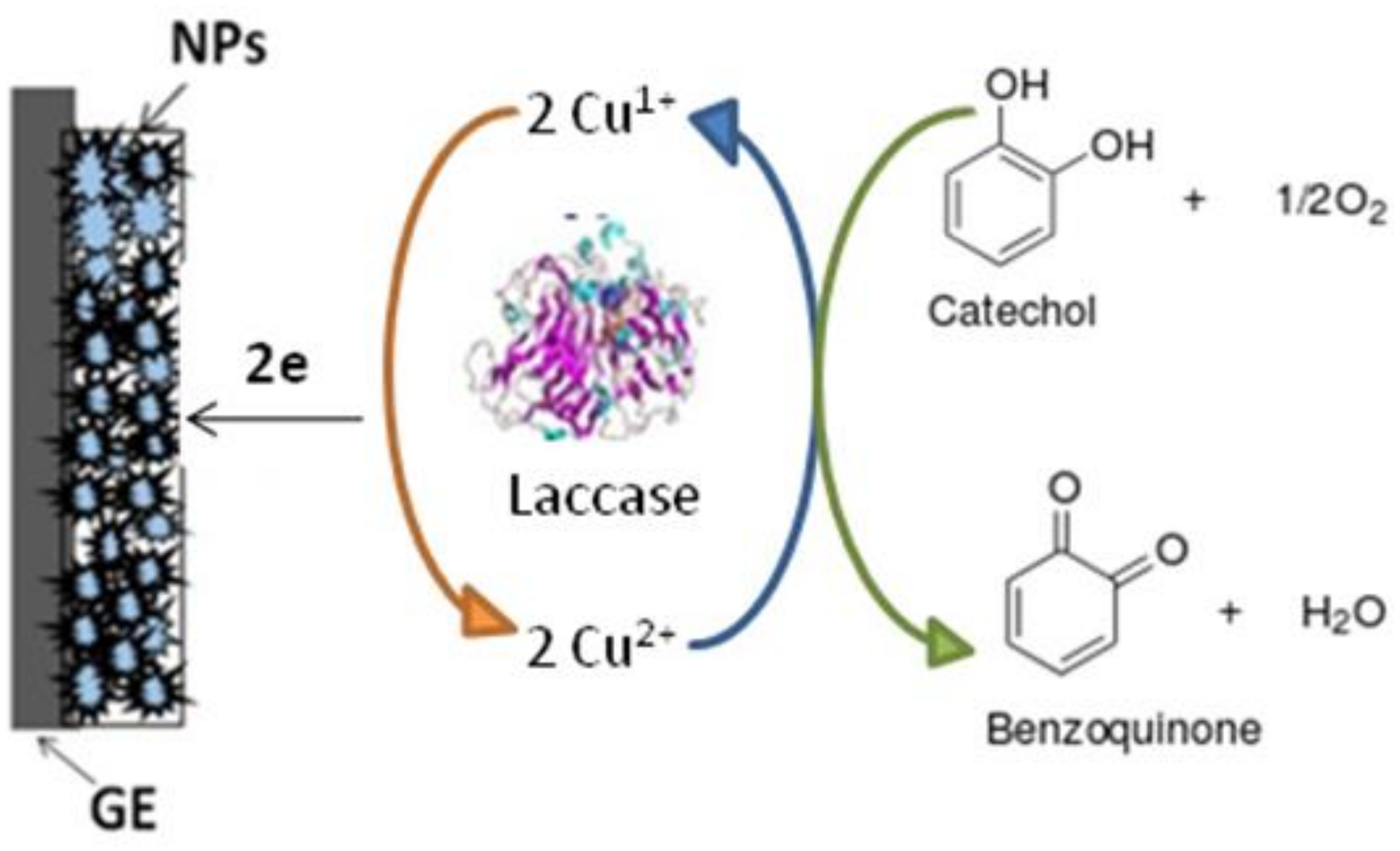
:1. Introduction
2. Materials and Methods
2.1. Reagents and Enzyme
2.2. Synthesis of NPs
2.3. Apparatus and Statistical Analysis
2.4. Characterization of the NPs for Their Redox Electroactivity
2.5. Fabrication and Characterization of the Laccase/NP-Modified Graphite Electrode
2.6. Application of the Laccase/CuCo-Based ABS for Catechol Assay in the Model and Real Samples
3. Results
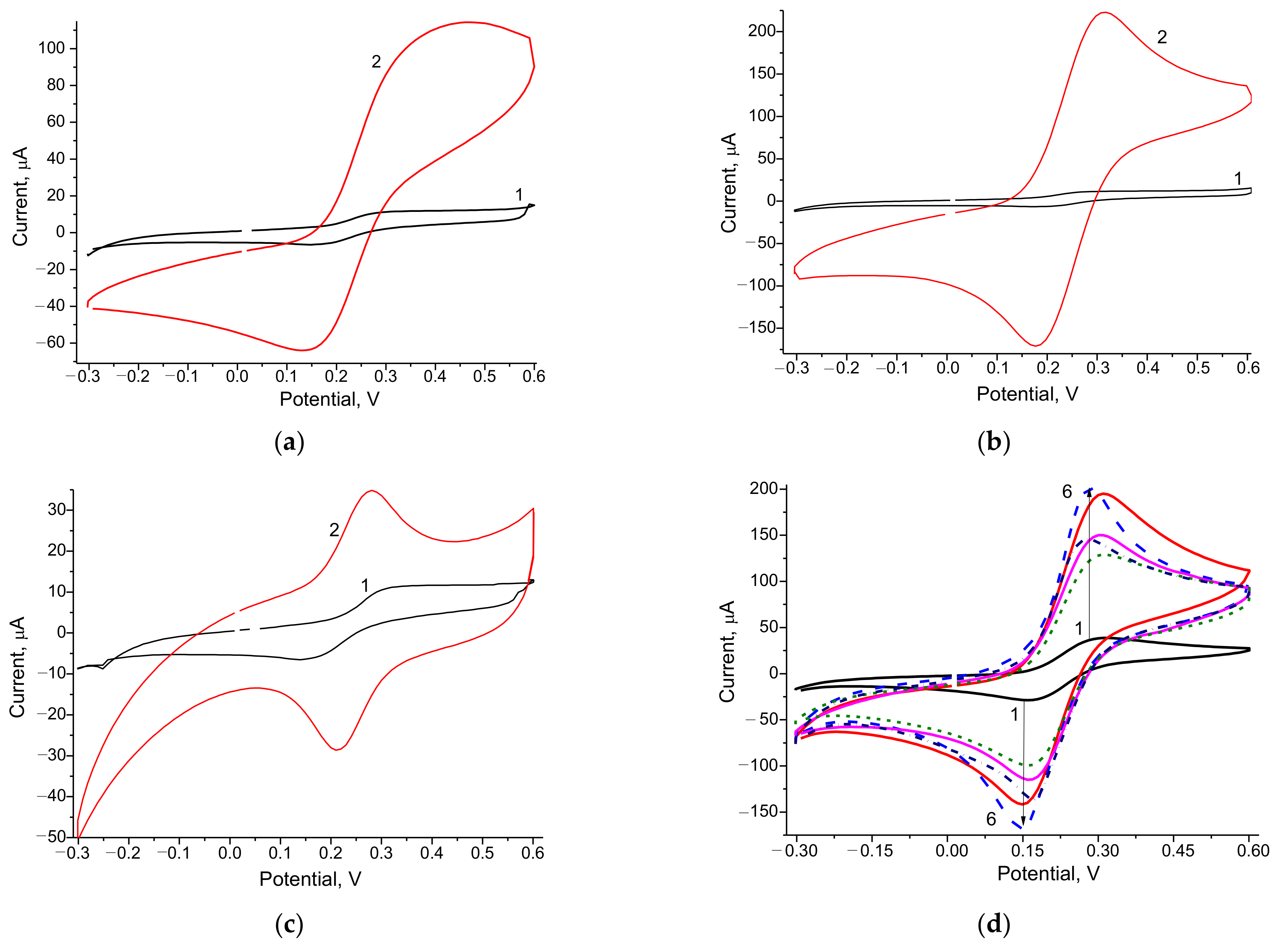
3.1. Selection of the Optimal Redox Nanomediators
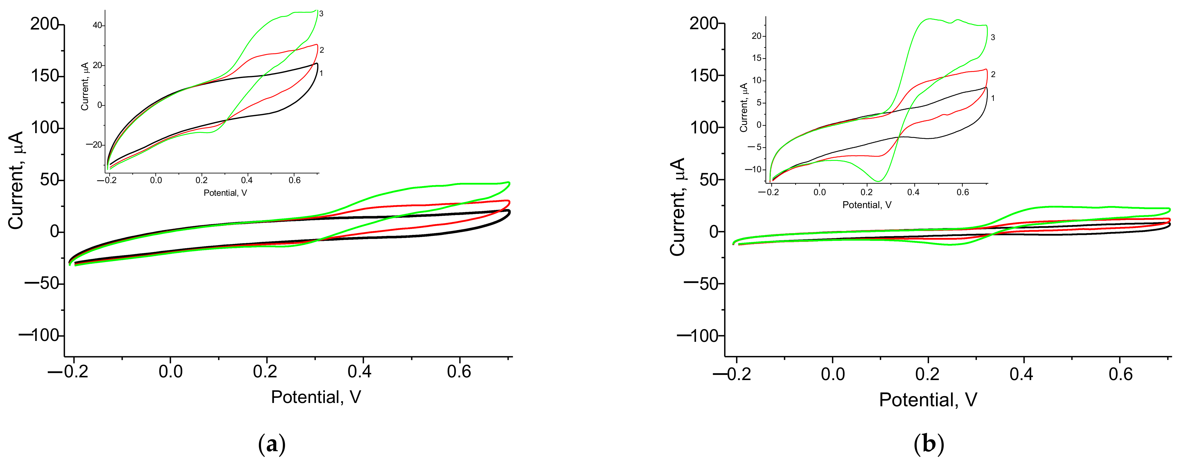
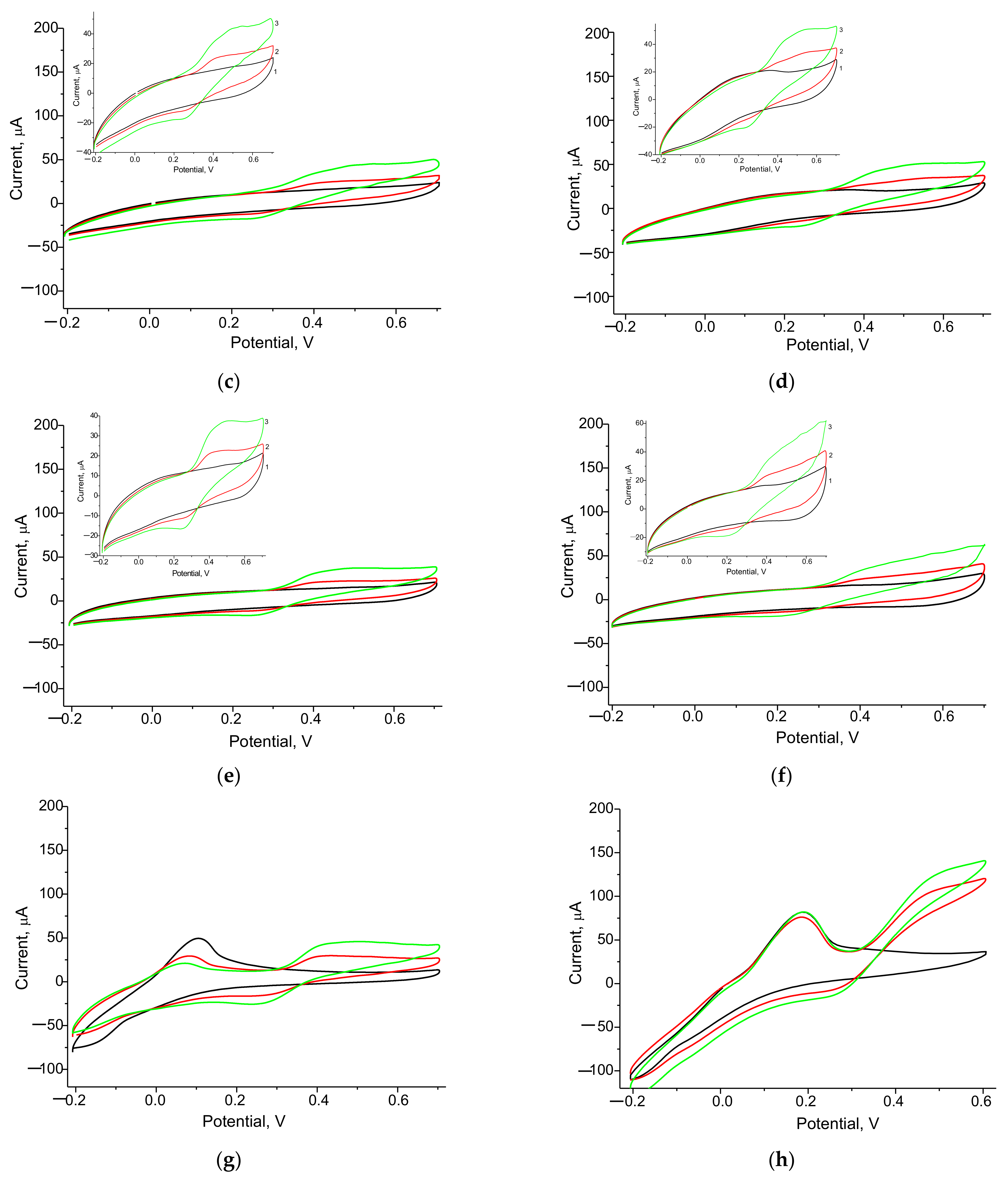
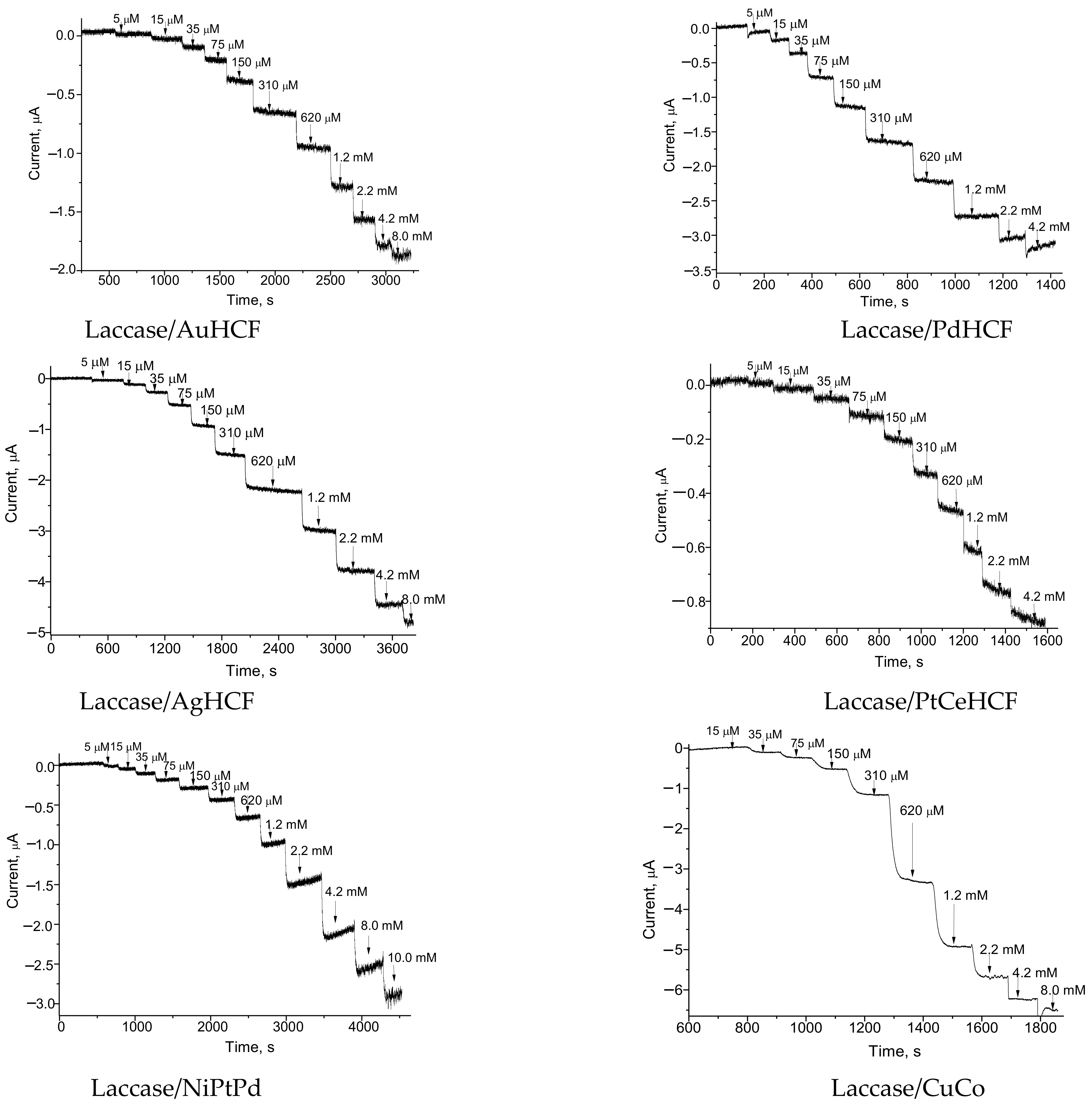
3.2. Development of Catechol-Sensitive Laccase-Based Amperometric Nanobiosensors
3.3. Properties of the Developed Laccase-Based ABS
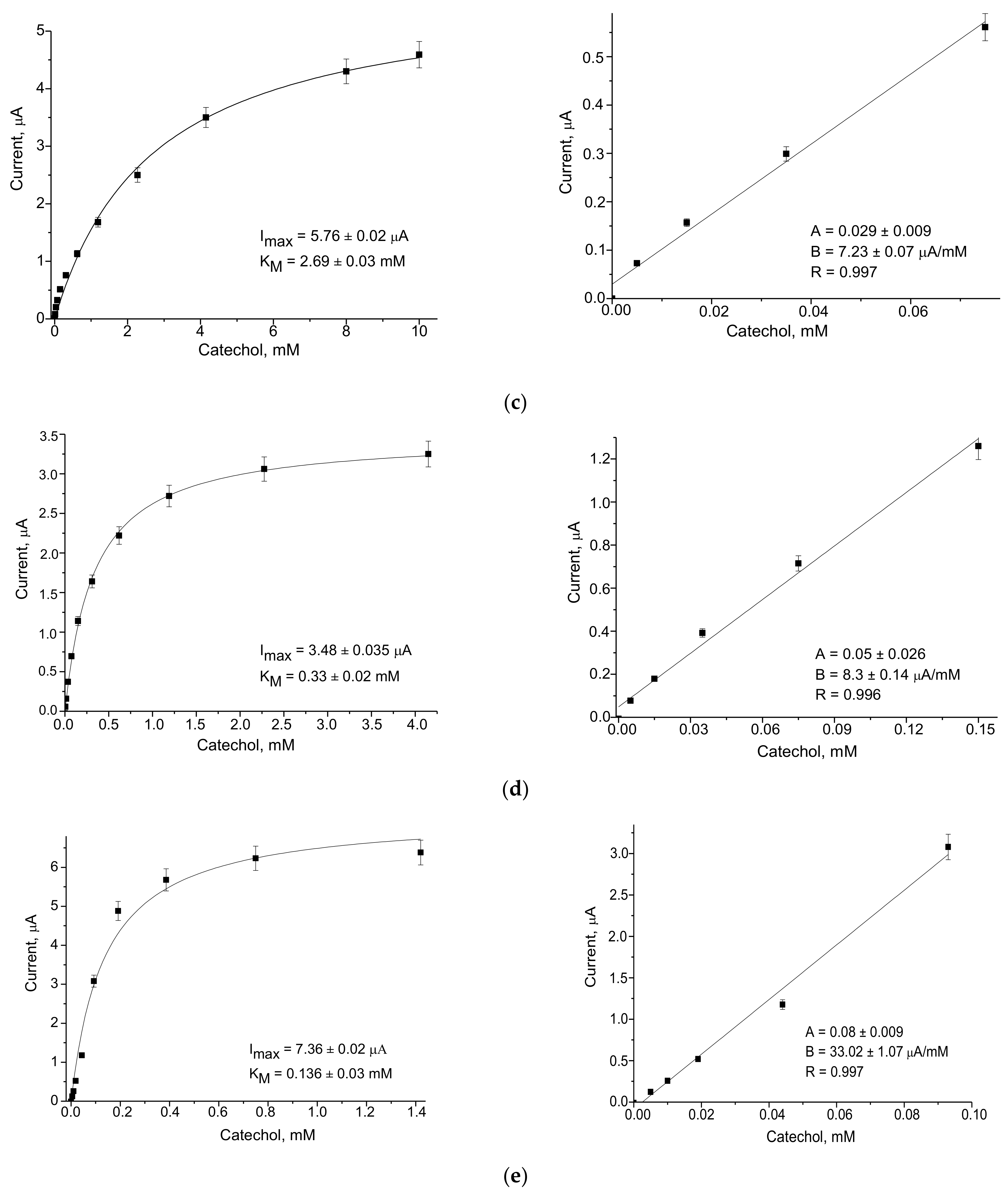
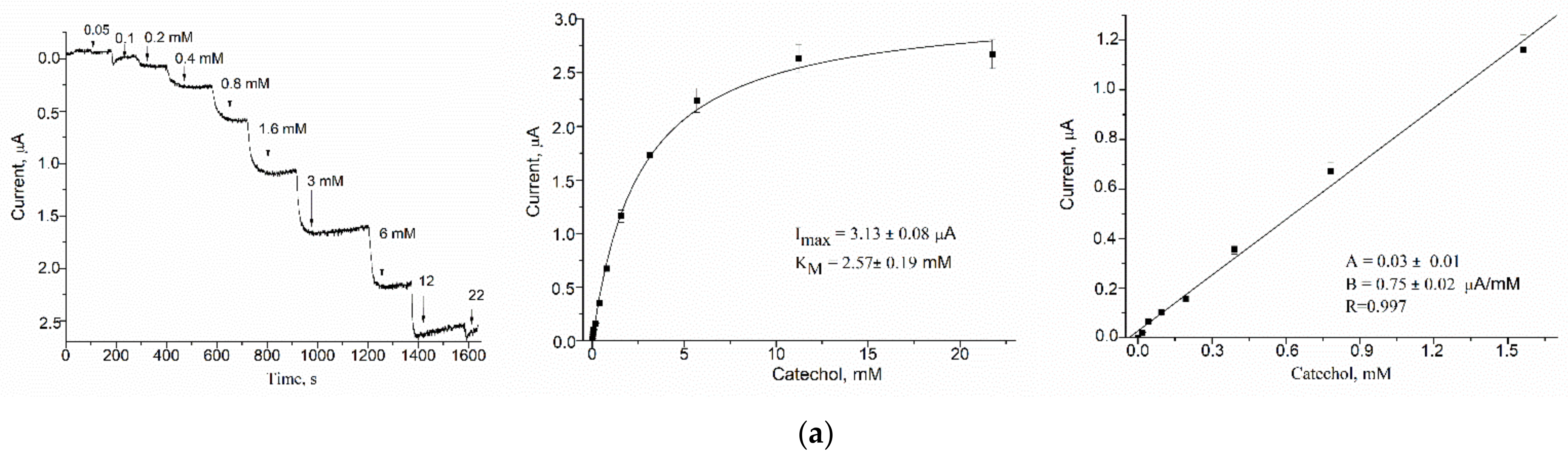
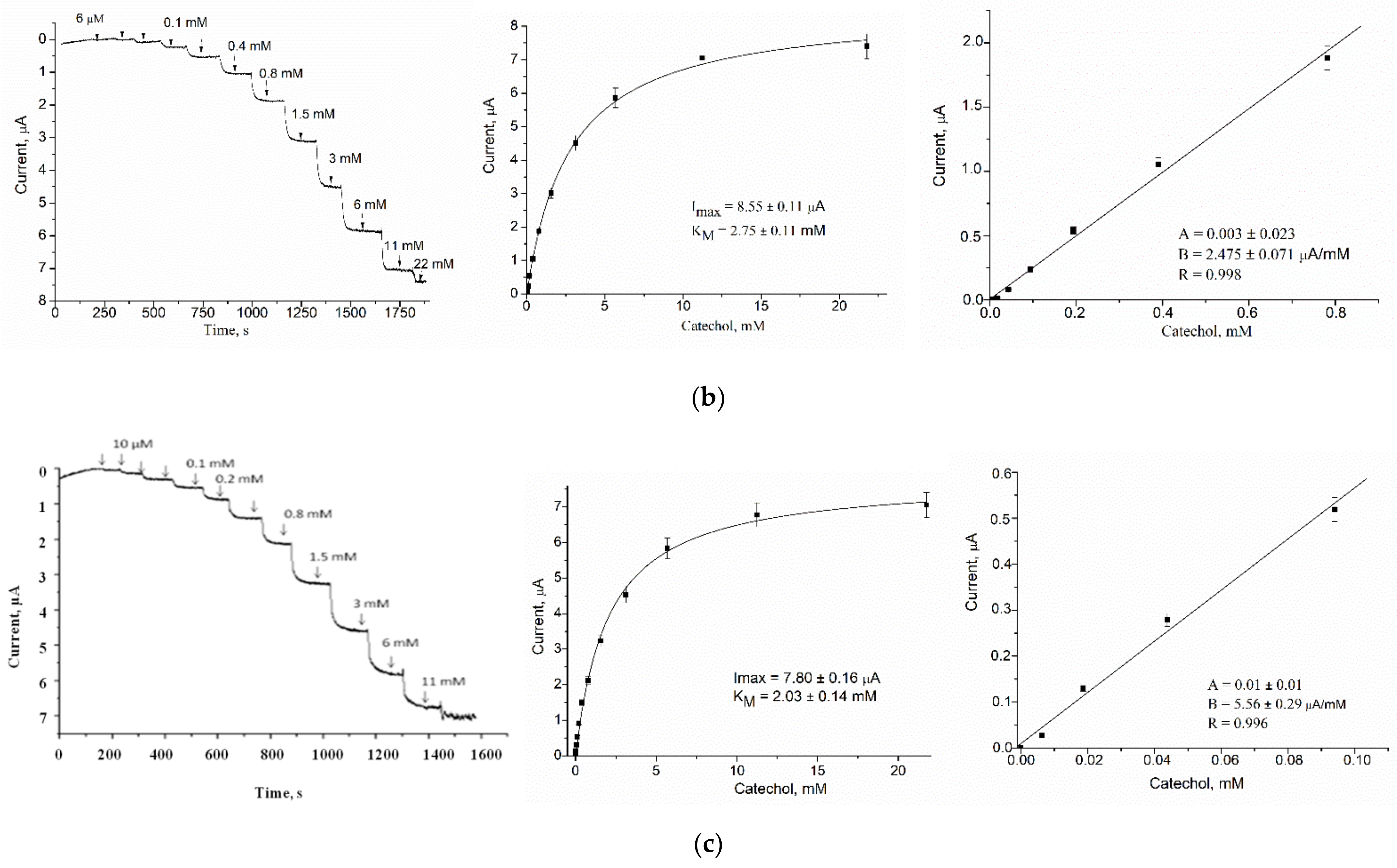
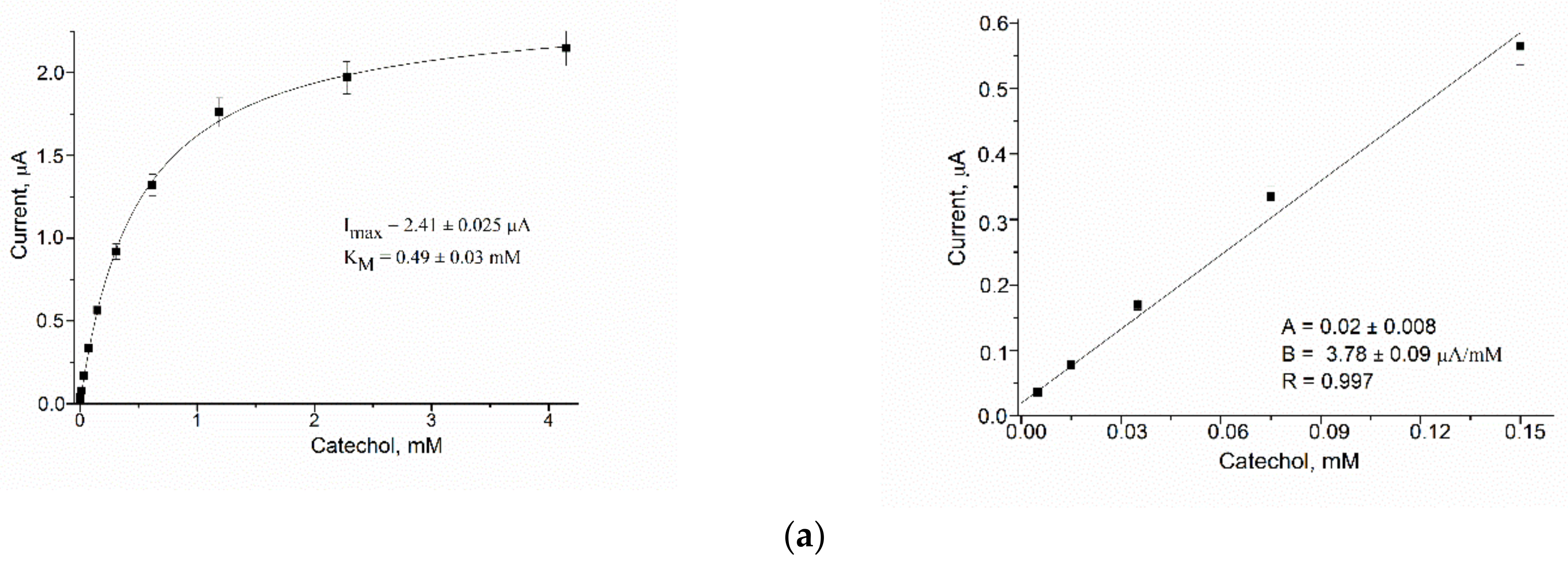
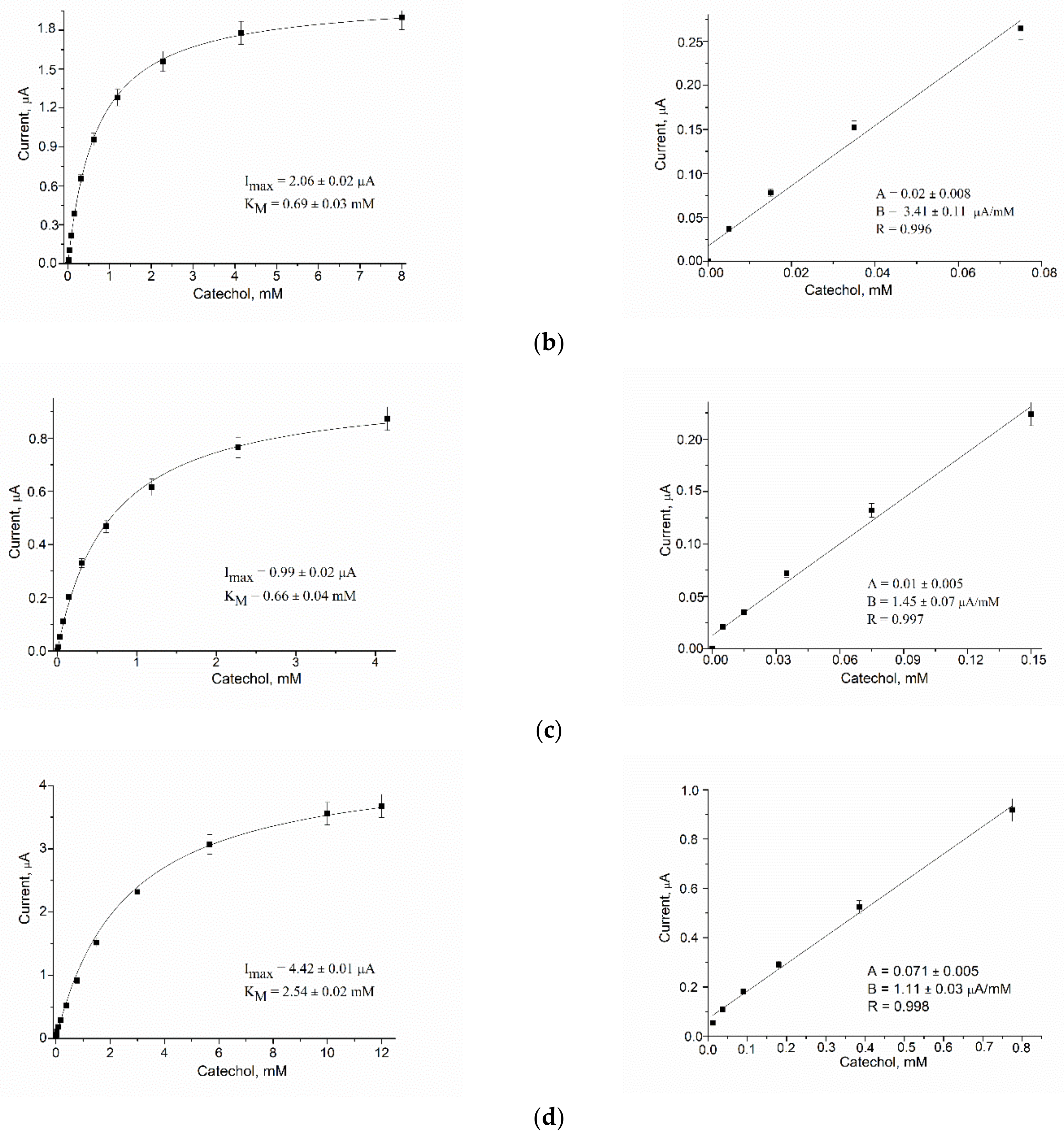
3.3.1. Analytical Characteristics
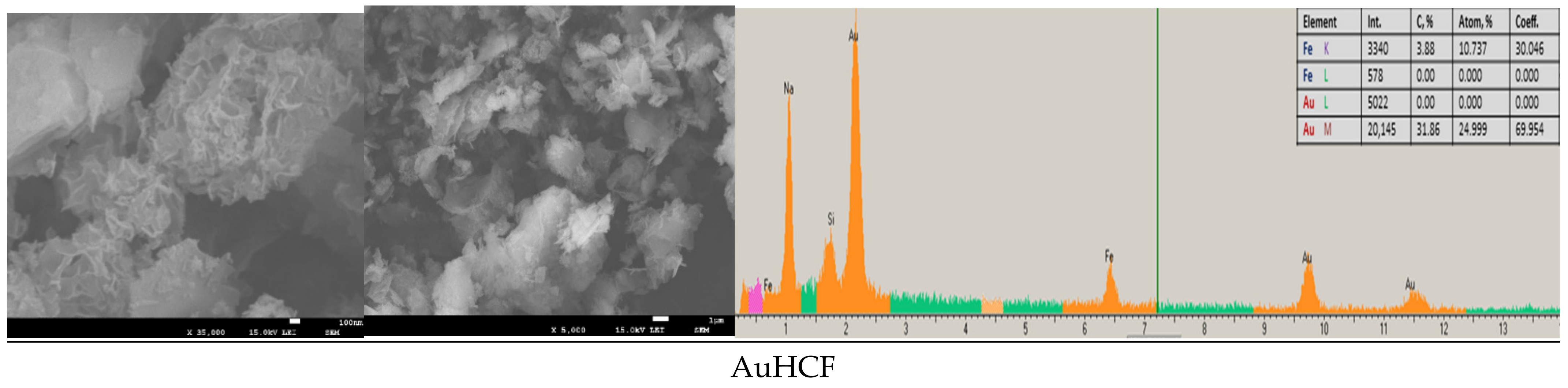
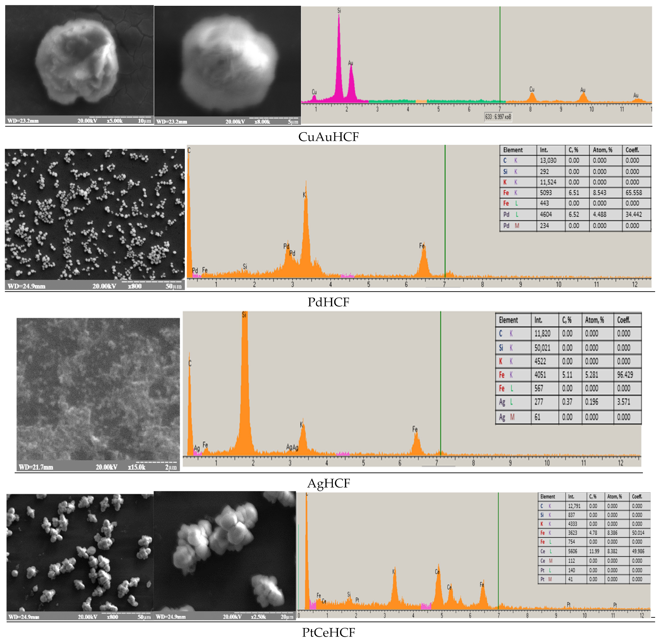
3.3.2. Morphologic Characterization of NPs
3.4. Properties of the Most Effective Laccase/CuCo-Based ABS
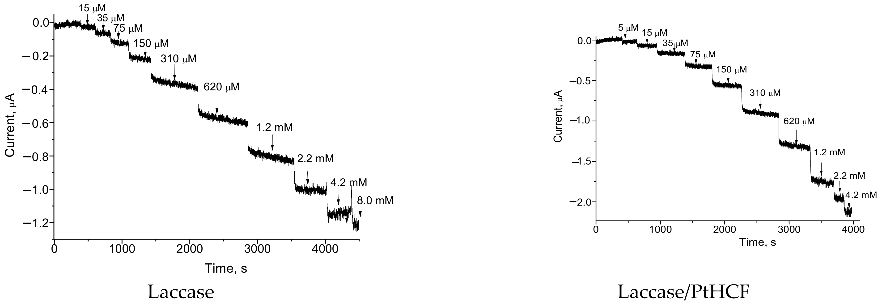
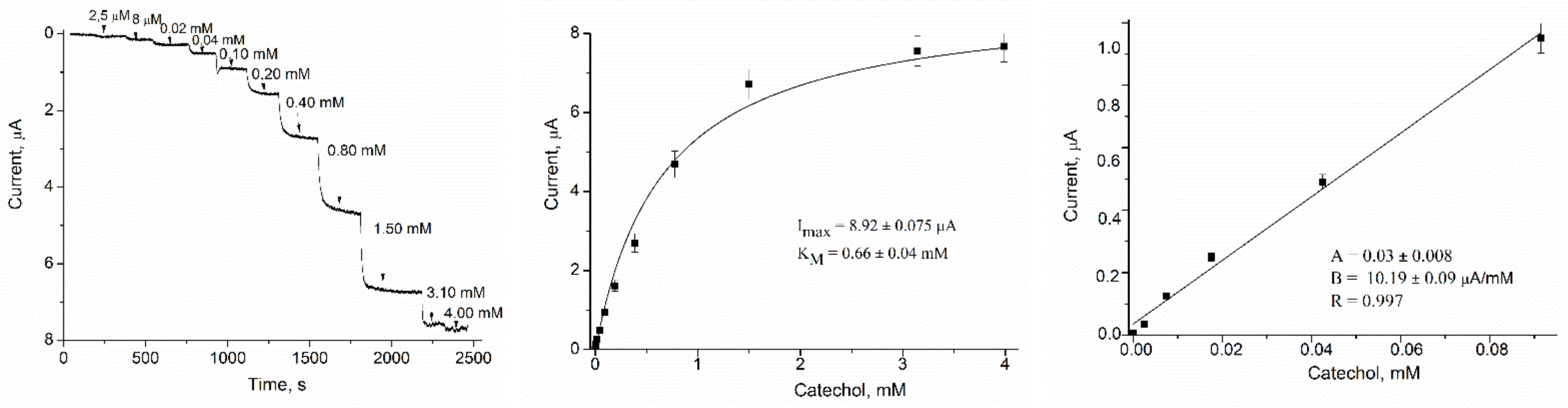
3.4.1. Optimization of Catechol Sensing
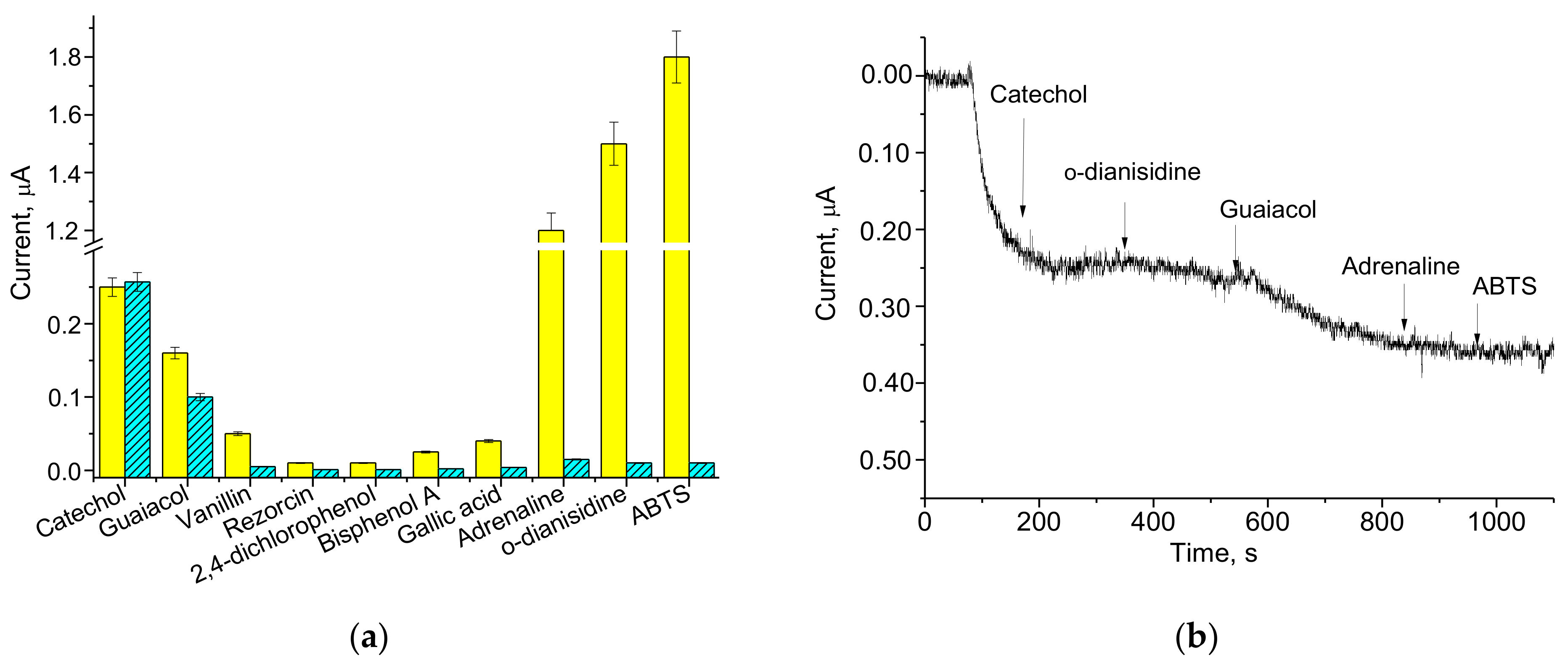
3.4.2. Selectivity
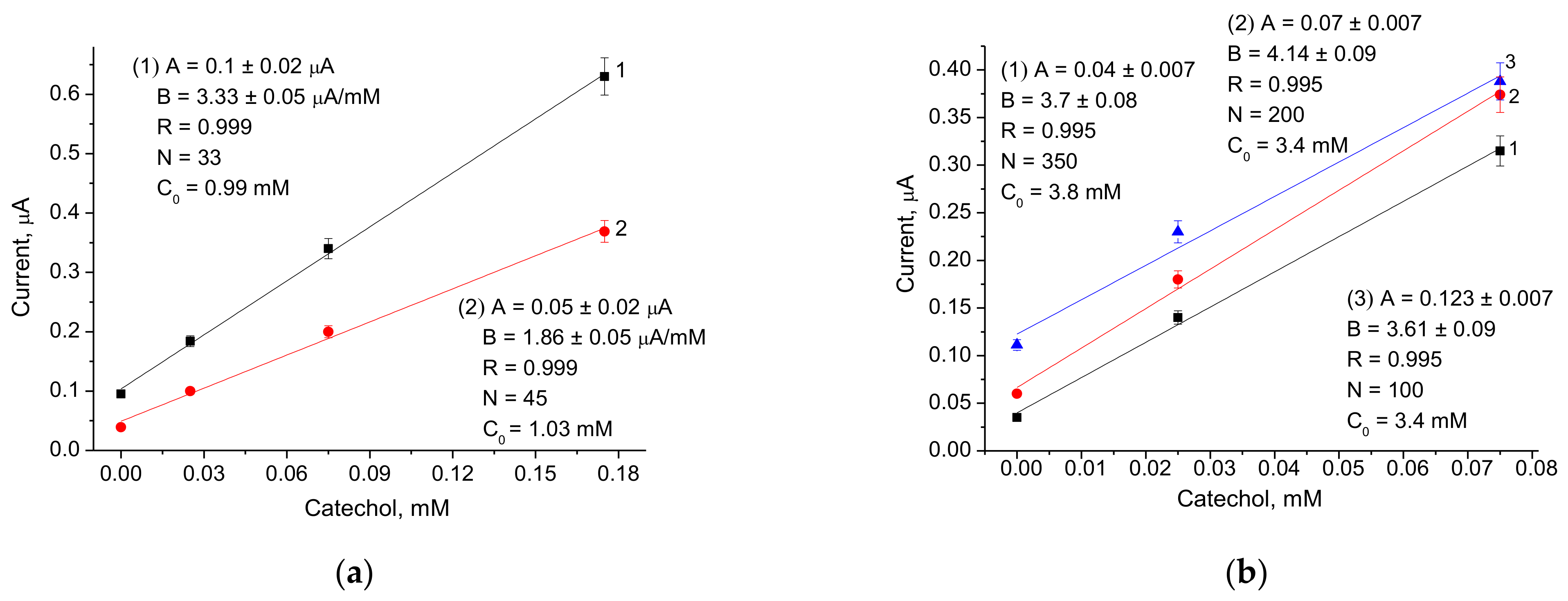
3.5. Application of the Laccase/CuCo-Based ABS for Catechol Assay in the Model and Real Samples
4. Discussion
Author Contributions
Funding
Institutional Review Board Statement
Informed Consent Statement
Data Availability Statement
Acknowledgments
Conflicts of Interest
Appendix A


References
- Jeon, J.-R.; Baldrian, P.; Murugesan, K.; Chang, Y.-S. Minireview Laccase-catalysed oxidations of naturally occurring phenols: From in vivo biosynthetic pathways to green synthetic applications. Microb. Biotechnol. 2012, 5, 318–332. [Google Scholar] [CrossRef]
- Reiss, R.; Ihssen, J.; Richter, M.; Eichhorn, E.; Schilling, B.; Thöny-Meyer, L. Laccase versus laccase-like multi-copper oxidase: A comparative study of similar enzymes with diverse substrate spectra. PLoS ONE 2013, 8, e65633. [Google Scholar] [CrossRef] [PubMed]
- Gulsunoglu-Konuskan, Z.; Kilic-Akyilmaz, M. Microbial Bioconversion of Phenolic Compounds in Agro-industrial Wastes: A Review of Mechanisms and Effective Factors. J. Agric. Food Chem. 2022, 70, 6901–6910. [Google Scholar] [CrossRef] [PubMed]
- Bhadra, F.; Gupta, A.; Vasundhara, M.; Reddy, M.S. Endophytic fungi: A potential source of industrial enzyme producers. 3 Biotech. 2022, 12, 86. [Google Scholar] [CrossRef] [PubMed]
- Mayolo-Deloisa, K.; González-González, M.; Rito-Palomares, M. Laccases in Food Industry: Bioprocessing, Potential Industrial and Biotechnological Applications. Front. Bioeng. Biotechnol. 2020, 8, 222. [Google Scholar] [CrossRef]
- Colella, A.; De Chiaro, A.; Lettera, V. In Situ Wood Fiber Dyeing Through Laccase Catalysis for Fiberboard Production. Front. Bioeng. Biotechnol. 2021, 9, 778971. [Google Scholar] [CrossRef]
- Perna, V.; Meyer, A.S.; Holck, J.; Eltis, L.D.; Eijsink, V.G.H.; Agger, J.W. Laccase-Catalyzed Oxidation of Lignin Induces Production of H2O2. ACS Sustain. Chem. Eng. 2020, 8, 831–841. [Google Scholar] [CrossRef]
- Shin, S.K.; Hyeon, J.E.; Joo, Y.-C.; Jeong, D.W.; You, S.K.; Han, S.O. Effective melanin degradation by a synergistic laccase-peroxidase enzyme complex for skin whitening and other practical applications. Int. J. Biol. Macromol. 2019, 129, 181–186. [Google Scholar] [CrossRef]
- Martini, M.C.; Berini, F.; Ausec, L.; Casciello, C.; Vacca, C.; Pistorio, M.; Lagares, A.; Mandic-Mulec, I.; Marinelli, F.; Del Papa, M.F. Identification and Characterization of a Novel Plasmid-Encoded Laccase-Like Multicopper Oxidase from Ochrobactrum sp. BF15 Isolated from an On-Farm Bio-Purification System. Food. Technol. Biotechnol. 2021, 59, 519–529. [Google Scholar] [CrossRef]
- Global “Laccase Market” (2022–2028) Report. Available online: https://www.wicz.com/story/45797138/global-laccase (accessed on 21 July 2022).
- Sakurai, T.; Kataoka, K. Basic and applied features of multicopper oxidases, CueO, bilirubin oxidase, and laccase. Chem Rec. 2007, 7, 220–229. [Google Scholar] [CrossRef] [Green Version]
- Bilal, M.; Ashraf, S.S.; Cui, J.; Lou, W.-Y.; Franco, M.; Mulla, S.I.; Iqbal, H.M.N. Harnessing the biocatalytic attributes and applied perspectives of nanoengineered laccases—A review. Int. J. Biol. Macromol. 2021, 166, 352–373. [Google Scholar] [CrossRef] [PubMed]
- Kumar, A.; Singh, D.; Sharma, K.K.; Arora, S.; Singh, A.K.; Gill, S.S.; Singhal, B. Gel-Based Purification and Biochemical Study of Laccase Isozymes from Ganoderma sp. and Its Role in Enhanced Cotton Callogenesis. Front. Microbiol. 2017, 8, 674. [Google Scholar] [CrossRef] [PubMed]
- Chiadò, A.; Bosco, F.; Bardelli, M.; Simonelli, L.; Pedotti, M.; Marmo, L.; Varani, L. Rational engineering of the lccβ T. versicolor laccase for the mediator-less oxidation of large polycyclic aromatic hydrocarbons. Comput. Struct. Biotechnol. J. 2021, 19, 2213–2222. [Google Scholar] [CrossRef] [PubMed]
- Wang, L.; Ding, X.; Huang, Q.; Hu, B.; Liang, L.; Wang, Q. Gllac7 Is Induced by Agricultural and Forestry Residues and Exhibits Allelic Expression Bias in Ganoderma lucidum. Front. Microbiol. 2022, 13, 890686. [Google Scholar] [CrossRef]
- Pezzella, C.; Guarino, L.; Piscitelli, A. How to enjoy laccases. Cell. Mol. Life Sci. 2015, 72, 923–940. [Google Scholar] [CrossRef]
- Yoshida, H. Chemistry of lacquer (urushi) part I. J. Chem. Soc. Trans. 1883, 43, 472–486. [Google Scholar] [CrossRef]
- Demkiv, O.M.; Gayda, G.Z.; Broda, D.; Gonchar, M.V. Extracellular laccase from Monilinia fructicola: Isolation, primary characterization and application. Cell Biol. Int. 2021, 45, 536–548. [Google Scholar] [CrossRef]
- Stasyuk, N.; Demkiv, O.; Gayda, G.; Zakalskiy, A.; Klepach, H.; Bisko, N.; Gonchar, M.; Nisnevitch, M. Highly Porous 3D Gold Enhances Sensitivity of Amperometric Biosensors Based on Oxidases and CuCe Nanoparticles. Biosensors 2022, 12, 472. [Google Scholar] [CrossRef]
- Miyata, M.; Kitazumi, Y.; Shirai, O.; Kataoka, K.; Kano, K. Diffusion-limited biosensing of dissolved oxygen by direct electron transfer-type bioelectrocatalysis of multi-copper oxidases immobilized on porous gold microelectrodes. J. Electroanal. Chem. 2020, 860, 113895. [Google Scholar] [CrossRef]
- Dinu, A.; Apetrei, C. Quantification of Tyrosine in Pharmaceuticals with the New Biosensor Based on Laccase-Modified Polypyrrole Polymeric Thin Film. Polymers. 2022, 14, 441. [Google Scholar] [CrossRef]
- Chen, H.; Simoska, O.; Lim, K.; Grattieri, M.; Yuan, M.; Dong, F.; Lee, Y.S.; Beaver, K.; Weliwatte, S.; Gaffney, E.M.; et al. Fundamentals, applications, and future directions of bioelectrocatalysis. Chem. Rev. 2020, 120, 12903–12993. [Google Scholar] [CrossRef] [PubMed]
- Kadam, A.A.; Saratale, G.D.; Ghodake, G.S.; Saratale, R.G.; Shahzad, A.; Magotra, V.K.; Kumar, M.; Palem, R.R.; Sung, J.-S. Recent Advances in the Development of Laccase-Based Biosensors via Nano-Immobilization Techniques. Chemosensors 2022, 10, 58. [Google Scholar] [CrossRef]
- Demkiv, O.; Stasyuk, N.; Serkiz, R.; Gayda, G.; Nisnevitch, M.; Gonchar, M. Peroxidase-Like Metal-Based Nanozymes: Synthesis, Catalytic Properties, and Analytical Application. Appl. Sci. 2021, 11, 777. [Google Scholar] [CrossRef]
- Flickinger, C.W. The benzenediols: Catechol, resorcinol and hydroquinone—A review of the industrial toxicology and current industrial exposure limits. Am. Ind. Hyg. Assoc. J. 1976, 37, 596–606. [Google Scholar] [CrossRef] [PubMed]
- Lee, B.P.; Birkedal, H.; Lee, H. Editorial: Catechol and Polyphenol Chemistry for Smart Polymers. Front. Chem. 2019, 7, 883. [Google Scholar] [CrossRef]
- Nsanzamahoro, S.; Mutuyimana, F.P.; Han, Y.; Ma, S.; Na, M.; Liu, J.; Ma, Y.; Ren, C.; Chen, H.; Chen, X. Highly selective and sensitive detection of catechol by one step synthesized highly fluorescent and water-soluble silicon nanoparticles. Sens. Actuat. B Chem. 2018, 281, 849–856. [Google Scholar] [CrossRef]
- Jabbari, S.; Dabirmanesh, B.; Khajeh, K. Specificity enhancement towards phenolic substrate by immobilization of laccase on surface plasmon resonance sensor chip. J. Mol. Catal. B Enzym. 2015, 121, 32–36. [Google Scholar] [CrossRef]
- Wang, Y.; Li, Y.; Bao, X.; Han, J.; Xia, J.; Tian, X.; Ni, L. A smartphone-based colorimetric reader coupled with a remote server for rapid on-site catechols analysis. Talanta 2016, 160, 194–204. [Google Scholar] [CrossRef]
- Castrovilli, M.C.; Bolognesi, P.; Chiarinelli, J.; Avaldi, L.; Cartoni, A.; Calandra, P.; Tempesta, E.; Giardi, M.T.; Antonacci, A.; Arduini, F.; et al. Electrospray deposition as a smart technique for laccase immobilisation on carbon black-nanomodified screen-printed electrodes. Biosens. Bioelectron. 2020, 163, 112299. [Google Scholar] [CrossRef]
- Li, Z.; Zeng, H.-Z.; Cao, X.-J.; Li, H.-B.; Long, Y.-W.; Feng, B.; Lv, S.-B. High-sensitive sensor for the simultaneous determination of phenolics based on multi-walled carbon nanotube/NiCoAl hydrotalcite electrode material. Microchim. Acta 2021, 188, 9. [Google Scholar] [CrossRef]
- Pillai, R.; Preetha, S.; Narasimhamurthy, B.; Lekshmi, I.C. Biosensing of catechol via amperometry using laccase immobilized nickel oxide/graphite modified screen-printed electrodes. Mater. Today: Proc. 2022, 62, 5434–5438. [Google Scholar] [CrossRef]
- Melak, F.; Redi, M.; Tessema, M.; Alemayehu, E. Electrochemical determination of catechol in tea samples using anthraquinone modified carbon paste electrode. Nat. Sci. 2013, 05, 888–894. [Google Scholar] [CrossRef]
- Broli, N.; Vallja, L.; Shehu, A.; Vasjar, M. Determination of catechol in extract of tea using carbon paste electrode modified with banana tissue. J. Food Process Preserv. 2018, 43, e13838. [Google Scholar] [CrossRef]
- Nellaiappan, S.; Kumar, S. Selective amperometric and flow injection analysis of 1,2-dihydroxy benzene isomer in presence of 1,3- and 1,4-dihydroxy benzene isomers using palladium nanoparticles-chitosan modified ITO electrode. Sens. Actuat. B Chem. 2018, 254, 820–826. [Google Scholar] [CrossRef]
- Zhou, Y.; Tang, L.; Zeng, G.; Chen, J.; Cai, Y.; Zhang, Y.; Yang, G.; Liu, Y.; Zhang, C.; Tang, W. Mesoporous carbon nitride based biosensor for highly sensitive and selective analysis of phenol and catechol in compost bioremediation. Biosens. Bioelectron. 2014, 61, 519–525. [Google Scholar] [CrossRef]
- Rodrigues, R.C.; Ortiz, C.; Berenguer-Murcia, Á.; Torres, R.; Fernández-Lafuente, R. Modifying enzyme activity and selectivity by immobilization. Chem. Soc. Rev. 2013, 42, 6290–6307. [Google Scholar] [CrossRef]
- Deniz, S.A.; Goker, S.; Toppare, L.; Soylemez, S. Fabrication of D–A–D type conducting polymer, carbon nanotubes and silica nanoparticle-based laccase biosensor for catechol detection. New J. Chem. 2022, 46, 15521–15529. [Google Scholar] [CrossRef]
- Qi, H.; Zhang, C. Simultaneous determination of hydroquinone and catechol at a glassy carbon electrode modified with multiwall carbon nanotubes. Electroanalysis 2005, 17, 832–838. [Google Scholar] [CrossRef]
- Hammani, H.; Laghrib, F.; Farahi, A.; Lahrich, S.; El Achaby, M.; El Harfi, K.; Aboulkas, A.; Bakasse, M.; El Mhammedi, M.A. Date stone based activated carbon/graphite electrode for catechol analysis: Physico-chemical properties and application in beverage samples. New J. Chem. 2018, 42, 13285–13296. [Google Scholar] [CrossRef]
- Arya Nair, J.S.; Saisree, S.; Sandhya, K.Y. Picomolar level electrochemical detection of hydroquinone, catechol and resorcinol simultaneously using a MoS2 nano-flower decorated graphene. Analyst 2022, 147, 2966–2979. [Google Scholar] [CrossRef]
- Harisha, B.K.; Kumara Swamy, V.E.; Ebenso, E.E. Poly (glycine) modified carbon paste electrode for simultaneous determination of catechol and hydroquinone: A voltammetric study. J. Electroanal. Chem. 2018, 823, 730–736. [Google Scholar] [CrossRef]
- Liu, H.; Zhou, P.; Wu, X.; Sun, J.; Chen, S. Radical Scavenging by Acetone: A New Perspective to Understand Laccase/ABTS Inactivation and to Recover Redox Mediator. Molecules 2015, 20, 19907–19913. [Google Scholar] [CrossRef] [PubMed]
- Gayda, G.Z.; Demkiv, O.M.; Gurianov, Y.; Serkiz, R.Y.; Klepach, H.M.; Gonchar, M.V.; Nisnevitch, M. “Green” Prussian Blue Analogues as Peroxidase Mimetics for Amperometric Sensing and Biosensing. Biosensors 2021, 11, 193. [Google Scholar] [CrossRef] [PubMed]
- Gayda, G.Z.; Demkiv, O.M.; Stasyuk, N.Y.; Serkiz, R.Y.; Gonchar, M.V.; Nisnevitch, M. Metallic nanoparticles obtained via “green” synthesis as a platform for biosensor construction. Appl. Sci. 2019, 9, 720. [Google Scholar] [CrossRef]
- Stasyuk, N.; Gayda, G.; Zakalskiy, A.; Zakalska, O.; Serkiz, R.; Gonchar, M. Amperometric biosensors based on oxidases and PtRu nanoparticles as artificial peroxidase. Food Chem. 2019, 285, 213–220. [Google Scholar] [CrossRef] [PubMed]
- Stasyuk, N.; Gayda, G.; Demkiv, O.; Darmohray, L.; Gonchar, M.; Nisnevitch, M. Amperometric biosensors for L-arginine determination based on L-arginine oxidase and peroxidase-like nanozymes. Appl. Sci. 2021, 11, 7024. [Google Scholar] [CrossRef]
- Stasyuk, N.; Demkiv, O.; Gayda, G.; Zakalska, O.; Zakalskiy, A.; Serkiz, R.; Kavetskyy, T.; Gonchar, M. Reusable alcohol oxidase-nPtCu/alginate beads for highly sensitive ethanol assay in beverages. RSC Adv. 2022, 12, 21103–21110. [Google Scholar] [CrossRef]
- Othman, A.M.; Wollenberger, U. Amperometric biosensor based on coupling aminated laccase to functionalized carbon nanotubes for phenolics detection. Int. J. Biol. Macromol. 2020, 15, 855–864. [Google Scholar] [CrossRef]
- Rodríguez-Delgado, M.M.; Alemán-Nava, G.S.; Rodríguez-Delgado, J.M.; Dieck-Assad, G.; Martínez-Chapa, S.O.; Barceló, D.; Parra, R. Laccase-based biosensors for detection of phenolic compounds. TrAC Trends Anal. Chem. 2015, 74, 21–45. [Google Scholar] [CrossRef]
- Thurston, C.F. The structure and function of fungal laccases. Microbiology 1994, 140, 19–26. [Google Scholar] [CrossRef] [Green Version]
- Schlosser, D.; Höfer, C. Laccase-Catalyzed Oxidation of Mn in the Presence of Natural Mn Chelators as a Novel Source of Extracellular H2O2 Production and Its Impact on Manganese Peroxidase. Appl. Environ. Microbiol. 2002, 68, 3514–3521. [Google Scholar] [CrossRef] [PubMed]
- Ming, C.; Lin, W.; Tian, T.; Xue-Cai, L.; Zai, Z.; Ruo-Chun, Y.; Ji-Hu, S.; Jiang-Feng, D. Radical Mechanism of Laccase-Catalyzed Catechol Ring-Opening. Acta Phys. Chim. Sin. 2017, 33, 620–626. [Google Scholar] [CrossRef]
- Jones, S.M.; Solomon, E.I. Electron transfer and reaction mechanism of laccases. Cell. Mol. Life Sci. 2015, 72, 869–883. [Google Scholar] [CrossRef] [PubMed]
- Clément, R.; Wang, X.; Biaso, F.; Ilbert, M.; Mazurenko, I.; Lojou, E. Mutations in the coordination spheres of T1 Cu affect Cu2+-activation of the laccase from Thermus thermophilus. Biochimie 2021, 182, 228–237. [Google Scholar] [CrossRef] [PubMed]
- Mazurenko, I.; Adachi, T.; Ezraty, B.; Ilbert, M.; Sowa, K.; Lojou, E. Electrochemistry of copper efflux oxidase-like multicopper oxidases involved in copper homeostasis. Curr. Opin. Electrochem. 2022, 32, 100919. [Google Scholar] [CrossRef]
- Ferraroni, M.; Myasoedova, N.M.; Schmatchenko, V.; Leontievsky, A.A.; Golovleva, L.A.; Scozzafava, A.; Briganti, F. Crystal structure of a blue laccase from Lentinus tigrinus: Evidences for intermediates in the molecular oxygen reductive splitting by multicopper oxidases. BMC Struct. Biol. 2007, 7, 60. [Google Scholar] [CrossRef]
- Davydov, R.; Herzog, A.E.; Jodts, R.J.; Karlin, K.D.; Hoffman, B.M. End-On Copper(I) Superoxo and Cu(II) Peroxo and Hydroperoxo Complexes Generated by Cryoreduction/Annealing and Characterized by EPR/ENDOR Spectroscopy. J. Am. Chem. Soc. 2022, 144, 377–389. [Google Scholar] [CrossRef]
- Giardina, P.; Faraco, V.; Pezzella, C.; Piscitelli, A.; Vanhulle, S.; Sannia, G. Laccases: A never-ending story. Cell. Mol. Life Sci. 2010, 67, 369–385. [Google Scholar] [CrossRef]


| Composition of ABS | Sensitivity, A·M−1·m−2 | Linear Range, up to, mM | LOD, µA | KMapp, mM | Imax, µA |
|---|---|---|---|---|---|
| Laccase/GE | 89 | 0.30 | 1 | 0.70 | 0.65 |
| Laccase/CuAuHCF/GE | 152 | 0.80 | 2 | 2.54 | 4.42 |
| Laccase/PtCeHCF/GE | 199 | 0.14 | 1 | 0.66 | 0.99 |
| Laccase/PtHCF/GE | 517 | 0.15 | 0.1 | 0.49 | 2.41 |
| Laccase/AuCo/GE | 630 | 0.06 | 0.1 | 1.10 | 2.05 |
| Laccase/AuHCF/GE | 757 | 0.08 | 0.2 | 1.27 | 6.02 |
| Laccase/AgHCF/GE | 881 | 0.15 | 0.2 | 0.80 | 5.24 |
| Laccase/NiPtPd/GE | 990 | 0.08 | 0.1 | 2.69 | 5.76 |
| Laccase/PdHCF/GE | 1137 | 0.10 | 0.2 | 0.33 | 3.48 |
| Laccase/CuCo/GE | 4523 | 0.09 | 0.2 | 0.14 | 7.36 |
| * Laccase/GE | 103 | 1.60 | 4 | 2.57 | 3.13 |
| * Laccase/gAu/GE | 295 | 0.40 | 1 | 1.56 | 3.96 |
| * Laccase/gFeHCF/GE | 339 | 0.80 | 1 | 2.75 | 8.55 |
| * Laccase/PtRu/GE | 551 | 0.40 | 1 | 1.66 | 9.14 |
| * Laccase/gCuHCF/GE | 762 | 0.20 | 0.5 | 2.03 | 7.31 |
| ABS, No | Sensing Layer | Sensitivity, A·M−1 m−2 | Linear Range, up to, mM | KMapp, mM | Imax, µM | |
|---|---|---|---|---|---|---|
| Laccase, m-Units | CuCo, µg | |||||
| 1 | 50 | 1 | 220 | 0.16 | 2.48 | 2.94 |
| 2 | 100 | 1 | 721 | 0.18 | 1.05 | 5.18 |
| 3 | 200 | 1 | 750 | 0.09 | 1.49 | 6.37 |
| 4 | 500 | 1 | 1441 | 0.09 | 0.66 | 8.92 |
| 5 | 500 | 5 | 3435 | 0.04 | 0.59 | 7.57 |
| 6 | 1000 | 5 | 4495 | 0.04 | 0.25 | 7.48 |
| 7 | 200 | 2 | 770 | 0.10 | 1.67 | 7.70 |
| 8 | 200 | 4 | 820 | 0.18 | 1.80 | 8.17 |
| 9 | 200 | 10 | 188 | 0.76 | 5.27 | 8.62 |
| 10 | 100 (Control) | 0 | 107 | 0.80 | 1.67 | 1.76 |
Publisher’s Note: MDPI stays neutral with regard to jurisdictional claims in published maps and institutional affiliations. |
© 2022 by the authors. Licensee MDPI, Basel, Switzerland. This article is an open access article distributed under the terms and conditions of the Creative Commons Attribution (CC BY) license (https://creativecommons.org/licenses/by/4.0/).
Share and Cite
Demkiv, O.; Gayda, G.; Stasyuk, N.; Brahinetz, O.; Gonchar, M.; Nisnevitch, M. Nanomaterials as Redox Mediators in Laccase-Based Amperometric Biosensors for Catechol Assay. Biosensors 2022, 12, 741. https://doi.org/10.3390/bios12090741
Demkiv O, Gayda G, Stasyuk N, Brahinetz O, Gonchar M, Nisnevitch M. Nanomaterials as Redox Mediators in Laccase-Based Amperometric Biosensors for Catechol Assay. Biosensors. 2022; 12(9):741. https://doi.org/10.3390/bios12090741
Chicago/Turabian StyleDemkiv, Olha, Galina Gayda, Nataliya Stasyuk, Olena Brahinetz, Mykhailo Gonchar, and Marina Nisnevitch. 2022. "Nanomaterials as Redox Mediators in Laccase-Based Amperometric Biosensors for Catechol Assay" Biosensors 12, no. 9: 741. https://doi.org/10.3390/bios12090741