Nanomaterial-Based Electrochemical Nanodiagnostics for Human and Gut Metabolites Diagnostics: Recent Advances and Challenges


 , and
, and
Abstract
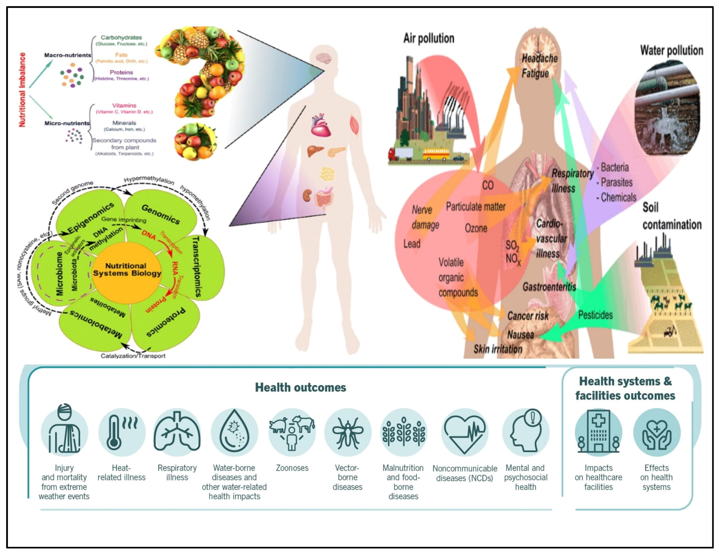
:1. Introduction

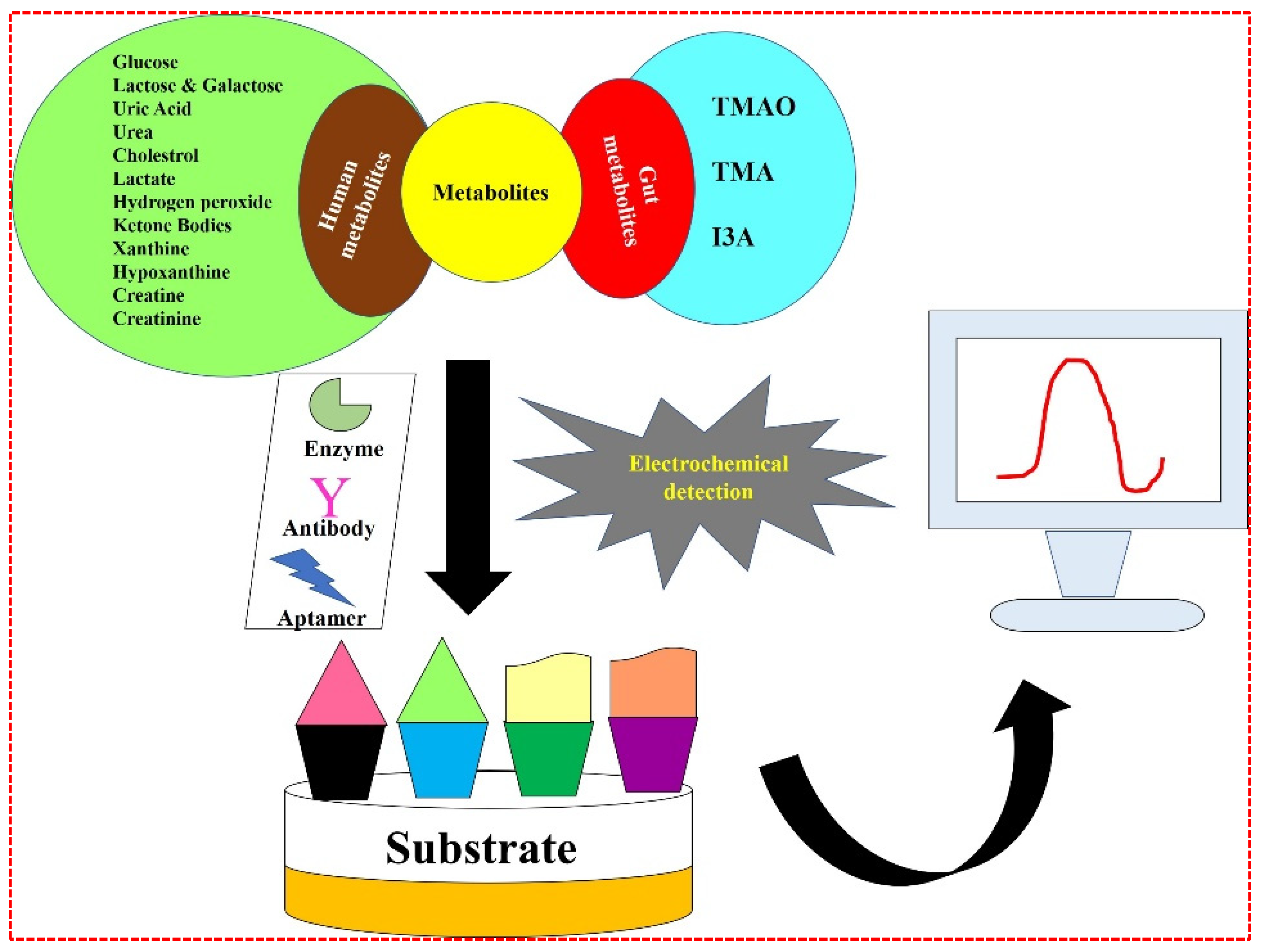
2. Nanomaterial-Based Biosensors for Metabolites Detection
2.1. Biosensors for Human Metabolites Detection
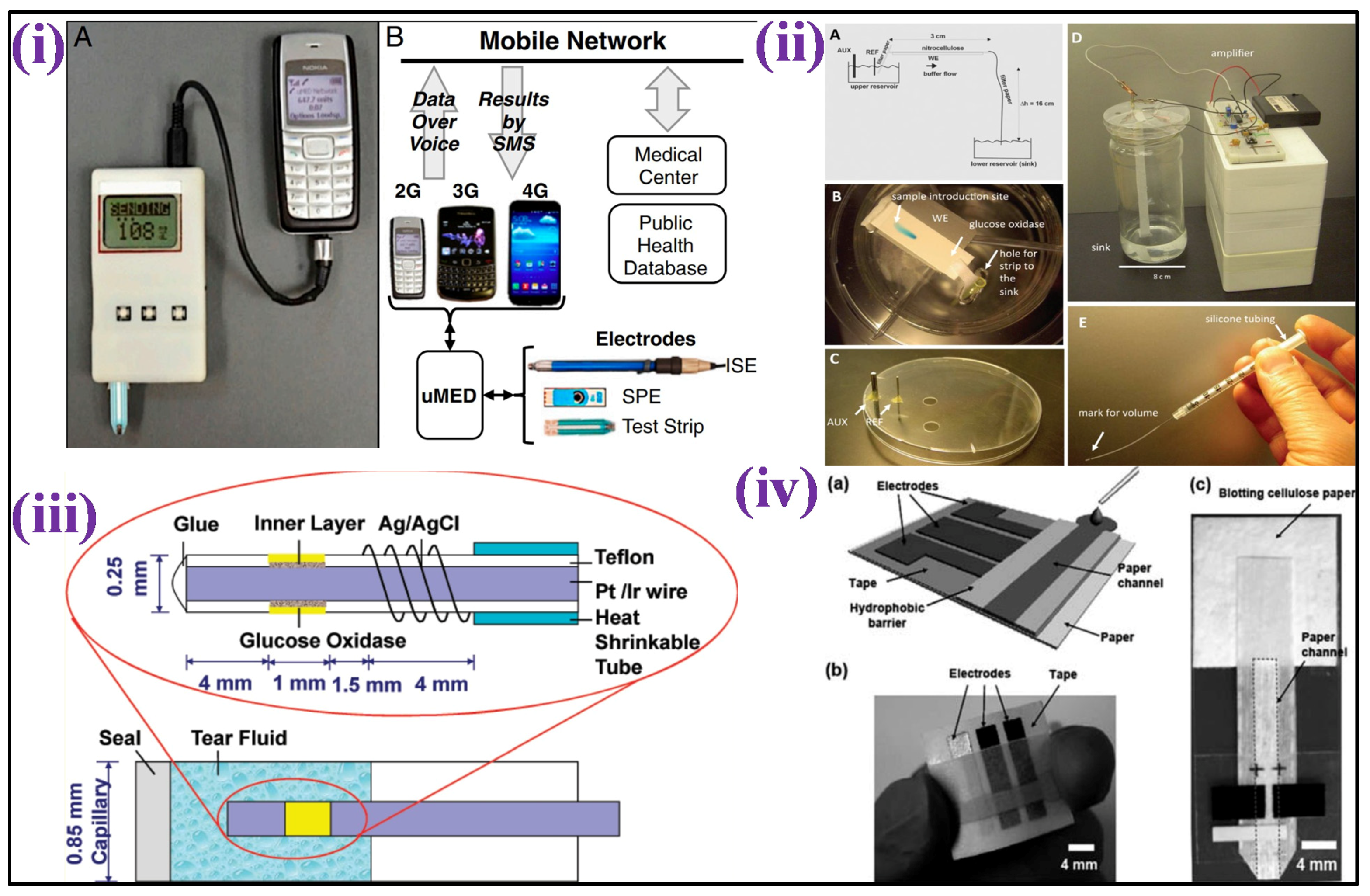
2.1.1. Glucose

2.1.2. Lactose and Galactose
2.1.3. Uric Acid (UA)s

2.1.4. Urea
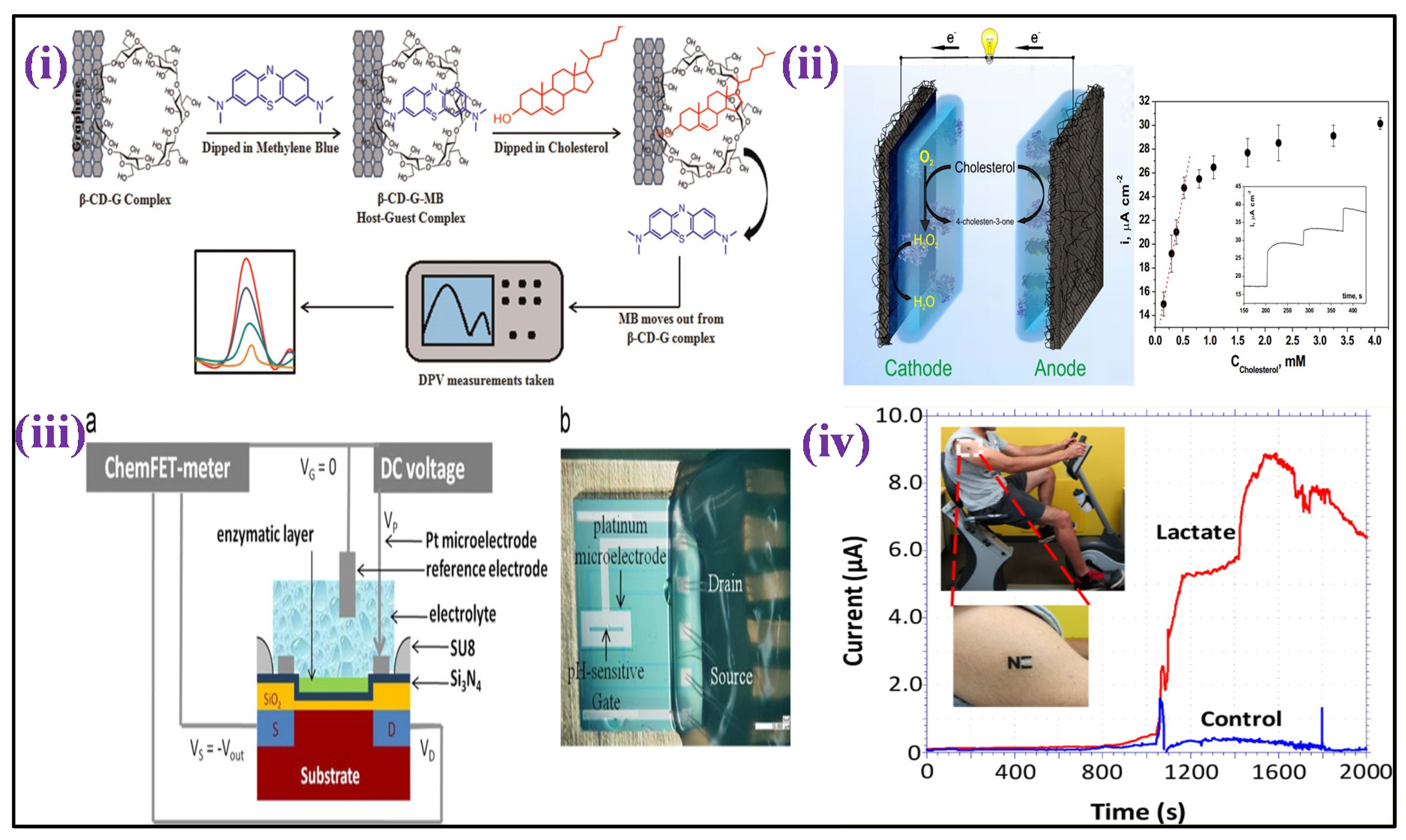
2.1.5. Cholesterol
2.1.6. Lactate

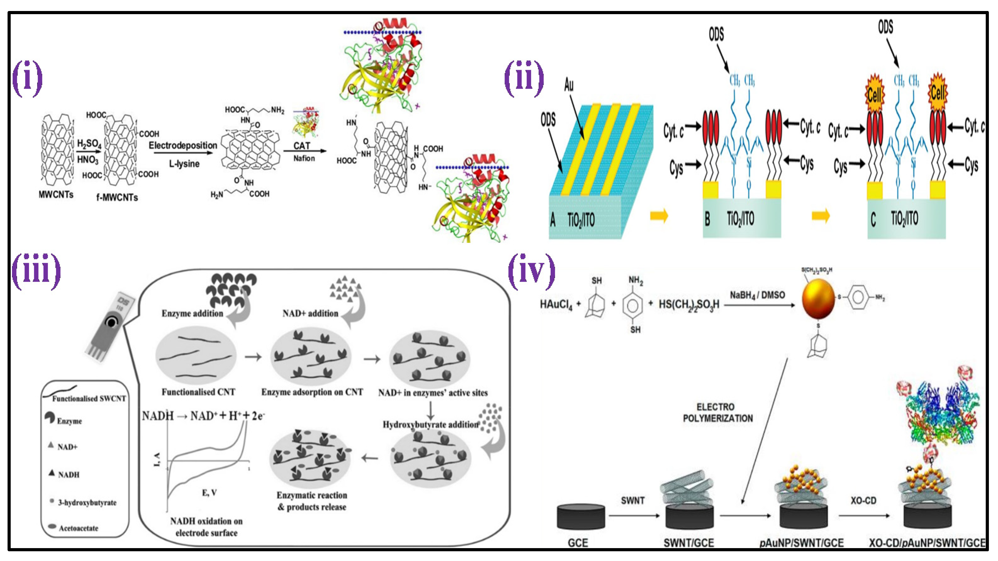
2.1.7. Hydrogen Peroxide (HP)
2.1.8. Ketone Bodies
2.1.9. Xanthine

2.1.10. Hypoxanthine
2.1.11. Creatine
2.1.12. Creatinine

| Metabolite | Nanomaterial Based Biosensing Platform | Electrochemical Technique | Limit of Detection (LOD) | Linear Detection Range (LDR) | Real Sample | Ref. |
|---|---|---|---|---|---|---|
| Glucose | Teflon-coated Pt/Ir wire | Amperometry | 1.5 μM | 5–800 μM | Tear | [46] |
| µPEDs | Chronoamperometric | 1.0 ppb | 0.2–22.6 mM | Urine | [47] | |
| PVA-CuO/ITO | Capacitance | - | 0.5–20 mM | - | [49] | |
| uMED | Chronoamperometry | - | 50–500 mg dL−1 | Blood | [51] | |
| Con A/Au | Capacitance | 1.0 × 10⁻⁶ M | 1.0 × 10⁻⁶ to 1.0 × 10⁻² M | - | [54] | |
| GOx/CeO2–TiO2/ITO | DPV | 10 mg dL−1 | 0.56–22.2 mM | - | [56] | |
| GOx/NS-PANI/ITO | DPV | 2.1 mM | up to 400 mg dL−1 | - | [57] | |
| GOx/CeO2/Pt bio- | DPV | 1.01 mM | 25–300 mg dL−1 | - | [58] | |
| ITO/PB/(PEI/PVS)1(PEI/β-Gal)30 | Amperometry | 1.13 mmol L−1 | 1.13 mmol L−1 | Milk | [63] | |
| Au-MPA-[MWCNT-P(AMB-A)-PDA]/CDH | Amperometry | - | 0–30 mM | - | [64] | |
| P3HT/SA/b-gal/GaO | Amperometry | - | 1–6 gdL−1 | [65] | ||
| Galactose | P3HT/SA/ITO | Amperometry | - | 1–4 gdL−1 | Milk | [66] |
| ITOP3HT/SA/GaO | Amperometry | - | 0.05–0.5 g galactoseL−1 | Blood | [67] | |
| Uric acid | Naf/UOx/Fc/GCE | CV and DPV | 230 nM | 500 nM to 600 μM | Blood | [72] |
| BDD | CV | 1 mM | 250–1250 μM | Urine | [73] | |
| S-Au electrode | Amperometry | 0.4 μM | 2.5 μM to 5 mM | urine | [74] | |
| GCE/AuNp@cysteamine/PAMAM | CV | 34.5 nM | - | Blood | [75] | |
| Uricase/MWCNT/PANI/ITO | CV | 5 μM | 0.005–0.6 mM | Serum | [76] | |
| Urea | CS-rGO/Con A | EIS | 1–7 mM | Serum | [82] | |
| Urs-GLDH/MLG/ITO | CV | 0.6 mM | 1.7–16.7 mM | - | [83] | |
| GA-NC/ITO | CV | - | 2–20 and 0.1–2 mM | - | [84] | |
| CdS QDs–MIPs/Au | DPV | 1.0 × 10⁻¹² M | 5.0 × 10⁻¹²–7.0 × 10⁻⁸ M | Serum | [86] | |
| Urs-GLDH/TiO2-ZrO2/ITO | CV | 0.44 mM | 5–100 mg dL−1 | - | [87] | |
| Urs-GLDH/GOS/ITO | CV | 2.1 mM | 3.3–19.9 mM | - | [88] | |
| Urs-GLDH/CDT/Au | CV | 9 mg dL−1 | 10–100 mg dL−1 | - | [89] | |
| Urs-GLDH/ZrO2/Au | CV | 5 mg dL−1 | 5–100 mg dL−1 | - | [90] | |
| Cholesterol | ChEt–ChOx/PPY–MWCNT/PTS/ITO | DPV | 0.04 mML−1 | 4 × 10⁻⁴–6.5 × 10⁻³ ML−1 | Serum | [93] |
| ChEt-ChOx/4-ATP/Au | CV | 1.34 mM and 1.06 mM | 25–400 mg dL−1 | - | [94] | |
| ChOx/nan-NiO-CHIT/ITO | CV | 43.4 mg dL−1 | 10–400 mg dL−1 | - | [95] | |
| ChOx/Glu/PANI-NT/ITO | LSV | 1.18 mM | 25–500 mg dL−1 | - | [96] | |
| ChOx/PANI-CMC/ITO | 1.31 mM | 0.5–22 mM | - | [97] | ||
| Lactate | Pt electrode | Amperometry | 0.8 μM | 2 to ~1000 μM | Whole blood | [110] |
| Fe3O4/MWCNT/LDH/NAD+/GC | DPV | 5 μM | 50–500 μM | Serum | [111] | |
| ElecFET | Amperometry | - | 1–6 mM | - | [114] | |
| sol–gel/PANI/LDH | Amperometry | - | 1–4 mM | - | [115] | |
| PPY–PVS–LDH | Amperometry | 1 × 10⁻⁴ M | 0.5–6 mM | - | [117] | |
| Hydrogen peroxide | HRP-PANI-ClO4−/ITO | CV | 1.984 mM | 3–136 mM | - | [120] |
| HRP/PANI-CeO2/ITO | CV | 50 mM | 50–500 mM | - | [121] | |
| HRP/NanoCeO2/ITO | CV | 0.5 μM | 1.0–170 μM | - | [122] | |
| MP-11/MWCNTs–BC | CV | 0.1 µM | 0.1–257.6 µM | - | [127] | |
| Au–TiO2/Cys | CV | 2 nM | 10−9–10−2 M | - | [129] | |
| Mb/CeO2/ITO | CV | 0.6 μM | 0.2–5 mM | - | [132] | |
| Ketone bodies | SWCNT-modified SPCE | CV | 80 μM | 0.1–2 mM | Serum | [140] |
| ZnO NPs | I–V | 68 μM | 130 μM–1330 mM | - | [141] | |
| Xanthine | XO-CD/pAuNP/SWNT/GCE | CV | 40 nM | 50 nM–9.5 μM | - | [144] |
| PAP/RGO/GC | LSV | 0.5 μM | 1.0–120 μM | - | [145] | |
| EPG/XDH | CV | 2.5 × 10⁻¹⁰ M | 1.0 ×10⁻⁵–1.8 × 10⁻³ M | - | [149] | |
| Creatinine | ZnO-NPs/CHIT/c-MWCNT/PANI/Pt | EIS | 0.5 μM | 10–650 μM | Blood | [152] |
| Fe3O4@PANI NPs | DPV | 0.35 nmol L−1 | 2.0×10⁸–1.0× 10⁶ mol L−1 | Urine and plasma | [153] | |
| Conducting polymer | Amperometric | 0.46 mg dL−1 | 0–11.33 mg dL−1 | Blood | [154] | |
| EPPG | CV | 0.27 mM | 7.5–11.5 mM | Urine | [155] | |
| Creatine | Fe3O4-CPEE | EIS | 2.0 × 10⁻⁷ mol L−1 | 2.0×10⁻⁷ to 3.8×10⁻⁶ mol L−1 | Commercial creatine powder | [151] |
2.2. Biosensors for Gut Metabolites Detection
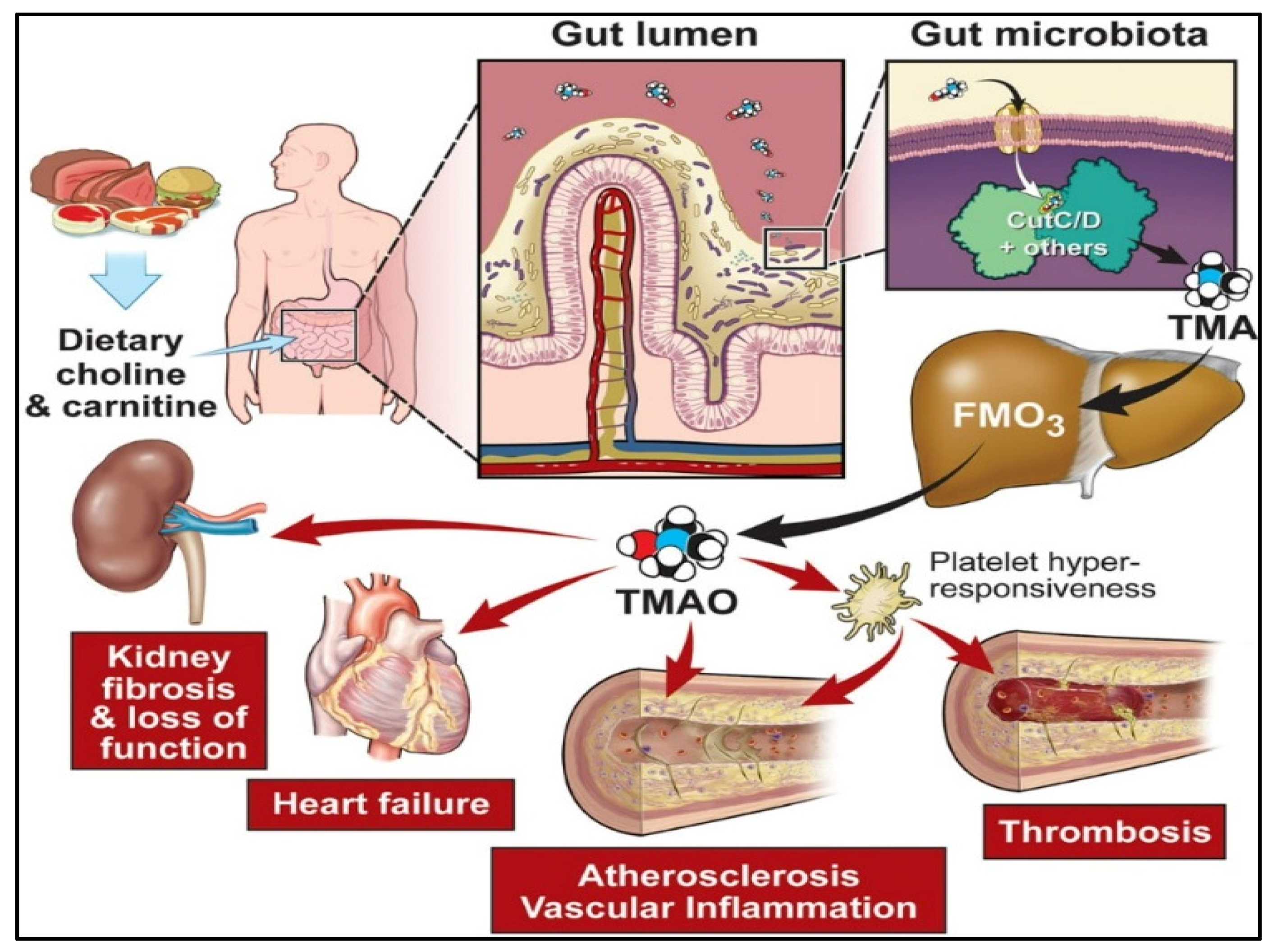
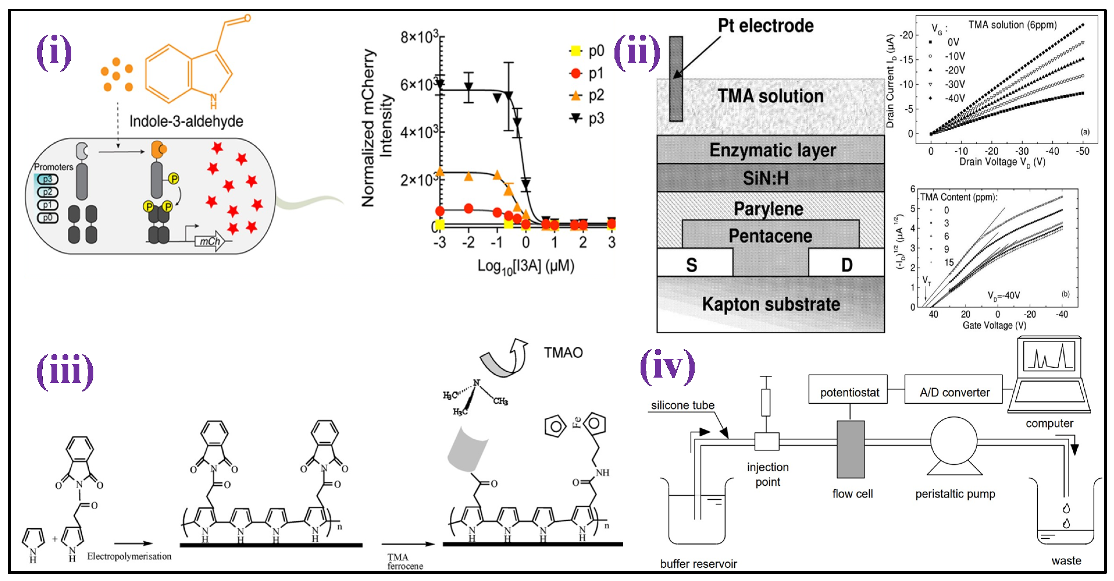
2.2.1. Trimethylamine-N-oxide (TMAO)
2.2.2. Other Gut Metabolites

| Metabolite | Nanomaterial Based Biosensing Platform | Electrochemical Technique | Limit of Detection (LOD) | Linear Detection Range (LDR) | Real Sample | Ref. |
|---|---|---|---|---|---|---|
| TMAO | MIP/ITO | DPV | 1 ppm | 1–15 ppm | Urine | [15] |
| PAH@MnO2 | - | <6.7 μM | 15.6 to 500 μM | Urine | [171] | |
| TorA/GOD/Cat | Amperometry | 10 µM | 2 µM–15 mM | Human serum | [172] | |
| S.loihica PV-4 | Chronoamperometry | 5.96 lM | 0 to 250 µM | Real serum | [173] | |
| Indoxyl sulfate (IS) | GR-SPE | SWV | 0.064 μM | 0.5–80 μM | Human serum and urine | [181] |
| Carbon composite film electrode | Voltammetric | 0.72 μmol L−1 | - | Urine | [182] | |
| Trimethylamine (TMA) | Organic field effect transistors (OFETs) | - | - | 0–8 ppm | Marine fishes and seafood | [183] |
| Poly(Py-FMO3-ferrocene-co-py | EIS | 0.4 g mL−1 | 0.4–80 gmL−1 | Fish extract | [184] | |
| FMO3 immobilized biosensor | - | - | 1.0–50.0 mmol L−1 | Fish-extract | [185] |
3. Summary and Future Directions
4. Conclusions
Author Contributions
Funding
Institutional Review Board Statement
Informed Consent Statement
Conflicts of Interest
Abbreviations
References
- Kelly, T.; Yang, W.; Chen, C.-S.; Reynolds, K.; He, J. Global Burden of Obesity in 2005 and Projections to 2030. Int. J. Obes. 2008, 32, 1431–1437. [Google Scholar] [CrossRef]
- Shaw, J.E.; Sicree, R.A.; Zimmet, P.Z. Global Estimates of the Prevalence of Diabetes for 2010 and 2030. Diabetes Res. Clin. Pract. 2010, 87, 4–14. [Google Scholar] [CrossRef]
- Chen, Y.; Michalak, M.; Agellon, L.B. Focus: Nutrition and Food Science: Importance of Nutrients and Nutrient Metabolism on Human Health. Yale J. Biol. Med. 2018, 91, 95. [Google Scholar]
- Misra, A.; Khurana, L. Obesity and the Metabolic Syndrome in Developing Countries. J. Clin. Endocrinol. Metab. 2008, 93, s9–s30. [Google Scholar] [CrossRef]
- GBD 2016 Risk Factor Collaborators. Global, Regional, and National Comparative Risk Assessment of 84 Behavioural, Environmental and Occupational, and Metabolic Risks or Clusters of Risks, 1990–2016: A Systematic Analysis for the Global Burden of Disease Study 2016. Lancet 2017, 390, 1345. [Google Scholar] [CrossRef]
- Jalandra, R.; Dalal, N.; Yadav, A.K.; Verma, D.; Sharma, M.; Singh, R.; Khosla, A.; Kumar, A.; Solanki, P.R. Emerging Role of Trimethylamine-N-Oxide (TMAO) in Colorectal Cancer. Appl. Microbiol. Biotechnol. 2021, 105, 7651–7660. [Google Scholar] [CrossRef] [PubMed]
- Dalal, N.; Jalandra, R.; Bayal, N.; Yadav, A.K.; Sharma, M.; Makharia, G.K.; Kumar, P.; Singh, R.; Solanki, P.R.; Kumar, A. Gut Microbiota-Derived Metabolites in CRC Progression and Causation. J. Cancer Res. Clin. Oncol. 2021, 147, 3141–3155. [Google Scholar] [CrossRef] [PubMed]
- Wang, Z.; Zhao, Y. Gut Microbiota Derived Metabolites in Cardiovascular Health and Disease. Protein Cell 2018, 9, 416–431. [Google Scholar] [CrossRef]
- Brial, F.; Le Lay, A.; Dumas, M.E.; Gauguier, D. Implication of Gut Microbiota Metabolites in Cardiovascular and Metabolic Diseases. Cell. Mol. Life Sci. 2018, 75, 3977–3990. [Google Scholar] [CrossRef]
- Zeng, H.; Umar, S.; Rust, B.; Lazarova, D.; Bordonaro, M. Secondary Bile Acids and Short Chain Fatty Acids in the Colon: A Focus on Colonic Microbiome, Cell Proliferation, Inflammation, and Cancer. Int. J. Mol. Sci. 2019, 20, 1214. [Google Scholar] [CrossRef]
- Caspani, G.; Kennedy, S.; Foster, J.A.; Swann, J. Gut Microbial Metabolites in Depression: Understanding the Biochemical Mechanisms. Microb. Cell 2019, 6, 454. [Google Scholar] [CrossRef] [PubMed]
- Louis, P.; Hold, G.L.; Flint, H.J. The Gut Microbiota, Bacterial Metabolites and Colorectal Cancer. Nat. Rev. Microbiol. 2014, 12, 661–672. [Google Scholar] [CrossRef] [PubMed]
- Vickers, N.J. Animal Communication: When i’m Calling You, Will You Answer Too? Curr. Biol. 2017, 27, R713–R715. [Google Scholar] [CrossRef]
- Yang, G.; Chen, S.; Deng, B.; Tan, C.; Deng, J.; Zhu, G.; Yin, Y.; Ren, W. Implication of G Protein-Coupled Receptor 43 in Intestinal Inflammation: A Mini-Review. Front. Immunol. 2018, 9, 1434. [Google Scholar] [CrossRef]
- Lakshmi, G.; Yadav, A.K.; Mehlawat, N.; Jalandra, R.; Solanki, P.R.; Kumar, A. Gut Microbiota Derived Trimethylamine N-Oxide (TMAO) Detection through Molecularly Imprinted Polymer Based Sensor. Sci. Rep. 2021, 11, 1338. [Google Scholar]
- Panahi, Z.; Custer, L.; Halpern, J.M. Recent Advances in Non-Enzymatic Electrochemical Detection of Hydrophobic Metabolites in Biofluids. Sens. Actuators Rep. 2021, 3, 100051. [Google Scholar] [CrossRef]
- Keshavarz, M.; Tan, B.; Venkatakrishnan, K. Label-Free SERS Quantum Semiconductor Probe for Molecular-Level and in Vitro Cellular Detection: A Noble-Metal-Free Methodology. ACS Appl. Mater. Interfaces 2018, 10, 34886–34904. [Google Scholar] [CrossRef]
- Morla-Folch, J.; Gisbert-Quilis, P.; Masetti, M.; Garcia-Rico, E.; Alvarez-Puebla, R.A.; Guerrini, L. Conformational SERS Classification of K-Ras Point Mutations for Cancer Diagnostics. Angew. Chemie 2017, 129, 2421–2425. [Google Scholar] [CrossRef]
- Li, W.; Wu, F.; Dai, Y.; Zhang, J.; Ni, B.; Wang, J. Poly (Octadecyl Methacrylate-Co-Trimethylolpropane Trimethacrylate) Monolithic Column for Hydrophobic in-Tube Solid-Phase Microextraction of Chlorophenoxy Acid Herbicides. Molecules 2019, 24, 1678. [Google Scholar] [CrossRef]
- Jalandra, R.; Yadav, A.K.; Verma, D.; Dalal, N.; Sharma, M.; Singh, R.; Kumar, A.; Solanki, P.R. Strategies and Perspectives to Develop SARS-CoV-2 Detection Methods and Diagnostics. Biomed. Pharmacother. 2020, 129, 110446. [Google Scholar] [CrossRef]
- Yadav, A.K.; Verma, D.; Kumar, A.; Kumar, P.; Solanki, P.R. The Perspectives of Biomarker-Based Electrochemical Immunosensors, Artificial Intelligence and the Internet of Medical Things TowardáCOVID-19 Diagnosis and Management. Mater. Today Chem. 2021, 20, 100443. [Google Scholar] [CrossRef] [PubMed]
- Yadav, A.K.; Dhiman, T.K.; Lakshmi, G.; Berlina, A.N.; Solanki, P.R. A Highly Sensitive Label-Free Amperometric Biosensor for Norfloxacin Detection Based on Chitosan-Yttria Nanocomposite. Int. J. Biol. Macromol. 2020, 151, 566–575. [Google Scholar] [CrossRef] [PubMed]
- Verma, D.; Yadav, A.K.; Mukherjee, M.D.; Solanki, P.R. Fabrication of a Sensitive Electrochemical Sensor Platform Using Reduced Graphene Oxide-Molybdenum Trioxide Nanocomposite for BPA Detection: An Endocrine Disruptor. J. Environ. Chem. Eng. 2021, 9, 105504. [Google Scholar] [CrossRef]
- Verma, D.; Chauhan, D.; Mukherjee, M.D.; Ranjan, K.R.; Yadav, A.K.; Solanki, P.R. Development of MWCNT Decorated with Green Synthesized AgNps-Based Electrochemical Sensor for Highly Sensitive Detection of BPA. J. Appl. Electrochem. 2021, 51, 447–462. [Google Scholar] [CrossRef]
- Yadav, A.K.; Verma, D.; Solanki, P.R. Electrophoretically Deposited L-Cysteine Functionalized MoS2@ MWCNT Nanocomposite Platform: A Smart Approach toward Highly Sensitive and Label-Free Detection of Gentamicin. Mater. Today Chem. 2021, 22, 100567. [Google Scholar] [CrossRef]
- Yadav, A.K.; Verma, D.; Lakshmi, G.; Eremin, S.; Solanki, P.R. Fabrication of Label-Free and Ultrasensitive Electrochemical Immunosensor Based on Molybdenum Disulfide Nanoparticles Modified Disposable ITO: An Analytical Platform for Antibiotic Detection in Food Samples. Food Chem. 2021, 363, 130245. [Google Scholar] [CrossRef]
- Chaudhary, N.; Yadav, A.K.; Sharma, J.G.; Solanki, P.R. Designing and Characterization of a Highly Sensitive and Selective Biosensing Platform for Ciprofloxacin Detection Utilizing Lanthanum Oxide Nanoparticles. J. Environ. Chem. Eng. 2021, 9, 106771. [Google Scholar] [CrossRef]
- Chauhan, D.; Yadav, A.K.; Solanki, P.R. Carbon Cloth-Based Immunosensor for Detection of 25-Hydroxy Vitamin D3. Microchim. Acta 2021, 188, 145. [Google Scholar] [CrossRef]
- Yadav, A.K.; Verma, D.; Chaudhary, N.; Kumar, A.; Solanki, P.R. Aptamer Based Switches: A Futuristic Approach for Helicobacter Pylori Detection. Mater. Lett. 2022, 308, 131239. [Google Scholar] [CrossRef]
- Yadav, A.K.; Verma, D.; Sharma, R.; Thakkar, A.; Solanki, P.R. Silane Modified Disposable ITO Electrode-Based Biosensor for Sperm Protein 17 Detection as a Novel Cancer Biomarker. SPAST Abstr. 2021, 1. [Google Scholar]
- Sen, R.K.; Prabhakar, P.; Bisht, N.; Patel, M.; Mishra, S.; Yadav, A.K.; Venu, D.V.; Gupta, G.K.; Solanki, P.R.; Ramakrishnan, S. 2D Materials-Based Aptamer Biosensors: Present Status and Way Forward. Curr. Med. Chem. 2021, 28. [Google Scholar] [CrossRef] [PubMed]
- Zhao, Y.; Barrere-Cain, R.E.; Yang, X. Nutritional Systems Biology of Type 2 Diabetes. Genes Nutr. 2015, 10, 31. [Google Scholar] [CrossRef] [PubMed]
- Hood, L.; Heath, J.R.; Phelps, M.E.; Lin, B. Systems Biology and New Technologies Enable Predictive and Preventative Medicine. Science 2004, 306, 640–643. [Google Scholar] [CrossRef] [PubMed] [Green Version]
- Sauer, R.T.; Pabo, C.O. Protein-DNA Recognition. Annu. Rev. Biochem. 1984, 53, 293–321. [Google Scholar]
- D’Orazio, P. Biosensors in Clinical Chemistry. Clin. Chim. acta 2003, 334, 41–69. [Google Scholar] [CrossRef]
- Parisi, O.I.; Dattilo, M.; Patitucci, F.; Malivindi, R.; Pezzi, V.; Perrotta, I.; Ruffo, M.; Amone, F.; Puoci, F. “Monoclonal-Type” Plastic Antibodies for SARS-CoV-2 Based on Molecularly Imprinted Polymers. BioRxiv 2020. [Google Scholar]
- Kelley, S.O. Disease Detector. Sci. Am. 2015, 313, 48–51. [Google Scholar] [CrossRef]
- Verma, D.; Dhiman, T.K.; Das Mukherjee, M.; Solanki, P.R. Electrophoretically Deposited Green Synthesized Silver Nanoparticles Anchored in Reduced Graphene Oxide Composite Based Electrochemical Sensor for Detection of Bisphenol A. J. Electrochem. Soc. 2021, 168, 097504. [Google Scholar] [CrossRef]
- Verma, D.; Ranjan, K.R.; Mukherjee, M.D.; Solanki, P.R. Bioinspired Synthesis of Hematite Nanoparticles-Reduced Graphene Oxide Composite for Application in Bisphenol a Detection: A New in-Sight. Biosens. Bioelectron. X 2022, 11, 100217. [Google Scholar] [CrossRef]
- Verma, D.; Kumar, A.; Rathee, G.; Dhingra, K.; Mukherjee, M.D.; Solanki, P.R. Prospects of Nanomaterial-Based Biosensors: A Smart Approach for Bisphenol-A Detection in Dental Sealants. J. Electrochem. Soc. 2022, 169, 027516. [Google Scholar] [CrossRef]
- Verma, D.; Singh, K.R.B.; Yadav, A.K.; Nayak, V.; Singh, J.; Solanki, P.R.; Singh, R.P. Internet of Things (IoT) in Nano-Integrated Wearable Biosensor Devices for Healthcare Applications. Biosens. Bioelectron. X 2022, 11, 100153. [Google Scholar] [CrossRef]
- Wang, J. Electrochemical Glucose Biosensors. Chem. Rev. 2008, 108, 814–825. [Google Scholar] [CrossRef] [PubMed]
- Heller, A.; Feldman, B. Electrochemical Glucose Sensors and Their Applications in Diabetes Management. Chem. Rev. 2008, 108, 2482–2505. [Google Scholar] [CrossRef] [PubMed]
- Vaddiraju, S.; Tomazos, I.; Burgess, D.J.; Jain, F.C.; Papadimitrakopoulos, F. Emerging Synergy between Nanotechnology and Implantable Biosensors: A Review. Biosens. Bioelectron. 2010, 25, 1553–1565. [Google Scholar] [CrossRef]
- Labib, M.; Hedström, M.; Amin, M.; Mattiasson, B. Competitive Capacitive Biosensing Technique (CCBT): A Novel Technique for Monitoring Low Molecular Mass Analytes Using Glucose Assay as a Model Study. Anal. Bioanal. Chem. 2010, 397, 1217–1224. [Google Scholar] [CrossRef]
- Yan, Q.; Peng, B.; Su, G.; Cohan, B.E.; Major, T.C.; Meyerhoff, M.E. Measurement of Tear Glucose Levels with Amperometric Glucose Biosensor/Capillary Tube Configuration. Anal. Chem. 2011, 83, 8341–8346. [Google Scholar] [CrossRef]
- Nie, Z.; Nijhuis, C.A.; Gong, J.; Chen, X.; Kumachev, A.; Martinez, A.W.; Narovlyansky, M.; Whitesides, G.M. Electrochemical Sensing in Paper-Based Microfluidic Devices. Lab Chip 2010, 10, 477–483. [Google Scholar] [CrossRef]
- Lankelma, J.; Nie, Z.; Carrilho, E.; Whitesides, G.M. Based Analytical Device for Electrochemical Flow-Injection Analysis of Glucose in Urine. Anal. Chem. 2012, 84, 4147–4152. [Google Scholar] [CrossRef]
- Dhiman, T.K.; Lakshmi, G.; Kumar, R.; Asokan, K.; Solanki, P.R. Non-Enzymatic Detection of Glucose Using a Capacitive Nanobiosensor Based on PVA Capped CuO Synthesized via Co-Precipitation Route. IEEE Sens. J. 2020, 20, 10415–10423. [Google Scholar] [CrossRef]
- Dhiman, T.K.; Poddar, M.; Lakshmi, G.; Kumar, R.; Solanki, P.R. Non-Enzymatic and Rapid Detection of Glucose on PVA-CuO Thin Film Using ARDUINO UNO Based Capacitance Measurement Unit. Biomed. Microdevices 2021, 23, 36. [Google Scholar] [CrossRef]
- Nemiroski, A.; Christodouleas, D.C.; Hennek, J.W.; Kumar, A.A.; Maxwell, E.J.; Fernández-Abedul, M.T.; Whitesides, G.M. Universal Mobile Electrochemical Detector Designed for Use in Resource-Limited Applications. Proc. Natl. Acad. Sci. USA 2014, 111, 11984–11989. [Google Scholar] [CrossRef] [PubMed]
- Bandodkar, A.J.; Jia, W.; Ramírez, J.; Wang, J. Biocompatible Enzymatic Roller Pens for Direct Writing of Biocatalytic Materials: “Do-it-Yourself” Electrochemical Biosensors. Adv. Healthc. Mater. 2015, 4, 1215–1224. [Google Scholar] [CrossRef] [PubMed]
- Schultz, J.S.; Mansouri, S.; Goldstein, I.J. Affinity Sensor: A New Technique for Developing Implantable Sensors for Glucose and Other Metabolites. Diabetes Care 1982, 5, 245–253. [Google Scholar] [CrossRef] [PubMed]
- Labib, M.; Hedström, M.; Amin, M.; Mattiasson, B. A Novel Competitive Capacitive Glucose Biosensor Based on Concanavalin A-Labeled Nanogold Colloids Assembled on a Polytyramine-Modified Gold Electrode. Anal. Chim. Acta 2010, 659, 194–200. [Google Scholar] [CrossRef] [PubMed]
- Rassaei, L.; Marken, F. Pulse-Voltammetric Glucose Detection at Gold Junction Electrodes. Anal. Chem. 2010, 82, 7063–7067. [Google Scholar] [CrossRef]
- Ansari, A.A.; Pandey, P.; Malhotra, B.D. Sol–Gel Derived Nanoporous Cerium Oxide–Titanium Oxide Platform for Glucose Sensor. Adv. Sci. Eng. Med. 2013, 5, 1113–1119. [Google Scholar] [CrossRef]
- Dhand, C.; Sumana, G.; Datta, M.; Malhotra, B.D. Electrophoretically Deposited Nano-Structured Polyaniline Film for Glucose Sensing. Thin Solid Films 2010, 519, 1145–1150. [Google Scholar] [CrossRef]
- Saha, S.; Arya, S.K.; Singh, S.P.; Sreenivas, K.; Malhotra, B.D.; Gupta, V. Nanoporous Cerium Oxide Thin Film for Glucose Biosensor. Biosens. Bioelectron. 2009, 24, 2040–2045. [Google Scholar] [CrossRef]
- Liu, W.; Zhao, X.; Guo, Q.; Dai, Y.; Tan, J.; Wang, M.; Qi, Y. Preparation of Electrochemical Sensor Based on the Novel NiO Quantum Dots Modified Cu/Cu2O 3D Hybrid Electrode and Its Application for Non-Enzymatic Detection of Glucose in Serums and Beverages. J. Alloys Compd. 2022, 895, 162573. [Google Scholar] [CrossRef]
- Lei, L.; Zhao, C.; Zhu, X.; Yuan, S.; Dong, X.; Zuo, Y.; Liu, H. Nonenzymatic Electrochemical Sensor for Wearable Interstitial Fluid Glucose Monitoring. Electroanalysis 2022, 34, 415–422. [Google Scholar] [CrossRef]
- Gopal, T.S.; Jeong, S.K.; Alrebdi, T.A.; Pandiaraj, S.; Alodhayb, A.; Muthuramamoorthy, M.; Grace, A.N. MXene-Based Composite Electrodes for Efficient Electrochemical Sensing of Glucose by Non-Enzymatic Method. Mater. Today Chem. 2022, 24, 100891. [Google Scholar] [CrossRef]
- Conzuelo, F.; Gamella, M.; Campuzano, S.; Ruiz, M.A.; Reviejo, A.J.; Pingarron, J.M. An Integrated Amperometric Biosensor for the Determination of Lactose in Milk and Dairy Products. J. Agric. Food Chem. 2010, 58, 7141–7148. [Google Scholar] [CrossRef] [PubMed]
- Campos, P.P.; Moraes, M.L.; Volpati, D.; Miranda, P.B.; Oliveira Jr, O.N.; Ferreira, M. Amperometric Detection of Lactose Using β-Galactosidase Immobilized in Layer-by-Layer Films. ACS Appl. Mater. Interfaces 2014, 6, 11657–11664. [Google Scholar] [CrossRef]
- Tanne, J.; Kracher, D.; Dietzel, B.; Schulz, B.; Ludwig, R.; Lisdat, F.; Scheller, F.W.; Bier, F.F. Carboxylated or Aminated Polyaniline—Multiwalled Carbon Nanotubes Nanohybrids for Immobilization of Cellobiose Dehydrogenase on Gold Electrodes. Biosensors 2014, 4, 370–386. [Google Scholar] [CrossRef] [PubMed]
- Sharma, S.K.; Singhal, R.; Malhotra, B.D.; Sehgal, N.; Kumar, A. Lactose Biosensor Based on Langmuir–Blodgett Films of Poly (3-Hexyl Thiophene). Biosens. Bioelectron. 2004, 20, 651–657. [Google Scholar] [CrossRef]
- Sharma, S.K.; Singhal, R.; Malhotra, B.D.; Sehgal, N.; Kumar, A. Langmuir–Blodgett Film Based Biosensor for Estimation of Galactose in Milk. Electrochim. Acta 2004, 49, 2479–2485. [Google Scholar] [CrossRef]
- Sharma, S.K.; Singhal, R.; Malhotra, B.D.; Sehgal, N.; Kumar, A. Biosensor Based on Langmuir–Blodgett Films of Poly (3-Hexyl Thiophene) for Detection of Galactose in Human Blood. Biotechnol. Lett. 2004, 26, 645–647. [Google Scholar] [CrossRef]
- Halpin, G.; McEntee, S.; Dwyer, C.; Lawless, F.; Dempsey, E. Lactose Biosensor Development and Deployment in Dairy Product Analysis. J. Electrochem. Soc. 2022, 169, 37528. [Google Scholar] [CrossRef]
- da Silva, J.L.; Buffon, E.; Beluomini, M.A.; Pradela-Filho, L.A.; Araújo, D.A.G.; Santos, A.L.; Takeuchi, R.M.; Stradiotto, N.R. Non-Enzymatic Lactose Molecularly Imprinted Sensor Based on Disposable Graphite Paper Electrode. Anal. Chim. Acta 2021, 1143, 53–64. [Google Scholar] [CrossRef]
- KojIma, T.; Nishina, T.; Kitamura, M.; Kamatani, N.; Nishioka, K. Reversed-Phase Liquid-Chromatographic Determination of Purine Compounds in Serum Applied to Studies of Hypouricemia. Clin. Chem. 1986, 32, 287–290. [Google Scholar] [CrossRef]
- Bravo, R.; Hsueh, C.; Brajter-Toth, A.; Jaramillo, A. Possibilities and Limitations in Miniaturized Sensor Design for Uric Acid. Analyst 1998, 123, 1625–1630. [Google Scholar] [CrossRef]
- Ghosh, T.; Sarkar, P.; Turner, A.P.F. A Novel Third Generation Uric Acid Biosensor Using Uricase Electro-Activated with Ferrocene on a Nafion Coated Glassy Carbon Electrode. Bioelectrochemistry 2015, 102, 1–9. [Google Scholar] [CrossRef]
- Kiran, R.; Scorsone, E.; Mailley, P.; Bergonzo, P. Quasi-Real Time Quantification of Uric Acid in Urine Using Boron Doped Diamond Microelectrode with in Situ Cleaning. Anal. Chem. 2012, 84, 10207–10213. [Google Scholar] [CrossRef]
- Miah, M.R.; Alam, M.T.; Ohsaka, T. Sulfur-Adlayer-Coated Gold Electrode for the in Vitro Electrochemical Detection of Uric Acid in Urine. Anal. Chim. Acta 2010, 669, 75–80. [Google Scholar] [CrossRef] [PubMed]
- Ramírez-Segovia, A.S.; Banda-Alemán, J.A.; Gutiérrez-Granados, S.; Rodríguez, A.; Rodríguez, F.J.; Godínez, L.A.; Bustos, E.; Manríquez, J. Glassy Carbon Electrodes Sequentially Modified by Cysteamine-Capped Gold Nanoparticles and Poly (Amidoamine) Dendrimers Generation 4.5 for Detecting Uric Acid in Human Serum without Ascorbic Acid Interference. Anal. Chim. Acta 2014, 812, 18–25. [Google Scholar] [CrossRef] [PubMed]
- Bhambi, M.; Sumana, G.; Malhotra, B.D.; Pundir, C.S. An Amperomertic Uric Acid Biosensor Based on Immobilization of Uricase onto Polyaniline-Multiwalled Carbon Nanotube Composite Film. Artif. Cells Blood Substit. Biotechnol. 2010, 38, 178–185. [Google Scholar] [CrossRef] [PubMed]
- Kulyk, B.; Pereira, S.O.; Fernandes, A.J.S.; Fortunato, E.; Costa, F.M.; Santos, N.F. Laser-Induced Graphene from Paper for Non-Enzymatic Uric Acid Electrochemical Sensing in Urine. Carbon N. Y. 2022, 197, 253–263. [Google Scholar] [CrossRef]
- Ahmed, J.; Faisal, M.; Alsareii, S.A.; Harraz, F.A. Highly Sensitive and Selective Non-Enzymatic Uric Acid Electrochemical Sensor Based on Novel Polypyrrole-Carbon Black-Co3O4 Nanocomposite. Adv. Compos. Hybrid Mater. 2022, 5, 1–14. [Google Scholar] [CrossRef]
- Monošík, R.; Stred’anský, M.; Šturdík, E. Application of Electrochemical Biosensors in Clinical Diagnosis. J. Clin. Lab. Anal. 2012, 26, 22–34. [Google Scholar] [CrossRef]
- Dhinasekaran, D.; Soundharraj, P.; Jagannathan, M.; Rajendran, A.R.; Rajendran, S. Hybrid ZnO Nanostructures Modified Graphite Electrode as an Efficient Urea Sensor for Environmental Pollution Monitoring. Chemosphere 2022, 296, 133918. [Google Scholar] [CrossRef]
- Ahmad, K.; Kim, H. Hydrothermally Synthesized MoSe2/RGO Composite as Electrode Modifier for the Construction of Non-Enzymatic Urea Sensor. Mater. Chem. Phys. 2022, 286, 126206. [Google Scholar] [CrossRef]
- Song, Y.; Liu, H.; Tan, H.; Xu, F.; Jia, J.; Zhang, L.; Li, Z.; Wang, L. PH-Switchable Electrochemical Sensing Platform Based on Chitosan-Reduced Graphene Oxide/Concanavalin a Layer for Assay of Glucose and Urea. Anal. Chem. 2014, 86, 1980–1987. [Google Scholar] [CrossRef] [PubMed]
- Srivastava, R.K.; Srivastava, S.; Narayanan, T.N.; Mahlotra, B.D.; Vajtai, R.; Ajayan, P.M.; Srivastava, A. Functionalized Multilayered Graphene Platform for Urea Sensor. ACS Nano 2012, 6, 168–175. [Google Scholar] [CrossRef] [PubMed]
- Sharma, A.; Rawat, K.; Bohidar, H.B.; Solanki, P.R. Studies on Clay-Gelatin Nanocomposite as Urea Sensor. Appl. Clay Sci. 2017, 146, 297–305. [Google Scholar] [CrossRef]
- Lakard, B.; Magnin, D.; Deschaume, O.; Vanlancker, G.; Glinel, K.; Demoustier-Champagne, S.; Nysten, B.; Jonas, A.M.; Bertrand, P.; Yunus, S. Urea Potentiometric Enzymatic Biosensor Based on Charged Biopolymers and Electrodeposited Polyaniline. Biosens. Bioelectron. 2011, 26, 4139–4145. [Google Scholar] [CrossRef]
- Lian, H.T.; Liu, B.; Chen, Y.P.; Sun, X.Y. A Urea Electrochemical Sensor Based on Molecularly Imprinted Chitosan Film Doping with CdS Quantum Dots. Anal. Biochem. 2012, 426, 40–46. [Google Scholar] [CrossRef]
- Srivastava, S.; Ali, M.A.; Solanki, P.R.; Chavhan, P.M.; Pandey, M.K.; Mulchandani, A.; Srivastava, A.; Malhotra, B.D. Mediator-Free Microfluidics Biosensor Based on Titania–Zirconia Nanocomposite for Urea Detection. Rsc Adv. 2013, 3, 228–235. [Google Scholar] [CrossRef]
- Abraham, S.; Ciobota, V.; Srivastava, S.; Srivastava, S.K.; Singh, R.K.; Dellith, J.; Malhotra, B.D.; Schmitt, M.; Popp, J.; Srivastava, A. Mesoporous Silica Particle Embedded Functional Graphene Oxide as an Efficient Platform for Urea Biosensing. Anal. Methods 2014, 6, 6711–6720. [Google Scholar] [CrossRef]
- Srivastava, S.; Solanki, P.R.; Kaushik, A.; Ali, M.A.; Srivastava, A.; Malhotra, B.D. A Self Assembled Monolayer Based Microfluidic Sensor for Urea Detection. Nanoscale 2011, 3, 2971–2977. [Google Scholar] [CrossRef]
- Sumana, G.; Das, M.; Srivastava, S.; Malhotra, B.D. A Novel Urea Biosensor Based on Zirconia. Thin Solid Films 2010, 519, 1187–1191. [Google Scholar] [CrossRef]
- Calabresi, L.; Gomaraschi, M.; Simonelli, S.; Bernini, F.; Franceschini, G. HDL and Atherosclerosis: Insights from Inherited HDL Disorders. Biochim. Biophys. Acta (BBA)-Mol. Cell Biol. Lipids 2015, 1851, 13–18. [Google Scholar] [CrossRef] [PubMed]
- Kaushik, P.; Solanki, P.R.; Chavhan, P.M.; Bhansali, S.; Malhotra, B.D. Sol-Gel Derived Nanostructured Zirconia for Cholesterol Detection. J. Nanosci. Lett. 2013, 3, 23. [Google Scholar]
- Singh, K.; Solanki, P.R.; Basu, T.; Malhotra, B.D. Polypyrrole/Multiwalled Carbon Nanotubes-based Biosensor for Cholesterol Estimation. Polym. Adv. Technol. 2012, 23, 1084–1091. [Google Scholar] [CrossRef]
- Matharu, Z.; Solanki, P.R.; Gupta, V.; Malhotra, B.D. Mediator Free Cholesterol Biosensor Based on Self-Assembled Monolayer Platform. Analyst 2012, 137, 747–753. [Google Scholar] [CrossRef] [PubMed]
- Singh, J.; Kalita, P.; Singh, M.K.; Malhotra, B.D. Nanostructured Nickel Oxide-Chitosan Film for Application to Cholesterol Sensor. Appl. Phys. Lett. 2011, 98, 123702. [Google Scholar] [CrossRef]
- Dhand, C.; Solanki, P.R.; Pandey, M.K.; Datta, M.; Malhotra, B.D. Electrophoretically Deposited Polyaniline Nanotubes Based Film for Cholesterol Detection. Electrophoresis 2010, 31, 3754–3762. [Google Scholar] [CrossRef]
- Barik, A.; Solanki, P.R.; Kaushik, A.; Ali, A.; Pandey, M.K.; Kim, C.G.; Malhotra, B.D. Polyaniline–Carboxymethyl Cellulose Nanocomposite for Cholesterol Detection. J. Nanosci. Nanotechnol. 2010, 10, 6479–6488. [Google Scholar] [CrossRef]
- Kaushik, A.; Solanki, P.R.; Kaneto, K.; Kim, C.G.; Ahmad, S.; Malhotra, B.D. Nanostructured Iron Oxide Platform for Impedimetric Cholesterol Detection. Electroanal. An Int. J. Devoted Fundam. Pract. Asp. Electroanal. 2010, 22, 1045–1055. [Google Scholar] [CrossRef]
- Kariz, D.G.A.; Darabi, E.; Elahi, S.M. Electrochemical Sensing of Cholesterol Based on MWCNTs Loaded Nanoparticles. Int. Nano Lett. 2022, 12, 113–123. [Google Scholar] [CrossRef]
- Shahriarinour, M.; Rahimi, F.; Siahbani, E.; Kochakinejad, R.; Kaki, S. A New Electrochemical Modified Graphite Pencil Electrode Developed for Cholesterol Assessing. J. Iran. Chem. Soc. 2022, 19, 159–171. [Google Scholar] [CrossRef]
- Tong, Y.; Li, H.; Guan, H.; Zhao, J.; Majeed, S.; Anjum, S.; Liang, F.; Xu, G. Electrochemical Cholesterol Sensor Based on Carbon Nanotube@ Molecularly Imprinted Polymer Modified Ceramic Carbon Electrode. Biosens. Bioelectron. 2013, 47, 553–558. [Google Scholar] [CrossRef] [PubMed]
- Eguílaz, M.; Villalonga, R.; Yáñez-Sedeño, P.; Pingarrón, J.M. Designing Electrochemical Interfaces with Functionalized Magnetic Nanoparticles and Wrapped Carbon Nanotubes as Platforms for the Construction of High-Performance Bienzyme Biosensors. Anal. Chem. 2011, 83, 7807–7814. [Google Scholar] [CrossRef] [PubMed]
- Sekretaryova, A.N.; Beni, V.; Eriksson, M.; Karyakin, A.A.; Turner, A.P.F.; Vagin, M.Y. Cholesterol Self-Powered Biosensor. Anal. Chem. 2014, 86, 9540–9547. [Google Scholar] [CrossRef] [PubMed]
- Pundir, C.S.; Singh, B.S.; Narang, J. Construction of an Amperometric Triglyceride Biosensor Using PVA Membrane Bound Enzymes. Clin. Biochem. 2010, 43, 467–472. [Google Scholar] [CrossRef]
- Nie, Z.; Deiss, F.; Liu, X.; Akbulut, O.; Whitesides, G.M. Integration of Paper-Based Microfluidic Devices with Commercial Electrochemical Readers. Lab Chip 2010, 10, 3163–3169. [Google Scholar] [CrossRef]
- Agnihotri, N.; Chowdhury, A.D.; De, A. Non-Enzymatic Electrochemical Detection of Cholesterol Using β-Cyclodextrin Functionalized Graphene. Biosens. Bioelectron. 2015, 63, 212–217. [Google Scholar] [CrossRef]
- Bakker, J.; Gris, P.; Coffernils, M.; Kahn, R.J.; Vincent, J.-L. Serial Blood Lactate Levels Can Predict the Development of Multiple Organ Failure Following Septic Shock. Am. J. Surg. 1996, 171, 221–226. [Google Scholar] [CrossRef]
- Madden, J.; Vaughan, E.; Thompson, M.; O’Riordan, A.; Galvin, P.; Iacopino, D.; Teixeira, S.R. Electrochemical Sensor for Enzymatic Lactate Detection Based on Laser-Scribed Graphitic Carbon Modified with Platinum, Chitosan and Lactate Oxidase. Talanta 2022, 246, 123492. [Google Scholar] [CrossRef]
- Zhu, C.; Xue, H.; Zhao, H.; Fei, T.; Liu, S.; Chen, Q.; Gao, B.; Zhang, T. A Dual-Functional Polyaniline Film-Based Flexible Electrochemical Sensor for the Detection of PH and Lactate in Sweat of the Human Body. Talanta 2022, 242, 123289. [Google Scholar] [CrossRef]
- Romero, M.R.; Ahumada, F.; Garay, F.; Baruzzi, A.M. Amperometric Biosensor for Direct Blood Lactate Detection. Anal. Chem. 2010, 82, 5568–5572. [Google Scholar] [CrossRef]
- Teymourian, H.; Salimi, A.; Hallaj, R. Low Potential Detection of NADH Based on Fe3O4 Nanoparticles/Multiwalled Carbon Nanotubes Composite: Fabrication of Integrated Dehydrogenase-Based Lactate Biosensor. Biosens. Bioelectron. 2012, 33, 60–68. [Google Scholar] [CrossRef] [PubMed]
- Jia, W.; Bandodkar, A.J.; Valdés-Ramírez, G.; Windmiller, J.R.; Yang, Z.; Ramírez, J.; Chan, G.; Wang, J. Electrochemical Tattoo Biosensors for Real-Time Noninvasive Lactate Monitoring in Human Perspiration. Anal. Chem. 2013, 85, 6553–6560. [Google Scholar] [CrossRef] [PubMed]
- Chen, J.; Jin, Y. Sensitive Lactate Determination Based on Acclimated Mixed Bacteria and Palygorskite Co-Modified Oxygen Electrode. Bioelectrochemistry 2011, 80, 151–154. [Google Scholar] [CrossRef] [PubMed]
- Diallo, A.K.; Djeghlaf, L.; Mazenq, L.; Launay, J.; Sant, W.; Temple-Boyer, P. Development of PH-Based ElecFET Biosensors for Lactate Ion Detection. Biosens. Bioelectron. 2013, 40, 291–296. [Google Scholar] [CrossRef] [PubMed]
- Chaubey, A.; Pande, K.K.; Malhotra, B.D. Application of Polyaniline/Sol-Gel Derived Tetraethylorthosilicate Films to an Amperometric Lactate Biosensor. Anal. Sci. 2003, 19, 1477–1480. [Google Scholar] [CrossRef]
- Chaubey, A.; Gerard, M.; Singh, V.S.; Malhotra, B.D. Immobilization of Lactate Dehydrogenase on Tetraethylorthosilicate-Derived Sol-Gel Films for Application to Lactate Biosensor. Appl. Biochem. Biotechnol. 2001, 96, 303–311. [Google Scholar] [CrossRef]
- Chaubey, A.; Gerard, M.; Singhal, R.; Singh, V.S.; Malhotra, B.D. Immobilization of Lactate Dehydrogenase on Electrochemically Prepared Polypyrrole–Polyvinylsulphonate Composite Films for Application to Lactate Biosensors. Electrochim. Acta 2001, 46, 723–729. [Google Scholar] [CrossRef]
- Chaubey, A.; Pande, K.K.; Singh, V.S.; Malhotra, B.D. Co-Immobilization of Lactate Oxidase and Lactate Dehydrogenase on Conducting Polyaniline Films. Anal. Chim. Acta 2000, 407, 97–103. [Google Scholar] [CrossRef]
- Trachootham, D.; Alexandre, J.; Huang, P. Targeting Cancer Cells by ROS-Mediated Mechanisms: A Radical Therapeutic Approach? Nat. Rev. Drug Discov. 2009, 8, 579–591. [Google Scholar] [CrossRef]
- Solanki, P.R.; Kaushik, A.; Ansari, A.A.; Sumana, G.; Malhotra, B.D. Horse Radish Peroxidase Immobilized Polyaniline for Hydrogen Peroxide Sensor. Polym. Adv. Technol. 2011, 22, 903–908. [Google Scholar] [CrossRef]
- Ansari, A.A.; Sumana, G.; Khan, R.; Malhotra, B.D. Polyaniline-Cerium Oxide Nanocomposite for Hydrogen Peroxide Sensor. J. Nanosci. Nanotechnol. 2009, 9, 4679–4685. [Google Scholar] [CrossRef]
- Ansari, A.A.; Solanki, P.R.; Malhotra, B.D. Hydrogen Peroxide Sensor Based on Horseradish Peroxidase Immobilized Nanostructured Cerium Oxide Film. J. Biotechnol. 2009, 142, 179–184. [Google Scholar] [CrossRef] [PubMed]
- Verma, S.; Mal, D.S.; de Oliveira, P.R.; Janegitz, B.C.; Prakash, J.; Gupta, R.K. A Facile Synthesis of Novel Polyaniline/Graphene Nanocomposite Thin Films for Enzyme-Free Electrochemical Sensing of Hydrogen Peroxide. Mol. Syst. Des. Eng. 2022, 7, 158–170. [Google Scholar] [CrossRef]
- Portorreal-Bottier, A.; Gutiérrez-Tarriño, S.; Calvente, J.J.; Andreu, R.; Roldán, E.; Oña-Burgos, P.; Olloqui-Sariego, J.L. Enzyme like Activity of Cobalt-MOF Nanosheets for Hydrogen Peroxide Electrochemical Sensing. Sensors Actuators B Chem. 2022, 368, 132129. [Google Scholar] [CrossRef]
- Suraniti, E.; Ben-Amor, S.; Landry, P.; Rigoulet, M.; Fontaine, E.; Bottari, S.; Devin, A.; Sojic, N.; Mano, N.; Arbault, S. Electrochemical Monitoring of the Early Events of Hydrogen Peroxide Production by Mitochondria. Angew. Chemie 2014, 126, 6773–6776. [Google Scholar] [CrossRef]
- Trifonov, A.; Herkendell, K.; Tel-Vered, R.; Yehezkeli, O.; Woerner, M.; Willner, I. Enzyme-Capped Relay-Functionalized Mesoporous Carbon Nanoparticles: Effective Bioelectrocatalytic Matrices for Sensing and Biofuel Cell Applications. ACS Nano 2013, 7, 11358–11368. [Google Scholar] [CrossRef] [PubMed]
- Zhang, B.; Zhou, J.; Li, S.; Zhang, X.; Huang, D.; He, Y.; Wang, M.; Yang, G.; Shen, Y. Hydrogen Peroxide Biosensor Based on Microperoxidase-11 Immobilized on Flexible MWCNTs-BC Nanocomposite Film. Talanta 2015, 131, 243–248. [Google Scholar] [CrossRef]
- Vilian, A.T.E.; Chen, S.M.; Lou, B.S. A Simple Strategy for the Immobilization of Catalase on Multi-Walled Carbon Nanotube/Poly (L-Lysine) Biocomposite for the Detection of H2O2 and Iodate. Biosens. Bioelectron. 2014, 61, 639–647. [Google Scholar] [CrossRef]
- Li, X.; Liu, Y.; Zhu, A.; Luo, Y.; Deng, Z.; Tian, Y. Real-Time Electrochemical Monitoring of Cellular H2O2 Integrated with in Situ Selective Cultivation of Living Cells Based on Dual Functional Protein Microarrays at Au− TiO2 Surfaces. Anal. Chem. 2010, 82, 6512–6518. [Google Scholar] [CrossRef]
- Salamifar, S.E.; Lee, S.; Lai, R.Y. Electrochemical Hydrogen Peroxide Sensors Fabricated Using Cytochrome c Immobilized on Macroelectrodes and Ultramicroelectrodes. Colloids Surf. B Biointerfaces 2014, 123, 866–869. [Google Scholar] [CrossRef]
- Ding, Y.; Wang, Y.; Li, B.; Lei, Y. Electrospun Hemoglobin Microbelts Based Biosensor for Sensitive Detection of Hydrogen Peroxide and Nitrite. Biosens. Bioelectron. 2010, 25, 2009–2015. [Google Scholar] [CrossRef]
- Yagati, A.K.; Lee, T.; Min, J.; Choi, J.W. An Enzymatic Biosensor for Hydrogen Peroxide Based on CeO2 Nanostructure Electrodeposited on ITO Surface. Biosens. Bioelectron. 2013, 47, 385–390. [Google Scholar] [CrossRef] [PubMed]
- Komori, K.; Terse-Thakoor, T.; Mulchandani, A. Bioelectrochemistry of Heme Peptide at Seamless Three-Dimensional Carbon Nanotubes/Graphene Hybrid Films for Highly Sensitive Electrochemical Biosensing. ACS Appl. Mater. Interfaces 2015, 7, 3647–3654. [Google Scholar] [CrossRef] [PubMed]
- Wu, H.; Fan, S.; Jin, X.; Zhang, H.; Chen, H.; Dai, Z.; Zou, X. Construction of a Zinc Porphyrin–Fullerene-Derivative Based Nonenzymatic Electrochemical Sensor for Sensitive Sensing of Hydrogen Peroxide and Nitrite. Anal. Chem. 2014, 86, 6285–6290. [Google Scholar] [CrossRef]
- Zhu, H.; Sigdel, A.; Zhang, S.; Su, D.; Xi, Z.; Li, Q.; Sun, S. Core/Shell Au/MnO Nanoparticles Prepared through Controlled Oxidation of AuMn as an Electrocatalyst for Sensitive H2O2Detection. Angew. Chemie Int. Ed. 2014, 53, 12508–12512. [Google Scholar]
- Xu, X.; Jiang, S.; Hu, Z.; Liu, S. Nitrogen-Doped Carbon Nanotubes: High Electrocatalytic Activity toward the Oxidation of Hydrogen Peroxide and Its Application for Biosensing. ACS Nano 2010, 4, 4292–4298. [Google Scholar] [CrossRef]
- Cao, X.; Zeng, Z.; Shi, W.; Yep, P.; Yan, Q.; Zhang, H. Three-Dimensional Graphene Network Composites for Detection of Hydrogen Peroxide. Small 2013, 9, 1703–1707. [Google Scholar] [CrossRef]
- Go, A.; Park, S.R.; Ku, Y.; Sun, M.; Yeon, S.; Lee, J.K.; Lee, S.W.; Lee, M.H. Highly Sensitive Electrochemical Sensor for Diagnosis of Diabetic Ketoacidosis (DKA) by Measuring Ketone Bodies in Urine. Sensors 2021, 21, 4902. [Google Scholar] [CrossRef]
- Verma, A.; Yadav, D.; Singh, A.; Gupta, M.; Thapa, K.B.; Yadav, B.C. Detection of Acetone via Exhaling Human Breath for Regular Monitoring of Diabetes by Low-Cost Sensing Device Based on Perovskite BaSnO3 Nanorods. Sensors Actuators B Chem. 2022, 361, 131708. [Google Scholar] [CrossRef]
- Khorsand, F.; Riahi, S.; Eynollahi Fard, S.; Kashanian, S.; Naeemy, A.; Larijani, B.; Omidfar, K. Development of 3-hydroxybutyrate Dehydrogenase Enzyme Biosensor Based on Carbon Nanotube-modified Screen-printed Electrode. IET Nanobiotechnol. 2013, 7, 1–6. [Google Scholar] [CrossRef]
- Khan, S.B.; Faisal, M.; Rahman, M.M.; Jamal, A. Low-Temperature Growth of ZnO Nanoparticles: Photocatalyst and Acetone Sensor. Talanta 2011, 85, 943–949. [Google Scholar] [CrossRef]
- Kim, K.Y.; Schumacher, H.R.; Hunsche, E.; Wertheimer, A.I.; Kong, S.X. A Literature Review of the Epidemiology and Treatment of Acute Gout. Clin. Ther. 2003, 25, 1593–1617. [Google Scholar] [CrossRef]
- Boulieu, R.; Bory, C.; Baltassat, P.; Gonnet, C. Hypoxanthine and Xanthine Levels Determined by High-Performance Liquid Chromatography in Plasma, Erythrocyte, and Urine Samples from Healthy Subjects: The Problem of Hypoxanthine Level Evolution as a Function of Time. Anal. Biochem. 1983, 129, 398–404. [Google Scholar] [CrossRef]
- Villalonga, R.; Diez, P.; Eguilaz, M.; Martinez, P.; Pingarron, J.M. Supramolecular Immobilization of Xanthine Oxidase on Electropolymerized Matrix of Functionalized Hybrid Gold Nanoparticles/Single-Walled Carbon Nanotubes for the Preparation of Electrochemical Biosensors. ACS Appl. Mater. Interfaces 2012, 4, 4312–4319. [Google Scholar] [CrossRef] [PubMed]
- Mu, S.; Shi, Q. Xanthine Biosensor Based on the Direct Oxidation of Xanthine at an Electrogenerated Oligomer Film. Biosens. Bioelectron. 2013, 47, 429–435. [Google Scholar] [CrossRef]
- Zhu, D.; Xin, J.; Li, X. Self-Assembly Encapsulation of Vanadium Tetrasulfide into Nitrogen Doped Biomass-Derived Porous Carbon as a High Performance Electrochemical Sensor for Xanthine Determination. New J. Chem. 2022, 46, 12773–12782. [Google Scholar] [CrossRef]
- Caussé, E.; Pradelles, A.; Dirat, B.; Negre-Salvayre, A.; Salvayre, R.; Couderc, F. Simultaneous Determination of Allantoin, Hypoxanthine, Xanthine, and Uric Acid in Serum/Plasma by CE. Electrophoresis 2007, 28, 381–387. [Google Scholar] [CrossRef]
- Saugstad, O.D. Hypoxanthine as an Indicator of Hypoxia: Its Role in Health and Disease through Free Radical Production. Pediatr. Res. 1988, 23, 143–150. [Google Scholar] [CrossRef]
- Kalimuthu, P.; Leimkühler, S.; Bernhardt, P. V Low-Potential Amperometric Enzyme Biosensor for Xanthine and Hypoxanthine. Anal. Chem. 2012, 84, 10359–10365. [Google Scholar] [CrossRef]
- Stefan, R.-I.; Bokretsion, R.G.; van Staden, J.F.; Aboul-Enein, H.Y. Simultaneous Determination of Creatine and Creatinine Using Amperometric Biosensors. Talanta 2003, 60, 1223–1228. [Google Scholar] [CrossRef]
- Kaçar, C.; Erden, P.E.; Pekyardimci, Ş.; Kiliç, E. An Fe3O4-Nanoparticles-Based Amperometric Biosensor for Creatine Determination. Artif. Cells Nanomed. Biotechnol. 2013, 41, 2–7. [Google Scholar] [CrossRef] [PubMed]
- Yadav, S.; Devi, R.; Kumar, A.; Pundir, C.S. Tri-Enzyme Functionalized ZnO-NPs/CHIT/c-MWCNT/PANI Composite Film for Amperometric Determination of Creatinine. Biosens. Bioelectron. 2011, 28, 64–70. [Google Scholar] [CrossRef] [PubMed]
- Wen, T.; Zhu, W.; Xue, C.; Wu, J.; Han, Q.; Wang, X.; Zhou, X.; Jiang, H. Novel Electrochemical Sensing Platform Based on Magnetic Field-Induced Self-Assembly of Fe3O4@ Polyaniline Nanoparticles for Clinical Detection of Creatinine. Biosens. Bioelectron. 2014, 56, 180–185. [Google Scholar] [CrossRef] [PubMed]
- Wei, F.; Cheng, S.; Korin, Y.; Reed, E.F.; Gjertson, D.; Ho, C.; Gritsch, H.A.; Veale, J. Serum Creatinine Detection by a Conducting-Polymer-Based Electrochemical Sensor to Identify Allograft Dysfunction. Anal. Chem. 2012, 84, 7933–7937. [Google Scholar] [CrossRef]
- Randviir, E.P.; Kampouris, D.K.; Banks, C.E. An Improved Electrochemical Creatinine Detection Method via a Jaffe-Based Procedure. Analyst 2013, 138, 6565–6572. [Google Scholar] [CrossRef]
- Dong, Y.; Luo, X.; Liu, Y.; Yan, C.; Li, H.; Lv, J.; Yang, L.; Cui, Y. A Disposable Printed Amperometric Biosensor for Clinical Evaluation of Creatinine in Renal Function Detection. Talanta 2022, 248, 123592. [Google Scholar] [CrossRef]
- Pedrozo-Penafiel, M.J.; Lopes, T.; Gutierrez-Beleno, L.M.; Da Costa, M.E.H.M.; Larrude, D.G.; Aucelio, R.Q. Voltammetric Determination of Creatinine Using a Gold Electrode Modified with Nafion Mixed with Graphene Quantum Dots-Copper. J. Electroanal. Chem. 2020, 878, 114561. [Google Scholar] [CrossRef]
- Fava, E.L.; do Prado, T.M.; Garcia-Filho, A.; Silva, T.A.; Cincotto, F.H.; de Moraes, F.C.; Faria, R.C.; Fatibello-Filho, O. Non-Enzymatic Electrochemical Determination of Creatinine Using a Novel Screen-Printed Microcell. Talanta 2020, 207, 120277. [Google Scholar] [CrossRef]
- Clark, J.A.; Coopersmith, C.M. Intestinal Crosstalk–a New Paradigm for Understanding the Gut as the “Motor” of Critical Illness. Shock 2007, 28, 384. [Google Scholar] [CrossRef]
- Honda, K.; Takeda, K. Regulatory Mechanisms of Immune Responses to Intestinal Bacteria. Mucosal Immunol. 2009, 2, 187–196. [Google Scholar] [CrossRef]
- Bischoff, S.C.; Krämer, S. Human Mast Cells, Bacteria, and Intestinal Immunity. Immunol. Rev. 2007, 217, 329–337. [Google Scholar] [CrossRef]
- Tsuji, M.; Suzuki, K.; Kinoshita, K.; Fagarasan, S. Dynamic Interactions between Bacteria and Immune Cells Leading to Intestinal IgA Synthesis. In Proceedings of the Seminars in Immunology; Elsevier: Amsterdam, The Netherlands, 2008; Volume 20, pp. 59–66. [Google Scholar]
- Müller, C.; Macpherson, A.J. Layers of Mutualism with Commensal Bacteria Protect Us from Intestinal Inflammation. Gut 2006, 55, 276–284. [Google Scholar] [CrossRef] [PubMed]
- Guarner, F.; Malagelada, J.R. Gut Flora in Health and Disease. Lancet 2003, 361, 512–519. [Google Scholar] [CrossRef]
- Witkowski, M.; Weeks, T.L.; Hazen, S.L. Gut Microbiota and Cardiovascular Disease. Circ. Res. 2020, 127, 553–570. [Google Scholar] [CrossRef] [PubMed]
- Zhu, Y.; Jameson, E.; Crosatti, M.; Schäfer, H.; Rajakumar, K.; Bugg, T.D.H.; Chen, Y. Carnitine Metabolism to Trimethylamine by an Unusual Rieske-Type Oxygenase from Human Microbiota. Proc. Natl. Acad. Sci. USA 2014, 111, 4268–4273. [Google Scholar] [CrossRef]
- Lever, M.; George, P.M.; Slow, S.; Bellamy, D.; Young, J.M.; Ho, M.; McEntyre, C.J.; Elmslie, J.L.; Atkinson, W.; Molyneux, S.L. Betaine and Trimethylamine-N-Oxide as Predictors of Cardiovascular Outcomes Show Different Patterns in Diabetes Mellitus: An Observational Study. PLoS ONE 2014, 9, e114969. [Google Scholar]
- Dambrova, M.; Latkovskis, G.; Kuka, J.; Strele, I.; Konrade, I.; Grinberga, S.; Hartmane, D.; Pugovics, O.; Erglis, A.; Liepinsh, E. Diabetes Is Associated with Higher Trimethylamine N-Oxide Plasma Levels. Exp. Clin. Endocrinol. Diabetes 2016, 124, 251–256. [Google Scholar] [CrossRef]
- Suzuki, T.; Heaney, L.M.; Jones, D.J.L.; Ng, L.L. Trimethylamine N-Oxide and Risk Stratification after Acute Myocardial Infarction. Clin. Chem. 2017, 63, 420–428. [Google Scholar] [CrossRef] [Green Version]
- Ottiger, M.; Nickler, M.; Steuer, C.; Odermatt, J.; Huber, A.; Christ-Crain, M.; Henzen, C.; Hoess, C.; Thomann, R.; Zimmerli, W. Trimethylamine-N-Oxide (TMAO) Predicts Fatal Outcomes in Community-Acquired Pneumonia Patients without Evident Coronary Artery Disease. Eur. J. Intern. Med. 2016, 36, 67–73. [Google Scholar] [CrossRef]
- Chang, Y.C.; Chu, Y.H.; Wang, C.C.; Wang, C.H.; Tain, Y.L.; Yang, H.W. Rapid Detection of Gut Microbial Metabolite Trimethylamine N-Oxide for Chronic Kidney Disease Prevention. Biosensors 2021, 11, 339. [Google Scholar] [CrossRef]
- Waffo, A.F.T.; Mitrova, B.; Tiedemann, K.; Iobbi-Nivol, C.; Leimkühler, S.; Wollenberger, U. Electrochemical Trimethylamine N-Oxide Biosensor with Enzyme-Based Oxygen-Scavenging Membrane for Long-Term Operation under Ambient Air. Biosensors 2021, 11, 98. [Google Scholar] [CrossRef] [PubMed]
- Yi, Y.; Liang, A.; Luo, L.; Zang, Y.; Zhao, H.; Luo, A. A Novel Real-Time TMAO Detection Method Based on Microbial Electrochemical Technology. Bioelectrochemistry 2022, 144, 108038. [Google Scholar] [CrossRef] [PubMed]
- Beaumont, M.; Neyrinck, A.M.; Olivares, M.; Rodriguez, J.; de Rocca Serra, A.; Roumain, M.; Bindels, L.B.; Cani, P.D.; Evenepoel, P.; Muccioli, G.G. The Gut Microbiota Metabolite Indole Alleviates Liver Inflammation in Mice. FASEB J. 2018, 32, 6681–6693. [Google Scholar] [CrossRef]
- Skye, S.M.; Hazen, S.L. Microbial Modulation of a Uremic Toxin. Cell Host Microbe 2016, 20, 691–692. [Google Scholar] [CrossRef]
- Zelante, T.; Puccetti, M.; Giovagnoli, S.; Romani, L. Regulation of Host Physiology and Immunity by Microbial Indole-3-Aldehyde. Curr. Opin. Immunol. 2021, 70, 27–32. [Google Scholar] [CrossRef] [PubMed]
- Laparra, J.M.; Olivares, M.; Gallina, O.; Sanz, Y. Bifidobacterium Longum CECT 7347 Modulates Immune Responses in a Gliadin-Induced Enteropathy Animal Model. PLoS ONE 2012, 7, e30744. [Google Scholar] [CrossRef]
- Yu, J.; Luo, Y.; Zhu, Z.; Zhou, Y.; Sun, L.; Gao, J.; Sun, J.; Wang, G.; Yao, X.; Li, W. A Tryptophan Metabolite of the Skin Microbiota Attenuates Inflammation in Patients with Atopic Dermatitis through the Aryl Hydrocarbon Receptor. J. Allergy Clin. Immunol. 2019, 143, 2108–2119. [Google Scholar] [CrossRef]
- Rothhammer, V.; Mascanfroni, I.D.; Bunse, L.; Takenaka, M.C.; Kenison, J.E.; Mayo, L.; Chao, C.-C.; Patel, B.; Yan, R.; Blain, M. Type I Interferons and Microbial Metabolites of Tryptophan Modulate Astrocyte Activity and Central Nervous System Inflammation via the Aryl Hydrocarbon Receptor. Nat. Med. 2016, 22, 586–597. [Google Scholar] [CrossRef]
- Wang, J.; Zhang, C.; Childers, W.S. A Biosensor for Detection of Indole Metabolites. ACS Synth. Biol. 2021, 10, 1605–1614. [Google Scholar] [CrossRef]
- Filik, H.; Avan, A.A.; Aydar, S. Voltammetric Sensing of Uremic Toxin Indoxyl Sulfate Using High Performance Disposable Screen-Printed Graphene Electrode. Curr. Pharm. Anal. 2016, 12, 36–42. [Google Scholar] [CrossRef]
- Bergerova, M.; Libansky, M.; Dejmkova, H. Determination of Indoxyl Sulphate on Carbon Film Composite Electrode and Carbon Paste Electrode. Curr. Anal. Chem. 2018, 14, 530–535. [Google Scholar] [CrossRef]
- Diallo, A.K.; Tardy, J.; Zhang, Z.Q.; Bessueille, F.; Jaffrezic-Renault, N.; Lemiti, M. Trimethylamine Biosensor Based on Pentacene Enzymatic Organic Field Effect Transistor. Appl. Phys. Lett. 2009, 94, 176. [Google Scholar] [CrossRef]
- Bourigua, S.; El Ichi, S.; Korri-Youssoufi, H.; Maaref, A.; Dzyadevych, S.; Renault, N.J. Electrochemical Sensing of Trimethylamine Based on Polypyrrole–Flavin-Containing Monooxygenase (FMO3) and Ferrocene as Redox Probe for Evaluation of Fish Freshness. Biosens. Bioelectron. 2011, 28, 105–111. [Google Scholar] [CrossRef]
- Mitsubayashi, K.; Kubotera, Y.; Yano, K.; Hashimoto, Y.; Kon, T.; Nakakura, S.; Nishi, Y.; Endo, H. Trimethylamine Biosensor with Flavin-Containing Monooxygenase Type 3 (FMO3) for Fish-Freshness Analysis. Sens. Actuators B Chem. 2004, 103, 463–467. [Google Scholar] [CrossRef]
- Kaushik, A.; Khan, R.; Solanki, P.; Gandhi, S.; Gohel, H.; Mishra, Y.K. From Nanosystems to a Biosensing Prototype for an Efficient Diagnostic: A Special Issue in Honor of Professor Bansi D. Malhotra. Biosensors 2021, 29, 359. [Google Scholar] [CrossRef]
- Kujawska, M.; Bhardwaj, S.K.; Mishra, Y.K.; Kaushik, A. Using graphene-based biosensors to detect dopamine for efficient parkinson’s disease diagnostics. Biosensors 2021, 11, 433. [Google Scholar] [CrossRef]
- Manickam, P.; Mariappan, S.A.; Murugesan, S.M.; Hansda, S.; Kaushik, A.; Shinde, R.; Thipperudraswamy, S.P. Artificial Intelligence (AI) and Internet of Medical Things (IoMT) Assisted Biomedical Systems for Intelligent Healthcare. Biosensors 2022, 12, 562. [Google Scholar] [CrossRef]


Publisher’s Note: MDPI stays neutral with regard to jurisdictional claims in published maps and institutional affiliations. |
© 2022 by the authors. Licensee MDPI, Basel, Switzerland. This article is an open access article distributed under the terms and conditions of the Creative Commons Attribution (CC BY) license (https://creativecommons.org/licenses/by/4.0/).
Share and Cite
Yadav, A.K.; Verma, D.; Sajwan, R.K.; Poddar, M.; Yadav, S.K.; Verma, A.K.; Solanki, P.R. Nanomaterial-Based Electrochemical Nanodiagnostics for Human and Gut Metabolites Diagnostics: Recent Advances and Challenges. Biosensors 2022, 12, 733. https://doi.org/10.3390/bios12090733
Yadav AK, Verma D, Sajwan RK, Poddar M, Yadav SK, Verma AK, Solanki PR. Nanomaterial-Based Electrochemical Nanodiagnostics for Human and Gut Metabolites Diagnostics: Recent Advances and Challenges. Biosensors. 2022; 12(9):733. https://doi.org/10.3390/bios12090733
Chicago/Turabian StyleYadav, Amit K., Damini Verma, Reena K. Sajwan, Mrinal Poddar, Sumit K. Yadav, Awadhesh Kumar Verma, and Pratima R. Solanki. 2022. "Nanomaterial-Based Electrochemical Nanodiagnostics for Human and Gut Metabolites Diagnostics: Recent Advances and Challenges" Biosensors 12, no. 9: 733. https://doi.org/10.3390/bios12090733