Communication

Dual Molecular Design toward a Lysosome-Tagged AIEgen and Heavy-Atom-Free Photosensitizers for Hypoxic Cancer Photodynamic Therapy

1
Institute for Tropical Technology, Vietnam Academy of Science and Technology, 18 Hoang Quoc Viet, Cau Giay, Hanoi 11300, Vietnam
2
Division of Chemical Engineering and Materials Science, Ewha Womans University, Seoul 03760, Korea
3
Department of Information Communication, Materials, and Chemistry Convergence Technology, Soongsil University, Seoul 06978, Korea
4
Industry 4.0 Convergence Bionics Engineering, Pukyong National University, Busan 48513, Korea
5
Department of Chemistry, Pukyong National University, Busan 48513, Korea
6
Laboratory of Veterinary Pharmacology, College of Veterinary Science and Research Institute for Veterinary Science, Seoul National University, Seoul 08826, Korea
7
Department of Chemistry, Pusan National University, Busan 46241, Korea
*
Author to whom correspondence should be addressed.
These authors contributed equally to this work.
Biosensors 2022, 12(6), 420; https://doi.org/10.3390/bios12060420
Submission received: 2 May 2022 / Revised: 10 June 2022 / Accepted: 13 June 2022 / Published: 15 June 2022

Abstract

:
To date, a large number of photosensitizers (PS) have introduced heavy atoms to improve the ISC process and 1O2 generation. However, they often show low efficiency in hypoxic conditions, aggregate states, and turn-off PDT in the dark. Besides that, the toxicity of heavy metals is also concerned. Therefore, we developed lysosome-targeted heavy-metal-free PS (3S and 4S) based on thionated naphthalimide for hypoxic cancer photodynamic therapy (PDT), not only under white light but also in the dark via thermal-induced 1O2 generation. AIEgen (3O and 4O) were prepared for studying the PDT action of PSs (3S and 4S) in lysosome and aggregate state. We also examined the photophysical properties of AIEgen (3O and 4O) and PS (3S and 4S) by UV–vis absorption, fluorescent emission spectra, and theoretical calculations.

1. Introduction

Sulfur-substituted nucleobases have been known for over half a century to be efficient photoactivable medicines with near-unity triplet quantum yields and are still being investigated for oncological uses [1]. Under UVA photoactivation, sulfur-substituted nucleobases have been widely explored for skin cancer treatment, but they generate harmful ROS via both type I and type II photochemical processes [2].
In addition, the effects of aggregation-induced emission (AIE) is a process opposite to the effects of aggregation-caused quenching (ACQ). AIE luminogens (AIEgens) are typically nonemissive in a good solvent but are induced to emit by aggregation [3,4,5,6,7]. Due to the high brightness and photostability of their solutions, AIEgens have emerged as promising fluorescent probes for various biological applications [8,9,10,11]. In particular, among previously reported AIEgens, several AIEgens show efficient photosensitizing ability in the aggregated state, which are beneficial to develop imaging-guided photodynamic therapy (PDT) for cancer treatment [11].
PDT is a medical treatment that has been approved in several countries for the treatment of certain malignancies and other disorders [12,13,14,15,16,17]. PDT requires three ingredients: a light-activated substance (photosensitizer, PS), light, and molecular oxygen [18,19]. The PS is activated to the excited singlet (S1) state upon photoexcitation at a specific wavelength; then, the S1 state decays back to the ground state, emitting fluorescent or undergoing rapid intersystem crossing (ISC) into the active triplet state (T1), producing reactive oxygen species (ROS) via type I and/or type II pathways for cancer photoresponsive treatment [20].
Although PDT has great advantages, it has several limitations for clinical application. One of the problems is dark toxicity from heavy-metal atoms, which are mainly used to accelerate the ISC process [21]. Despite the use of heavy-metal atoms, PDT efficiency is low under hypoxic conditions and turn off in the dark. Unfortunately, cancer cells have many regions that lack oxygen because blood vessels grow slowly but rapidly divide [22]. Moreover, PDT is oxygen-dependent and rapidly consumes intracellular oxygen as PDT progresses. This reduces PDT efficiency in cancer cells. So, it is most important to overcome hypoxia. Another problem is inefficient ROS generation of PS in aggregate condition, which also is one of the typical characteristics of cancer cells and tumors [23]. Many AIE PS have been developed; however, they lack clear evidence of ROS production in aggregate state [24,25]. Herein, we propose that if singlet oxygen could be generated directly in the desired region (in the cancer cells) by a remotely controlled mechanisms, all of these issues would be splendidly avoided.
In this research, we rationally designed and successfully synthesized 4S composed of naphthalimide and pyridine-2(1H)-thione derivatives as a heavy-atom-free PS that can be activated even in hypoxia and aggregate state. The morpholine group in 4S plays a role in the ability to target the lysosome—an important organelle—and the push–pull effect. As a result, the efficiency of PDT increases and a red shift appears. Type-I and Type-II PDT are possible by substituting sulfur for oxygen in the naphthalimide carbonyl group. The pyridone moiety that reacts with singlet oxygen can form endoperoxide. In the absence of light, endoperoxide from pyridone releases stored singlet oxygen through thermal cycloreversion without other side reactions and returns to its original form of pyridone. During this dark process, it reduces oxygen starvation caused by PDT and allows time for oxygen replenishment. Therefore, the PDT process occurs effectively and continuously in the light/dark cycle. Finally, we demonstrate that the amount of singlet oxygen produced in this way is sufficient for triggering apoptosis in cell cultures.

2. Materials and Methods

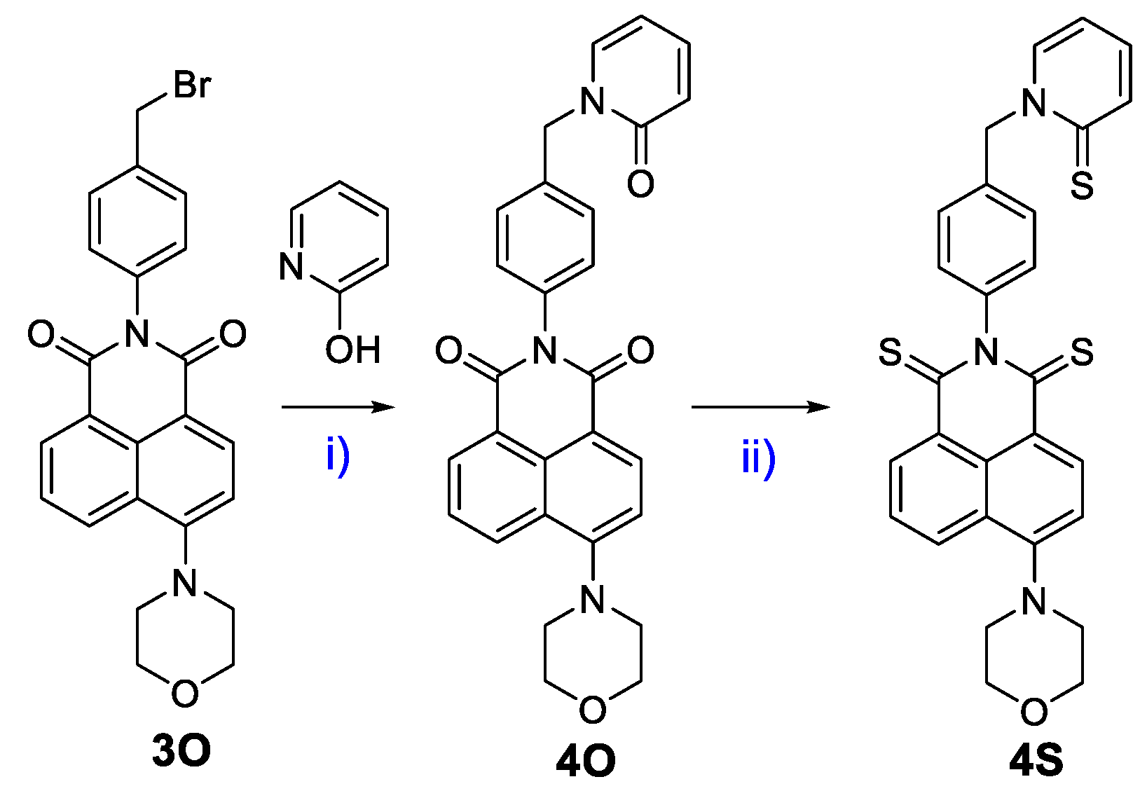
2.1. Synthesis of 4S

4O (1.0 mmol) and Lawesson’s reagent (3.0 mmol) in Toluene (15 mL) was refluxed for 12 h. After solvent was evaporated, it was diluted in DW and extracted 3 times with MC. The organic layer was collected by column chromatography on silica gel using Hexane/Ethyl acetate (4/1) as eluent. The product was dried to afford a red solid of 4S (yield ~70%). 1H NMR (400 MHz, Chloroform-d) δ 8.91 (dd, J = 7.7, 1.2 Hz, 1H), 8.86 (d, J = 8.6 Hz, 1H), 8.39 (dd, J = 8.6, 1.3 Hz, 1H), 7.77 (dd, J = 8.7, 1.6 Hz, 1H), 7.67–7.58 (m, 2H), 7.53–7.45 (m, 2H), 7.16 (dp, J = 8.7, 2.1 Hz, 4H), 6.64 (td, J = 6.8, 1.6 Hz, 1H), 5.90 (s, 2H), 4.01 (dd, J = 5.7, 3.4 Hz, 4H), 3.35–3.26 (m, 4H); 13C NMR (101 MHz, Chloroform-d) δ 192.30, 191.24, 181.26, 156.24, 146.26, 139.97, 139.44, 137.85, 136.53, 135.05, 133.81, 130.52, 130.40, 129.95, 129.24, 126.46, 125.88, 125.44, 125.07, 116.00, 113.86, 66.93, 58.37, 53.34; ESI HRMS m/z = 514.1077 [M+H]+, calc. for C28H23N3OS3 = 513.1003.

2.2. Cell Experiments

HeLa cells (human cervical cancer cells) were cultured in DMEM (Dulbecco’s Modified Eagle Medium) supplemented with a 10% FBS solution and 1% penicillin-streptomycin (v/v) and kept in 5% CO2 at 37 °C.

2.3. Confocal Microscopy Cell Imaging

The HeLa cells were resuspended in confocal dishes to a final density of ~5 × 10⁴ cells/2 mL of the DMEM medium. After overnight culture, the cells were washed twice with DPBS before the fresh culture medium containing 3O and 4O (10 μM) was added for 30 min. After washing with DPBS, images were recorded using a confocal microscope equipped with 405-nm excitation and 600-nm emission filters by confocal microscopy Zeiss LSM 900.
HeLa cells were incubated with 5-μM 3S and 4S, respectively, and costained with 10 μM DCFH-DA for 30 min. Then, cells were irradiated with a green LED (20 mW/cm2, 5 min). After washing with DPBS, fluorescence images were acquired by confocal microscopy.

2.4. Cell Viability

Cells were seeded in a 96-well plate to a final density of ∼5 × 10³ cells/well with culture media. After overnight culture, HeLa cells were incubated with different concentrations (0–50 μM) of 3S and 4S for 1 h. After washing with DPBS, cells were irradiated by a green LED (20 mW/cm2, 15 min) and incubated for another 24 h. The samples were subsequently combined with a D-Plus™CCK solution and incubated under normoxia conditions (37 °C, 5% CO2) for another 4 h period.
Hypoxic condition was applied by incubation in mixed gas (5% CO2, 1% O2, 94% N2) for 24 h and all media were bubbled with mixed gas beforehand. After incubation with 3S and 4S for 1h, green LED was irradiated for 15 min. Then, the cells were incubated for 24 h. The samples were subsequently combined with a D-Plus™CCK solution and incubated under hypoxia conditions (37 °C, 1% O2) for another 4 h period.

2.5. Theoretical Calculation

Computational calculation is detailed in the supporting information. In the system, crossing rate ( k I S C n m ) between excited singlet state (Sn) to the excited triplet state (Tm) were calculated using Fermi’s Golden rule [26]:
k I S C n m = 2 π ρ F C S n H S O C T m 2
where 〈Sn|HSOC|Tm〉 is the spin–orbit coupling (SOC)-matrix element between Sn and Tm and ρFC is Franck–Condon weighted density of states, which was calculated in the framework of the Marcus theory [27]:
ρ F C = 1 4 π λ M k B T e x p E S T + λ M 2 4 π λ M k B T
where ∆EST, T, kB and λM are singlet-triplet energy gap, temperature, Boltzmann constant, and Marcus reorganization energy, respectively.

3. Results and Discussion

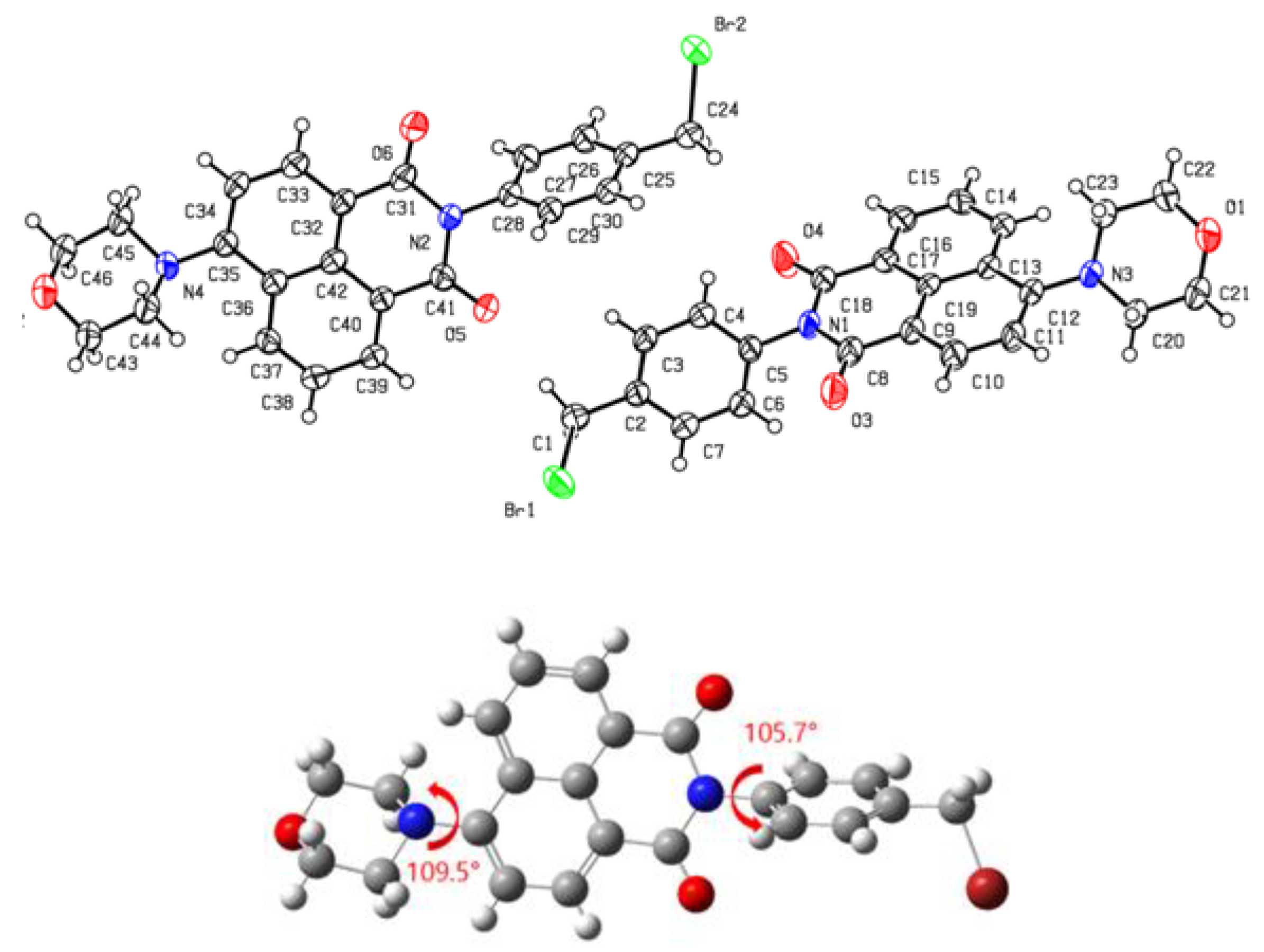
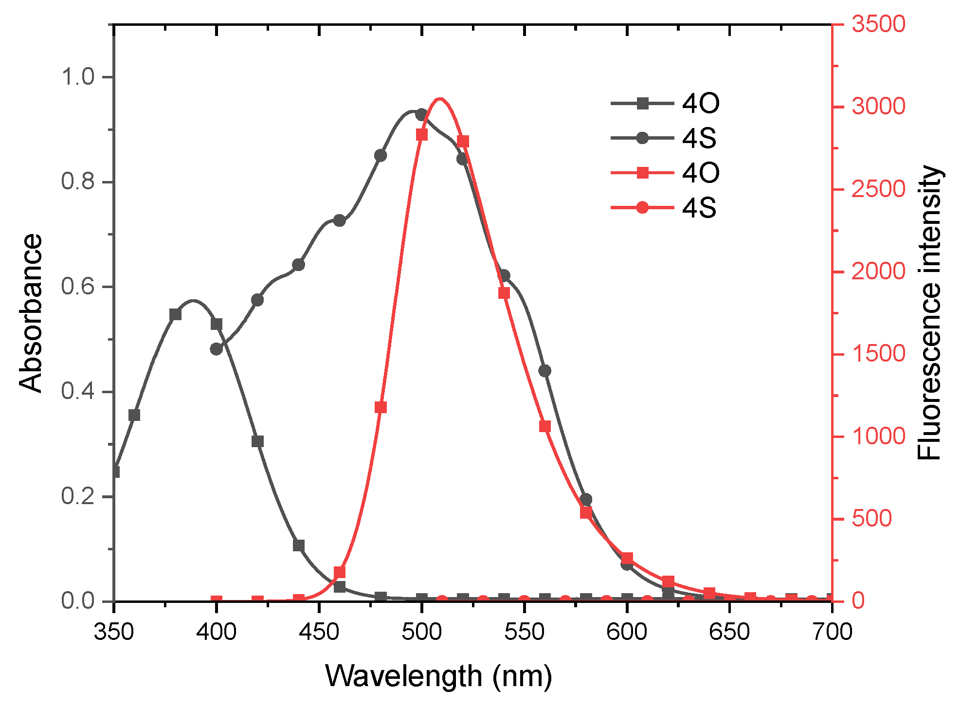
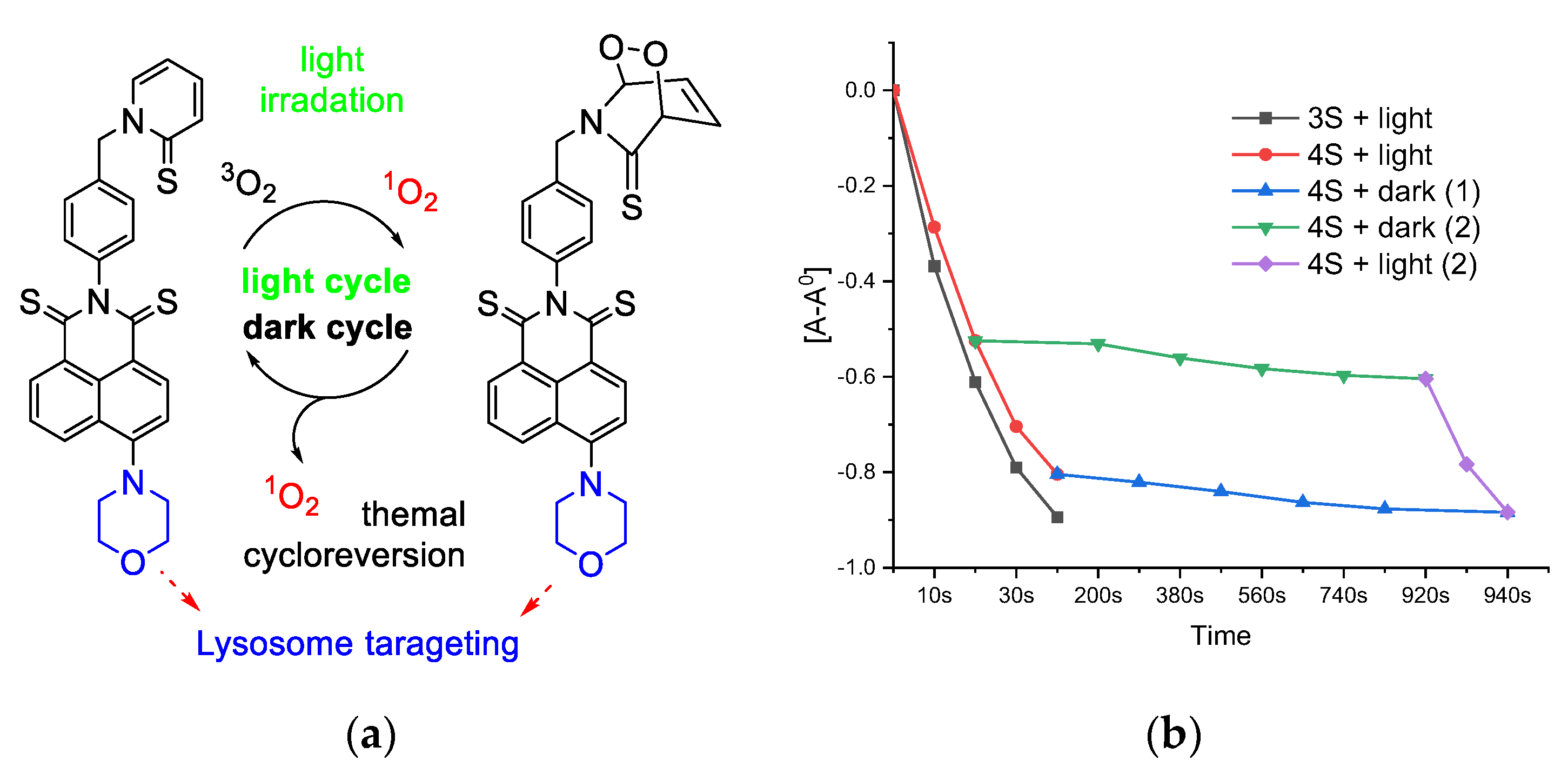
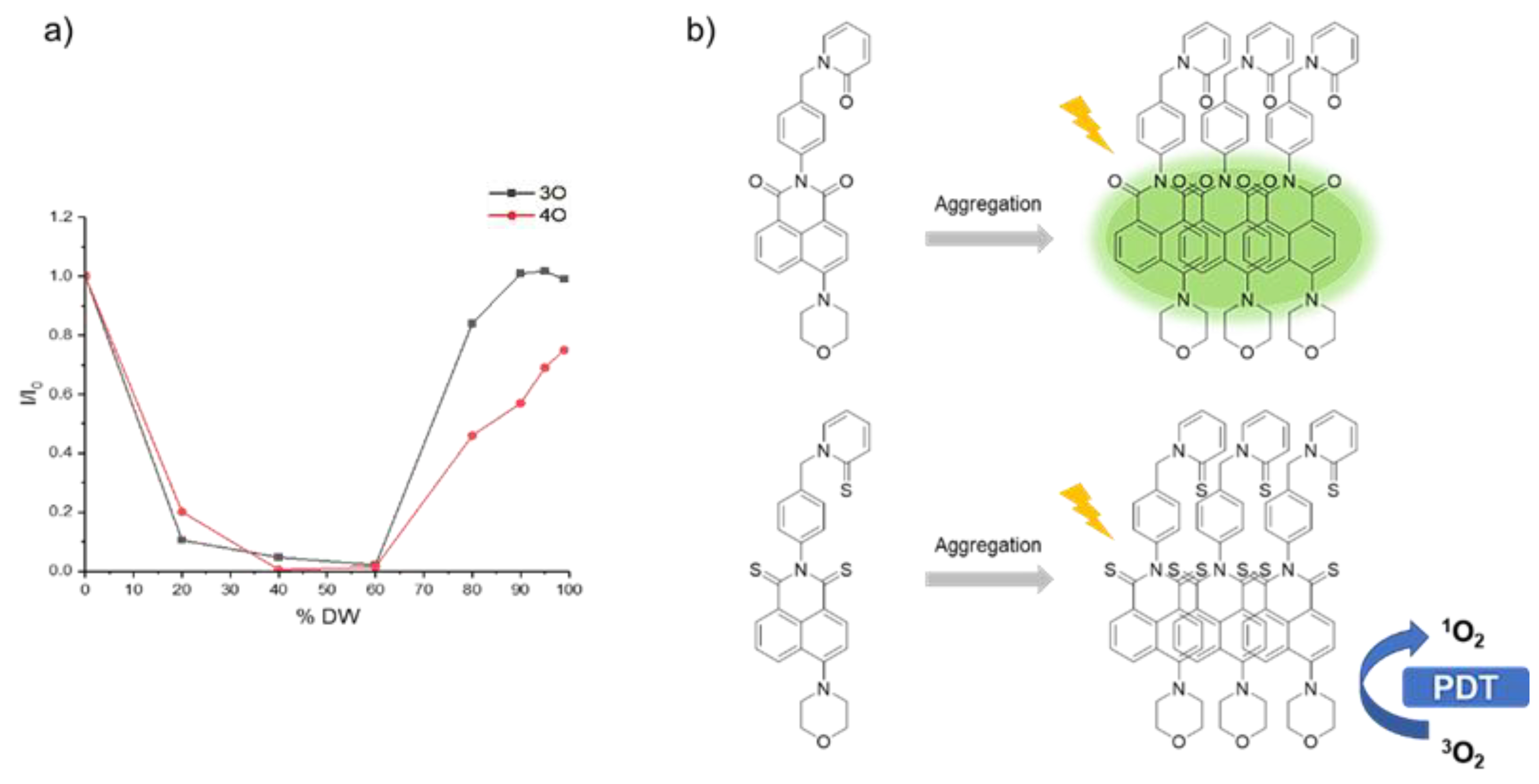
3.1. Molecular Design, Synthesis Process, and Photophysical Properties

As in Scheme 1, we prepared 4O from the reaction of 3O and 2-hyroxypyridine by adding 18-crown-6 and KI catalyst. Then, the carbonyl group of 4O was converted to the thiocarbonyl group in the presence of Lawesson’s reagent to form 4S. We also prepared the thionated form of 3O (3S) by the same method and introduced morpholine to direct the targeting ability of fluorophore and PS toward the lysosome [28]. 4O and 4S were conjugated to 2-pyridone and its thiol form, respectively, which are expected to release thermal-induced 1O2 [29]. All reactions showed high yields (>70%), and products were confirmed by 1HNMR, 13CNMR, and HR-MS spectra (Supplementary Materials). We observed the 13C signal of 3S and 4S at about 192 ppm, which confirmed the carbonyl to thiocarbonyl conversion. Notably, the X-ray single-crystal structure of 3O was recorded (Figure 1 and Figure S17) and found to be similar to the optimized geometry by DFT calculation.
Then, we examined the UV–VIS absorption and fluorescent spectra of 3O, 3S, 4O, and 4S in several solvents (Toluene, THF, and ACN) (Figure 2, Figures S18 and S19). 3O and 4O, 3S, and 4S show similar UV–Vis absorption bands with peaks near 388 nm and 496 nm, respectively. However, the thionation form shows a higher molar absorption coefficient that corresponds with previous thionated PSs. 3O and 4O exhibit a strong green emission (λems ~ 509 nm; ΦF > 0.94) with a large stock shift of 120 nm (Table 1), which is very suitable for fluorescent bioimaging. In sharp contrast, 3S and 4S do not show fluorescent emission, which predicts a strong ISC process as well as singlet oxygen quantum yield [30]. By computational calculation, the spin–orbit coupling (SOC) of the energy gap between the singlet and triplet states (ΔEST) was generated (Tables S4 and S5); then, we calculated the ISC constants (kISC). Due to the thionation, the SOC constants of 4S and 3S are significantly higher than that of 4O and 3O, respectively. Besides that, the energy gap between the singlet and triplet states of the thiocarbonyl form is also smaller than that of the corresponding the carbonyl form, leading to much larger single ISC constants (kISC) of each singlet–triplet pathway. As a result, the total ISC constants of 4S and 3S are significantly higher than those of 4O and 3O, respectively. The increased ISC process of thionation facilitates the popular triplet states of 4S and 3S, which were confirmed by strong 1O2 generation quantum yield (ΦΔ = 0.50 and 0.56, respectively). Additionally, the total ISC constant of 3S (9.7 × 10¹²), higher than that of 4S (5.6 × 10¹²), completely corresponds with the higher 1O2 generation quantum yield, which confirms the well-matched experimental/theoretical results.
The ROS generation of 4S was caused not only by 3O21O2 conversion but also by biomolecules → O2•− production under green light irradiation. The fluorescent emission of dihydroethidium (O2•− probe) in degassed DW (10% fetal bovine serum) increased in the presence of 4S and white light during 20 min (Figure S20e,f), which confirmed the ROS generation in aggregate state and type I caused by the sensitive excited C=S bond and amino groups [30]. The type I ROS generation, which helps the PSs work well in the hypoxic condition, has been desired in recent years. On the other hand, the pyridine-2(1H)-thione introduced in 4S can store the generated 1O2 in the light irradiation phase and thermally release it in the dark phase (Figure 3a). In the presence of 4S, the UV–Vis absorption spectra of the 1O2 detector (DPBF) quickly decay under white-light irradiation and slowly decrease in the dark and 37 °C (Figure 3b). After the 940 s light/dark condition, its decreasing of the UV–Vis absorbance of DPBF was similar to that of 3S in 40 s light irradiation. Maintaining 1O2 release facilitates the killing of cancer in the dark. Besides that, C=S boned-based chemosensors have been developed for detecting OCl [31]. However, 3S and 4S shows stability in the presence of various ROS types and pH ranges, which is observed by negligible change in UV–Vis absorption and FL emission spectra (Figures S22 and S23). The stability of PS facilitates an efficient PDT action in cancer cell.

3.2. In Vitro Experiment
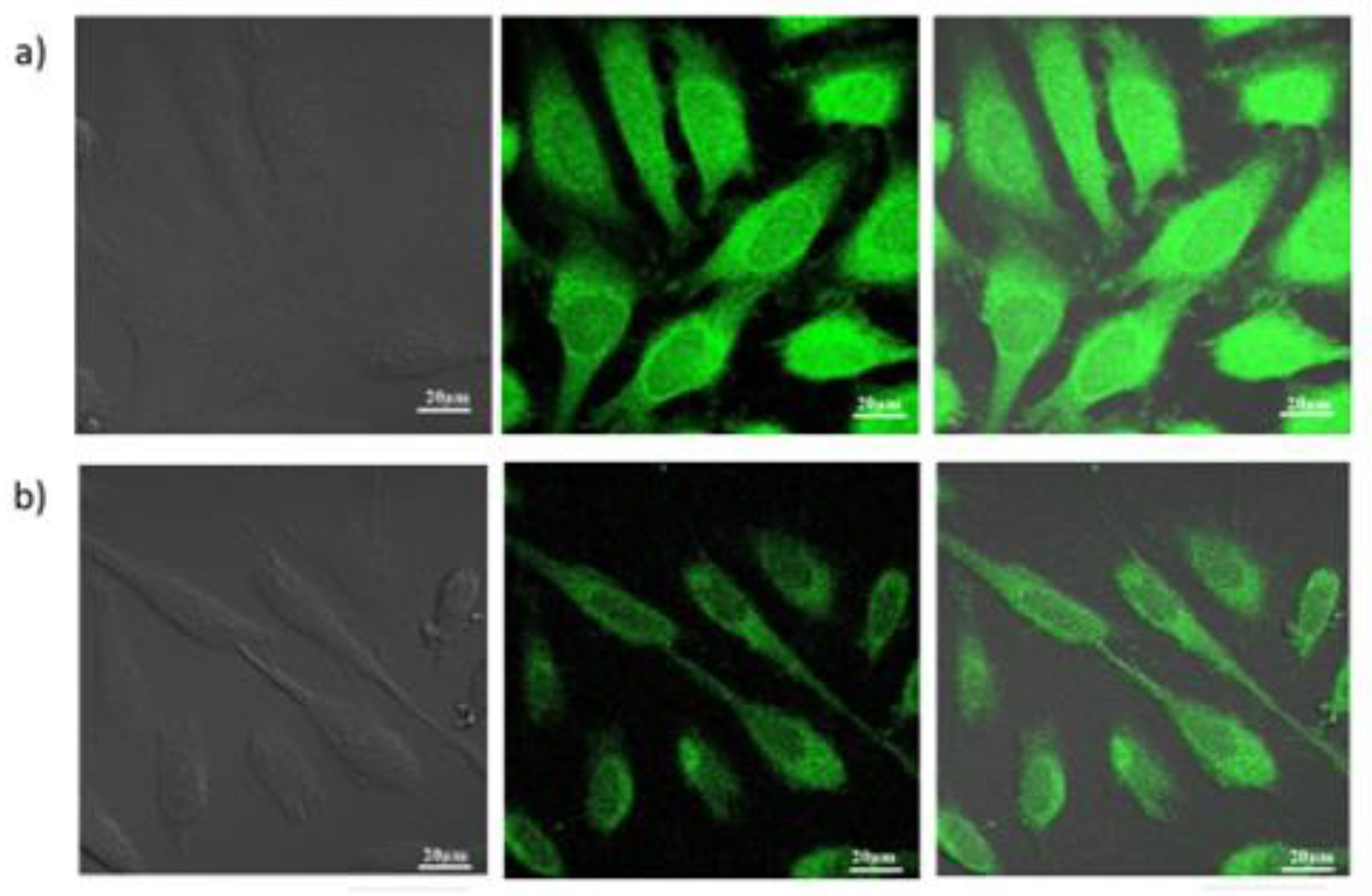
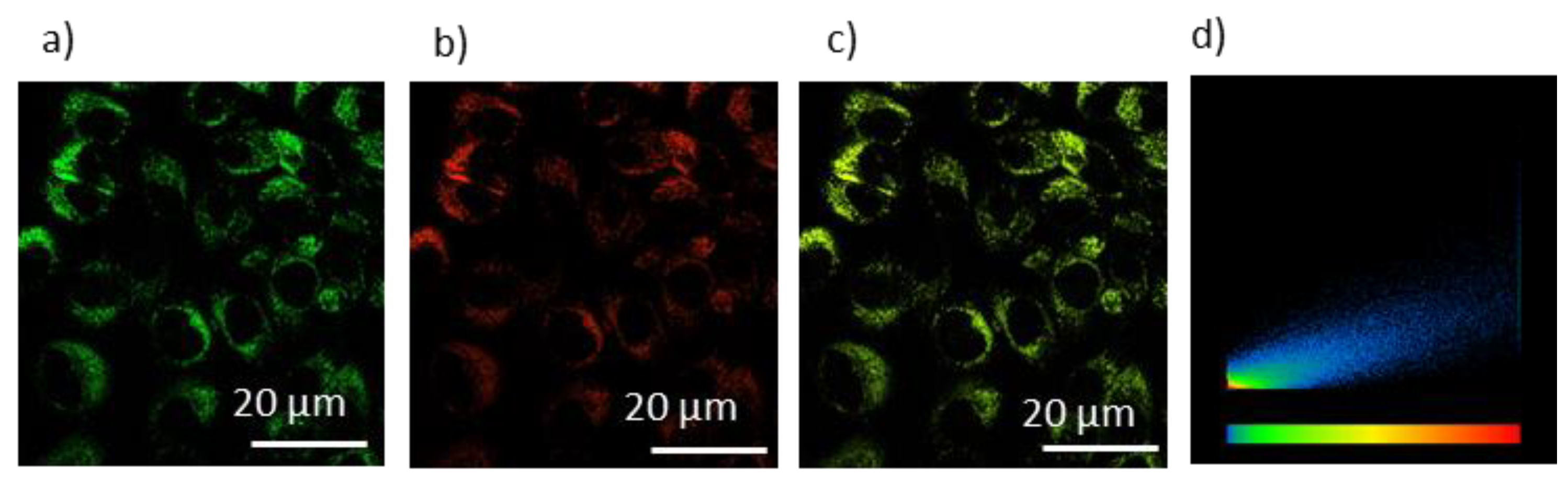
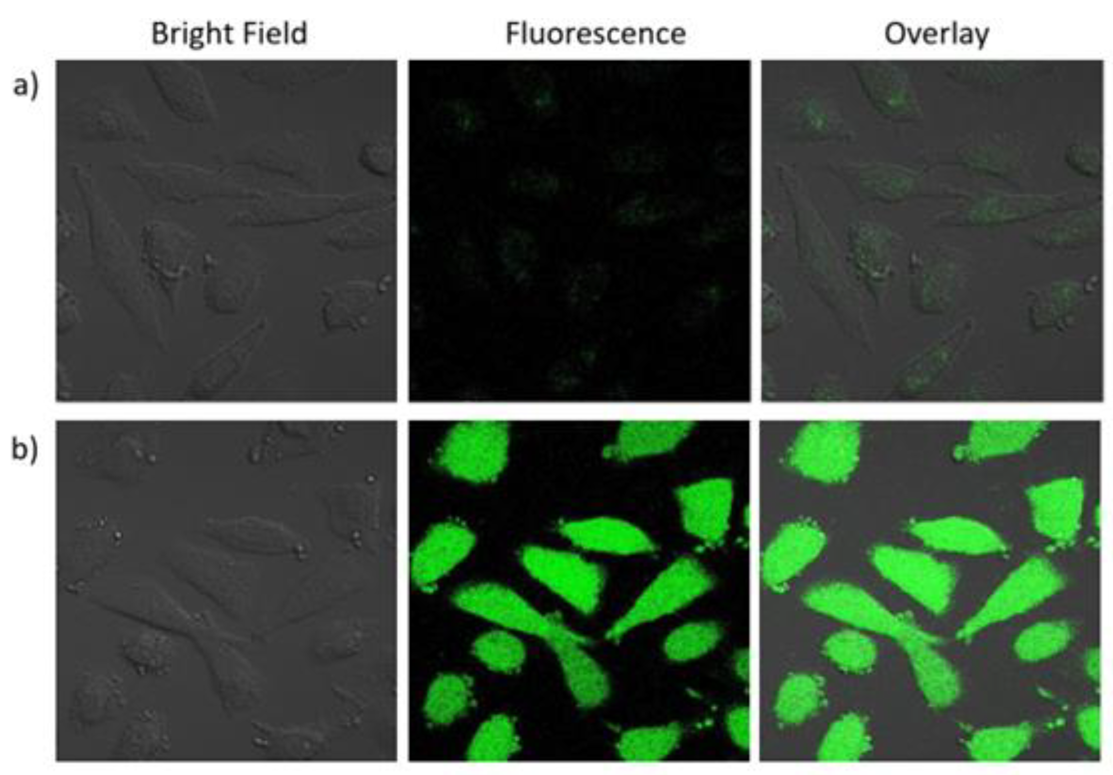
3.2.1. Cell Imaging

Due to strong fluorescent emission (ΦF > 0.94), 3O and 4O may be usable for cell imaging applications. We further examined their emissions in the aggregate state. Interestingly, the fluorescent intensity of 3O and 4O was recovered with an increasing water content (80–99%) in the THF solution (Figure 4 and Figure S21), which indicates AIE properties because of the restricted intramolecular rotation between the naphthalimide and morpholine groups—that is, the naphthalimide and benzene ring (Figure 1b). This also provides alternative evidence of ROS generation of 3S and 4S in aggregate state due to their similar structures. Encouraged by the excellent emission, we further evaluated the cellular uptake of 3O and 4O in HeLa living cells by confocal fluorescent imaging. 3O exhibited a brighter green emission than did 4O because of a stronger AIE effect (Figure 4). The cell imaging and viability of HeLa was retained in the presence of 3O and 4O (0–50 µM) (Figure 5 and Figure S22a,c), indicating their excellent biosafety and biocompatibility. On the other hand, their structures introduced morpholine-lysosome targeting groups, which may be useful as a lysosome-targeted fluorescent probe [28]. The emission signal of parent 3O and 4O overlapped well with that of commercial LysoTracker deep red with a high Pearson’s coefficient value (RP = 0.85 and 0.82, respectively) (Figure 6 and Figure S25), suggesting specific localization of 3O and 4O in lysosomes.

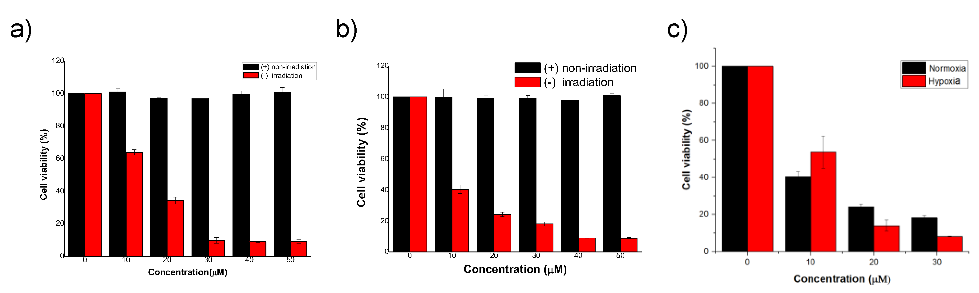
3.2.2. PDT in Normoxia and Hypoxia

To demonstrate their potential for photoresponsive therapy, we explored the anticancer efficacy of 3S and 4S toward HeLa cells via methyl thiazolyl tetrazolium (MTT) assays. The viability of HeLa cells was retained when 3S and 4S (0–50 µM) were increased without light irradiation (Figure S22b,d), indicating the negligible dark cytotoxicity. Under light irradiation, the viability decreased in the presence of 3S and 4S from 0 to 50 µM (Figure 7a,b). At the 10 µM concentration, the viability of HeLa cells toward 3S and 4S was 63.9% and 40.5%, respectively. Due to the specific localization of 3O and 4O in lysosomes, we speculate that 3S and 4S with highly similar structures will destroy cancer cell lysosomes under irradiation to induce severe apoptotic cell death [28]. Interestingly, 4S showed high photocytotoxicity not only in normoxia but also in hypoxia, with a similar cell viability (Figure 7c). The ROS generation of PSs in HeLa cells during PDT was confirmed by turn-on of fluorescent emission of the DCHF-DA detector (Figure 8). The efficient PDT action of 4S in both conditions can be explained by the dual Type I + II ROS generation because of the efficient formation of triplet states in aggregate state [32], introduction of sensitive excited C=S bond and amino groups [30], and thermal-induced releasing of 1O2 in the dark [29].

4. Conclusions

We examined the photophysical properties and excited states of fluorophore (3O and 4O) and PSs (3S and 4S) by joint experimental/theoretical studies. 3O and 4O showed intense fluorescent visualization in HeLa cells due to AIE effect, whereas 4S PSs exhibited absorbing/releasing cycloreversion of 1O2 and strong ROS generation even in the aggregate condition because of the strong ISC process and sensitivity of the excited C=S bond and amino groups. 4S can exhibit a lysosome-targeted PDT efficiency in both normoxic and hypoxic environments.

Supplementary Materials

The following supporting information can be downloaded at: https://www.mdpi.com/article/10.3390/bios12060420/s1, Figures S1–S16: NMR and Mass spectra data; Table S1: Crystal data and structure refinement for 3O; Figure S17: X-ray result; Figures S18–S21: UV–vis and Fluorescence spectra analysis; Tables S2–S5: Computational calculation results; Figure S22: Cell experiment results.

Author Contributions

Conceptualization, T.C.P., S.-W.J., O.-S.J. and S.L. (Songyi Lee); methodology, T.T.H.H., Y.C., G.K. and D.K.; software, T.C.P.; formal analysis, T.C.P., Y.C. and D.K.; writing—original draft preparation, T.C.P.; writing—review and editing, S.L. (Seongman Lee); visualization, T.C.P.; supervision, S.L. (Seongman Lee); project administration, S.L. (Seongman Lee); funding acquisition, S.L. (Seongman Lee). All authors have read and agreed to the published version of the manuscript.

Funding

This work was supported by the National Research Foundation of Korea (NRF) grant funded by the Korea government (MSIT) (No. 2022R1C1C1007740 and 2022R1A5A8023404) for S. L.

Institutional Review Board Statement

Not applicable.

Informed Consent Statement

Not applicable.

Data Availability Statement

Not applicable.

Conflicts of Interest

The authors declare no conflict of interest.

References

  1. Farrell, K.M.; Brister, M.M.; Pittelkow, M.; Sølling, T.I.; Crespo-Hernandez, C.E. Heavy-Atom-Substituted Nucleobases in Photodynamic Applications: Substitution of Sulfur with Selenium in 6-Thioguanine Induces a Remarkable Increase in the Rate of Triplet Decay in 6-Selenoguanine. J. Am. Chem. Soc. 2018, 140, 11214–11218. [Google Scholar] [CrossRef] [PubMed]
  2. Karran, P.; Attard, N. Thiopurines in current medical practice: Molecular mechanisms and contributions to therapy-related cancer. Rev. Cancer 2008, 8, 24–36. [Google Scholar] [CrossRef] [PubMed]
  3. Hong, Y.; Lam, J.W.; Tang, B.Z. Aggregation-induced emission: Phenomenon, mechanism and applications. Chem. Commun. 2009, 4332–4353. [Google Scholar] [CrossRef] [PubMed]
  4. Hong, Y.; Lam, J.Z.; Tang, B.Z. Aggregation-induced emission. Chem. Soc. Rev. 2011, 40, 5361–5388. [Google Scholar] [CrossRef] [PubMed] [Green Version]
  5. Yuan, Y.; Feng, G.; Qin, W.; Tang, B.Z.; Liu, B. Targeted and image-guided photodynamic cancer therapy based on organic nanoparticles with aggregation-induced emission characteristics. Chem. Commun. 2014, 50, 8757–8760. [Google Scholar] [CrossRef] [PubMed] [Green Version]
  6. Luo, J.; Xie, Z.; Lam, J.W.; Cheng, L.; Tang, B.Z.; Chen, H.; Qiu, C.; Kwok, H.S.; Zhan, X.; Liu, Y.; et al. Aggregation-induced emission of 1-methyl-1,2,3,4,5-pentaphenylsilole. Chem. Commun. 2001, 1740–1741. [Google Scholar] [CrossRef]
  7. Mei, J.; Hong, Y.; Lam, J.W.; Qin, A.; Tang, T.; Tang, B.Z. Aggregation-induced emission: The whole is more brilliant than the parts. Adv. Mater. 2014, 26, 5429–5479. [Google Scholar] [CrossRef]
  8. Hu, F.; Huang, Y.; Zhang, G.; Zhao, G.; Yang, H.; Zhang, D. Targeted Bioimaging and Photodynamic Therapy of Cancer Cells with an Activatable Red Fluorescent Bioprobe. Anal. Chem. 2014, 86, 7987–7995. [Google Scholar] [CrossRef]
  9. Chang, C.C.; Hsieh, M.C.; Lin, J.C.; Chang, T.C. Selective photodynamic therapy based on aggregation-induced emission enhancement of fluorescent organic nanoparticles. Biomaterials 2012, 33, 897–906. [Google Scholar] [CrossRef]
  10. Hsieh, M.-C.; Chien, C.-H.; Chang, C.-C.; Chang, T.-C. Aggregation induced photodynamic therapy enhancement based on linear and nonlinear excited FRET of fluorescent organic nanoparticles. J. Mater. Chem. B 2013, 1, 2350–2357. [Google Scholar] [CrossRef] [Green Version]
  11. Pandey, N.K.; Xiong, W.; Wang, L.; Chen, W.; Bui, B.; Yang, J.; Amador, E.; Chen, M.; Xing, C.; Athavale, A.A.; et al. Aggregation-induced emission luminogens for highly effective microwave dynamic therapy. Bioact. Mater. 2022, 7, 112–125. [Google Scholar] [CrossRef] [PubMed]
  12. Dolmans, D.E.J.G.J.; Fukumura, D.; Jain, R.K. Photodynamic therapy for cancer. Nat. Rev. Cancer 2003, 3, 380–387. [Google Scholar] [CrossRef] [PubMed]
  13. Castano, A.P.; Mroz, P.; Hamblin, M.R. Photodynamic therapy and anti-tumour immunity. Nat. Rev. Cancer 2006, 6, 535–545. [Google Scholar] [CrossRef] [PubMed] [Green Version]
  14. Agostinis, P.; Berg, K.; Cengel, K.A.; Foster, T.H.; Girotti, A.W.; Gollnick, S.O.; Hahn, S.M.; Hamblin, M.R.; Juzeniene, A.; Kessel, D.; et al. Photodynamic therapy of cancer: An update. CA Cancer J. Clin. 2011, 61, 250–281. [Google Scholar] [CrossRef]
  15. Liu, H.-W.; Hu, X.-X.; Li, K.; Liu, Y.; Rong, Q.; Zhu, L.; Yuan, L.; Qu, F.-L.; Zhang, X.-B.; Tan, W. A mitochondrial-targeted prodrug for NIR imaging guided and synergetic NIR photodynamic-chemo cancer therapy. Chem. Sci. 2017, 8, 7689–7695. [Google Scholar] [CrossRef] [Green Version]
  16. Nguyen, V.-N.; Yan, Y.; Zhao, J.; Yoon, J. Heavy-Atom-Free Photosensitizers: From Molecular Design to Applications in the Photodynamic Therapy of Cancer. Acc. Chem. Res. 2021, 54, 207–220. [Google Scholar] [CrossRef]
  17. Nguyen, V.-N.; Yim, Y.; Kim, S.; Ryu, B.; Swamy, K.M.K.; Kim, G.; Kwon, N.; Kim, C.-Y.; Park, S.; Yoon, J. Molecular Design of Highly Efficient Heavy-Atom-Free Triplet BODIPY Derivatives for Photodynamic Therapy and Bioimaging. Angew. Chem. Int. Ed. 2020, 59, 8957–8962. [Google Scholar] [CrossRef]
  18. Li, X.; Lee, S.; Yoon, J. Supramolecular photosensitizers rejuvenate photodynamic therapy. Chem. Soc. Rev. 2018, 47, 1174–1188. [Google Scholar] [CrossRef]
  19. Li, X.; Kolemen, S.; Yoon, J.; Akkaya, E.U. Activatable Photosensitizers: Agents for Selective Photodynamic Therapy. Adv. Funct. Mater. 2017, 27, 1604053–1604063. [Google Scholar] [CrossRef]
  20. Luby, B.M.; Walsh, C.D.; Zheng, G. Advanced Photosensitizer Activation Strategies for Smarter Photodynamic Therapy Beacons. Angew. Chem. Int. Ed. 2019, 58, 2558–2569. [Google Scholar] [CrossRef]
  21. Gorman, A.; Killoran, J.; O’Shea, C.; Kenna, T.; Gallagher, W.M.; O’Shea, D.F. In Vitro Demonstration of the Heavy-Atom Effect for Photodynamic Therapy. J. Am. Chem. Soc. 2004, 126, 10619–10631. [Google Scholar] [CrossRef] [PubMed]
  22. Bertout, J.A.; Patel, S.A.; Simon, M.C. The impact of O2 availability on human cancer. Nat. Rev. Cancer 2008, 8, 967–975. [Google Scholar] [CrossRef] [PubMed] [Green Version]
  23. Northcott, J.M.; Dean, I.S.; Mouw, J.K.; Weaver, V.M. Feeling Stress: The Mechanics of Cancer Progression and Aggression. Front. Cell Dev. Biol. 2018, 6, 17. [Google Scholar] [CrossRef] [PubMed]
  24. Hu, F.; Xu, S.; Liu, B. Photosensitizers with Aggregation-Induced Emission: Materials and Biomedical Applications. Adv. Mater. 2018, 30, 1801350. [Google Scholar] [CrossRef] [PubMed]
  25. Pham, T.C.; Nguyen, V.-N.; Choi, Y.; Lee, S.; Yoon, J. Recent strategies to develop innovative photosensitizers for enhanced photodynamic therapy. Chem. Rev. 2021, 121, 13454–13619. [Google Scholar] [CrossRef] [PubMed]
  26. Lawetz, V.; Orlandi, G.; Siebrand, W. Theory of Intersystem Crossing in Aromatic Hydrocarbons. J. Chem. Phys. 1971, 56, 4058. [Google Scholar] [CrossRef]
  27. Beljonne, D.; Shuai, Z.; Pourtois, G.; Bredas, J.L. Spin-Orbit Coupling and Intersystem Crossing in Conjugated Polymers:  A Configuration Interaction Description. J. Phys. Chem. A 2001, 105, 3899–3907. [Google Scholar] [CrossRef]
  28. Nguyen, V.-N.; Baek, G.; Qi, S.; Heo, S.; Yim, Y.; Yoon, J. A lysosome-localized thionaphthalimide as a potential heavy-atom-free photosensitizer for selective photodynamic therapy. Dyes Pigm. 2020, 177, 108265. [Google Scholar] [CrossRef]
  29. Turan, I.S.; Yildiz, D.; Turksoy, A.; Gunaydin, G.; Akkaya, E.U. A Bifunctional Photosensitizer for Enhanced Fractional Photodynamic Therapy: Singlet Oxygen Generation in the Presence and Absence of Light. Angew. Chem. Int. Ed. 2016, 55, 2875–2878. [Google Scholar] [CrossRef]
  30. Pham, T.C.; Heo, S.; Nguyen, V.-N.; Lee, M.W.; Yoon, J.; Lee, S. Molecular Design toward Heavy-Atom-free Photosensitizers Based on the C=S Bond and their Dual Functions in Hypoxia Photodynamic Cancer Therapy and ClO Detection. ACS Appl. Mater. Interfaces 2021, 13, 13949–13957. [Google Scholar] [CrossRef]
  31. Pham, T.C.; Nguyen, V.-N.; Choi, Y.; Kim, D.; Jung, O.-S.; Lee, D.J.; Kim, H.J.; Lee, M.W.; Yoon, J.; Kim, H.M.; et al. Hypochlorite-Activated Fluorescence Emission and Antibacterial Activities of Imidazole Derivatives for Biological Applications. Front. Chem. 2021, 9, 713078. [Google Scholar] [CrossRef] [PubMed]
  32. Nguyen, V.-N.; Qi, S.; Kim, S.; Kwon, N.; Kim, G.; Yim, Y.; Park, S.; Yoon, J. An Emerging Molecular Design Approach to Heavy-Atom-Free Photosensitizers for Enhanced Photodynamic Therapy under Hypoxia. J. Am. Chem. Soc. 2019, 141, 16243–16248. [Google Scholar] [CrossRef] [PubMed]
Scheme 1. Synthesis of 4S. (i) K2CO3, 18-crown-6, KI, Acetone, reflux, N2; (ii) and Lawesson’s reagent, Toluene, reflux, 12 h.
Biosensors 12 00420 sch001
Figure 1. X-ray crystal structure of 3O.
Biosensors 12 00420 g001
Figure 2. UV–vis absorption (black line) and PL spectra (red line) of 4O and 4S in THF (40 µM, λex at maximum absorption wavelength, slit 5/5).
Biosensors 12 00420 g002
Figure 3. (a) Proposed mechanism of light/dark controlled release of lysosome-targeted heavy-metal-free PSs; (b) the decreased absorbance of DPBF (1O2 generation detector) (50 µM) in the presence of 3S (20 µM) and 4S (20 µM) under light/dark conditions.
Biosensors 12 00420 g003
Figure 4. (a) Fluorescent emission of 3O and 4O at 508 nm in THF/DW (0–99%); (b) proposed scheme of AIE/PDT active fluorescent probes.
Biosensors 12 00420 g004
Figure 5. Fluorescent image of HeLa cell after incubation with (a) 3O and (b) 4O.
Biosensors 12 00420 g005
Figure 6. Hela cells costained with (a) 10 μM 4Oex = 405 nm, λem = 600 nm) for 30 min and (b) 50 nM LysoTracker deep red for 1 h (λex = 647 nm, λem = 655 nm) in PBS and fluorescence images acquired by confocal microscopy. (c) An overlay. (d) Colocalization analysis plot of the LysoTracker and 4O. Pearson’s overlap coefficient, RP = 0.82.
Biosensors 12 00420 g006
Figure 7. Viability of HeLa cells in the presence of (a) 3S and (b) 4S with and without light irradiation; (c) viability of HeLa cells in the presence of 4S with light irradiation under normoxia and hypoxia.
Biosensors 12 00420 g007
Figure 8. Fluorescent image of HeLa cell after incubation with (a) 3O and (b) 4O. Fluorescent image of HeLa cells in the presence of DCFH-DA and 4S (a) before and (b) after irradiation.
Biosensors 12 00420 g008
Table 1. Photophysical and photosensitizing properties of 4O and 4S.
λabsa
(nm)
ԑ × 103
(M−1 cm−1)
λems a
(nm)
∆v b
(nm)
ՓF a,cEg d
(eV)
kISCeՓ f
3O38811.965091210.943.585.1 × 1010-
3S49620.20---2.479.7 × 10120.56
4O38913.485091200.983.403.9 × 108-
4S49623.37---2.295.6 × 10120.50
a in THF. b Stock shift. c Fluorescence quantum yield evaluated with Rhodamine 6G as the standard (ՓF = 0.94 in Ethanol). d Eg, the energy difference between the HOMO and LUMO levels. e k I S C = 2 π ρ F C S n H S O C T m 2 . f Singlet oxygen quantum yield was determined with respect to Ru(bpy)32+ = 0.57 in ACN)—not observed).
Publisher’s Note: MDPI stays neutral with regard to jurisdictional claims in published maps and institutional affiliations.

