Terahertz Metamaterial Sensor for Sensitive Detection of Citrate Salt Solutions

 ,
,
Abstract
:1. Introduction
2. Simulation and Experimental Methods
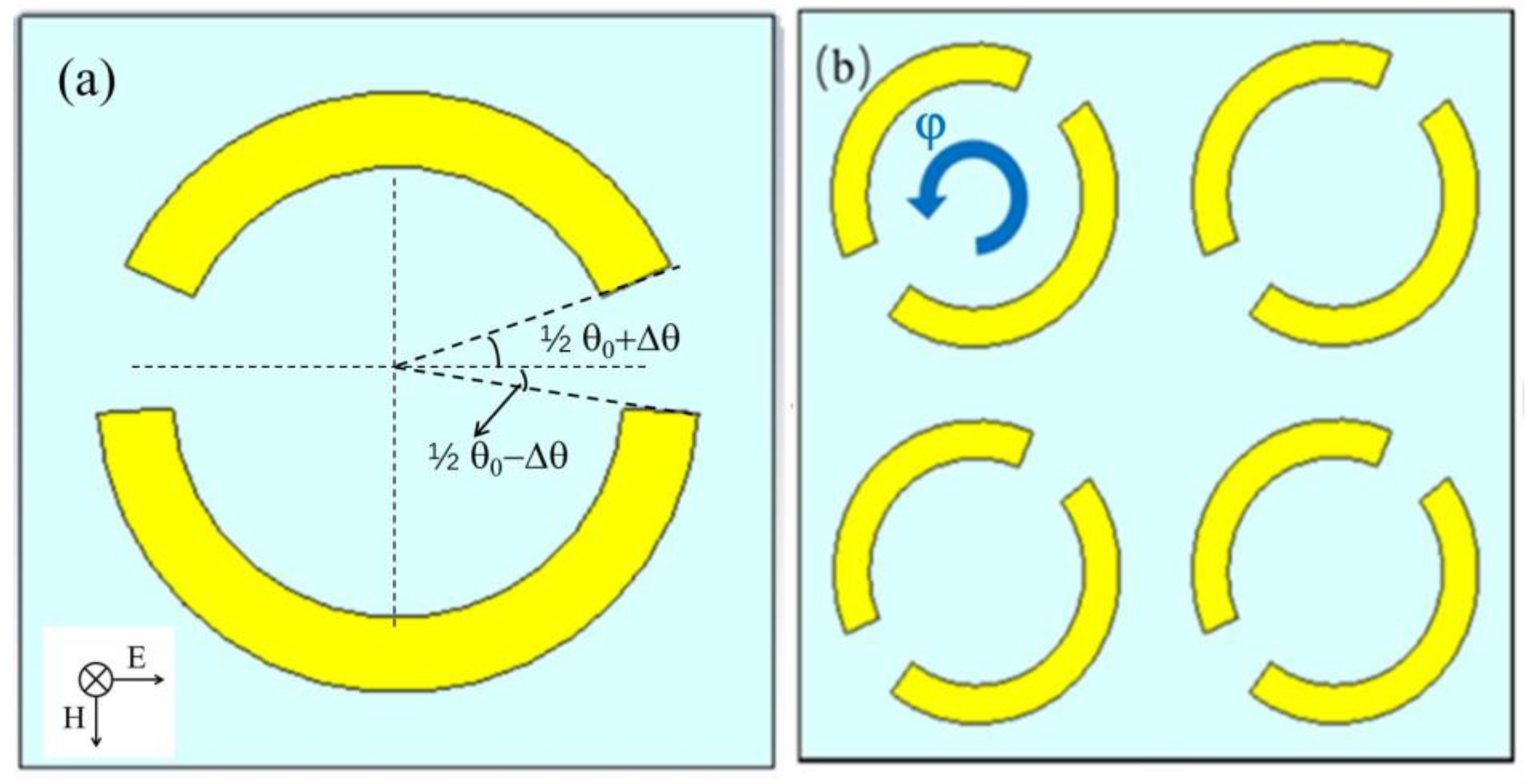
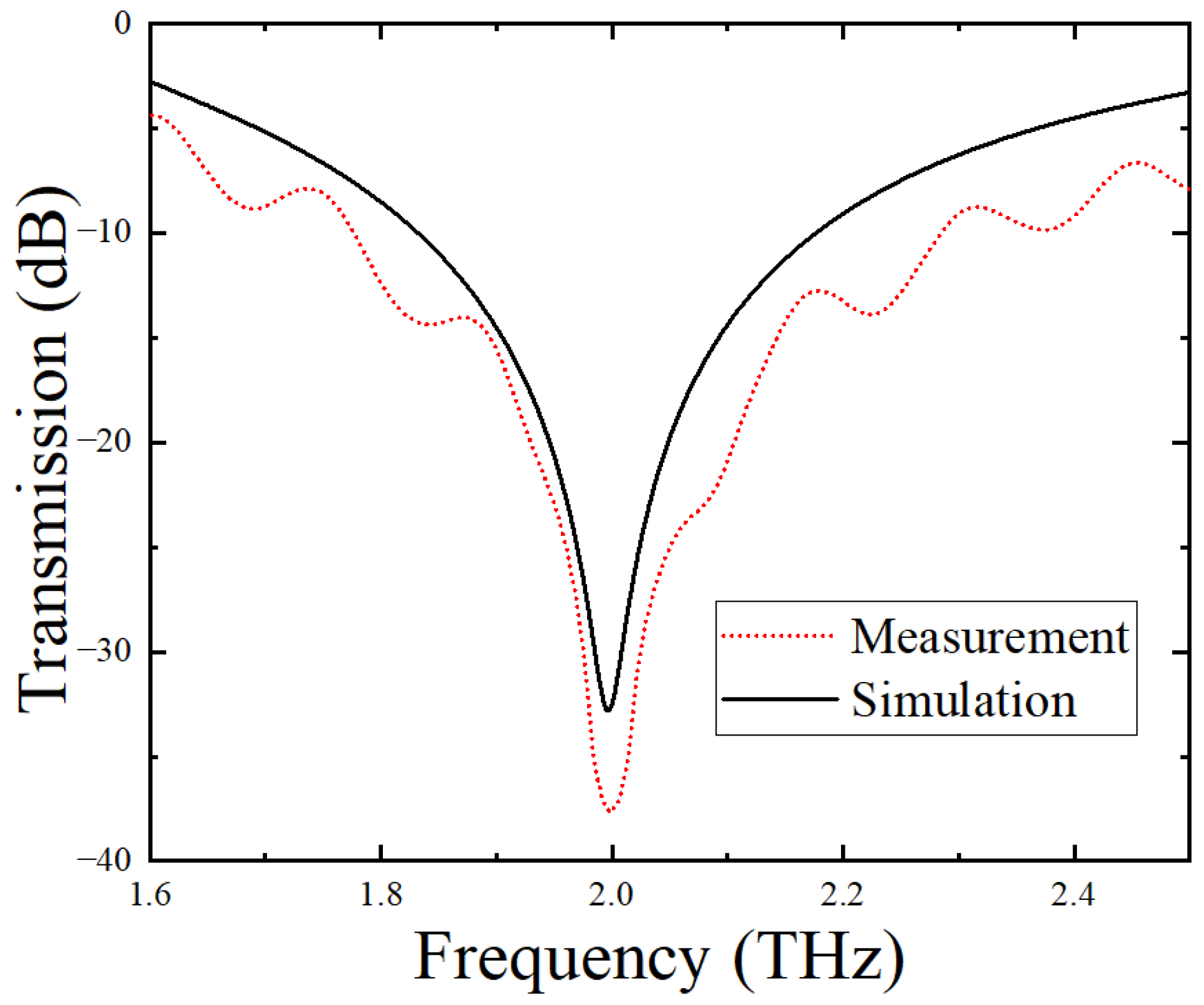
2.1. Design of the Metamaterial Sensor
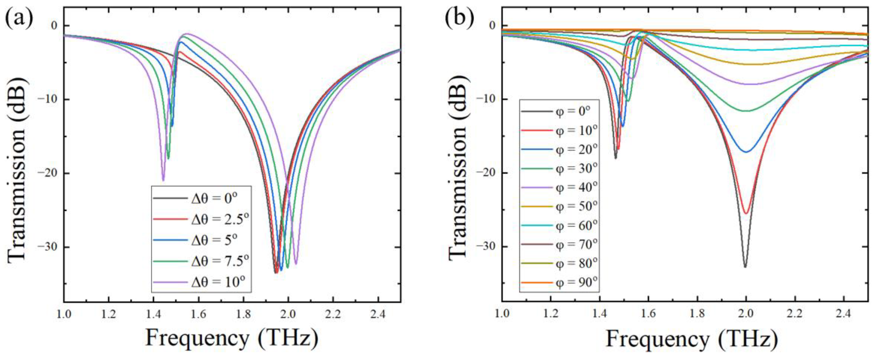
2.1.1. Effect of the Opening Position of the Metal Ring
2.1.2. Effect of the Arrangement of the Metal Ring Units
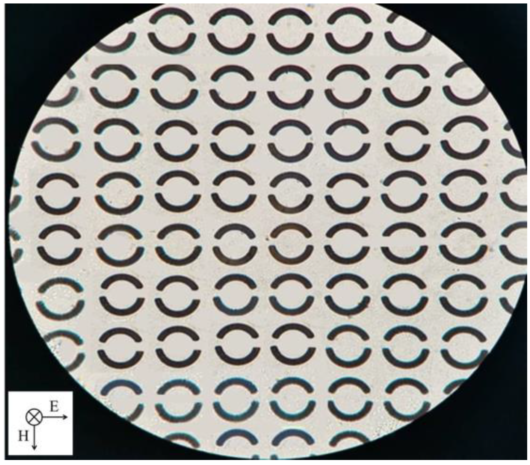
2.2. Fabrication of Metamaterial Sensor
2.3. Terahertz Spectroscopy Measurements of Citrate Salt Samples
3. Results and Discussion
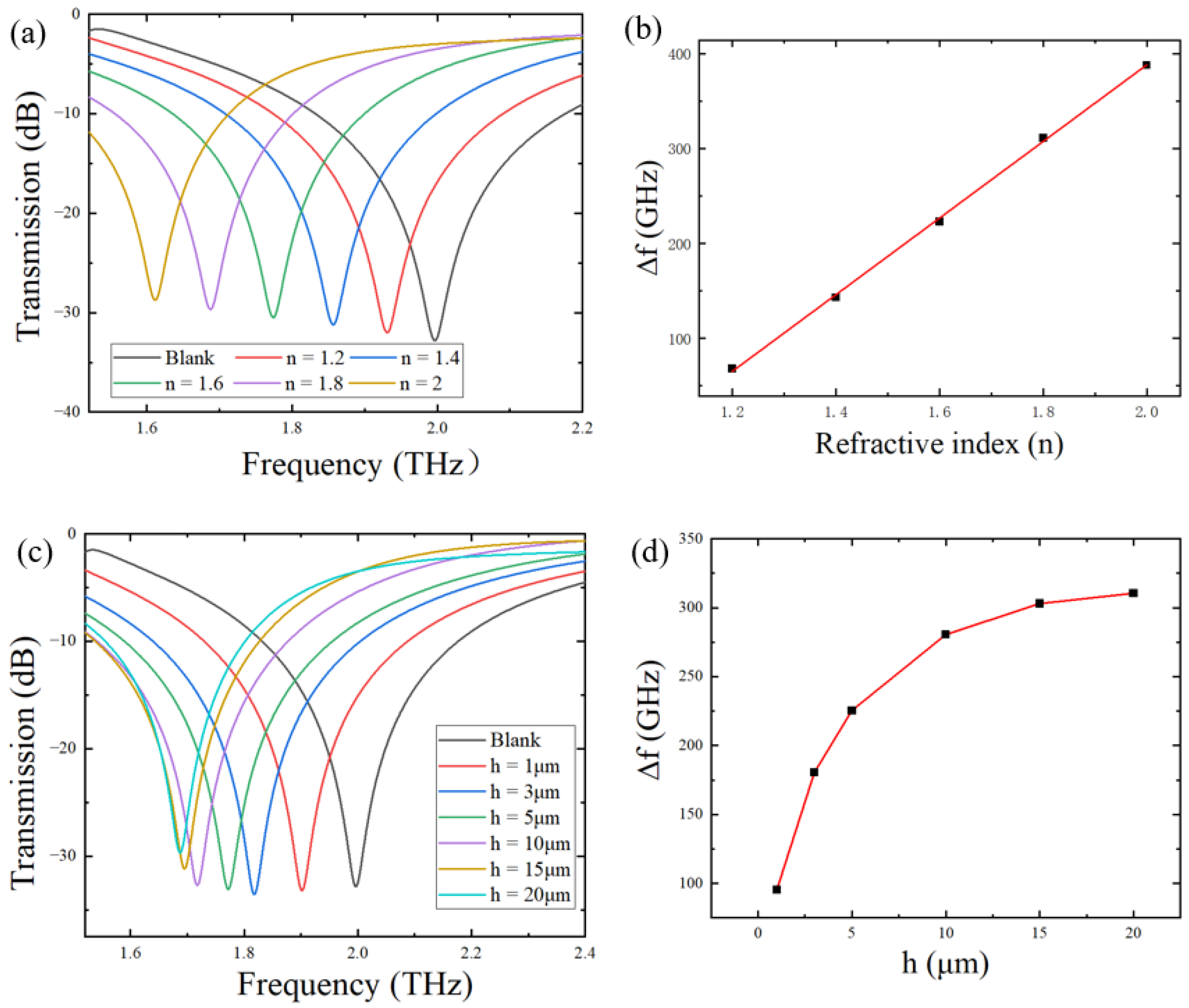
3.1. Factors That Influence Sensitivity of the Metamaterial Sensor Covered with Analytes
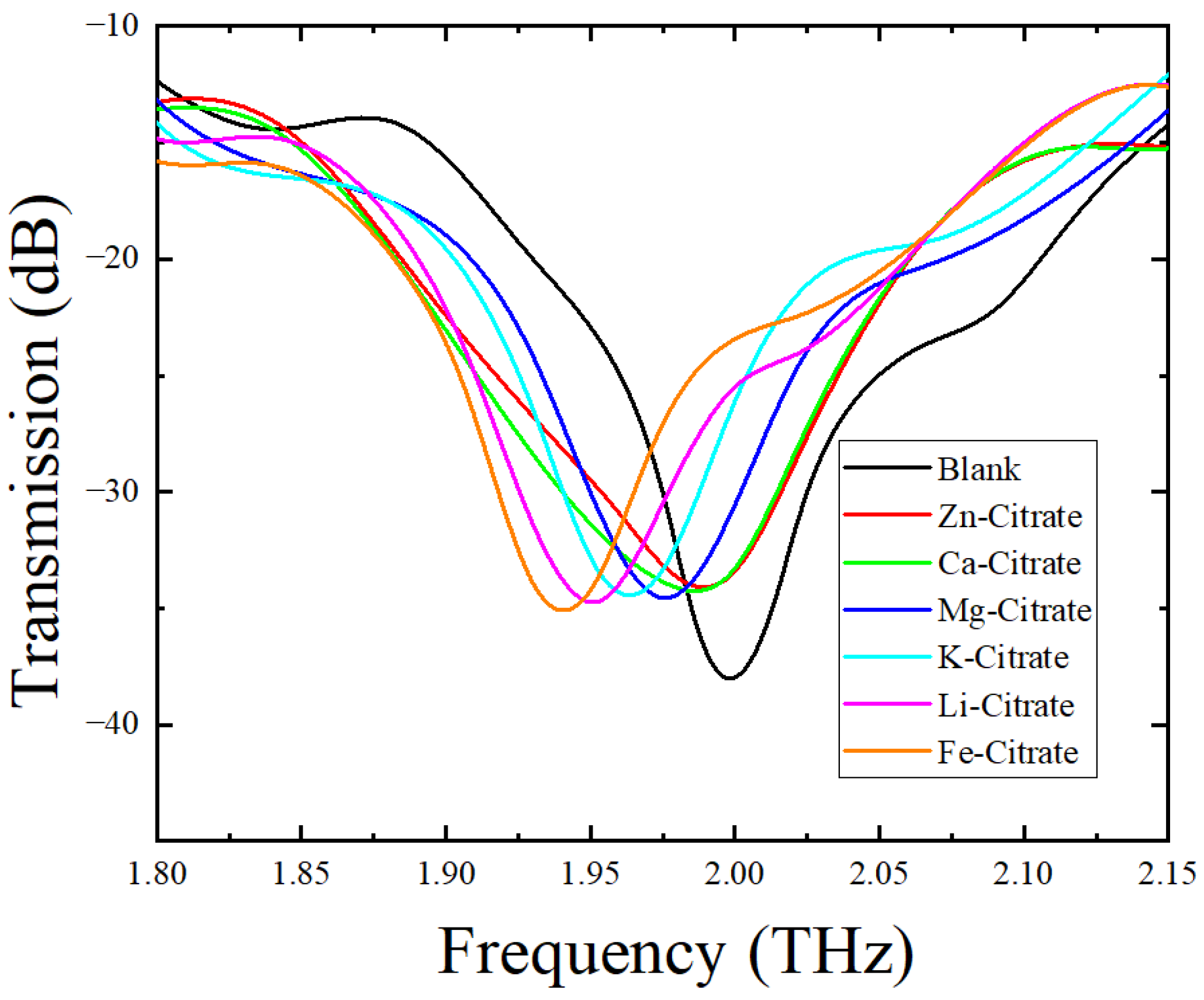
3.2. THz Spectroscopy Detection of Citrate Salts Using the Metamaterial Sensor
4. Conclusions
Author Contributions
Funding
Institutional Review Board Statement
Informed Consent Statement
Data Availability Statement
Conflicts of Interest
References
- Icard, P.; Coquerel, A.; Wu, Z.; Gligorov, J.; Fuks, D.; Fournel, L.; Lincet, H.; Simula, L. Understanding the Central Role of Citrate in the Metabolism of Cancer Cells and Tumors: An Update. Int. J. Mol. Sci. 2021, 22, 6587. [Google Scholar] [CrossRef]
- dos Santos, W.M.; de Souza, M.L.; Nóbrega, F.P.; de Sousa, A.L.M.D.; de França, E.J.; Rolim, L.A.; Neto, P.J.R. A Review of Analytical Methods for Calcium Salts and Cholecalciferol in Dietary Supplements. Crit. Rev. Anal. Chem. 2022, 52, 697–711. [Google Scholar] [CrossRef]
- Pardo, M.R.; Vilar, E.G.; Martín, I.S.M.; Martín, M.A.C. Bioavailability of magnesium food supplements: A systematic review. Nutrition 2021, 89, 111294. [Google Scholar] [CrossRef] [PubMed]
- Aguilar-Ramirez, D.; Alegre-Díaz, J.; Herrington, W.G.; Staplin, N.; Ramirez-Reyes, R.; Gnatiuc, L.; Hill, M.; Romer, F.; Torres, J.; Trichia, E.; et al. Association of Kidney Function with NMR-Quantified Lipids, Lipoproteins, and Metabolic Measures in Mexican Adults. J. Clin. Endocrinol. Metab. 2021, 106, 2828–2839. [Google Scholar] [CrossRef] [PubMed]
- Alenezi, N.A.; Zanaty, F.; Hodhod, A.; El-Gharabawy, M.; El-Sherif, E.; Badawy, A.; El-Shazly, M. The safety of ureteral stenting with the use of potassium citrate for management of renal uric acid stones. Urol. Ann. 2020, 12, 37–41. [Google Scholar] [CrossRef] [PubMed]
- Awan, S.; Abelleira, A.; Khehra, L.; Hieber, R. Undetectable serum lithium concentrations after coadministration of liquid lithium citrate and apple juice: A case report. Ment. Health Clin. 2021, 11, 27–30. [Google Scholar] [CrossRef] [PubMed]
- Block, G.A.; Pergola, P.E.; Fishbane, S.; Martins, J.G.; LeWinter, R.D.; Uhlig, K.; Neylan, J.F.; Chertow, G.M. Effect of ferric citrate on serum phosphate and fibroblast growth factor 23 among patients with nondialysis-dependent chronic kidney disease: Path analyses. Nephrol. Dial. Transplant. 2018, 34, 1115–1124. [Google Scholar] [CrossRef]
- Hu, D.; Sreenivasan, P.K.; Zhang, Y.P.; De Vizio, W. The effects of a zinc citrate dentifrice on bacteria found on oral surfaces. Oral Health Prev. Dent. 2010, 8, 47–53. [Google Scholar]
- Straub, D.A. Calcium Supplementation in Clinical Practice: A Review of Forms, Doses, and Indications. Nutr. Clin. Pract. 2007, 22, 286–296. [Google Scholar] [CrossRef]
- Avila, J.G. Pharmacologic treatment of constipation in cancer patients. Cancer Control 2004, 11, 10–18. [Google Scholar] [CrossRef]
- Orsag, A.; Bozic-Mijovski, M.; Hudoklin, S.; Simcic, S.; Gubensek, J. Biocompatibility Parameters with Standard and Increased Dose of Citrate in Hemodialysis—A Randomized Trial. J. Clin. Med. 2021, 10, 2987. [Google Scholar] [CrossRef] [PubMed]
- Hamm, R.E.; Shull, C.M.; Grant, D.M. Citrate Complexes with Iron(II) and Iron(III)1. J. Am. Chem. Soc. 1954, 76, 2111–2114. [Google Scholar] [CrossRef]
- Rheims, J.; Köser, J.; Wriedt, T. Refractive-index measurements in the near-IR using an Abbe refractometer. Meas. Sci. Technol. 1997, 8, 601–605. [Google Scholar] [CrossRef]
- Hopkins, A.; McNeal, K.; Soli, A.; Byrne, R. In-situ spectrophotometric pH measurements: The effect of pressure on thymol blue protonation and absorbance characteristics. Mar. Chem. 2000, 71, 103–109. [Google Scholar] [CrossRef]
- Guidotti, G.G.; Borghetti, A.F.; Loreti, L. A colorimetric method for the quantitative determination of thiazolidine-4-carboxylic acids. Anal. Biochem. 1966, 17, 513–520. [Google Scholar] [CrossRef]
- Wallace, V.P.; Fitzgerald, A.J.; Shankar, S.; Flanagan, N.; Pye, R.; Cluff, J.; Arnone, D.D. Terahertz pulsed imaging of basal cell carcinoma ex vivo and in vivo. Br. J. Dermatol. 2004, 151, 424–432. [Google Scholar] [CrossRef] [PubMed]
- Kaltenecker, K.J.; König-Otto, J.C.; Mittendorff, M.; Winnerl, S.; Schneider, H.; Helm, M.; Helm, H.; Walther, M.; Fischer, B.M. Gouy phase shift of a tightly focused, radially polarized beam. Optica 2016, 3, 35–41. [Google Scholar] [CrossRef]
- Cheng, Y.; Wang, Y.; Niu, Y.; Zhao, Z. Concealed object enhancement using multi-polarization information for passive millimeter and terahertz wave security screening. Opt. Express 2020, 28, 6350–6366. [Google Scholar] [CrossRef]
- Ibrahim, M.E.; Headland, D.; Withayachumnankul, W.; Wang, C.H. Nondestructive Testing of Defects in Polymer–Matrix Composite Materials for Marine Applications Using Terahertz Waves. J. Nondestruct. Eval. 2021, 40, 37. [Google Scholar] [CrossRef]
- Tekbiyik, K.; Ekti, A.R.; Kurt, G.K.; Gorcin, A.; Yarkan, S. Modeling and Analysis of Short Distance Sub-Terahertz Communication Channel via Mixture of Gamma Distribution. IEEE Trans. Veh. Technol. 2021, 70, 2945–2954. [Google Scholar] [CrossRef]
- Shelby, R.A.; Smith, D.R.; Schultz, S. Experimental Verification of a Negative Index of Refraction. Science 2001, 292, 77–79. [Google Scholar] [CrossRef] [Green Version]
- Shelby, R.A.; Smith, D.R.; Nemat-Nasser, S.C.; Schultz, S. Microwave transmission through a two-dimensional, isotropic, left-handed metamaterial. Appl. Phys. Lett. 2001, 78, 489–491. [Google Scholar] [CrossRef] [Green Version]
- Smith, D.R.; Padilla, W.J.; Vier, D.C.; Nemat-Nasser, S.C.; Schultz, S. Composite Medium with Simultaneously Negative Permeability and Permittivity. Phys. Rev. Lett. 2000, 84, 4184–4187. [Google Scholar] [CrossRef] [PubMed] [Green Version]
- Pendry, J.B. Negative refraction. Contemp. Phys. 2004, 45, 191–202. [Google Scholar] [CrossRef] [Green Version]
- Pendry, J.B.; Holden, A.J.; Robbins, D.J.; Stewart, W.J. Magnetism from conductors and enhanced nonlinear phenomena. IEEE Trans. Microw. Theory Tech. 1999, 47, 2075–2084. [Google Scholar] [CrossRef] [Green Version]
- Sakai, O.; Tachibana, K. Plasmas as metamaterials: A review. Plasma Sources Sci. Technol. 2012, 21, 013001. [Google Scholar] [CrossRef]
- Li, W.; Valentine, J. Metamaterial Perfect Absorber Based Hot Electron Photodetection. Nano Lett. 2014, 14, 3510–3514. [Google Scholar] [CrossRef]
- Simovski, C.R.; Belov, P.A.; Atrashchenko, A.V.; Kivshar, Y.S. Wire Metamaterials: Physics and Applications. Adv. Mater. 2012, 24, 4229–4248. [Google Scholar] [CrossRef]
- Zhang, X.; Tian, Z.; Yue, W.; Gu, J.; Zhang, S.; Han, J.; Zhang, W. Broadband Terahertz Wave Deflection Based on C-shape Complex Metamaterials with Phase Discontinuities. Adv. Mater. 2013, 25, 4567–4572. [Google Scholar] [CrossRef]
- Cong, L.; Cao, W.; Tian, Z.; Gu, J.; Han, J.; Zhang, W. Manipulating polarization states of terahertz radiation using metamaterials. New J. Phys. 2012, 14, 115013. [Google Scholar] [CrossRef]
- Liu, S.; Cui, T.J.; Xu, Q.; Bao, D.; Du, L.; Wan, X.; Tang, W.X.; Ouyang, C.; Zhou, X.Y.; Yuan, H.; et al. Anisotropic coding metamaterials and their powerful manipulation of differently polarized terahertz waves. Light. Sci. Appl. 2016, 5, e16076. [Google Scholar] [CrossRef] [PubMed]
- Chen, H.-T.; Padilla, W.; Zide, J.; Gossard, A.C.; Taylor, A.J.; Averitt, R.D. Active terahertz metamaterial devices. Nature 2006, 444, 597–600. [Google Scholar] [CrossRef] [PubMed] [Green Version]
- Yen, T.J.; Padilla, W.J.; Fang, N.; Vier, D.C.; Smith, D.R.; Pendry, J.B.; Basov, D.N.; Zhang, X. Terahertz Magnetic Response from Artificial Materials. Science 2004, 303, 1494–1496. [Google Scholar] [CrossRef] [PubMed] [Green Version]
- Singh, R.; Cao, W.; Al-Naib, I.; Cong, L.; Withayachumnankul, W.; Zhang, W. Ultrasensitive terahertz sensing with high-Q Fano resonances in metasurfaces. Appl. Phys. Lett. 2014, 105, 171101. [Google Scholar] [CrossRef] [Green Version]
- Hu, F.; Guo, E.; Xu, X.; Li, P.; Xu, X.; Yin, S.; Wang, Y.; Chen, T.; Yin, X.; Zhang, W. Real-timely monitoring the interaction between bovine serum albumin and drugs in aqueous with terahertz metamaterial biosensor. Opt. Commun. 2017, 388, 62–67. [Google Scholar] [CrossRef]
- Cheng, R.; Xu, L.; Yu, X.; Zou, L.; Shen, Y.; Deng, X. High-sensitivity biosensor for identification of protein based on terahertz Fano resonance metasurfaces. Opt. Commun. 2020, 473, 125850. [Google Scholar] [CrossRef]
- Zhang, J.; Mu, N.; Liu, L.; Xie, J.; Feng, H.; Yao, J.; Chen, T.; Zhu, W. Highly sensitive detection of malignant glioma cells using metamaterial-inspired THz biosensor based on electromagnetically induced transparency. Biosens. Bioelectron. 2021, 185, 113241. [Google Scholar] [CrossRef]
- He, J.; He, X.; Dong, T.; Wang, S.; Fu, M.; Zhang, Y. Recent progress and applications of terahertz metamaterials. J. Phys. D Appl. Phys. 2021, 55, 123002. [Google Scholar] [CrossRef]
- Shen, S.; Liu, X.; Shen, Y.; Qu, J.; Pickwell-MacPherson, E.; Wei, X.; Sun, Y. Recent Advances in the Development of Materials for Terahertz Metamaterial Sensing. Adv. Opt. Mater. 2021, 10, 2101008. [Google Scholar] [CrossRef]
- Lee, Y.; Kim, S.-J.; Park, H.; Lee, B. Metamaterials and Metasurfaces for Sensor Applications. Sensors 2017, 17, 1726. [Google Scholar] [CrossRef] [Green Version]
- Xu, W.; Xie, L.; Ying, Y. Mechanisms and applications of terahertz metamaterial sensing: A review. Nanoscale 2017, 9, 13864–13878. [Google Scholar] [CrossRef] [PubMed]
- Ahmadivand, A.; Gerislioglu, B.; Ahuja, R.; Mishra, Y.K. Terahertz plasmonics: The rise of toroidal metadevices towards immunobiosensings. Mater. Today 2020, 32, 108–130. [Google Scholar] [CrossRef]
- Cui, N.; Guan, M.; Xu, M.; Fang, W.; Zhang, Y.; Zhao, C.; Zeng, Y. Design and application of terahertz metamaterial sensor based on DSRRs in clinical quantitative detection of carcinoembryonic antigen. Opt. Express 2020, 28, 16834. [Google Scholar] [CrossRef]
- Janneh, M.; de Marcellis, A.; Palange, E.; Tenggara, A.T.; Byun, D. Design of a metasurface-based dual-band Terahertz perfect absorber with very high Q-factors for sensing applications. Opt. Commun. 2018, 416, 152–159. [Google Scholar] [CrossRef]
- Abu Safia, O.; Talbi, L.; Hettak, K. A New Type of Transmission Line-Based Metamaterial Resonator and Its Implementation in Original Applications. IEEE Trans. Magn. 2013, 49, 968–973. [Google Scholar] [CrossRef]
- Wang, S.; Xia, L.; Mao, H.; Jiang, X.; Yan, S.; Wang, H.; Wei, D.; Cui, H.-L.; Du, C. Terahertz biosensing based on a polarization-insensitive metamaterial. IEEE Photonics Technol. Lett. 2016, 28, 986–989. [Google Scholar] [CrossRef]
- Xu, W.; Xie, L.; Zhu, J.; Tang, L.; Singh, R.; Wang, C.; Ma, Y.; Chen, H.-T.; Ying, Y. Terahertz biosensing with a graphene-metamaterial heterostructure platform. Carbon 2019, 141, 247–252. [Google Scholar] [CrossRef]
- Zhang, R.; Chen, Q.; Liu, K.; Chen, Z.; Li, K.; Zhang, X.; Xu, J.; Pickwell-MacPherson, E. Terahertz Microfluidic Metamaterial Biosensor for Sensitive Detection of Small-Volume Liquid Samples. IEEE Trans. Terahertz Sci. Technol. 2019, 9, 209–214. [Google Scholar] [CrossRef]
- Konopsky, V.N.; Alieva, E.V. Optical Biosensors Based on Photonic Crystal Surface Waves. Methods Mol. Biol. 2009, 503, 49–64. [Google Scholar]
- Lidiya, A.E.; Raja, R.V.J.; Pham, V.D.; Ngo, Q.M.; Vigneswaran, D. Detecting hemoglobin content blood glucose using surface plasmon resonance in D-shaped photonic crystal fiber. Opt. Fiber Technol. 2019, 50, 132–138. [Google Scholar] [CrossRef]
- Arunkumar, R.; Suaganya, T.; Robinson, S. Design and Analysis of 2D Photonic Crystal Based Biosensor to Detect Different Blood Components. Photonic Sens. 2019, 9, 69–77. [Google Scholar] [CrossRef]
- Mollah, M.A.; Yousufali, M.; Faysal, M.R.B.A.; Hasan, M.R.; Hossain, M.B.; Amiri, I.S. Highly sensitive photonic crystal fiber salinity sensor based on Sagnac interferometer. Results Phys. 2020, 16, 103022. [Google Scholar] [CrossRef]
- Bijalwan, A.; Singh, B.K.; Rastogi, V. Analysis of one-dimensional photonic crystal based sensor for detection of blood plasma and cancer cells. Optik 2021, 226, 165994. [Google Scholar] [CrossRef]
- Cao, L.; Jia, S.; Thomson, M.D.; Meng, F.; Roskos, H.G. Can a terahertz metamaterial sensor be improved by ultra-strong coupling with a high-Q photonic resonator? Opt. Express 2022, 30, 13659–13672. [Google Scholar] [CrossRef]


| Δθ | Resonant Frequency (THz) | Q Value |
|---|---|---|
| 0° | 1.943 | 11 |
| 2.5° | 1.950 | 12 |
| 5° | 1.968 | 12 |
| 7.5° | 1.998 | 13 |
| 10° | 2.035 | 13 |
| Rotation Angle | Resonant Frequency (THz) | Q Value |
|---|---|---|
| 0° | 1.998 | 13 |
| 10° | 2.000 | 8 |
| 20° | 2.000 | 5 |
| 30° | 2.000 | 3 |
| 40° | 2.019 | 3 |
| 50° | 2.028 | 2 |
| 60° | 2.028 | 2 |
| Structure | Sensitivity (GHz/RIU) | Detection Target | Metal and Reference |
|---|---|---|---|
| Asymmetric split-circle ring | 402 | Citrate salts | Au, this work |
| Asymmetric split-square ring | 36.7 | Photoresist | Al [34] |
| Asymmetric split-circle ring | 240 | Protein | Cu [36] |
| Asymmetric split-square ring | 387 | Carcinoembryonic antigen | Au [43] |
| Concentric square rings and a cylinder positioned at their center | 360 | Thin films | Ag [44] |
| Name | Resonant Frequency (THz) | Frequency Shift (GHz) |
|---|---|---|
| Zn-citrate | 1.989 ± 0.002 | 9 |
| Ca-citrate | 1.985 ± 0.004 | 13 |
| Mg-citrate | 1.976 ± 0.003 | 22 |
| K-citrate | 1.961 ± 0.005 | 37 |
| Li-citrate | 1.950 ± 0.005 | 48 |
| Fe-citrate | 1.945 ± 0.006 | 56 |
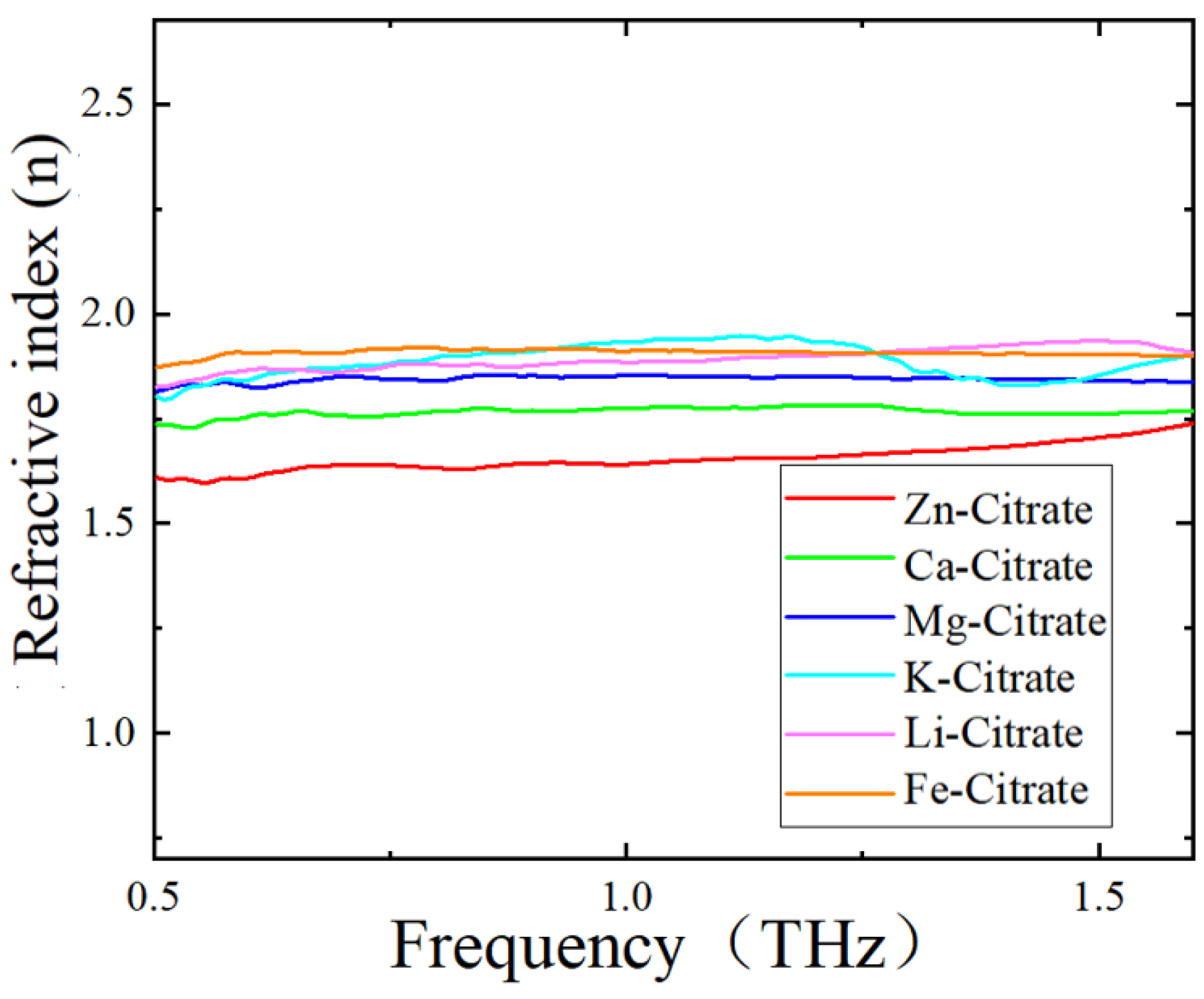
| Zn-Citrate | Ca-Citrate | Mg-Citrate | K-Citrate | Li-Citrate | Fe-Citrate |
|---|---|---|---|---|---|
| 1.65 ± 0.03 | 1.76 ± 0.01 | 1.84 ± 0.07 | 1.88 ± 0.03 | 1.89 ± 0.01 | 1.91 ± 0.06 |
Publisher’s Note: MDPI stays neutral with regard to jurisdictional claims in published maps and institutional affiliations. |
© 2022 by the authors. Licensee MDPI, Basel, Switzerland. This article is an open access article distributed under the terms and conditions of the Creative Commons Attribution (CC BY) license (https://creativecommons.org/licenses/by/4.0/).
Share and Cite
Deng, X.; Shen, Y.; Liu, B.; Song, Z.; He, X.; Zhang, Q.; Ling, D.; Liu, D.; Wei, D. Terahertz Metamaterial Sensor for Sensitive Detection of Citrate Salt Solutions. Biosensors 2022, 12, 408. https://doi.org/10.3390/bios12060408
Deng X, Shen Y, Liu B, Song Z, He X, Zhang Q, Ling D, Liu D, Wei D. Terahertz Metamaterial Sensor for Sensitive Detection of Citrate Salt Solutions. Biosensors. 2022; 12(6):408. https://doi.org/10.3390/bios12060408
Chicago/Turabian StyleDeng, Xinxin, Yanchun Shen, Bingwei Liu, Ziyu Song, Xiaoyong He, Qinnan Zhang, Dongxiong Ling, Dongfeng Liu, and Dongshan Wei. 2022. "Terahertz Metamaterial Sensor for Sensitive Detection of Citrate Salt Solutions" Biosensors 12, no. 6: 408. https://doi.org/10.3390/bios12060408