Current Advancements and Future Road Map to Develop ASSURED Microfluidic Biosensors for Infectious and Non-Infectious Diseases


Abstract
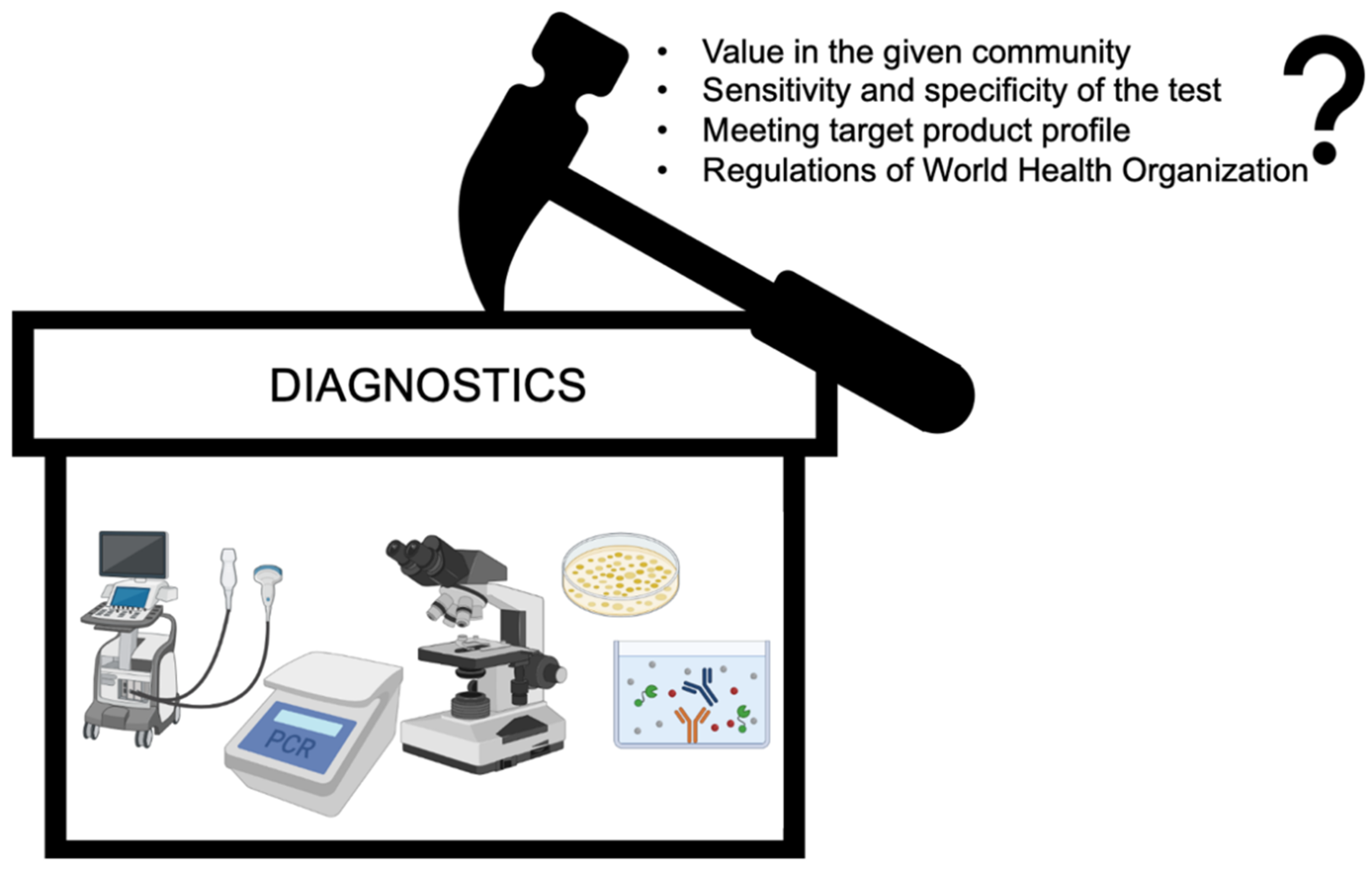
:1. Introduction
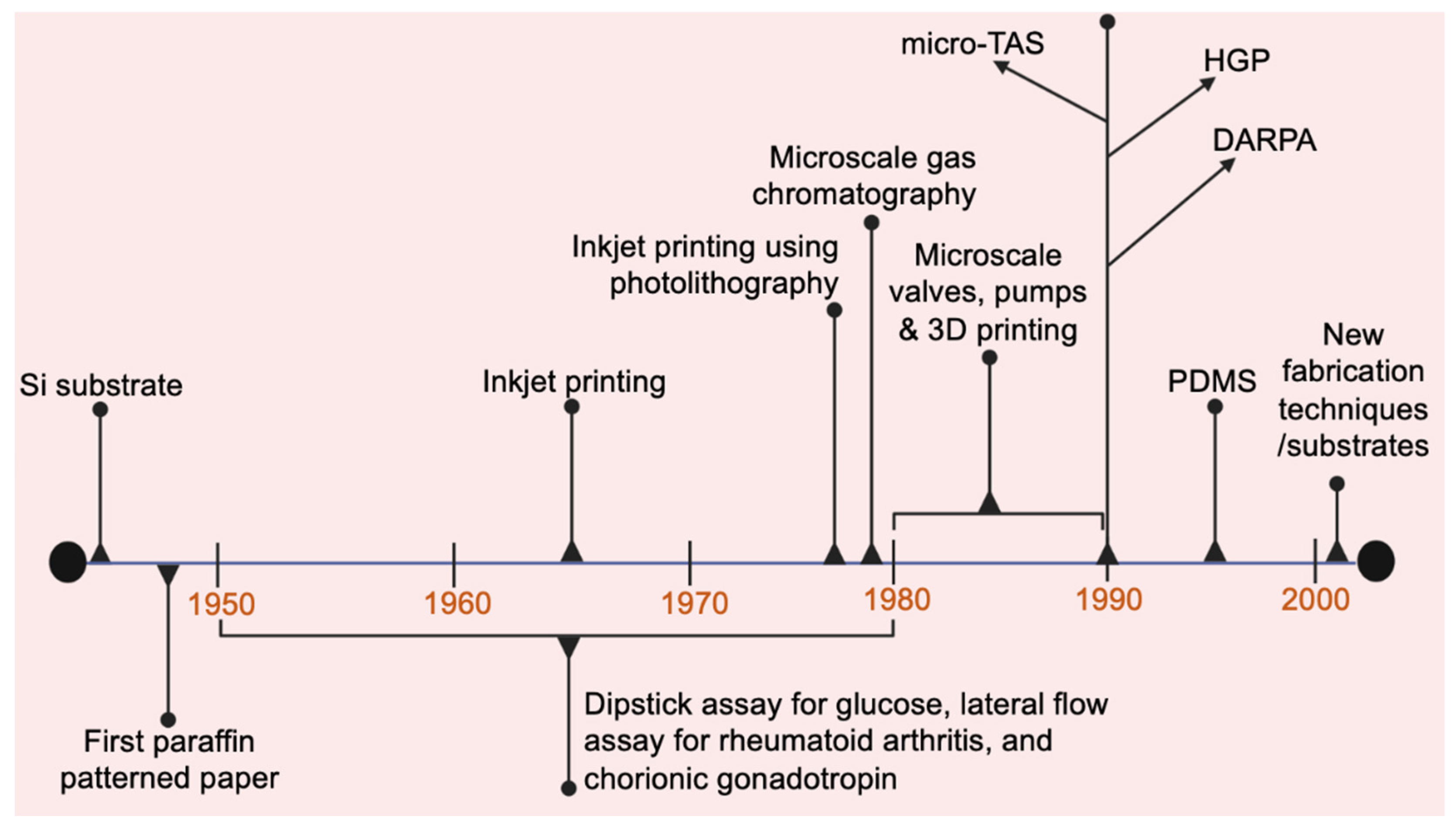
2. Brief History of Microfluidics
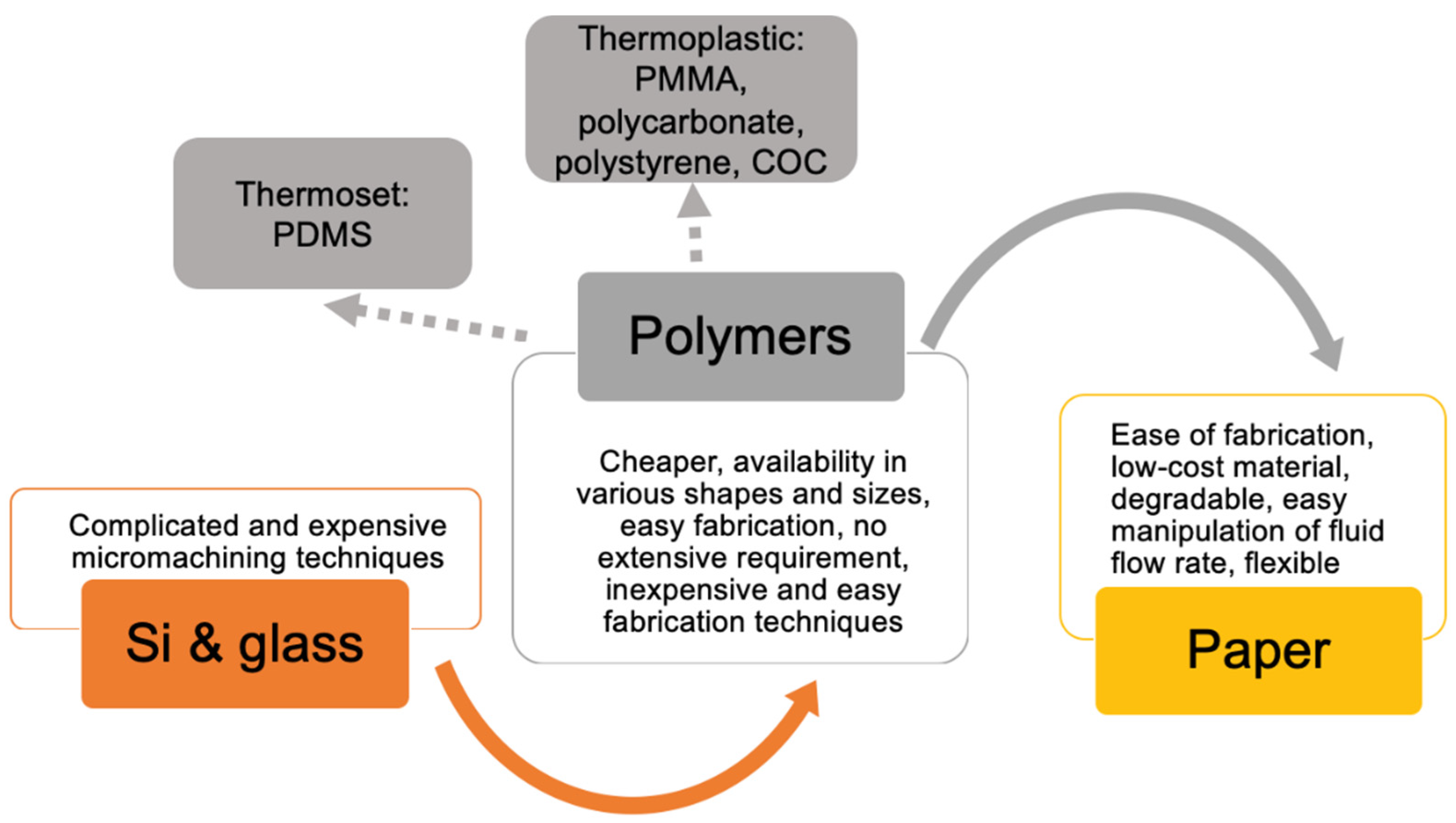
3. Current Practices in the Selection of Materials for Microfluidic Devices
4. Microfluidic Chip Biosensors for Diagnosis of Infectious Diseases
4.1. Malaria
4.1.1. Immunoassay-Based Microfluidic Chip Biosensors
4.1.2. Nucleic Acid-Based Microfluidic Chip Biosensors
4.2. Sepsis
4.2.1. Immunoassay-Based Microfluidic Chip Biosensors
4.2.2. Nucleic Acid-Based Microfluidic Chip Biosensors
4.3. AIDS
4.3.1. Immunoassay-Based Microfluidic Chip Biosensors
4.3.2. Nucleic Acid-Based Microfluidic Chip Biosensors
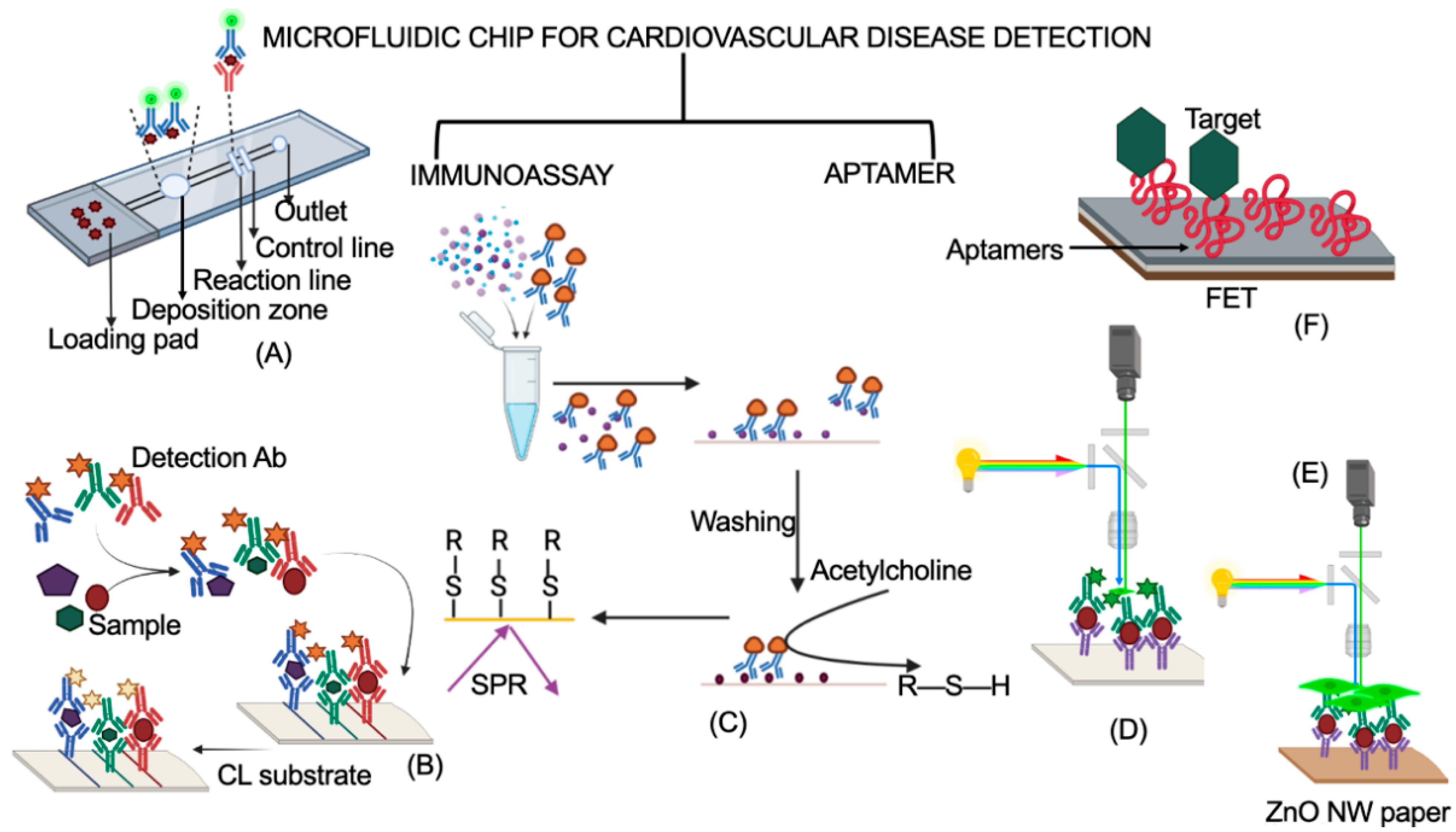
5. Microfluidic Chips for Diagnosis of Non-Infectious Diseases
5.1. Immunoassay-Based Microfluidic Chip Biosensors
5.2. Aptamer-Based Microfluidic Chip Biosensors
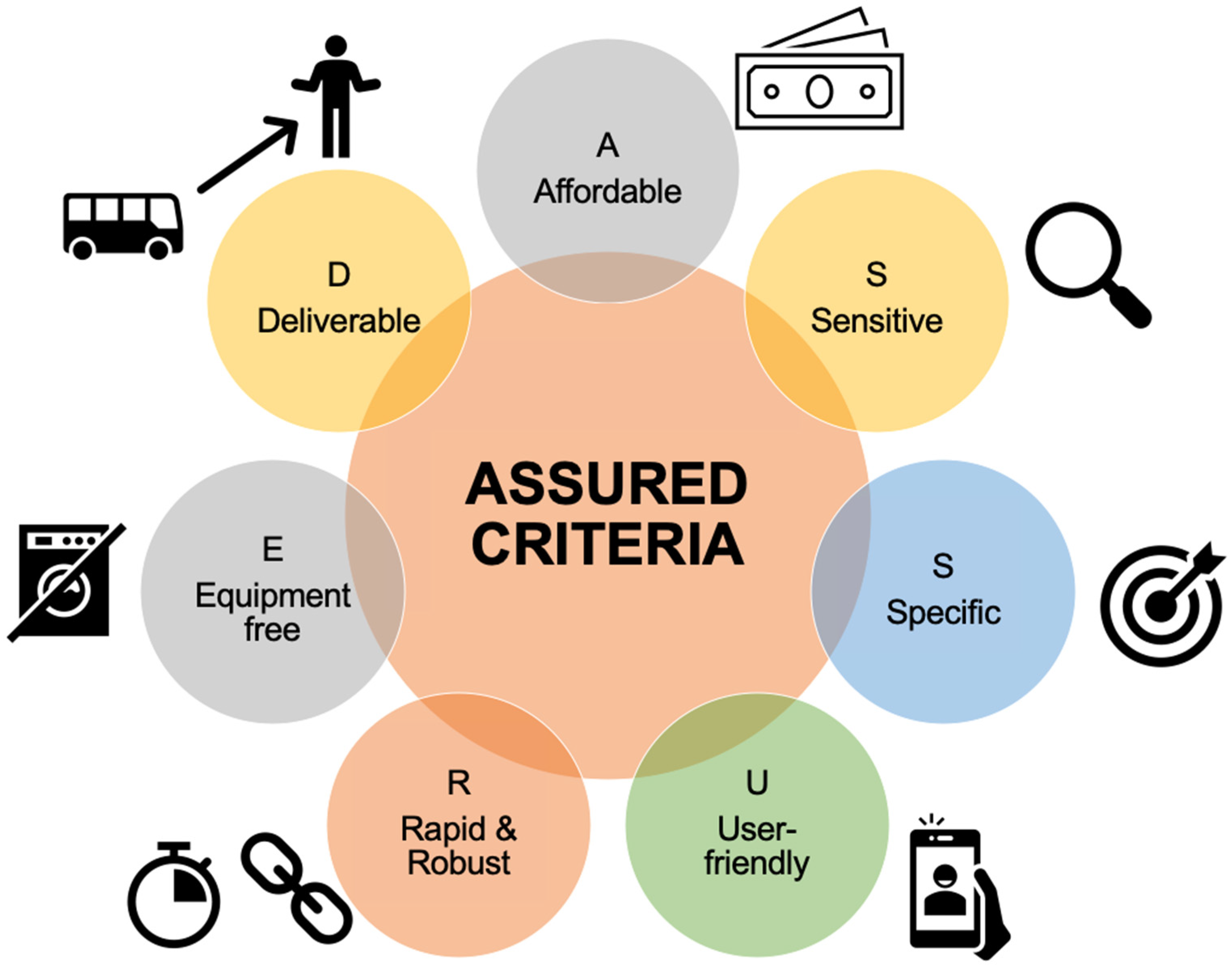
6. Future Roadmap
7. Conclusions
Author Contributions
Funding
Institutional Review Board Statement
Informed Consent Statement
Data Availability Statement
Conflicts of Interest
References
- Martinez, A.W.; Phillips, S.T.; Whitesides, G.M.; Carrilho, E. Diagnostics for the Developing World: Microfluidic Paper-Based Analytical Devices. Anal. Chem. 2010, 82, 3–10. [Google Scholar] [CrossRef] [PubMed]
- Srivastava, S.; Singh, P.K.; Vatsalya, V.; Karch, R.C. Developments in the Diagnostic Techniques of Infectious Diseases: Rural and Urban Prospective. Adv. Infect. Dis. 2018, 8, 121–138. [Google Scholar] [CrossRef] [PubMed] [Green Version]
- Kaur, H.; Chaterjee, B.; Bruno, J.G.; Kumar Sharma, T. Defining Target Product Profiles (TPPs) for Aptamer-Based Diagnostics; Springer Nature: Cham, Switzerland, 2019. [Google Scholar]
- Tay, A.; Pavesi, A.; Yazdi, S.R.; Lim, C.T.; Warkiani, M.E. Advances in Microfluidics in Combating Infectious Diseases. Biotechnol. Adv. 2016, 34, 404–421. [Google Scholar] [CrossRef] [PubMed]
- Mark, D.; Haeberle, S.; Roth, G.; Von Stetten, F.; Zengerle, R. Microfluidic Lab-on-a-Chip Platforms: Requirements, Characteristics and Applications. Chem. Soc. Rev. 2010, 39, 1153–1182. [Google Scholar] [CrossRef] [Green Version]
- Fiorini, G.S.; Chiu, D.T. Disposable Microfluidic Devices: Fabrication, Function, and Application. BioTechniques 2005, 446, 429–446. [Google Scholar] [CrossRef] [Green Version]
- Bhardwaj, T. Review on Biosensor Technologies. Int. J. Adv. Res. Eng. Technol. 2015, 6, 36–62. [Google Scholar]
- Taylor, B.J.; Howell, A.; Martin, K.A.; Manage, D.P.; Gordy, W.; Campbell, S.D.; Lam, S.; Jin, A.; Polley, S.D.; Samuel, R.A.; et al. A Lab-on-Chip for Malaria Diagnosis and Surveillance. Malar. J. 2014, 13, 179. [Google Scholar] [CrossRef] [Green Version]
- Caliendo, A.M.; Gilbert, D.N.; Ginocchio, C.C.; Hanson, K.E.; May, L.; Quinn, T.C.; Tenover, F.C.; Alland, D.; Blaschke, A.J.; Bonomo, R.A.; et al. Better Tests, Better Care: Improved Diagnostics for Infectious Diseases. Clin. Infect. Dis. 2013, 57 (Suppl. S3), S139–S170. [Google Scholar] [CrossRef] [Green Version]
- Wang, C.; Liu, M.; Wang, Z.; Li, S.; Deng, Y.; He, N. Point-of-Care Diagnostics for Infectious Diseases: From Methods to Devices. Nano Today 2021, 37, 101092. [Google Scholar] [CrossRef]
- Hocking, L.; George, J.; Broberg, E.K.; Struelens, M.J.; Leitmeyer, K.C.; Deshpande, A.; Parkinson, S.; Francombe, J.; Morley, K.I.; de Carvalho Gomes, H. Point of Care Testing for Infectious Disease in Europe: A Scoping Review and Survey Study. Front. Public Health 2021, 9, 722943. [Google Scholar] [CrossRef]
- Malcolm, S.; Cadet, J.; Crompton, L.; DeGennaro, V. A Model for Point of Care Testing for Non-Communicable Disease Diagnosis in Resource-Limited Countries. Glob. Health Epidemiol. Genom. 2019, 4, e7. [Google Scholar] [CrossRef] [PubMed] [Green Version]
- Hu, J.; Cui, X.; Gong, Y.; Xu, X.; Gao, B.; Wen, T.; Lu, T.J.; Xu, F. Portable Microfluidic and Smartphone-Based Devices for Monitoring of Cardiovascular Diseases at the Point of Care. Biotechnol. Adv. 2016, 34, 305–320. [Google Scholar] [CrossRef] [PubMed]
- Ma, Q.; Ma, H.; Xu, F.; Wang, X.; Sun, W. Microfluidics in Cardiovascular Disease Research: State of the Art and Future Outlook. Microsyst. Nanoeng. 2021, 7, 19. [Google Scholar] [CrossRef]
- Mohammed, M.I.; Desmulliez, M.P.Y. Lab-on-a-Chip Based Immunosensor Principles and Technologies for the Detection of Cardiac Biomarkers: A Review. Lab Chip 2011, 11, 569–595. [Google Scholar] [CrossRef] [PubMed]
- Convery, N.; Gadegaard, N. 30 Years of Microfluidics. Micro Nano Eng. 2019, 2, 76–91. [Google Scholar] [CrossRef]
- Warner, J. Microelectronics: Its Unusual Origin and Personality. IEEE Trans. Electron Devices 2001, 48, 2457–2467. [Google Scholar] [CrossRef]
- Lei, K.F. Introduction: The Origin, Current Status, and Future of Microfluidics. In Microfluidics: Fundamental, Devices and Applications; Wiley: Hoboken, NJ, USA, 2018; pp. 1–18. [Google Scholar]
- Kyrylchuk, A. Microfluidics—A Review. J. Micromech. Microeng. 1993, 3, 168. [Google Scholar]
- Sweet, R.G. High Frequency Recording with Electrostatically Deflected Ink Jets. Rev. Sci. Instrum. 1965, 36, 131–136. [Google Scholar] [CrossRef]
- Bassous, E.; Taub, H.H.; Kuhn, L. Ink Jet Printing Nozzle Arrays Etched in Silicon. Appl. Phys. Lett. 1977, 31, 135–137. [Google Scholar] [CrossRef]
- Whitesides, G.M. The Origins and the Future of Microfluidics. Nature 2006, 442, 368–373. [Google Scholar] [CrossRef]
- Terry, S.; Jerman, J.H.; Angell, J.B. A Gas Chromatographic Air Analyzer Fabricated on a Silicon Wafer. IEEE Trans. Electron Devices 1979, 26, 1880–1886. [Google Scholar] [CrossRef]
- Manz, A.; Graber, N.; Widmer, H.M. Miniaturized Total Chemical Analysis Systems: A Novel Concept for Chemical Sensing. Sensors Actuators B. Chem. 1990, 1, 244–248. [Google Scholar] [CrossRef]
- Collins, F.S.; Morgan, M.; Patrinos, A. The Human Genome Project: Lessons from Large-Scale Biology. Science 2003, 300, 286–290. [Google Scholar] [CrossRef] [PubMed] [Green Version]
- Swerdlow, H.; Gesteland, R. Capillary Gel Electrophoresis for Rapid, High Resolution DNA Sequencing. Nucleic Acids Res. 1990, 18, 1415–1419. [Google Scholar] [CrossRef] [PubMed] [Green Version]
- Northrup, M.A.; Ching, M.T.; White, R.M.; Watson, R.T. DNA Amplification with a Microfabricated Reaction Chamber. In Proceedings of the 7th International Conference on Solid-State Sensors and Actuators, Yokohama, Japan, 7–10 June 1993; pp. 925–926. [Google Scholar]
- Müller, R.H.; Clegg, D.L. Automatic Paper Chromatography. Ann. N. Y. Acad. Sci. 1951, 53, 1108–1118. [Google Scholar] [CrossRef]
- Lee, J.H. Paper-Based Medical Diagnostic Devices: As a Part of Bioanalysis-Advanced Materials, Methods, and Device; Springer Nature: Berlin, Germany, 2020. [Google Scholar]
- Dabbagh, S.R.; Becher, E.; Ghaderinezhad, F.; Havlucu, H.; Ozcan, O.; Ozkan, M.; Yetisen, A.K.; Tasoglu, S. Increasing the Packing Density of Assays in Paper-Based Microfluidic Devices. Biomicrofluidics 2021, 15, 40–42. [Google Scholar] [CrossRef]
- Barry, R.; Ivanov, D. Microfluidics in Biotechnology. J. Nanobiotechnol. 2004, 2, 2. [Google Scholar] [CrossRef] [Green Version]
- Fallahi, H.; Zhang, J.; Phan, H.P.; Nguyen, N.T. Flexible Microfluidics: Fundamentals, Recent Developments, and Applications. Micromachines 2019, 10, 830. [Google Scholar] [CrossRef] [Green Version]
- Beebe, D.J.; Mensing, G.A.; Walker, G.M. Physics and Applications of Microfluidics in Biology. Annu. Rev. Biomed. Eng. 2002, 4, 261–286. [Google Scholar] [CrossRef]
- Roberts, M.A.; Rossier, J.S.; Bercier, P.; Girault, H. UV Laser Machined Polymer Substrates for the Development of Microdiagnostic Systems. Anal. Chem. 1997, 69, 2035–2042. [Google Scholar] [CrossRef]
- Soper, S.A.; Ford, S.M.; Qi, S.; McCarley, R.L.; Kelly, K.; Murphy, M.C. Peer Reviewed: Polymeric Microelectromechanical Systems. Anal. Chem. 2000, 72, 642 A–651 A. [Google Scholar] [CrossRef] [PubMed]
- Ansari, M.I.H.; Hassan, S.; Qurashi, A.; Khanday, F.A. Microfluidic-Integrated DNA Nanobiosensors. Biosens. Bioelectron. 2016, 85, 247–260. [Google Scholar] [CrossRef] [PubMed]
- Attia, U.M.; Marson, S.; Alcock, J.R. Micro-Injection Moulding of Polymer Microfluidic Devices. Microfluid. Nanofluid. 2009, 7, 1–28. [Google Scholar] [CrossRef] [Green Version]
- Greener, J.; Li, W.; Ren, J.; Voicu, D.; Pakharenko, V.; Tang, T.; Kumacheva, E. Rapid, Cost-Efficient Fabrication of Microfluidic Reactors in Thermoplastic Polymers by Combining Photolithography and Hot Embossing. Lab Chip 2010, 10, 522–524. [Google Scholar] [CrossRef] [PubMed]
- Guckenberger, D.J.; De Groot, T.E.; Wan, A.M.D.; Beebe, D.J.; Young, E.W.K. Micromilling: A Method for Ultra-Rapid Prototyping of Plastic Microfluidic Devices. Lab Chip 2015, 15, 2364–2378. [Google Scholar] [CrossRef] [Green Version]
- Hong, T.F.; Ju, W.J.; Wu, M.C.; Tai, C.H.; Tsai, C.H.; Fu, L.M. Rapid Prototyping of PMMA Microfluidic Chips Utilizing a CO2 Laser. Microfluid. Nanofluid. 2010, 9, 1125–1133. [Google Scholar] [CrossRef]
- Friend, J.; Yeo, L. Fabrication of Microfluidic Devices Using Polydimethylsiloxane. Biomicrofluidics 2010, 4, 1–5. [Google Scholar] [CrossRef] [Green Version]
- Giannitsis, A.T. Microfabrication of Biomedical Lab-on-Chip Devices. A Review. Est. J. Eng. 2011, 17, 109–139. [Google Scholar] [CrossRef] [Green Version]
- Gervais, L.; De Rooij, N.; Delamarche, E. Microfluidic Chips for Point-of-Care Immunodiagnostics. Adv. Mater. 2011, 23, H151–H176. [Google Scholar] [CrossRef]
- Faustino, V.; Catarino, S.O.; Lima, R.; Minas, G. Biomedical Microfluidic Devices by Using Low-Cost Fabrication Techniques: A Review. J. Biomech. 2016, 49, 2280–2292. [Google Scholar] [CrossRef] [Green Version]
- Jigar Panchal, H.; Kent, N.J.; Knox, A.J.S.; Harris, L.F. Microfluidics in Haemostasis: A Review. Molecules 2020, 25, 833. [Google Scholar] [CrossRef] [PubMed] [Green Version]
- Bernard, A.; Michel, B.; Delamarche, E. Micromosaic Immunoassays. Anal. Chem. 2001, 73, 8–12. [Google Scholar] [CrossRef] [PubMed]
- Chou, H.P.; Spence, C.; Scherer, A.; Quake, S. A Microfabricated Device for Sizing and Sorting DNA Molecules. Proc. Natl. Acad. Sci. USA 1999, 96, 11–13. [Google Scholar] [CrossRef] [PubMed] [Green Version]
- Ismagilov, R.F.; Ng, J.M.K.; Kenis, P.J.A.; Whitesides, G.M. Microfluidic Arrays of Fluid-Fluid Diffusional Contacts as Detection Elements and Combinatorial Tools. Anal. Chem. 2001, 73, 5207–5213. [Google Scholar] [CrossRef]
- DeBusschere, B.D.; Kovacs, G.T.A. Portable Cell-Based Biosensor System Using Integrated CMOS Cell-Cartridges. Biosens. Bioelectron. 2001, 16, 543–556. [Google Scholar] [CrossRef]
- Toepke, M.W.; Beebe, D.J. PDMS Absorption of Small Molecules and Consequences in Microfluidic Applications. Lab Chip 2006, 6, 1484–1486. [Google Scholar] [CrossRef]
- Li, P.C.H. Microfluidic Lab-on-a-Chip for Chemical and Biological Analysis and Discovery; CRC Press: Boca Raton, FL, USA, 2006. [Google Scholar]
- Vullev, V.I.; Wan, J.; Heinrich, V.; Landsman, P.; Bower, P.E.; Xia, B.; Millare, B.; Jones, G. Nonlithographic Fabrication of Microfluidic Devices. J. Am. Chem. Soc. 2006, 128, 16062–16072. [Google Scholar] [CrossRef]
- Sia, S.K.; Whitesides, G.M. Microfluidic Devices Fabricated in Poly(Dimethylsiloxane) for Biological Studies. Electrophoresis 2003, 24, 3563–3576. [Google Scholar] [CrossRef]
- Bhattacharya, S.; Kumar, S.; Agarwal, A. Paper Microfluidics—Theory and Applications; Springer: Singapore, 2019; ISBN 9789811610929. [Google Scholar]
- Chami Khazraji, A.; Robert, S. Self-Assembly and Intermolecular Forces When Cellulose and Water Interact Using Molecular Modeling. J. Nanomater. 2013, 10, 745979. [Google Scholar] [CrossRef]
- Quinn, M.J. Wildlife Toxicity Assessments for Chemicals of Military Concern; Williams, M.A., Reddy, G., Quinn, M.J., Johnson, M.S., Eds.; Elsevier: Amsterdam, The Netherlands, 2015. [Google Scholar]
- Mahoney, E.; Kun, J.; Smieja, M.; Fang, Q. Review—Point-of-Care Urinalysis with Emerging Sensing and Imaging Technologies. J. Electrochem. Soc. 2020, 167, 037518. [Google Scholar] [CrossRef]
- Smith, S.; Korvink, J.G.; Mager, D.; Land, K. The Potential of Paper-Based Diagnostics to Meet the ASSURED Criteria. RSC Adv. 2018, 8, 34012–34034. [Google Scholar] [CrossRef] [PubMed] [Green Version]
- Anand, A.; Chatterjee, B.; Dhiman, A.; Goel, R.; Khan, E.; Malhotra, A.; Santra, V.; Salvi, N.; Khadilkar, M.V.; Bhatnagar, I.; et al. Complex Target SELEX-Based Identification of DNA Aptamers against Bungarus Caeruleus Venom for the Detection of Envenomation Using a Paper-Based Device. Biosens. Bioelectron. 2021, 193, 113523. [Google Scholar] [CrossRef] [PubMed]
- Zhang, A.L.; Zha, Y. Fabrication of Paper-Based Microfluidic Device Using Printed Circuit Technology. AIP Adv. 2012, 2, 022171. [Google Scholar] [CrossRef] [Green Version]
- Khan, S.M.; Nassar, J.M.; Hussain, M.M. Paper as a Substrate and an Active Material in Paper Electronics. ACS Appl. Electron. Mater. 2021, 3, 30–52. [Google Scholar] [CrossRef]
- Park, J.; Shin, P.; Kim, J.; Park, H.; Baek, Y.; DeVries, L. Evaluation of Interfacial and Micro-Damage Sensing of Composites via Pencil Lead Drawing Paper Sensor (PLDPS) and Electrical Resistance (ER) Mapping. In Proceedings of the Sensors and Smart Structures Technologies for Civil, Mechanical, and Aerospace Systems, Denver, CO, USA, 4–7 March 2019; pp. 357–363. [Google Scholar]
- Kim, Y.S.; Yang, Y.; Henry, C.S. Laminated and Infused Parafilm®—Paper for Paper-Based Analytical Devices. Sens. Actuators B Chem. 2018, 255, 3654–3661. [Google Scholar] [CrossRef]
- Ramana, L.N.; Mathapati, S.S.; Salvi, N.; Khadilkar, M.V.; Malhotra, A.; Santra, V.; Sharma, T.K. A Paper Microfluidic Device Based Colorimetric Sensor for the Detection and Discrimination of Elapid: Versus Viper Envenomation. Analyst 2022, 147, 685–694. [Google Scholar] [CrossRef]
- Nishat, S.; Jafry, A.T.; Martinez, A.W.; Awan, F.R. Paper-Based Microfluidics: Simplified Fabrication and Assay Methods. Sens. Actuators B Chem. 2021, 336, 129681. [Google Scholar] [CrossRef]
- Zargaryan, A.; Farhoudi, N.; Haworth, G.; Ashby, J.F.; Au, S.H. Hybrid 3D Printed-Paper Microfluidics. Sci. Rep. 2020, 10, 18379. [Google Scholar] [CrossRef]
- Asano, H.; Shiraishi, Y. Development of Paper-Based Microfluidic Analytical Device for Iron Assay Using Photomask Printed with 3D Printer for Fabrication of Hydrophilic and Hydrophobic Zones on Paper by Photolithography. Anal. Chim. Acta 2015, 883, 55–60. [Google Scholar] [CrossRef]
- Li, X.; Tian, J.; Nguyen, T.; Shen, W. Paper-Based Microfluidic Devices by Plasma Treatment. Anal. Chem. 2008, 80, 9131–9134. [Google Scholar] [CrossRef]
- Haller, P.D.; Flowers, C.A.; Gupta, M. Three-Dimensional Patterning of Porous Materials Using Vapor Phase Polymerization. Soft Matter 2011, 7, 2428–2432. [Google Scholar] [CrossRef]
- Mathaweesansurn, A.; Thongrod, S.; Khongkaew, P.; Phechkrajang, C.M.; Wilairat, P.; Choengchan, N. Simple and Fast Fabrication of Microfluidic Paper-Based Analytical Device by Contact Stamping for Multiple-Point Standard Addition Assay: Application to Direct Analysis of Urinary Creatinine. Talanta 2020, 210, 120675. [Google Scholar] [CrossRef] [PubMed]
- Noviana, E.; Ozer, T.; Carrell, C.S.; Link, J.S.; McMahon, C.; Jang, I.; Henry, C.S. Microfluidic Paper-Based Analytical Devices: From Design to Applications. Chem. Rev. 2021, 121, 11835–11885. [Google Scholar] [CrossRef]
- Trieu, P.T.; Lee, N.Y. Paper-Based All-in-One Origami Microdevice for Nucleic Acid Amplification Testing for Rapid Colorimetric Identification of Live Cells for Point-of-Care Testing. Anal. Chem. 2019, 91, 11013–11022. [Google Scholar] [CrossRef] [PubMed]
- Tseng, C.-C.; Kung, C.-T.; Chen, R.-F.; Tsai, M.-H.; Chao, H.-R.; Wang, Y.-N.; Fu, L.-M. Recent Advances in Microfluidic Paper-Based Assay Devices for Diagnosis of Human Diseases Using Saliva, Tears and Sweat Samples. Sens. Actuators B Chem. 2021, 342, 130078. [Google Scholar] [CrossRef]
- Yang, X.; Forouzan, O.; Brown, T.P.; Shevkoplyas, S.S. Integrated Separation of Blood Plasma from Whole Blood for Microfluidic Paper-Based Analytical Devices. Lab Chip 2012, 12, 274–280. [Google Scholar] [CrossRef]
- Park, C.; Kim, H.R.; Kim, S.K.; Jeong, I.K.; Pyun, J.C.; Park, S. Three-Dimensional Paper-Based Microfluidic Analytical Devices Integrated with a Plasma Separation Membrane for the Detection of Biomarkers in Whole Blood. ACS Appl. Mater. Interfaces 2019, 11, 36428–36434. [Google Scholar] [CrossRef]
- Bandara, G.C.; Unitan, L.J.; Kremer, M.H.; Shellhammer, O.T.; Bracha, S.; Remcho, V.T. Wicking Microfluidic Approach to Separate Blood Plasma from Whole Blood to Facilitate Downstream Assays. Anal. Bioanal. Chem. 2021, 413, 4511–4520. [Google Scholar] [CrossRef]
- Ren, K.; Zhou, J.; Wu, H. Materials for Microfluidic Chip Fabrication. Acc. Chem. Res. 2013, 46, 2396–2406. [Google Scholar] [CrossRef]
- Buckner, C.A.; Lafrenie, R.M.; Dénommée, J.A.; Caswell, J.M.; Want, D.A.; Gan, G.G.; Leong, Y.C.; Bee, P.C.; Chin, E.; Teh, A.K.H.; et al. Overview of Materials for Microfluidic Applications; IntechOpen: London, UK, 2016; Volume 11, p. 13. ISBN 0000957720. [Google Scholar]
- Conzone, S.D.; Pantano, C.G. Glass Slides to DNA Microarrays. Mater. Today 2004, 7, 20–26. [Google Scholar] [CrossRef]
- Luka, G.; Ahmadi, A.; Najjaran, H.; Alocilja, E.; Derosa, M.; Wolthers, K.; Malki, A.; Aziz, H.; Althani, A.; Hoorfar, M. Microfluidics Integrated Biosensors: A Leading Technology towards Lab-on-A-Chip and Sensing Applications. Sensors 2015, 15, 30011–30031. [Google Scholar] [CrossRef] [PubMed] [Green Version]
- Saxena, S.; Joshi, R. Microfluidic Devices: Applications and Role of Surface Wettability in Its Fabrication; Intech: London, UK, 2016; Volume i, p. 13. [Google Scholar]
- Bhardwaj, T.; Dalal, P.; Rathore, A.S.; Jha, S.K. An Aptamer Based Microfluidic Chip for Impedimetric Detection of Ranibizumab in a Bioreactor. Sens. Actuators B Chem. 2020, 312, 127941. [Google Scholar] [CrossRef]
- Lee, W.S.; Kang, T.; Kwak, K.J.; Park, K.; Yi, S.Y.; Lee, U.J. Simple, Rapid, and Accurate Malaria Diagnostic Platform Using Microfluidic—Based Immunoassay of Plasmodium Falciparum Lactate Dehydrogenase. Nano Converg. 2020, 134, 333–340. [Google Scholar] [CrossRef] [PubMed] [Green Version]
- Ruiz-Vega, G.; Arias-Alpízar, K.; de la Serna, E.; Borgheti-Cardoso, L.N.; Sulleiro, E.; Molina, I.; Fernàndez-Busquets, X.; Sánchez-Montalvá, A.; del Campo, F.J.; Baldrich, E. Electrochemical POC Device for Fast Malaria Quantitative Diagnosis in Whole Blood by Using Magnetic Beads, Poly-HRP and Microfluidic Paper Electrodes. Biosens. Bioelectron. 2020, 150, 111925. [Google Scholar] [CrossRef] [PubMed]
- Xu, G.; Nolder, D.; Reboud, J.; Oguike, M.C.; van Schalkwyk, D.A.; Sutherland, C.J.; Cooper, J.M. Paper-Origami-Based Multiplexed Malaria Diagnostics from Whole Blood. Angew. Chem. 2016, 128, 15476–15479. [Google Scholar] [CrossRef] [Green Version]
- Wang, S.Q.; Inci, F.; Chaunzwa, T.L.; Ramanujam, A.; Vasudevan, A.; Subramanian, S.; Fai Ip, A.C.; Sridharan, B.; Gurkan, U.A.; Demirci, U. Portable Microfluidic Chip for Detection of Escherichia Coli in Produce and Blood. Int. J. Nanomed. 2012, 7, 2591–2600. [Google Scholar] [CrossRef] [Green Version]
- Giannetti, A.; Trono, C.; Porro, G.; Domenici, C.; Puntoni, M.; Baldini, F. Towards an Integrated System as Point-of-Care Device for the Optical Detection of Sepsis Biomarkers. Chemosensors 2020, 8, 12. [Google Scholar] [CrossRef] [Green Version]
- Liu, D.; Li, X.; Zhou, J.; Liu, S.; Tian, T.; Song, Y.; Zhu, Z.; Zhou, L.; Ji, T.; Yang, C. A Fully Integrated Distance Readout ELISA-Chip for Point-of-Care Testing with Sample-in-Answer-out Capability. Biosens. Bioelectron. 2017, 96, 332–338. [Google Scholar] [CrossRef]
- Verma, M.S.; Tsaloglou, M.N.; Sisley, T.; Christodouleas, D.; Chen, A.; Milette, J.; Whitesides, G.M. Sliding-Strip Microfluidic Device Enables ELISA on Paper. Biosens. Bioelectron. 2018, 99, 77–84. [Google Scholar] [CrossRef]
- Patterson, A.S.; Heithoff, D.M.; Ferguson, B.S.; Tom Soh, H.; Mahan, M.J.; Plaxco, K.W. Microfluidic Chip-Based Detection and Intraspecies Strain Discrimination of Salmonella Serovars Derived from Whole Blood of Septic Mice. Appl. Environ. Microbiol. 2013, 79, 2302–2311. [Google Scholar] [CrossRef] [Green Version]
- Fang, Y.L.; Wang, C.H.; Chen, Y.S.; Chien, C.C.; Kuo, F.C.; You, H.L.; Lee, M.S.; Lee, G. Bin An Integrated Microfluidic System for Early Detection of Sepsis-Inducing Bacteria. Lab Chip 2021, 21, 113–121. [Google Scholar] [CrossRef] [PubMed]
- Sia, S.K.; Linder, V.; Parviz, B.A.; Siegel, A.; Whitesides, G.M. An Integrated Approach to a Portable and Low-Cost Immunoassay for Resource-Poor Settings. Angew. Chem. Int. Ed. 2004, 43, 498–502. [Google Scholar] [CrossRef] [PubMed]
- Klostranec, J.M.; Xiang, Q.; Farcas, G.A.; Lee, J.A.; Rhee, A.; Lafferty, E.I.; Perrault, S.D.; Kain, K.C.; Chan, W.C.W. Convergence of Quantum Dot Barcodes with Microfluidics and Signal Processing for Multiplexed High-Throughput Infectious Disease Diagnostics. Nano Lett. 2007, 7, 2812–2818. [Google Scholar] [CrossRef] [PubMed]
- Cheng, C.M.; Martinez, A.W.; Gong, J.; Mace, C.R.; Phillips, S.T.; Carrilho, E.; Mirka, K.A.; Whitesides, G.M. Paper-Based Elisa. Angew. Chem. Int. Ed. 2010, 49, 4771–4774. [Google Scholar] [CrossRef]
- Phillips, E.A.; Moehling, T.J.; Ejendal, K.F.K.; Hoilett, O.S.; Byers, K.M.; Basing, L.A.; Jankowski, L.A.; Bennett, J.B.; Lin, L.K.; Stanciu, L.A.; et al. Microfluidic Rapid and Autonomous Analytical Device (MicroRAAD) to Detect HIV from Whole Blood Samples. Lab Chip 2019, 19, 3375–3386. [Google Scholar] [CrossRef] [Green Version]
- Rohrman, B.A.; Richards-Kortum, R.R. A Paper and Plastic Device for Performing Recombinase Polymerase Amplification of HIV DNA. Lab Chip 2012, 12, 3082–3088. [Google Scholar] [CrossRef]
- Gervais, L.; Delamarche, E. Toward One-Step Point-of-Care Immunodiagnostics Using Capillary-Driven Microfluidics and PDMS Substrates. Lab Chip 2009, 9, 3330–3337. [Google Scholar] [CrossRef]
- Kurita, R.; Yokota, Y.; Sato, Y.; Mizutani, F.; Niwa, O. On-Chip Enzyme Immunoassay of a Cardiac Marker Using a Microfluidic Device Combined with a Portable Surface Plasmon Resonance System. Anal. Chem. 2006, 78, 5525–5531. [Google Scholar] [CrossRef]
- Yin, B.; Wan, X.; Qian, C.; Sohan, A.S.M.M.F.; Wang, S.; Zhou, T. Point-of-Care Testing for Multiple Cardiac Markers Based on a Snail-Shaped Microfluidic Chip. Front. Chem. 2021, 9, 741058. [Google Scholar] [CrossRef]
- Mohammed, M.I.; Desmulliez, M.P.Y. Autonomous Capillary Microfluidic System with Embedded Optics for Improved Troponin I Cardiac Biomarker Detection. Biosens. Bioelectron. 2014, 61, 478–484. [Google Scholar] [CrossRef]
- Guo, X.; Zong, L.; Jiao, Y.; Han, Y.; Zhang, X.; Xu, J.; Li, L.; Zhang, C.W.; Liu, Z.; Ju, Q.; et al. Signal-Enhanced Detection of Multiplexed Cardiac Biomarkers by a Paper-Based Fluorogenic Immunodevice Integrated with Zinc Oxide Nanowires. Anal. Chem. 2019, 91, 9300–9307. [Google Scholar] [CrossRef] [PubMed]
- Sinha, A.; Tai, S.-Y.; Li, K.-H.; Gopinathan, P.; Chung, Y.-D.; Sarangadharan, I.; Ma, H.-P.; Huang, P.-C.; Shiesh, S.-C.; Wang, Y.-L.; et al. An Integrated Microfluidic System with Field-Effect-Transistor Sensor Arrays for Detecting Multiple Cardiovascular Biomarkers from Clinical Samples. Biosens. Bioelectron. 2019, 129, 155–163. [Google Scholar] [CrossRef] [PubMed]
- World Health Organization. World Malaria Report 2021; World Health Organization: Geneva, Switzerland, 2021. [Google Scholar]
- Reboud, J.; Xu, G.; Garrett, A.; Adriko, M.; Yang, Z.; Tukahebwa, E.M.; Rowell, C.; Cooper, J.M. Paper-Based Microfluidics for DNA Diagnostics of Malaria in Low Resource Underserved Rural Communities. Proc. Natl. Acad. Sci. USA 2019, 116, 4834–4842. [Google Scholar] [CrossRef] [PubMed] [Green Version]
- Kong, T.F.; Ye, W.; Peng, W.K.; Hou, H.W.; Marcos; Preiser, P.R.; Nguyen, N.T.; Han, J. Enhancing Malaria Diagnosis through Microfluidic Cell Enrichment and Magnetic Resonance Relaxometry Detection. Sci. Rep. 2015, 5, 11425. [Google Scholar] [CrossRef] [Green Version]
- Pham, N.M.; Karlen, W.; Beck, H.P.; Delamarche, E. Malaria and the “last” Parasite: How Can Technology Help? Malar. J. 2018, 17, 260. [Google Scholar] [CrossRef] [Green Version]
- Markwalter, C.F.; Davis, K.M.; Wright, D.W. Immunomagnetic Capture and Colorimetric Detection of Malarial Biomarker Plasmodium Falciparum Lactate Dehydrogenase. Anal. Biochem. 2016, 493, 30–34. [Google Scholar] [CrossRef] [Green Version]
- Markwalter, C.F.; Ricks, K.M.; Bitting, A.L.; Mudenda, L.; Wright, D.W. Simultaneous Capture and Sequential Detection of Two Malarial Biomarkers on Magnetic Microparticles. Talanta 2016, 161, 443–449. [Google Scholar] [CrossRef] [Green Version]
- Sergeyeva, T.; Yarynka, D.; Piletska, E.; Linnik, R.; Zaporozhets, O.; Brovko, O.; Piletsky, S.; El’skaya, A. Development of a Smartphone-Based Biomimetic Sensor for Aflatoxin B1 Detection Using Molecularly Imprinted Polymer Membranes. Talanta 2019, 201, 204–210. [Google Scholar] [CrossRef]
- Kang, D.K.; Ali, M.M.; Zhang, K.; Huang, S.S.; Peterson, E.; Digman, M.A.; Gratton, E.; Zhao, W. Rapid Detection of Single Bacteria in Unprocessed Blood Using Integrated Comprehensive Droplet Digital Detection. Nat. Commun. 2014, 5, 5427. [Google Scholar] [CrossRef] [Green Version]
- Singhal, C.; Bruno, J.G.; Kaushal, A.; Sharma, T.K. Recent Advances and a Roadmap to Aptamer-Based Sensors for Bloodstream Infections. ACS Appl. Bio Mater. 2021, 4, 3962–3984. [Google Scholar] [CrossRef]
- Kaur, H.; Bruno, J.G.; Kumar, A.; Sharma, T.K. Aptamers in the Therapeutics and Diagnostics Pipelines. Theranostics 2018, 8, 4016–4032. [Google Scholar] [CrossRef] [PubMed]
- Alba-Patiño, A.; Russell, S.M.; Borges, M.; Pazos-Pérez, N.; Álvarez-Puebla, R.A.; De La Rica, R. Nanoparticle-Based Mobile Biosensors for the Rapid Detection of Sepsis Biomarkers in Whole Blood. Nanoscale Adv. 2020, 2, 1253–1260. [Google Scholar] [CrossRef] [Green Version]
- Liu, J.; Du, B.; Zhang, P.; Haleyurgirisetty, M.; Zhao, J.; Ragupathy, V.; Lee, S.; DeVoe, D.L.; Hewlett, I.K. Development of a Microchip Europium Nanoparticle Immunoassay for Sensitive Point-of-Care HIV Detection. Biosens. Bioelectron. 2014, 61, 177–183. [Google Scholar] [CrossRef] [PubMed]
- Eid, J.; Mougel, M.; Socol, M. Advances in Continuous Microfluidics-Based Technologies for the Study of HIV Infection. Viruses 2020, 12, 982. [Google Scholar] [CrossRef]
- Chen, C.A.; Yuan, H.; Chen, C.W.; Chien, Y.S.; Sheng, W.H.; Chen, C.F. An Electricity- And Instrument-Free Infectious Disease Sensor Based on a 3D Origami Paper-Based Analytical Device. Lab Chip 2021, 21, 1908–1915. [Google Scholar] [CrossRef]
- Ryu, Y.; Jin, Z.; Kang, M.S.; Kim, H.S. Increase in the Detection Sensitivity of a Lateral Flow Assay for a Cardiac Marker by Oriented Immobilization of Antibody. Biochip J. 2011, 5, 193–198. [Google Scholar] [CrossRef]
- Zhu, J.; Zou, N.; Zhu, D.; Wang, J.; Jin, Q.; Zhao, J.; Mao, H. Simultaneous Detection of High-Sensitivity Cardiac Troponin I and Myoglobin by Modified Sandwich Lateral Flow Immunoassay: Proof of Principle. Clin. Chem. 2011, 57, 1732–1738. [Google Scholar] [CrossRef]
- Gervais, L.; Hitzbleck, M.; Delamarche, E. Capillary-Driven Multiparametric Microfluidic Chips for One-Step Immunoassays. Biosens. Bioelectron. 2011, 27, 64–70. [Google Scholar] [CrossRef]
- Available online: https://www.mordorintelligence.com/industry-reports/microfluidics-market (accessed on 16 May 2022).
- Sachdeva, S.; Davis, R.W.; Saha, A.K. Microfluidic Point-of-Care Testing: Commercial Landscape and Future Directions. Front. Bioeng. Biotechnol. 2021, 8, 602659. [Google Scholar] [CrossRef]
- Available online: https://www.marketsandmarkets.com/market-reports/microfluidics-market-1305.html (accessed on 16 May 2022).
- Available online: https://www.globenewswire.com/news-release/2021/11/29/2341819/28124/en/global-microfluidics-market-report-2021-rapid-testing-and-improved-portability-through-microfluidic-chip-miniaturization.html (accessed on 16 May 2022).
- Mejía-Salazar, J.R.; Cruz, K.R.; Vásques, E.M.M.; de Oliveira, O.N. Microfluidic Point-of-Care Devices: New Trends and Future Prospects for Ehealth Diagnostics. Sensors 2020, 20, 1951. [Google Scholar] [CrossRef] [Green Version]
- Chaudhary, V.; Kaushik, A.; Furukawa, H.; Khosla, A. Review—Towards 5th Generation AI and IoT Driven Sustainable Intelligent Sensors Based on 2D MXenes and Borophene. ECS Sens. Plus 2022, 1, 013601. [Google Scholar] [CrossRef]
- Kaushik, A.; Khan, R.; Solanki, P.; Gandhi, S.; Gohel, H.; Mishra, Y.K. From Nanosystems to a Biosensing Prototype for an Efficient Diagnostic: A Special Issue in Honor of Professor Bansi D. Malhotra. Biosensors 2021, 11, 359. [Google Scholar] [CrossRef] [PubMed]
- Ayodele, O.O.; Adesina, A.O.; Pourianejad, S.; Averitt, J.; Ignatova, T. Recent Advances in Nanomaterial-Based Aptasensors in Medical Diagnosis and Therapy. Nanomaterials 2021, 11, 932. [Google Scholar] [CrossRef] [PubMed]
- Kujawska, M.; Bhardwaj, S.K.; Mishra, Y.K.; Kaushik, A. Using Graphene-Based Biosensors to Detect Dopamine for Efficient Parkinson’s Disease Diagnostics. Biosensors 2021, 11, 433. [Google Scholar] [CrossRef]


| Target | Material | Principle | Detection | LOD | ASSURED Criteria (Fulfilled or Not) | Ref. |
|---|---|---|---|---|---|---|
| Malaria | ||||||
| pfLDH | Polystyrene | Immunoassay | Chemiluminiscence | 1 pg/µL | Affordable, rapid, user-friendly, sensitivity | [83] |
| pfLDH | Paper | Immunoassay | Electrochemical (amperometric) | 300 parasites/µL | Affordable, rapid, sensitivity | [84] |
| P. falciparum and P. vivax | Plastic (cyclic olefin polymer) | Nucleic acid | Fluorescence | 2 parasites/μL | Affordable, user-friendly, portable | [8] |
| P. falciparum and P. vivax | Paper | Nucleic acid | Fluorescence | 5 parasites/µL | Affordable, user-friendly, sensitivity | [85] |
| Sepsis | ||||||
| E.coli | PMMA/glass | Immunoassay | Fluorescence imaging | 50 CFUs/mL | Low fabrication cost, user-friendly | [86] |
| CRP and NP | PMMA | Immunoassay | Fluorescence | CRP:10 μg/L NP:2.1 μg/L | Rapid, simple, user-friendly, portable | [87] |
| CRP | PMMA | Immunoassay | Colorimetric (visual analysis) | 0.01 μg/mL | Affordable, portable, equipment-free | [88] |
| CRP | Paper | Immunoassay | Colorimetric | 40 ng/mL | Affordable, portable, equipment-free, user-friendly | [89] |
| S.enterica | PDMS and glass | Nucleic acid (LAMP) | Amperometric | <1000 CFU/mL | User-friendly, affordable | [90] |
| E. coli, K. pneumoniae, P. aeruginosa, S. epidermidis, and S. saprophyticus | PMMA, glass, and PDMS | Nucleic acid (PCR) | Fluorescence | 5 CFU/mL | Affordable, user-friendly, sensitivity | [91] |
| AIDS | ||||||
| Anti-HIV-1-antibodies | PDMS and polystyrene | Immunoassay | Optical | 5 pg/mL | Portable, affordable | [92] |
| HBsAg, NSP4 and gp41 | PDMS | Immunoassay | Fluorescence | Sensitivtiy range HBsAg and gp41: 10−10–10−12 M NSP4: pM range | Rapid, required small sample | [93] |
| p24 protein | Paper | Immunoassay | Colorimetric | 54 fmol | User-friendly, equipment free | [94] |
| HIV-RNA | Membranes, paper and plastic | Nucleic acid (RT-LAMP) | LFIA | 2.3 × 10⁷ virus copies/mL | Affordable, user-friendly, sensitive, portable | [95] |
| HIV-DNA | Paper, glass and plastic | Nucleic acid (Isothermal enzymatic amplification) | LFIA | 10 copies of HIV DNA | Rapid, sensitive, portable, user-friendly | [96] |
| Cardiovascular diseases | ||||||
| CRP | Silicon | Immunoassay | Fluorescence | 1 ng/mL | Sensitive | [97] |
| BNP | Glass and PDMS | Immunoassay | SPR | 15 fg | Sensitive, simple | [98] |
| CK-MB, cTnI and myoglobin | Silicon and PDMS | Immunoassay | Chemiluminescence | cTnI: 1.02 pg/mL, CK-MB: 1.37 pg/mL Myo: 4.15 pg/mL | Rapid, sensitive, portable | [99] |
| cTnI | PMMA | Immunoassay | Fluorescence | 24 pg/mL | Affordable, portable, sensitive, rapid | [100] |
| FABP, cTnI and myoglobin | Paper | Immunoassay | Fluorescence | FABP: 1.36 ng/mL cTnI: 1.00 ng/mL, Myo: 2.38 ng/mL | Simple, affordable, rapid, robust, portable | [101] |
| CRP, NT-proBNP, cTnI and fibrinogen | PDMS | Aptamers-based assay | Potentiometric | CRP: 0.14 mg/L NT-proBNP: 0.832 pg/mL cTnI: 0.394 pg/mL Fibrinogen: 20.2 mg/dL | Portable, user-friendly, rapid, robust | [102] |
Publisher’s Note: MDPI stays neutral with regard to jurisdictional claims in published maps and institutional affiliations. |
© 2022 by the authors. Licensee MDPI, Basel, Switzerland. This article is an open access article distributed under the terms and conditions of the Creative Commons Attribution (CC BY) license (https://creativecommons.org/licenses/by/4.0/).
Share and Cite
Bhardwaj, T.; Ramana, L.N.; Sharma, T.K. Current Advancements and Future Road Map to Develop ASSURED Microfluidic Biosensors for Infectious and Non-Infectious Diseases. Biosensors 2022, 12, 357. https://doi.org/10.3390/bios12050357
Bhardwaj T, Ramana LN, Sharma TK. Current Advancements and Future Road Map to Develop ASSURED Microfluidic Biosensors for Infectious and Non-Infectious Diseases. Biosensors. 2022; 12(5):357. https://doi.org/10.3390/bios12050357
Chicago/Turabian StyleBhardwaj, Tanu, Lakshmi Narashimhan Ramana, and Tarun Kumar Sharma. 2022. "Current Advancements and Future Road Map to Develop ASSURED Microfluidic Biosensors for Infectious and Non-Infectious Diseases" Biosensors 12, no. 5: 357. https://doi.org/10.3390/bios12050357