Nanomaterial-Based Label-Free Electrochemical Aptasensors for the Detection of Thrombin


Abstract
:1. Introduction
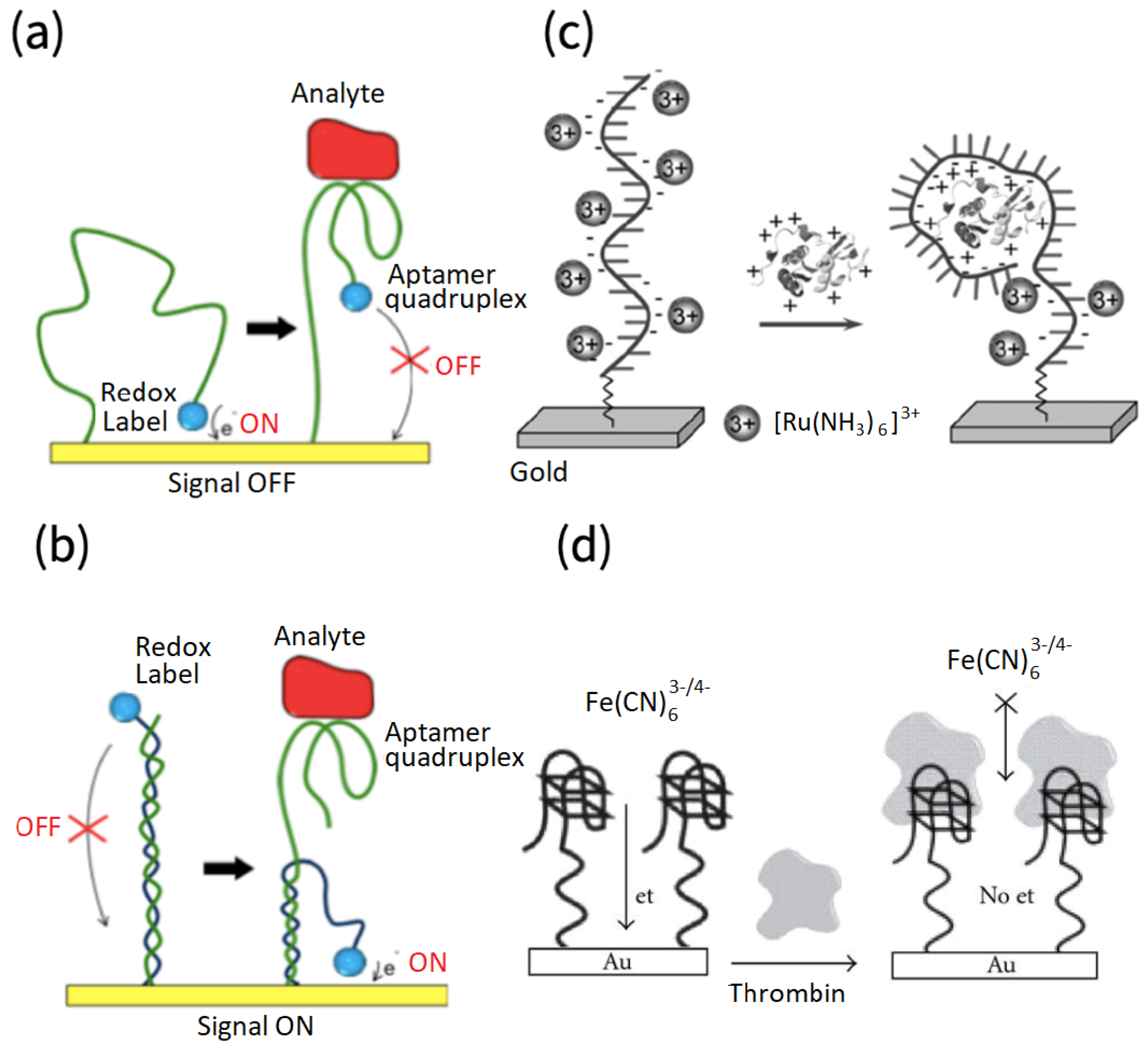
2. Sensor Structure and Sensing Principle
3. Structure and Immobilization of Aptamers
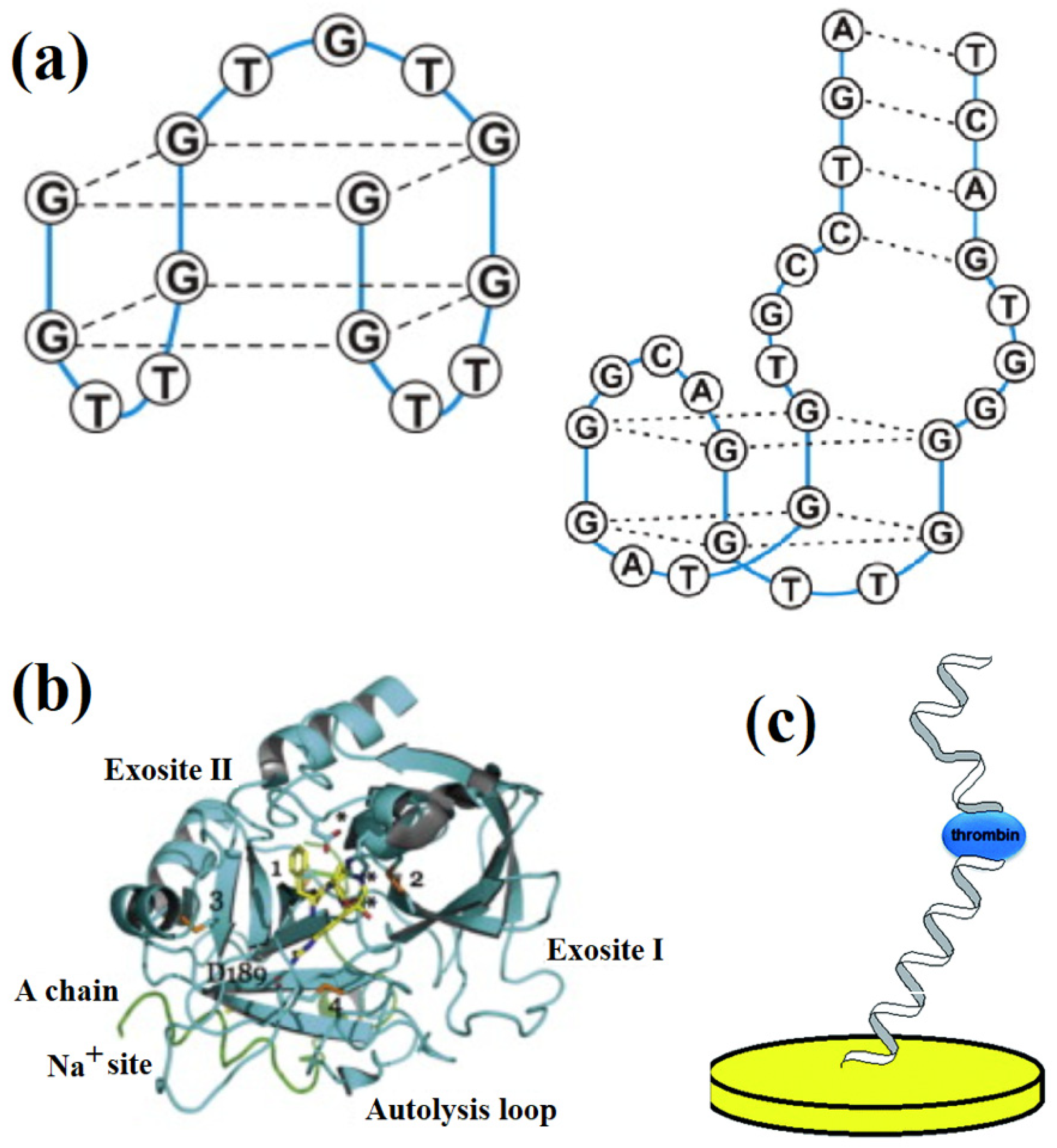
3.1. Structure and Function of Thrombin Aptamers
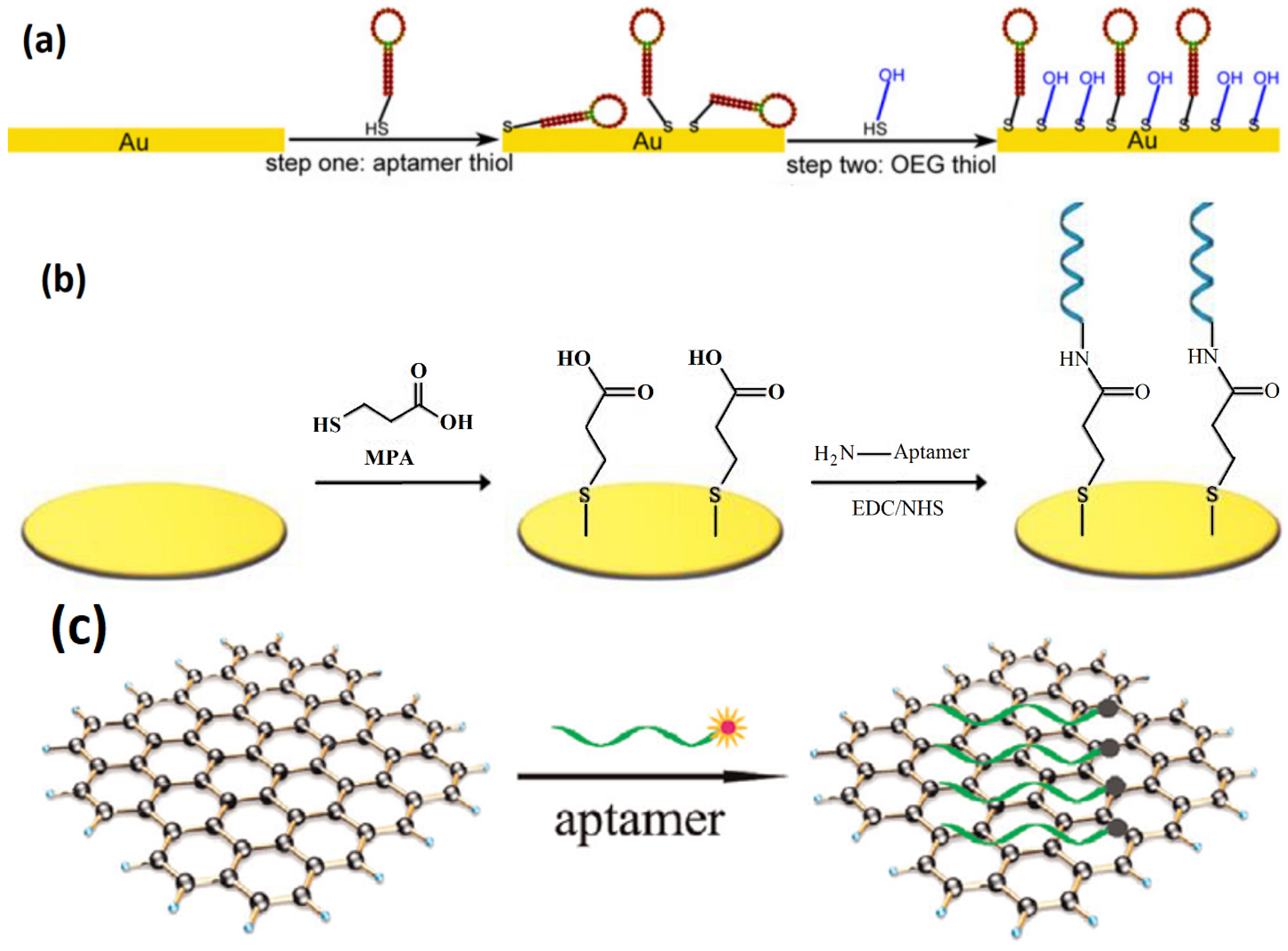
3.2. Immobilization of Aptamers
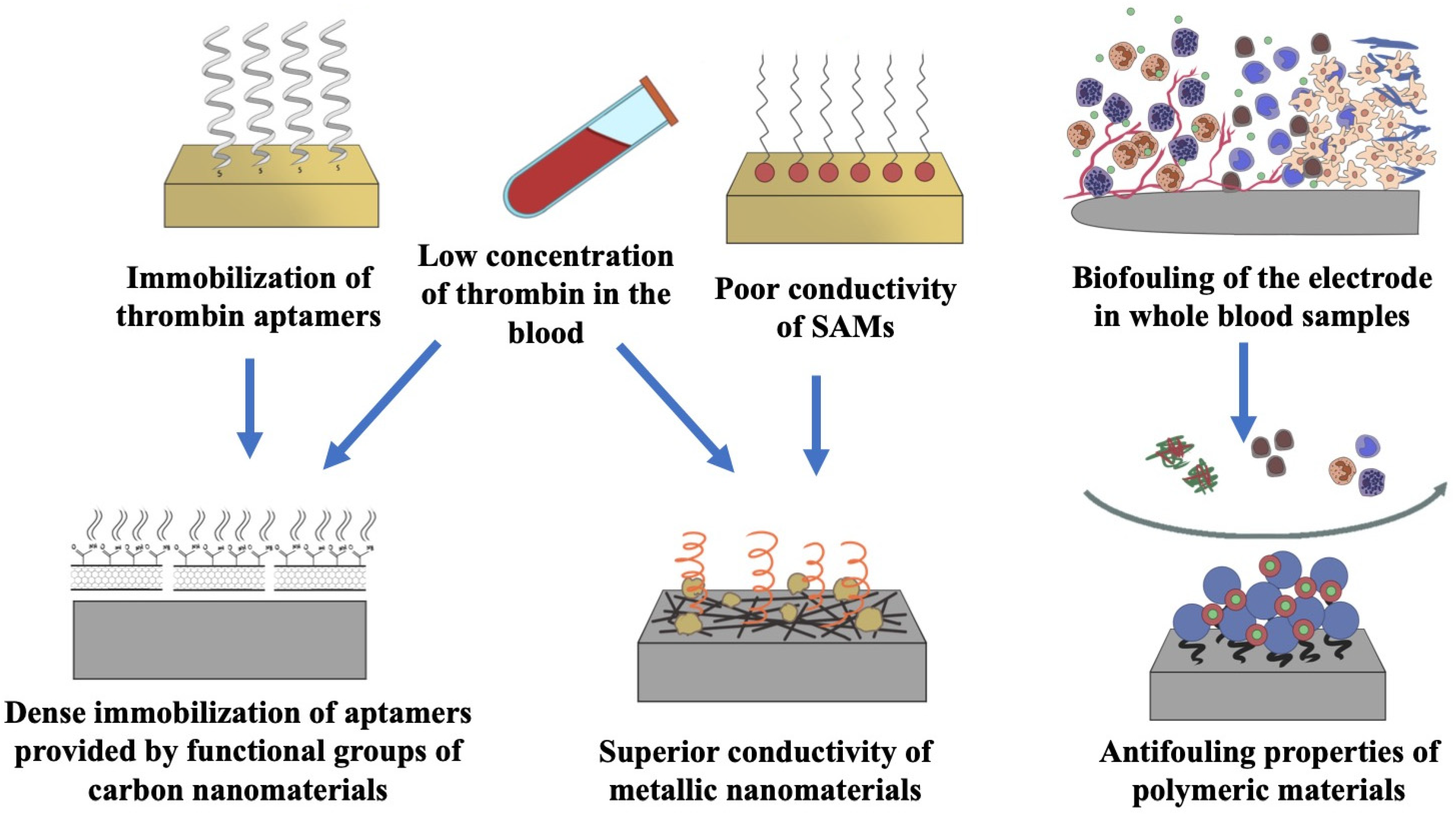
4. Nanomaterial-Based Thrombin Electrochemical Aptasensors
4.1. Low Dimensional Metallic Nanomaterial-Based Thrombin Electrochemical Aptasensors
4.2. Porous Nanomaterial-Based Thrombin Electrochemical Aptasensors
4.3. Carbon Nanomaterial-Based Thrombin Electrochemical Aptasensors
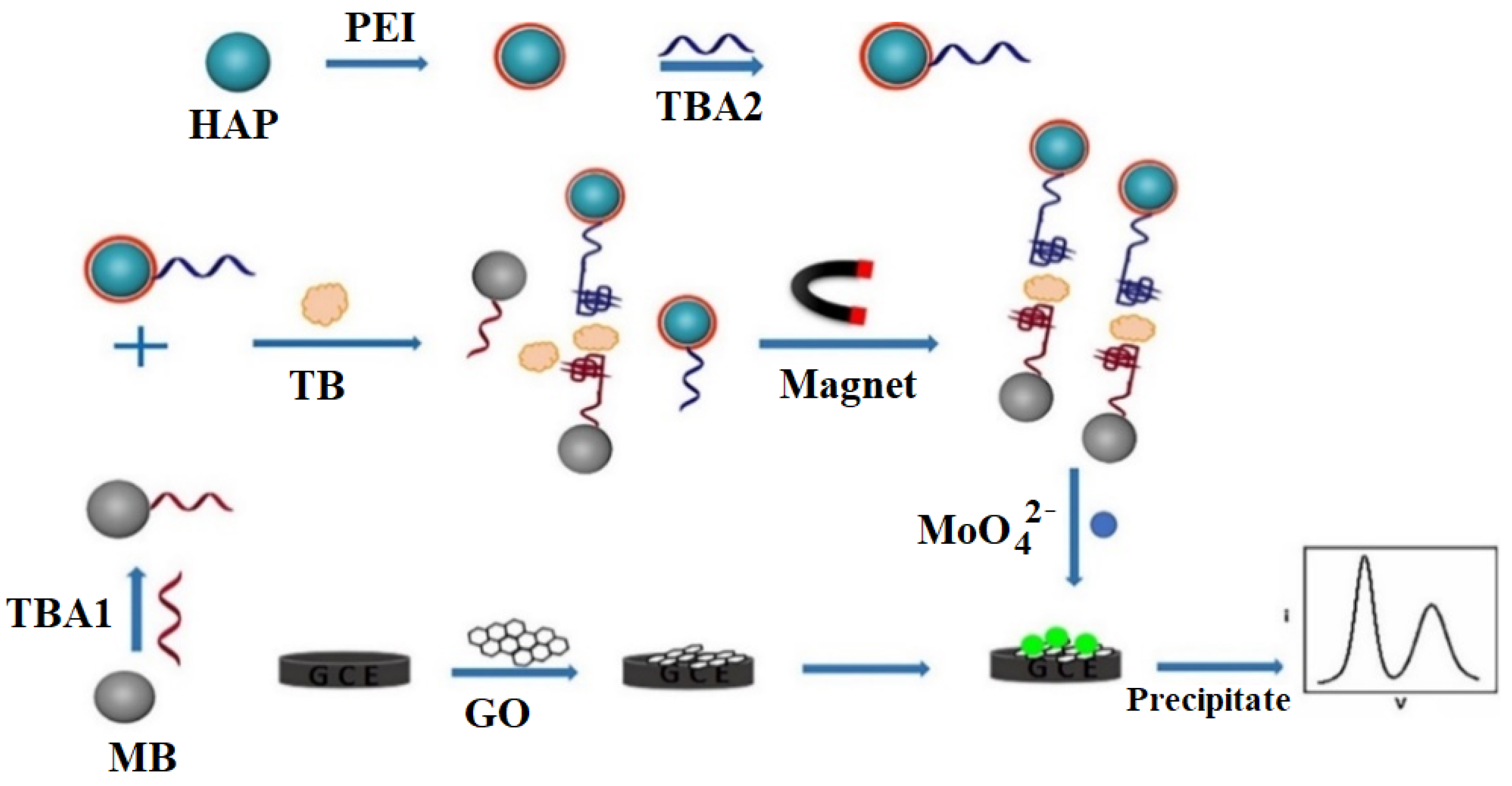
4.4. Magnetic Nanoparticle (MNP)-Enhanced Thrombin Electrochemical Aptasensors
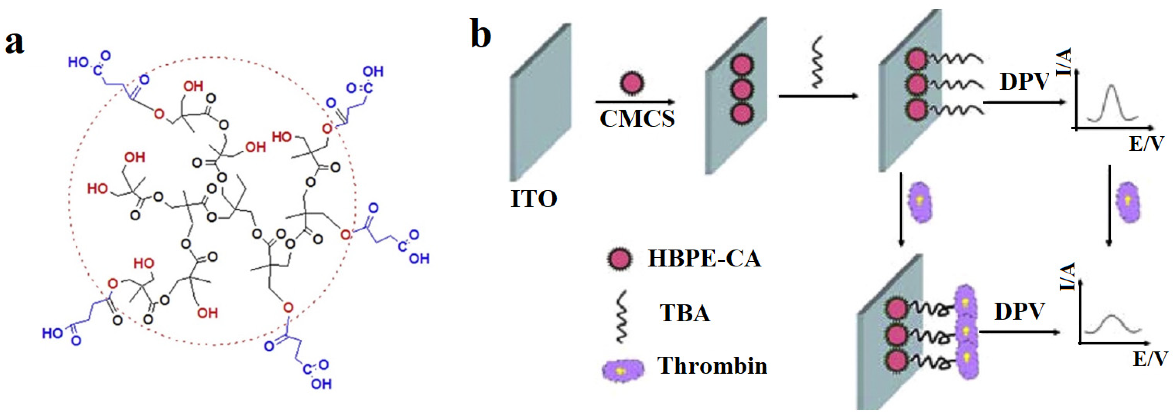
4.5. Polymer-Based Thrombin Electrochemical Aptasensors
5. Conclusions
Author Contributions
Funding
Institutional Review Board Statement
Informed Consent Statement
Conflicts of Interest
Nomenclature
| ABA | 3-aminophe- nylboronic |
| ADH | Alcohol dehydrogenase |
| AN | Aniline |
| BSA | Bovine serum albumin |
| CHIT-SB | Chitosan and a synthetic Schiff base |
| CM-PEG-CM | Carboxymethyl-PEG- carboxymethyl |
| CNC | Carbon nanocages |
| CNT | Carbon Nanotubes |
| CSPH | Conductive supramolecular polymer hydrogel |
| CV | Cyclic voltammetry |
| DA | Dopamine |
| DPV | Differential pulse voltammetry |
| EIS | Electrochemical impedance spectroscopy |
| GCE | Glassy carbon electrode |
| GDH | Glucose dehydrogenase |
| GN-Por | Graphene-porphyrin |
| GO | Graphene oxide |
| H3TCA | Tricarboxytriphenyl- amine |
| HAP | Hydroxyapatite |
| HBPE-CA | Hyperbranched polyester microspheres with carboxylic acid functional groups |
| HBPE-SO3 NPs | Heparin-mimicking hyperbranched polyester nanoparticles |
| HRP | Horseradish peroxidase |
| ITO | Indium tin oxide |
| LDH | Layered double hydroxides |
| LOD | Limit of detection |
| MBs | Magnetic beads |
| MCH | Mercapto-hexanol |
| MOF | Metal organic framework |
| MWCNTs | Multi-walled carbon nanotubes |
| N-GO | N-doped graphene oxide |
| NH2-H2BDC | 2-amino terephthalic acid |
| NPs | Nanoparticles |
| NWs | Nanowires |
| PAA | Porous anodic alumina |
| PEI | Polyethylenimine |
| PET | Polyester terephthalate |
| PGE | Pencil graphite electrode |
| PQdot | Polymer quantum dots |
| PVA | Polyvinyl alcohol |
| rGO | Reduced graphene oxide |
| SA-ALP | Streptavidin-conjugated alkaline phosphatase |
| SAMs | Self-assembled monolayers |
| SPCE | Screen printed carbon electrode |
| SWCNTs | Single-walled carbon nanotubes |
| SWV | Square wave voltammetry |
| TBA | Thrombin-binding aptamer |
| TMDC | Transition-metal dichalcogenide |
| UME | Ultra micro electrode |
References
- Di Cera, E. Thrombin. Mol. Asp. Med. 2008, 29, 203–254. [Google Scholar] [CrossRef] [PubMed]
- Yang, C.; Li, Z.; Tian, Y.; Guo, Q.; Nie, G. A simple label-free photoelectrochemical aptasensor for ultrasensitive detection of thrombin. Microchem. J. 2020, 159, 105452. [Google Scholar] [CrossRef]
- Deng, B.; Lin, Y.; Wang, C.; Li, F.; Wang, Z.; Zhang, H.; Li, X.F.; Le, X.C. Aptamer binding assays for proteins: The thrombin example—A review. Anal. Chim. Acta 2014, 837, 1–15. [Google Scholar] [CrossRef] [PubMed]
- Dahlbäck, B. Blood Coagulation. Haematology 2000, 355, 1627–1632. [Google Scholar] [CrossRef]
- Crawley, J.T.B.; Zanardelli, S.; Chion, C.K.N.K.; Lane, D.A. The central role of thrombin in hemostasis. J. Thromb. Haemost. 2007, 5, 95–101. [Google Scholar] [CrossRef]
- Shen, G.; Zhang, H.; Yang, C.; Yang, Q.; Tang, Y. Thrombin ultrasensitive detection based on chiral supramolecular assembly signal-amplified strategy induced by thrombin-binding aptamer. Anal. Chem. 2017, 89, 548–551. [Google Scholar] [CrossRef] [Green Version]
- Riccardi, C.; Napolitano, E.; Platella, C.; Musumeci, D.; Montesarchio, D. G-Quadruplex-based aptamers targeting human thrombin: Discovery, chemical modifications and antithrombotic effects. Pharmacol. Ther. 2021, 217, 107649. [Google Scholar] [CrossRef]
- Campello, E.; Bulato, C.; Spiezia, L.; Boscolo, A.; Poletto, F.; Cola, M.; Gavasso, S.; Simion, C.; Radu, C.M.; Cattelan, A.; et al. Thrombin generation in patients with COVID-19 with and without Thromboprophylaxis. Clin. Chem. Lab. Med. 2021, 59, 1323–1330. [Google Scholar] [CrossRef]
- Ranucci, M.; Sitzia, C.; Baryshnikova, E.; Di Dedda, U.; Cardani, R.; Martelli, F.; Corsi Romanelli, M. COVID-19-associated coagulopathy: Biomarkers of thrombin generation and fibrinolysis leading the outcome. J. Clin. Med. 2020, 9, 3487. [Google Scholar] [CrossRef]
- Cataldo, R.; Leuzzi, M.; Alfinito, E. Modelling and development of electrical aptasensors: A short review. Chemosensors 2018, 6, 20. [Google Scholar] [CrossRef] [Green Version]
- Yoo, H.; Jo, H.; Oh, S.S. Detection and beyond: Challenges and advances in aptamer-based biosensors. Mater. Adv. 2020, 1, 2663–2687. [Google Scholar] [CrossRef]
- Ikebukuro, K.; Kiyohara, C.; Sode, K. Electrochemical detection of protein using a double aptamer sandwich. Anal. Lett. 2004, 37, 2901–2909. [Google Scholar] [CrossRef]
- Ștefan, G.; Hosu, O.; De Wael, K.; Lobo-Castañón, M.J.; Cristea, C. Aptamers in biomedicine: Selection strategies and recent advances. Electrochim. Acta 2021, 376, 137994. [Google Scholar] [CrossRef]
- Shaban, S.M.; Kim, D.H. Recent advances in aptamer sensors. Sensors 2021, 21, 979. [Google Scholar] [CrossRef] [PubMed]
- Kim, S.M.; Kim, J.; Noh, S.; Sohn, H.; Lee, T. Recent development of aptasensor for influenza virus detection. BioChip J. 2020, 14, 327–339. [Google Scholar] [CrossRef]
- Navani, N.K.; Li, Y. Nucleic acid aptamers and enzymes as sensors. Curr. Opin. Chem. Biol. 2006, 10, 272–281. [Google Scholar] [CrossRef]
- Li, Z.; Mohamed, M.A.; Vinu Mohan, A.M.; Zhu, Z.; Sharma, V.; Mishra, G.K.; Mishra, R.K. Application of electrochemical aptasensors toward clinical diagnostics, food, and environmental monitoring: Review. Sensors 2019, 19, 5435. [Google Scholar] [CrossRef] [Green Version]
- Villalonga, A.; Pérez-Calabuig, A.M.; Villalonga, R. Electrochemical biosensors based on nucleic acid aptamers. Anal. Bioanal. Chem. 2020, 412, 55–72. [Google Scholar] [CrossRef]
- Sun, G.; Huang, Y.; Zheng, L.; Zhan, Z.; Zhang, Y.; Pang, J.H.L.; Wu, T.; Chen, P. Ultra-sensitive and wide-dynamic-range sensors based on dense arrays of carbon nanotube tips. Nanoscale 2011, 3, 4854–4858. [Google Scholar] [CrossRef]
- Grieshaber, D.; MacKenzie, R.; Vörös, J.; Reimhult, E. Electrochemical biosensors—Sensor principles and architectures. Sensors 2008, 8, 1400–1458. [Google Scholar] [CrossRef]
- Ronkainen, N.J.; Halsall, H.B.; Heineman, W.R. Electrochemical biosensors. Chem. Soc. Rev. 2010, 39, 1747–1763. [Google Scholar] [CrossRef] [PubMed]
- Sun, C.; Han, Q.; Wang, D.; Xu, W.; Wang, W.; Zhao, W.; Zhou, M. A Label-free and high sensitive aptamer biosensor based on hyperbranched polyester microspheres for thrombin detection. Anal. Chim. Acta 2014, 850, 33–40. [Google Scholar] [CrossRef] [PubMed]
- Park, K.; Kwon, S.J.; Kwak, J. A Label-free electrochemical aptasensor for thrombin using a Single-Wall Carbon Nanotube (SWCNT) Casted Glassy Carbon Electrode (GCE). Electroanalysis 2014, 26, 513–520. [Google Scholar] [CrossRef]
- Qin, B.; Yang, K. Voltammetric aptasensor for thrombin by using a gold microelectrode modified with graphene oxide decorated with silver nanoparticles. Microchim. Acta 2018, 185, 407. [Google Scholar] [CrossRef] [PubMed]
- Zoski, C.G. Ultramicroelectrodes: Design, fabrication, and characterization. Electroanalysis 2002, 14, 1041–1051. [Google Scholar] [CrossRef]
- Spychalska, K.; Zając, D.; Baluta, S.; Halicka, K.; Cabaj, J. Functional polymers structures for (bio) sensing application—A review. Polymers 2020, 12, 1154. [Google Scholar] [CrossRef]
- Holzinger, M.; Le Goff, A.; Cosnier, S. Nanomaterials for biosensing applications: A review. Front. Chem. 2014, 2, 63. [Google Scholar] [CrossRef] [Green Version]
- Yang, Y.; Yang, Z.; Lv, J.; Yuan, R.; Chai, Y. Thrombin aptasensor enabled by pt nanoparticles-functionalized co-based metal organic frameworks assisted electrochemical signal amplification. Talanta 2017, 169, 44–49. [Google Scholar] [CrossRef]
- Zhang, Y.; Liu, Z.Y.; Wang, Y.G.; Kuang, X.; Ma, H.M.; Wei, Q. Directly assembled electrochemical sensor by combining self-supported con nanoarry platform grown on carbon cloth with molecularly imprinted polymers for the detection of tylosin. J. Hazard. Mater. 2020, 398, 1227788. [Google Scholar] [CrossRef]
- Zhao, A.S.; Lin, T.; Xu, Y.; Zhang, W.G.; Asif, M.; Sun, Y.M.; Xiao, F. Integrated electrochemical microfluidic sensor with hierarchically porous nanoarrays modified graphene fiver microelectrode for bioassay. Biosens. Bioelectron. 2022, 205, 114095. [Google Scholar] [CrossRef]
- Asif, M.; Aziz, A.; Ashraf, G.; Iftikhar, T.; Sun, Y.M.; Liu, H.F. Turning the page: Advancing detection platforms for sulfate reducing bacteria and their perks. Chem. Rec. 2022, 22, e202100166. [Google Scholar] [CrossRef] [PubMed]
- Radi, A.-E.; Abd-Ellatief, M.R. Electrochemical aptasensors: Current status and future perspectives. Diagnostics 2021, 11, 104. [Google Scholar] [CrossRef] [PubMed]
- Oberhaus, F.V.; Frense, D.; Beckmann, D. Immobilization techniques for aptamers on gold electrodes for the electrochemical detection of proteins: A review. Biosensors 2020, 10, 45. [Google Scholar] [CrossRef] [PubMed]
- Yan, S.R.; Foroughi, M.M.; Safaei, M.; Jahani, S.; Ebrahimpour, N.; Borhani, F.; Rezaei Zade Baravati, N.; Aramesh-Boroujeni, Z.; Foong, L.K. A review: Recent advances in ultrasensitive and highly specific recognition aptasensors with various detection strategies. Int. J. Biol. Macromol. 2020, 155, 184–207. [Google Scholar] [CrossRef]
- Beitollahi, H.; Tajik, S.; Dourandish, Z.; Zhang, K.; Van Le, Q.; Jang, H.W.; Kim, S.Y.; Shokouhimehr, M. Recent Advances in the aptamer-based electrochemical biosensors for detecting aflatoxin B1 and its pertinent metabolite aflatoxin M1. Sensors 2020, 20, 3256. [Google Scholar] [CrossRef] [PubMed]
- Rozenblum, G.T.; Pollitzer, I.G.; Radrizzani, M. Challenges in electrochemical aptasensors and current sensing architectures using flat gold surfaces. Chemosensors 2019, 7, 57. [Google Scholar] [CrossRef] [Green Version]
- Dekanski, A.; Stevanović, J.; Stevanović, R.; Nikolić, B.Ž.; Jovanović, V.M. Glassy carbon electrodes: I. Characterization and electrochemical activation. Carbon 2001, 39, 1195–1205. [Google Scholar] [CrossRef]
- Zhang, Q.; Li, W.; Zhao, F.; Xu, C.; Fan, G.; Liu, Q. Colloids and surfaces a: Physicochemical and engineering aspects electrochemical sandwich-type thrombin aptasensor based on silver nanowires & particles decorated electrode and the signal amplifier of pt loaded hollow zinc ferrite. Colloids Surf. A Physicochem. Eng. Asp. 2021, 611, 125804. [Google Scholar] [CrossRef]
- Chiorcea-Paquim, A.M.; Oliveira-Brett, A.M. Guanine quadruplex electrochemical aptasensors. Chemosensors 2016, 4, 13. [Google Scholar] [CrossRef] [Green Version]
- Xu, Y.; Cheng, G.; He, P.; Fang, Y. A Review: Electrochemical aptasensors with various detection strategies. Electroanalysis 2009, 21, 1251–1259. [Google Scholar] [CrossRef]
- Radi, A.-E. Electrochemical aptamer-based biosensors: Recent advances and perspectives. Int. J. Electrochem. 2011, 2011, 863196. [Google Scholar] [CrossRef] [Green Version]
- De-Los-Santos-Álvarez, N.; Lobo-Castañón, M.J.; Miranda-Ordieres, A.J.; Tuñón-Blanco, P. Aptamers as recognition elements for label-free analytical devices. Trends Anal. Chem. 2008, 27, 437–446. [Google Scholar] [CrossRef]
- Rhouati, A.; Catanante, G.; Nunes, G.; Hayat, A.; Marty, J.-L. Label-free aptasensors for the detection of mycotoxins. Sensors 2016, 16, 2178. [Google Scholar] [CrossRef] [PubMed]
- Li, L.; Zhao, H.; Chen, Z.; Mu, X.; Guo, L. Aptamer-based electrochemical approach to the detection of thrombin by modification of gold nanoparticles. Anal. Bioanal. Chem. 2010, 398, 563–570. [Google Scholar] [CrossRef] [PubMed]
- Jamei, H.R.; Rezaei, B.; Ensafi, A.A. Ultra-sensitive and selective electrochemical biosensor with aptamer recognition surface based on polymer quantum dots and C60/MWCNTs-polyethylenimine nanocomposites for analysis of thrombin protein. Bioelectrochemistry 2021, 138, 107701. [Google Scholar] [CrossRef] [PubMed]
- Ohuchi, S. Cell-selex technology. Biores 2012, 1, 265–272. [Google Scholar] [CrossRef]
- Tuerk, C.; Gold, L. Systematic evolution of ligands by exponential enrichment: RNA ligands to bacteriophage T4 DNA polymerase. Science 1990, 249, 505–510. [Google Scholar] [CrossRef]
- Ellington, A.D.; Szostak, J.W. In vitro selection of RNA molecules that bind specific ligands. Nature 1990, 346, 818–822. [Google Scholar] [CrossRef]
- Zhuo, Z.; Yu, Y.; Wang, M.; Li, J.; Zhang, Z.; Liu, J.; Wu, X.; Lu, A.; Zhang, G.; Zhang, B. Recent advances in SELEX technology and aptamer applications in biomedicine. Int. J. Mol. Sci. 2017, 18, 2142. [Google Scholar] [CrossRef] [Green Version]
- Zhou, J.; Rossi, J. Aptamers as targeted therapeutics: Current potential and challenges. Nat. Rev. Drug Discov. 2017, 16, 181–202. [Google Scholar] [CrossRef] [Green Version]
- Canoura, J.; Yu, H.; Alkhamis, O.; Roncancio, D.; Farhana, R.; Xiao, Y. Accelerating post-SELEX aptamer engineering using exonuclease digestion. J. Am. Chem. Soc. 2021, 143, 805–816. [Google Scholar] [CrossRef] [PubMed]
- Klug, S.J.; Famulok, M. All you wanted to know about SELEX. Mol. Biol. Rep. 1994, 20, 97–107. [Google Scholar] [CrossRef] [PubMed]
- Bock, L.C.; Griffin, L.C.; Latham, J.A.; Vermaas, E.H.; Toole, J.J. Selection of single-stranded DNA molecules that bind and inhibit human thrombin. Nature 1992, 355, 564–566. [Google Scholar] [CrossRef] [PubMed]
- Wang, Y.H.; Xia, H.; Huang, K.J.; Wu, X.; Ma, Y.Y.; Deng, R.; Lu, Y.F.; Han, Z.W. Ultrasensitive determination of thrombin by using an electrode modified with WSe 2 and gold nanoparticles, aptamer-thrombin-aptamer sandwiching, redox cycling, and signal enhancement by alkaline phosphatase. Microchim. Acta 2018, 185, 502. [Google Scholar] [CrossRef]
- Bochman, M.L.; Paeschke, K.; Zakian, V.A. DNA secondary structures: Stability and function of g-quadruplex structures. Nat. Rev. Genet. 2012, 13, 770–780. [Google Scholar] [CrossRef] [Green Version]
- Baldrich, E.; O’Sullivan, C.K. Ability of thrombin to act as molecular chaperone, inducing formation of quadruplex structure of thrombin-binding aptamer. Anal. Biochem. 2005, 341, 194–197. [Google Scholar] [CrossRef]
- Nishio, M.; Tsukakoshi, K.; Ikebukuro, K. G-quadruplex: Flexible conformational changes by cations, pH, crowding and its applications to biosensing. Biosens. Bioelectron. 2021, 178, 113030. [Google Scholar] [CrossRef]
- Chen, J.; Hickey, B.L.; Wang, L.; Lee, J.; Gill, A.D.; Favero, A.; Pinalli, R.; Dalcanale, E.; Hooley, R.J.; Zhong, W. Selective discrimination and classification of g-quadruplex structures with a host–guest sensing array. Nat. Chem. 2021, 13, 488–495. [Google Scholar] [CrossRef]
- Vairamani, M.; Gross, M.L. G-quadruplex formation of thrombin-binding aptamer detected by electrospray ionization mass spectrometry. J. Am. Chem. Soc. 2003, 125, 42–43. [Google Scholar] [CrossRef]
- Macaya, R.F.; Schultze, P.; Smith, F.W.; Roe, J.A.; Feigon, J. Thrombin-binding DNA aptamer forms a unimolecular quadruplex structure in solution. Proc. Natl. Acad. Sci. USA 1993, 90, 3745–3749. [Google Scholar] [CrossRef] [Green Version]
- Jarczewska, M.; Górski, Ł.; Malinowska, E. Electrochemical aptamer-based biosensors as potential tools for clinical diagnostics. Anal. Methods 2016, 8, 3861–3877. [Google Scholar] [CrossRef] [Green Version]
- Tasset, D.M.; Kubik, M.F.; Steiner, W. Oligonucleotide inhibitors of human thrombin that bind distinct epitopes. J. Mol. Biol. 1997, 272, 688–698. [Google Scholar] [CrossRef] [PubMed]
- Sun, C.; Wang, X.; Yang, X.; Xing, L.; Zhao, B.; Yang, X.; Mao, C. A Label-free electrochemical aptasensor for sensitive thrombin detection in whole blood. Electrochim. Acta 2013, 106, 327–332. [Google Scholar] [CrossRef]
- Bin Seo, H.; Gu, M.B. Aptamer-based sandwich-type biosensors. J. Biol. Eng. 2017, 11, 11. [Google Scholar] [CrossRef] [PubMed] [Green Version]
- Chung, S.; Moon, J.M.; Choi, J.; Hwang, H.; Shim, Y.B. Magnetic force assisted electrochemical sensor for the detection of thrombin with aptamer-antibody sandwich formation. Biosens. Bioelectron. 2018, 117, 480–486. [Google Scholar] [CrossRef] [PubMed]
- Mishra, G.K.; Sharma, V.; Mishra, R.K. Electrochemical aptasensors for food and environmental safeguarding: A review. Biosensors 2018, 8, 28. [Google Scholar] [CrossRef] [Green Version]
- Zhou, L.; Wang, M.H.; Wang, J.P.; Ye, Z.Z. Application of biosensor surface immobilization methods for aptamer. Fenxi Huaxue/Chin. J. Anal. Chem. 2011, 39, 432–438. [Google Scholar] [CrossRef]
- Zhang, X.; Yadavalli, V.K. Surface immobilization of DNA aptamers for biosensing and protein interaction analysis. Biosens. Bioelectron. 2011, 26, 3142–3147. [Google Scholar] [CrossRef]
- Zhang, H.; Shuang, S.; Sun, L.; Chen, A.; Qin, Y.; Dong, C. Label-free aptasensor for thrombin using a glassy carbon electrode modified with a graphene-porphyrin composite. Microchim. Acta 2014, 181, 189–196. [Google Scholar] [CrossRef]
- Ping, J.; Zhou, Y.; Wu, Y.; Papper, V.; Boujday, S.; Marks, R.S.; Steele, T.W.J. Recent advances in aptasensors based on graphene and graphene-like nanomaterials. Biosens. Bioelectron. 2015, 64, 373–385. [Google Scholar] [CrossRef]
- Wang, X.; Gao, F.; Gong, Y.; Liu, G.; Zhang, Y.; Ding, C. Electrochemical aptasensor based on conductive supramolecular polymer hydrogels for thrombin detection with high selectivity. Talanta 2019, 205, 120140. [Google Scholar] [CrossRef]
- Doria, G.; Conde, J.; Veigas, B.; Giestas, L.; Almeida, C.; Assunção, M.; Rosa, J.; Baptista, P.V. Noble metal nanoparticles for biosensing applications. Sensors 2012, 12, 1657–1687. [Google Scholar] [CrossRef] [PubMed]
- Pan, M.; Yang, J.; Liu, K.; Yin, Z.; Ma, T.; Liu, S.; Xu, L.; Wang, S. Noble metal nanostructured materials for chemical and biosensing systems. Nanomaterials 2020, 10, 209. [Google Scholar] [CrossRef] [PubMed] [Green Version]
- Vinci, G.; Rapa, M. Noble metal nanoparticles applications: Recent trends in food control. Bioengineering 2019, 6, 10. [Google Scholar] [CrossRef] [Green Version]
- Wang, J. Electrochemical biosensing based on noble metal nanoparticles. Microchim. Acta 2012, 177, 245–270. [Google Scholar] [CrossRef]
- Li, L.; Zhao, H.; Chen, Z.; Mu, X.; Guo, L. Aptamer biosensor for label-free impedance spectroscopy detection of thrombin based on gold nanoparticles. Sens. Actuators B 2011, 157, 189–194. [Google Scholar] [CrossRef]
- Chen, Y.; Xiang, J.; Liu, B.; Chen, Z.; Zuo, X. Gold nanoparticle-engineered electrochemical aptamer biosensor for ultrasensitive detection of thrombin. Anal. Methods 2020, 12, 3729–3733. [Google Scholar] [CrossRef]
- Xu, Q.; Wang, G.; Zhang, M.; Xu, G.; Lin, J.; Luo, X. Aptamer based label free thrombin assay based on the use of silver nanoparticles incorporated into self-polymerized dopamine. Microchim. Acta 2018, 185, 2–8. [Google Scholar] [CrossRef]
- Wanekaya, A.K.; Chen, W.; Myung, N.V.; Mulchandani, A. Nanowire-based electrochemical biosensors. Electroanalysis 2006, 18, 533–550. [Google Scholar] [CrossRef]
- Zheng, Y.; Chai, Y.; Yuan, Y.; Yuan, R. A Pseudo triple-enzyme electrochemical aptasensor based on the amplification of pt-pd nanowires and hemin/g-quadruplex. Anal. Chim. Acta 2014, 834, 45–50. [Google Scholar] [CrossRef]
- Sun, A.; Qi, Q.; Wang, X.; Bie, P. Porous platinum nanotubes labeled with hemin/g-quadruplex based electrochemical aptasensor for sensitive thrombin analysis via the cascade signal amplification. Biosens. Bioelectron. 2014, 57, 16–21. [Google Scholar] [CrossRef] [PubMed]
- Wen, W.; Song, Y.; Yan, X.; Zhu, C.; Du, D.; Wang, S.; Asiri, A.M.; Lin, Y. Recent advances in emerging 2D nanomaterials for biosensing and bioimaging applications. Mater. Today 2018, 21, 164–177. [Google Scholar] [CrossRef]
- Evtugyn, G.; Porfireva, A.; Shamagsumova, R.; Hianik, T. Advances in electrochemical aptasensors based on carbon nanomaterials. Chemosensors 2020, 8, 96. [Google Scholar] [CrossRef]
- Manzeli, S.; Ovchinnikov, D.; Pasquier, D.; Yazyev, O.V.; Kis, A. 2D transition metal dichalcogenides. Nat. Rev. Mater. 2017, 2, 17033. [Google Scholar] [CrossRef]
- Wang, Y.H.; Huang, K.J.; Wu, X. Recent advances in transition-metal dichalcogenides based electrochemical biosensors: A review. Biosens. Bioelectron. 2017, 97, 305–316. [Google Scholar] [CrossRef]
- Huang, Y.; Guo, J.; Kang, Y.; Ai, Y.; Li, C.M. Two dimensional atomically thin MoS2 nanosheets and their sensing applications. Nanoscale 2015, 7, 19358–19376. [Google Scholar] [CrossRef]
- Lin, K.C.; Jagannath, B.; Muthukumar, S.; Prasad, S. Sub-picomolar label-free detection of thrombin using electrochemical impedance spectroscopy of aptamer-functionalized MoS2. Analyst 2017, 142, 2770–2780. [Google Scholar] [CrossRef]
- Song, X.; Xu, W.; Su, D.; Tang, J.; Liu, X. The synthesis of hollow/porous Cu2O nanoparticles by ion-pairing behavior control. ACS Omega 2020, 5, 1879–1886. [Google Scholar] [CrossRef] [Green Version]
- Fu, C.; Wang, Y. Nanostructured porous materials for biosensor applications. In The world Scientific Encyclopedia of Nanomedicine and Bioengineering; World Scientific Publishing: Singapore, 2016; pp. 245–290. [Google Scholar] [CrossRef]
- Shuai, H.L.; Wu, X.; Huang, K.J. Molybdenum disulfide sphere-based electrochemical aptasensors for protein detection. J. Mater. Chem. B 2017, 5, 5362–5372. [Google Scholar] [CrossRef]
- Zhang, Z.; Zhang, S.; Liu, S.; Wang, M.; Fu, G.; He, L.; Yang, Y.; Fang, S. Electrochemical aptasensor based on one-step synthesis of Cu2O@aptamer nanospheres for sensitive thrombin detection. Sens. Actuators B Chem. 2015, 220, 184–191. [Google Scholar] [CrossRef]
- Du, L.; Chen, W.; Zhu, P.; Tian, Y.; Chen, Y.; Wu, C. Applications of functional metal-organic frameworks in biosensors. Biotechnol. J. 2021, 16, 1900424. [Google Scholar] [CrossRef] [PubMed]
- Xie, S.; Ye, J.; Yuan, Y.; Chai, Y.; Yuan, R. A Multifunctional hemin@metal-organic framework and its application to construct an electrochemical aptasensor for thrombin detection. Nanoscale 2015, 7, 18232–18238. [Google Scholar] [CrossRef] [PubMed] [Green Version]
- Wu, H.; Li, M.; Wang, Z.; Yu, H.; Han, J.; Xie, G.; Chen, S. Analytica chimica acta highly stable Ni-MOF comprising triphenylamine moieties as a high- performance redox indicator for sensitive aptasensor construction. Anal. Chim. Acta 2019, 1049, 74–81. [Google Scholar] [CrossRef]
- De La Escosura-Muñiz, A.; Merkoçi, A. Nanochannels preparation and application in biosensing. ACS Nano 2012, 6, 7556–7583. [Google Scholar] [CrossRef] [PubMed]
- Meervelt, V.; Soskine, M.; Maglia, G. Detection of two isomeric binding configurations in a protein-aptamer complex with a biological nanopore. ACS Nano 2014, 8, 12826–12835. [Google Scholar] [CrossRef] [Green Version]
- Zhao, X.P.; Cao, J.; Nie, X.G.; Wang, S.S.; Wang, C.; Xia, X.H. Label-free monitoring of the thrombin–aptamer recognition reaction using an array of nanochannels coupled with electrochemical detection. Electrochem. Commun. 2017, 81, 5–9. [Google Scholar] [CrossRef]
- Li, L.D.; Mu, X.J.; Peng, Y.; Chen, Z.B.; Guo, L.; Jiang, L. Signal-on architecture for electrochemical aptasensors based on multiple ion channels. Anal. Chem. 2012, 84, 10554–10559. [Google Scholar] [CrossRef]
- Krishnan, S.K.; Singh, E.; Singh, P.; Meyyappan, M.; Nalwa, H.S. A review on graphene-based nanocomposites for electrochemical and fluorescent biosensors. RSC Adv. 2019, 9, 8778–8781. [Google Scholar] [CrossRef]
- Amiri, M.; Nekoueian, K.; Saberi, R.S. Graphene-family materials in electrochemical aptasensors. Anal. Bioanal. Chem. 2021, 413, 673–699. [Google Scholar] [CrossRef]
- Liu, Y.; Dong, Y.; Guo, C.X.; Cui, Z.; Zheng, L.; Li, C.M. Protein-directed in situ synthesis of gold nanoparticles on reduced graphene oxide modified electrode for nonenzymatic glucose sensing. Electroanalysis 2012, 24, 2348–2353. [Google Scholar] [CrossRef]
- Xu, J.; Wang, Y.; Hu, S. Nanocomposites of graphene and graphene oxides: Synthesis, molecular functionalization and application in electrochemical sensors and biosensors. A review. Microchim. Acta 2017, 184, 1–44. [Google Scholar] [CrossRef]
- Ahour, F.; Ahsani, M.K. An electrochemical label-free and sensitive thrombin aptasensor based on graphene oxide modified pencil graphite electrode. Biosens. Bioelectron. 2016, 86, 764–769. [Google Scholar] [CrossRef] [PubMed]
- Zhang, H.; Zhang, B.; Chen, A.; Qin, Y. Controllable: N -Fe2O3@graphene nanomaterials by ALD applied in an aptasensor with enhanced electrochemical performance for thrombin detection. Dalt. Trans. 2017, 46, 7434–7440. [Google Scholar] [CrossRef] [PubMed]
- Rao, N.; Singh, R.; Bashambu, L. Carbon-based nanomaterials: Synthesis and prospective applications. Mater. Today Proc. 2021, 44, 608–614. [Google Scholar] [CrossRef]
- Sanati, A.; Jalali, M.; Raeissi, K.; Karimzadeh, F.; Kharaziha, M.; Mahshid, S.S.; Mahshid, S. A Review on recent advancements in electrochemical biosensing using carbonaceous nanomaterials. Microchim. Acta 2019, 186, 773. [Google Scholar] [CrossRef]
- Liu, Y.; Sun, G.; Jiang, C.; Zheng, X.T.; Zheng, L.; Li, C.M. Highly sensitive detection of hydrogen peroxide at a carbon nanotube fiber microelectrode coated with palladium nanoparticles. Microchim. Acta 2014, 181, 63–70. [Google Scholar] [CrossRef]
- Appaturi, J.N.; Pulingam, T.; Thong, K.L.; Muniandy, S.; Ahmad, N.; Leo, B.F. Rapid and sensitive detection of salmonella with reduced graphene oxide-carbon nanotube based electrochemical aptasensor. Anal. Biochem. 2020, 589, 113489. [Google Scholar] [CrossRef]
- Zhu, Z. An overview of carbon nanotubes and graphene for biosensing applications. Nano-Micro Lett. 2017, 9, 25. [Google Scholar] [CrossRef] [Green Version]
- Tîlmaciu, C.M.; Morris, M.C. Carbon nanotube biosensors. Front. Chem. 2015, 3, 59. [Google Scholar] [CrossRef] [Green Version]
- Heydari-Bafrooei, E.; Amini, M.; Ardakani, M.H. An electrochemical aptasensor based on TiO2/MWCNT and a novel synthesized schiff base nanocomposite for the ultrasensitive detection of thrombin. Biosens. Bioelectron. 2016, 85, 828–836. [Google Scholar] [CrossRef]
- Konari, M.; Heydari-Bafrooei, E.; Dinari, M. Efficient immobilization of aptamers on the layered double hydroxide nanohybrids for the electrochemical proteins detection. Int. J. Biol. Macromol. 2021, 166, 54–60. [Google Scholar] [CrossRef] [PubMed]
- Gao, F.; Du, L.; Zhang, Y.; Zhou, F.; Tang, D. A Sensitive sandwich-type electrochemical aptasensor for thrombin detection based on platinum nanoparticles decorated carbon nanocages as signal labels. Biosens. Bioelectron. 2016, 86, 185–193. [Google Scholar] [CrossRef] [PubMed]
- Rocha-Santos, T.A.P. Sensors and biosensors based on magnetic nanoparticles. Trends Anal. Chem. 2014, 62, 28–36. [Google Scholar] [CrossRef]
- Zhang, Y.; Xia, J.; Zhang, F.; Wang, Z.; Liu, Q. Ultrasensitive label-free homogeneous electrochemical aptasensor based on sandwich structure for thrombin detection. Sens. Actuators B Chem. 2018, 267, 412–418. [Google Scholar] [CrossRef]
- Zhu, C.; Zhu, W.; Xu, L.; Zhou, X. A Label-free electrochemical aptasensor based on magnetic biocomposites with pb 2+-dependent DNAzyme for the detection of thrombin. Anal. Chim. Acta 2019, 1047, 21–27. [Google Scholar] [CrossRef] [PubMed]
- Ionita, I.; Grigorita, L.; Miloicov, C.B.; Petre, I.; Bernad, E.; Craina, M.; Diaconu, M.; Citu, C.; Radu, F.; Oros, D.; et al. The role of thrombophilia in pregnancy. Rev. Chim. 2016, 67, 2643–2647. [Google Scholar] [CrossRef] [Green Version]
- Hui, N.; Sun, X.; Niu, S.; Luo, X. PEGylated polyaniline nanofibers: Antifouling and conducting biomaterial for electrochemical DNA sensing. ACS Appl. Mater. Interfaces 2017, 9, 2914–2923. [Google Scholar] [CrossRef] [PubMed]
- Campuzano, S.; Pedrero, M.; Yáñez-Sedeño, P.; Pingarrón, J.M. Antifouling (bio) materials for electrochemical (bio) sensing. Int. J. Mol. Sci. 2019, 20, 423. [Google Scholar] [CrossRef] [Green Version]
- Zhao, C.; Li, L.; Guo, M.; Zheng, J. Functional polymer thin films designed for antifouling materials and biosensors. Chem. Pap. 2012, 66, 323–339. [Google Scholar] [CrossRef]
- Gereadr, M.; Choubey, A.; Malhotra, B. Review: Application of conducting polymer to biosensors. Biosens. Bioelectron. 2001, 17, 345–359. [Google Scholar]
- Niu, Y.; Chu, M.; Xu, P.; Meng, S.; Zhou, Q.; Zhao, W.; Zhao, B.; Shen, J. An aptasensor based on heparin-mimicking hyperbranched polyester with anti-biofouling interface for sensitive thrombin detection. Biosens. Bioelectron. 2018, 101, 174–180. [Google Scholar] [CrossRef] [PubMed]


| Material Category | Detailed Electrode Material | Aptamer Sequence | Analytical Method | LOD (fM) | Linear Range | Others | Reference |
|---|---|---|---|---|---|---|---|
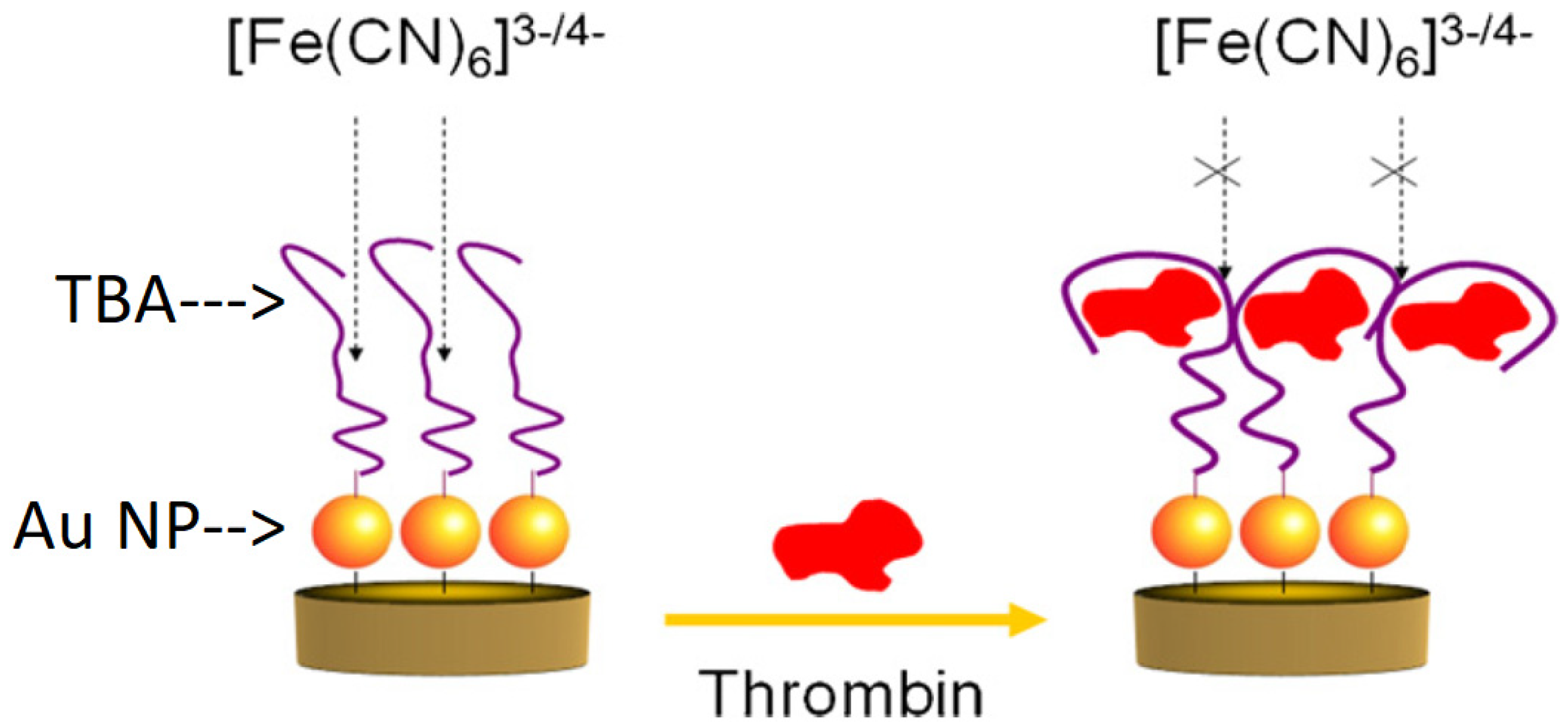
| Metallic nanoparticle (0D) | Au NPs (on Au electrode) | TBA1: 5′-SH-(CH2)6-GGT TGG TGT GGT TGG-3′ TBA2: 5′-SH-AGT CCG TGG TAG GGC AGG TTG GGG TGA CT-3′ | DPV | 0.1429 | 1 fM to 6 pM | Directly bound TBA | [77] |
| Ag NPs @ dopamine (on GCE) | 5′-SH-(CH2)6-GGT TGG TGT GGT TGG-3′ | EIS | 36 | 0.1 pM to 5.0 nM | Conductive and hydrophilic | [78] | |
| Metallic nanowire/tube(1D) | Ag NWs&NPs/ZnFe2O4 NPs (on ITO) | Apt1: 5′-NH2-(CH2)6-GGT TGG TGT GGT TGG-3′ Apt2: 5′-SH—(CH2)6—AGT CCGTGG TAG GGC AGG TTG GGG TGA CT-3′ | Amperometric I-t | 16 | 0.05 pM to 35 nM | Sandwich assay design | [38] |
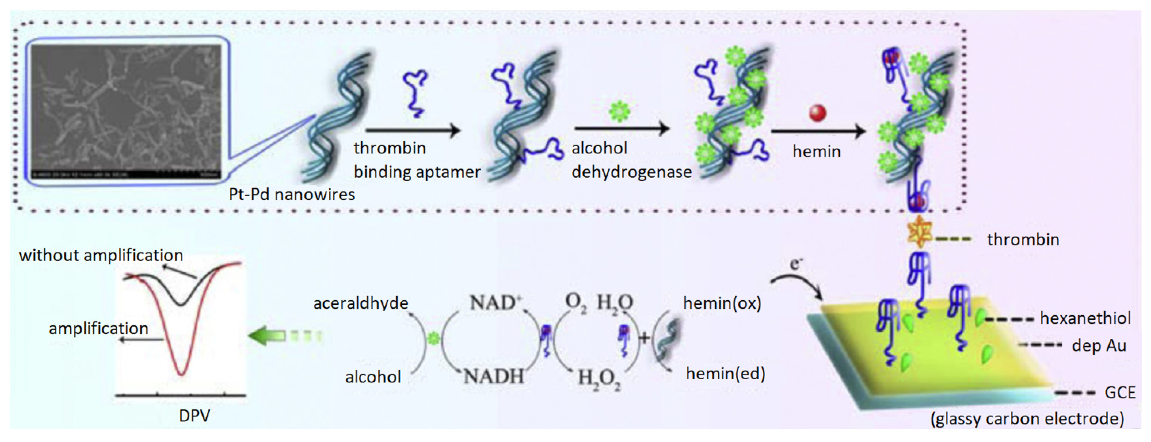
| Pt-Pd NWs (on GCE) | 5′-SH-(CH2)6-GGT TGG TGT GGT TGG-3′ | DPV | 67 | 0.2 pM to 20 nM | Triple enzyme cascade | [80] | |
| Pt Nanotubes (on GCE) | 5′-SH–(CH2)6–GGT TGG TGT GGT TGG-3′ | DPV | 150 | 0.4 pM to 30 nM | Sandwich assay design | [81] | |
| Metallic nanosheet (2D) | Au NPs/WSe2 (on GCE) | TBA1: 5′-biotin-TEG linker-GGT TGG TGT GGT TGG-3′ TBA2: 5′-NH2-TEG linker-AGT CCG TGG TAG GGC AGG TTG GGG TGA CT-3′ | DPV | 190 | 0–1 ng/mL | Sandwich assay design | [54] |
| MoS2 (on Pt) | TBA (12T): 5′-(Thiol-C6) TTT TTT TTT TTT GGT TGG TGT GGT TGG-3′ | EIS | 267 | 2.67 pM to 267 pM | TMDC semiconductor behavior | [87] |
| Material Category | Detailed Electrode Material | Aptamer Sequence | Analytical Method | LOD (fM) | Linear Range | Others | Reference |
|---|---|---|---|---|---|---|---|
| Hollow and porous nanomaterials | N- GO and Au NPs (on GCE) | TBA1: 5′-SH-(CH2)6-TTT TTT TTT TTT GGT TGG TGT GGT TGG-3′ TBA2: 5′-SH-(CH2)6-GGT TGG TGT GGT TGG-3′ | DPV | 0.027 | 0.1 fM to 0.1 nM | MnO2 nanospheres in a sandwich assay design | [90] |
| CuO2@aptamer (on Au) | 5′-TCT CTC AGT CCG TGG TAG GGC AGG GTT GGG GTG ACT-3′ | EIS | 330 | 0.1 to 50 ng mL−1 | Cu2O Nanospheres | [91] | |
| PtNPs@Co(II)MOFs@PtNPs (on GCE) | TBA1: 5′-NH2-(CH2)6-GGT TGG TGT GGT TGG-3′ TBA2: 5′-NH2-AGT CCG TGG TAG GGC AGG TTG GGG TGA CT-3′ | DPV | 33 | 0.1 pM to 50 nM | MOF/Sandwich design | [28] | |
| Au/hemin@MOFs (on GCE) | 5′-NH2-(CH2)6-GGT TGG TGT GGT TGG-3′ | DPV | 68 | 0.1 pM to 30 nM | MOF/Sandwich design | [93] | |
| AuNPs/Ni-MOFs (on GCE) | TBA1: 5′-SH-(CH2)6-GGT TGG TGT GGT TGG-3′ TBA2: 5′-NH2-(CH2)6-AGT CCG TGG TAG GGC AGG TTG GGG TGA CT- 3′ | DPV | 16 | 0.05 pM to 50 nM | MOF/Sandwich design | [94] | |
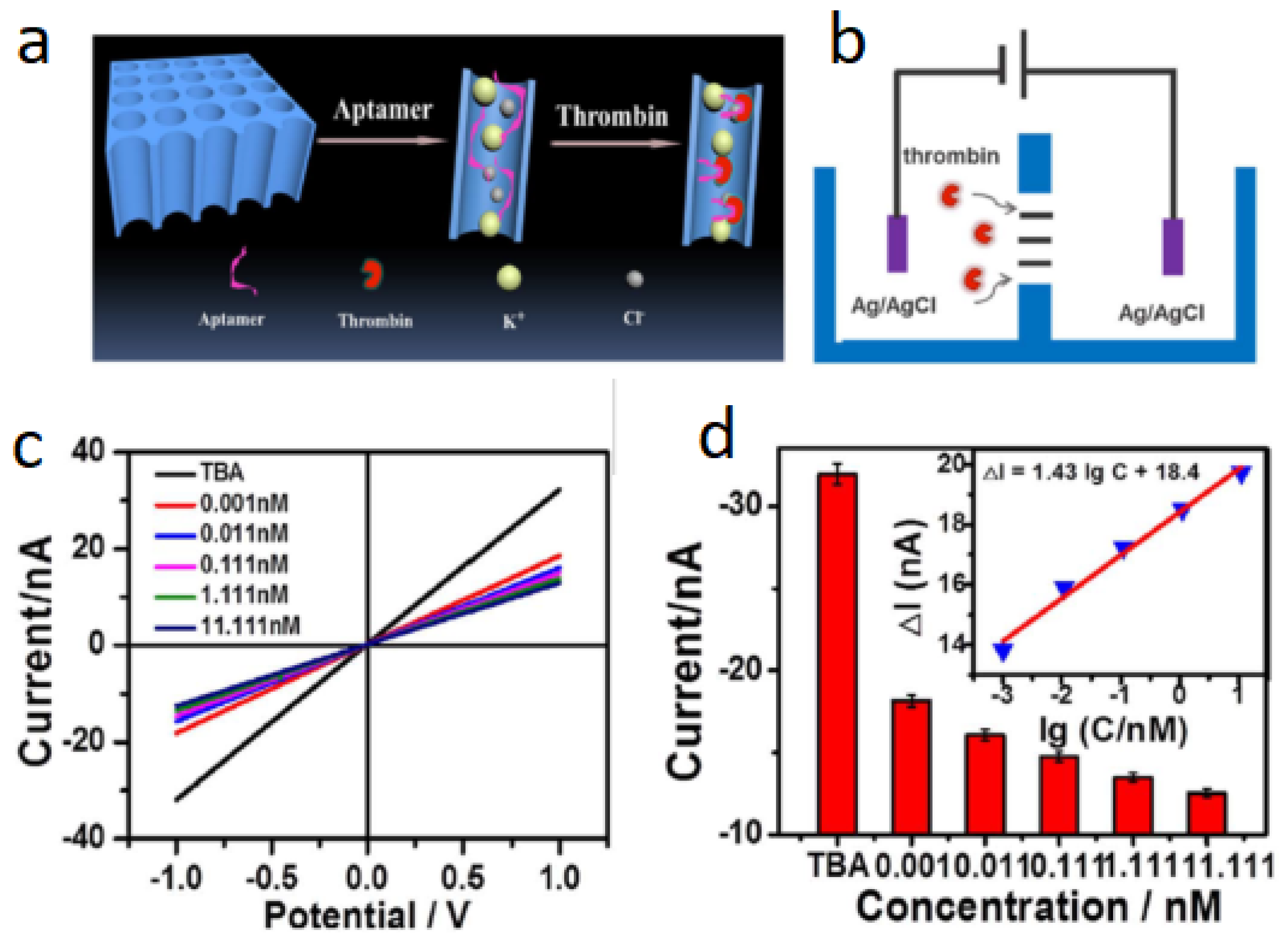
| Nanochannels | PAA nanochannels as separator | GGT TGG TGT GGT TGG | CV | 1000 | 1 pM to 11.111 nM | PAA with Nanochannels | [97] |
| Au NPs coated PET membrane with multiple ion channels | 5′-(NH2)-(CH2)6- CCA TCT CCA CTT GGT TGG TGT GGT TGG-3 | CV | 600 | 3 to 50 nM | Signal-on mechanism | [98] |
| Material Category | Detailed Electrode Material | Aptamer Sequence | Analytical Method | LOD (fM) | Linear Range | Others | Reference |
|---|---|---|---|---|---|---|---|
| Graphene based | GO (on GCE) | 5′-GGT TGG TGT GGT TGG-3′ | DPV | 7.0 × 10⁴ | 0.1 nM to 10 nM | Easy and cheap | [103] |
| n-Fe2O3/graphene (on GCE) | 5′-GGT TGG TGT GGT TGG-3′ | DPV | 1000 | 10 pM to 4.0 nM | Easy immobilization | [104] | |
| GCE with Porphyrin/graphene (on GCE) | 5′- GGT TGG TGT GGT TGG-3′ | DPV | 2.0 × 10⁵ | 5 nM to 1.5 μM | Short incubation time | [69] | |
| Ag NPs/GO (on Au) | TBA1: 5′-SH-(CH2)6-AGT CCG TGG TAG GGC AGG TTG GGG TGA CT-3′ TBA2: 5′-SH-(CH2)6-GGT TGG TGT GGT TGG-3′ | SWV | 3.0 × 10⁴ | 0.05 nM to 5 nM | Sandwich assay design | [24] | |
| CNT based | SWCNT (on GCE) | 5′-AGT CCG TGG TAG GGC AGG TTG GGG TGA CT-3′ | CV | 1.0 × 10⁷ | 10 nM to 100 μM | Simple and cheap design | [23] |
| TiO2-MWCNT/CHIT-SB (on GCE) | 5′-AGT CCG TGG TAG GGC AGG TTG GGG TGA CT-3′ | DPV | 1.0 | 0.00005 nM to 10 nM | Complex electrode | [111] | |
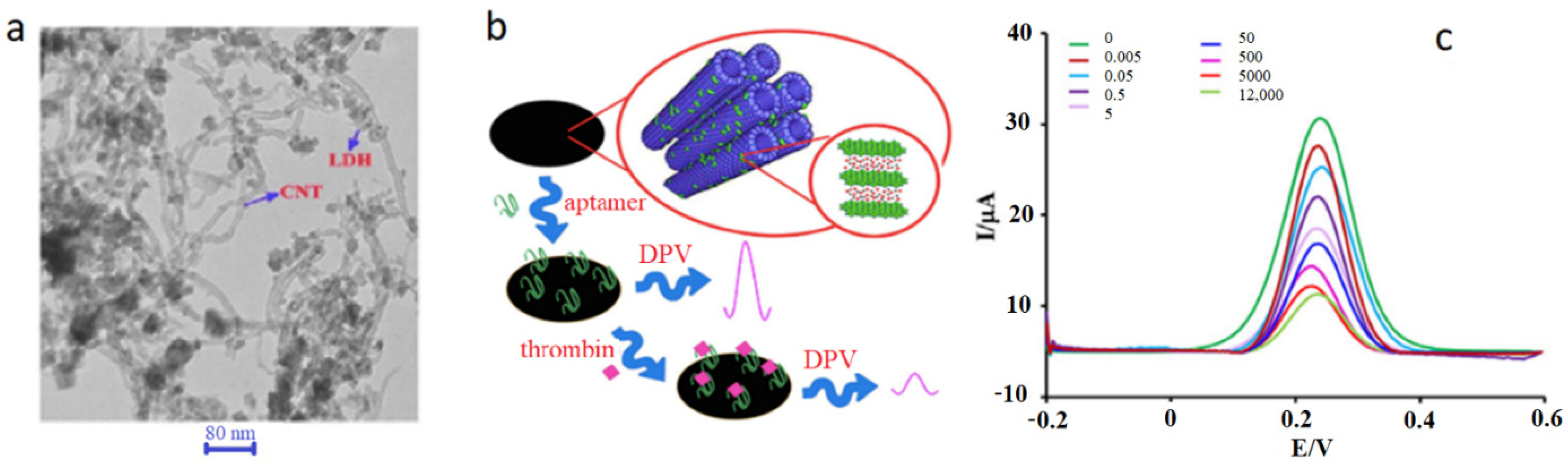
| CNT/ZnCr-LDH (on Au) | 5′-NH2-AGT CCG TGG TAG GGC AGG TTG GGG TGA CT-3′ | DPV | 0.1 | 5 fM to 12 nM | Pre-concentration | [112] | |
| C60/MWCNTs-PEI/PQdot (on SPCE) | 5′–NH2-AGT CCG TGG TAG GGC AGG TTG GGG TGA CT-3′ | DPV | 5 | 50 fM to 20 nM | Large surface area | [45] | |
| Carbon Nanocages | Pt NPs/CNCs (on Au) | 5′-SH-(CH2)6 GGT TGG TGT GGT TGG-3 | DPV | 10 | 0.05 pM to 20 nM | Sandwich assay design | [113] |
| Material Category | Detailed Electrode Material | Aptamer Sequence | Analytical Method | LOD (fM) | Linear Range | Others | Reference |
|---|---|---|---|---|---|---|---|
| Polymer-based | CM-PEG-CM (on GCE) | 5′-NH2-GGT TGG TGT GGT TGG-3′ | DPV | 15.6 | 1 pM to 160 nM | Biocompat-ibility and antibiofouling | [63] |
| HBPE-CA (on ITO) | 5′-NH2-GGT TGG TGT GGT TGG-3′ | DPV | 0.90 | 10 fM to 100 nM | Whole blood analysis | [22] | |
| HBPE-SO3 NPs (on GCE) | 5′-GGT TGG TGT GGT TGG-3′ | DPV | 31 | 2.70 pM to 270 nM | Anticoagulant | [122] | |
| CSPH (on GCE) | TBA1: 5′-COOH-(CH2)10-GGT TGG TGT GGT TGG-3′ TBA2: 5′-NH2-(CH2)6-AGT CCG TGG TAG GGC AGG TTG GGG TGA CT-3′ | DPV | 640 | 1 pM to 10 nM | Conductive and antifouling | [71] |
Publisher’s Note: MDPI stays neutral with regard to jurisdictional claims in published maps and institutional affiliations. |
© 2022 by the authors. Licensee MDPI, Basel, Switzerland. This article is an open access article distributed under the terms and conditions of the Creative Commons Attribution (CC BY) license (https://creativecommons.org/licenses/by/4.0/).
Share and Cite
Yousef, H.; Liu, Y.; Zheng, L. Nanomaterial-Based Label-Free Electrochemical Aptasensors for the Detection of Thrombin. Biosensors 2022, 12, 253. https://doi.org/10.3390/bios12040253
Yousef H, Liu Y, Zheng L. Nanomaterial-Based Label-Free Electrochemical Aptasensors for the Detection of Thrombin. Biosensors. 2022; 12(4):253. https://doi.org/10.3390/bios12040253
Chicago/Turabian StyleYousef, Hibba, Yang Liu, and Lianxi Zheng. 2022. "Nanomaterial-Based Label-Free Electrochemical Aptasensors for the Detection of Thrombin" Biosensors 12, no. 4: 253. https://doi.org/10.3390/bios12040253