Article

Green Synthesis of Silver Nanoparticles Using Jasminum nudiflorum Flower Extract and Their Antifungal and Antioxidant Activity

Guizhou Key Laboratory for Tobacco Quality Research, College of Tobacco Science, Guizhou University, Huaxi District, Guiyang 550025, China
*
Author to whom correspondence should be addressed.
Nanomaterials 2023, 13(18), 2558; https://doi.org/10.3390/nano13182558
Submission received: 22 August 2023 / Revised: 12 September 2023 / Accepted: 12 September 2023 / Published: 14 September 2023

Abstract

:
The synthesis of metal nanomaterials is a timely topic due to their widespread use in fields such as crop protection, the environment, medicine, and engineering. Green synthesis of nanoparticles, which uses plant extracts instead of industrial chemical agents to reduce metal ions, has been developed to decrease costs, reduce pollution, and improve environmental and human health safety. In this paper, silver nanoparticles (AgNPs) were synthesized from the flower extract of Jasminum nudiflorum. The green synthesized AgNPs were characterized by UV-Vis, FTIR, XRD, SEM, and other technologies. The antifungal activity of the prepared AgNPs against Alternaria longipes was tested using the plate method, the concentration dilution method, and other methods, and the antioxidant activity of the prepared AgNPs was evaluated by DPPH and hydroxyl free scavenging methods. The results showed that AgNPs synthesized from J. nudiflorum flower extract have a face-centered cubic structure (fcc), and the average grain size of the nanoparticles is 13 nm; they are also mainly spherical in shape. Additionally, the concentration of AgNPs (ranging from 16 to 128 μg/mL) significantly inhibited the mycelial growth of A. longipes in comparison to the control. The inhibitory rate gradually increased with increasing AgNP concentration, ranging from 70.64% to 79.60% at a concentration of 128 μg/mL. The minimum inhibitory concentration was observed at 32 μg/mL. AgNPs induced overaccumulation of MDA in A. longipes, resulting in cell membrane damage and nucleic acid leakage. Moreover, the AgNPs have significant antioxidant properties, which increase with increasing concentration. The clearance rate of DPPH was 25.46 ± 0.90% when the concentration of AgNPs was 8 μg/mL, and the clearance rate of the hydroxyl radical was 28.62 ± 0.59% when the concentration of AgNPs was 128 μg/mL. Thus, the flower extract from J. nudiflorum holds potential as an environmentally friendly and green alternative for the synthesis of AgNPs, which have antifungal and antioxidant potential.

1. Introduction

Nanotechnology is a branch of materials science and is considered to be one of the key technologies of the future [1]. The emerging field of nanotechnology research refers to the implementation of nanostructures or nanomaterials in the design, development, and application [2]. Nanomaterials refer to substances with a size of about one billionth of a meter and a length of approximately 100 nm [3,4]. These nanomaterials are classified as zero-dimensional, one-dimensional, two-dimensional, and three-dimensional nanomaterials [2,5]. In the context of nanotechnology, zero-dimensional nanoparticles are materials that possess a nanoscale dimension (below 100 nm) in all three dimensions of space [5]. Nanomaterials exhibit distinct properties, such as special surface area, volume, quantum size, and macroscopic tunneling effects, owing to their small particle size. Their unique optical, mechanical, catalytic, and biological properties make nanoparticles useful in various fields, such as biomedicine, industry, and agriculture [6,7,8].
Nanoparticles can be synthesized using physical, chemical, or biological methods. Physical methods encompass laser ablation, evaporation condensation, and mechanical ball milling [9,10,11,12]. On the other hand, chemical methods involve sol gel, microwave-assisted, and microemulsion techniques [8,13,14,15]. These physical and chemical methods commonly employ reducing agents, such as hydrogen peroxide, carbon monoxide, hydroxylamine hydrochloride, dimethylformamide, sodium borohydride, and hydrazine, for nanoparticle synthesis. However, it is important to note that these chemicals can be hazardous. In contrast, biological methods employ microorganisms or plants as a means to produce nanoparticles, utilizing natural components within plants as both reducing and capping agents. Biosynthetic methods offer several advantages, including simplicity, environmental friendliness, low cost, biocompatibility, scalability for large-scale production, and biodegradability [8,16].
Inorganic materials, particularly metal nanoparticles (NPs), have been found to possess antibacterial properties and exhibit greater stability under harsh conditions and high temperatures compared to organic materials [17]. AgNPs are one of the most commonly used nanomaterials because of their antimicrobial properties against a variety of pathogenic bacteria, and silver ions released from AgNPs are thought to play a crucial role in antibacterial activity [18,19]. The effects and toxicity of silver nanoparticles (AgNPs) on microorganisms, plants, and the environment have been extensively researched. These include their mode of action, transformation, transportation within the food chain, and their toxic effects on various plant systems. This also extends to their applications in biomedical and agricultural technology [20]. Studies have demonstrated that AgNPs enhance the germination rate of cabbage seeds, seedling growth, and yield. Furthermore, silver (Ag) does not bioaccumulate in edible tissues, thereby affirming the biosafety of AgNPs [21]. Xiang et al. observed that the life activities of crucian carp in both the clean water control group and the AgNP treatment group remained normal within a 96 h period, with no signs of restlessness or erratic swimming behavior [22]. AgNPs synthesized from petal extract of Cydonia oblonga exhibited an inhibitory zone diameter of 10 ± 0.80 mm against Erwinia amovora, a pathogenic bacterium that causes fire blight. Furthermore, AgNPs significantly reduced both the diameter and depth of the necrotic area of immature Pyrus communis fruits. In comparison, the necrotic zone diameter on fruits inoculated with pathogens alone was 32 ± 0.45 mm, almost eight times larger than that observed when bacterial and AgNPs suspensions were applied [23]. AgNPs also demonstrated antifungal potential. Green synthesis of AgNPs from leaves of Pongamia glabra resulted in a significant reduction in the weight of mycelium and spore count of the plant pathogen Rhizopus stolonifera [24]. AgNPs synthesized from rice leaves displayed antimicrobial activity against Rhizoctonia solani. At a concentration of 10 μg/mL, AgNPs exhibited maximum inhibition of 96.7% against TS-06, followed by 93% inhibition against TS-22. The minimum inhibitory concentration (MIC) values of AgNP on mycelium and sclerotia of R. solani were 5–10 and 15–20 μg/mL, respectively. AgNP treatment resulted in a significant decrease in percent disease incidence (PDI) values under in vitro conditions, and Ag NP at a concentration of 20 μg/mL completely inhibited the incidence of all R. solani isolates. [25]. AgNPs derived from wheat leaf extract demonstrated resistance against four fungi. The MIC of AgNPs against Fusarium oxysporum was found to be 41.7 μg/mL, while for Aspergillus versicolor, A. niger, and A. brasiliensis, it was determined to be 208.3 μg/mL [18]. Compared with the control, the green synthesis of AgNPs with 10.7 μg/mL Citrus limetta peel extract increased the content of protein and inhibited bacterial and yeast pathogens. AgNPs would damage the cell membrane integrity of bacteria and candida, cause changes in cell morphology, and lead to cell foaming and thick exudate deposition around cells [26].
Plant-based synthesis of silver nanoparticles has been achieved using a variety of plant materials, including rice leaves, wheat leaves, the leaves of Pongamia glabra, petals of C. oblonga, Citrus limetta peel, Cleome viscosa, and other plants [18,23,24,25,26,27]. Plant extracts abundant in phytoconstituents, such as alkaloids, flavonoids, and terpenoids, have the ability to reduce metal salts from Ag+ to Ag0, leading to the synthesis of AgNPs [28]. Due to the presence of diverse biomolecules in different plants, nanoparticles generated from distinct plant sources may demonstrate varying antimicrobial properties. Hence, it is imperative to explore new plant extract sources for nanoparticle synthesis to enhance the antimicrobial efficacy of the nanoparticles [17]. Jasminum nudiflorum Lindl, commonly cultivated as an ornamental plant and used in traditional medicine in China, is distinguished by the presence of dicyclic iridoid glycosides, pigments, flavonoids, and fatty acids in its flowers [29]. This study aimed to characterize the size and shape of green-synthesized silver nanoparticles (AgNPs) using an extract from J. nudiflorum flowers. Additionally, the study sought to assess the antibacterial activity of these green-synthesized AgNPs against Alternaria longipes and to evaluate their antioxidant activity. The research is distinguished by its validation of a more optimized synthesis scheme, which includes the volume ratio of plant extract to silver nitrate solution and reaction time. The AgNPs were synthesized from the J. nudiflorum flower extract without the need for catalysts or surfactants. The results indicate that these biosynthetic AgNPs can significantly inhibit A. longipes, the primary pathogen of tobacco leaf spot (tobacco brown spot), as well as exhibit antioxidant activity.

2. Materials and Methods

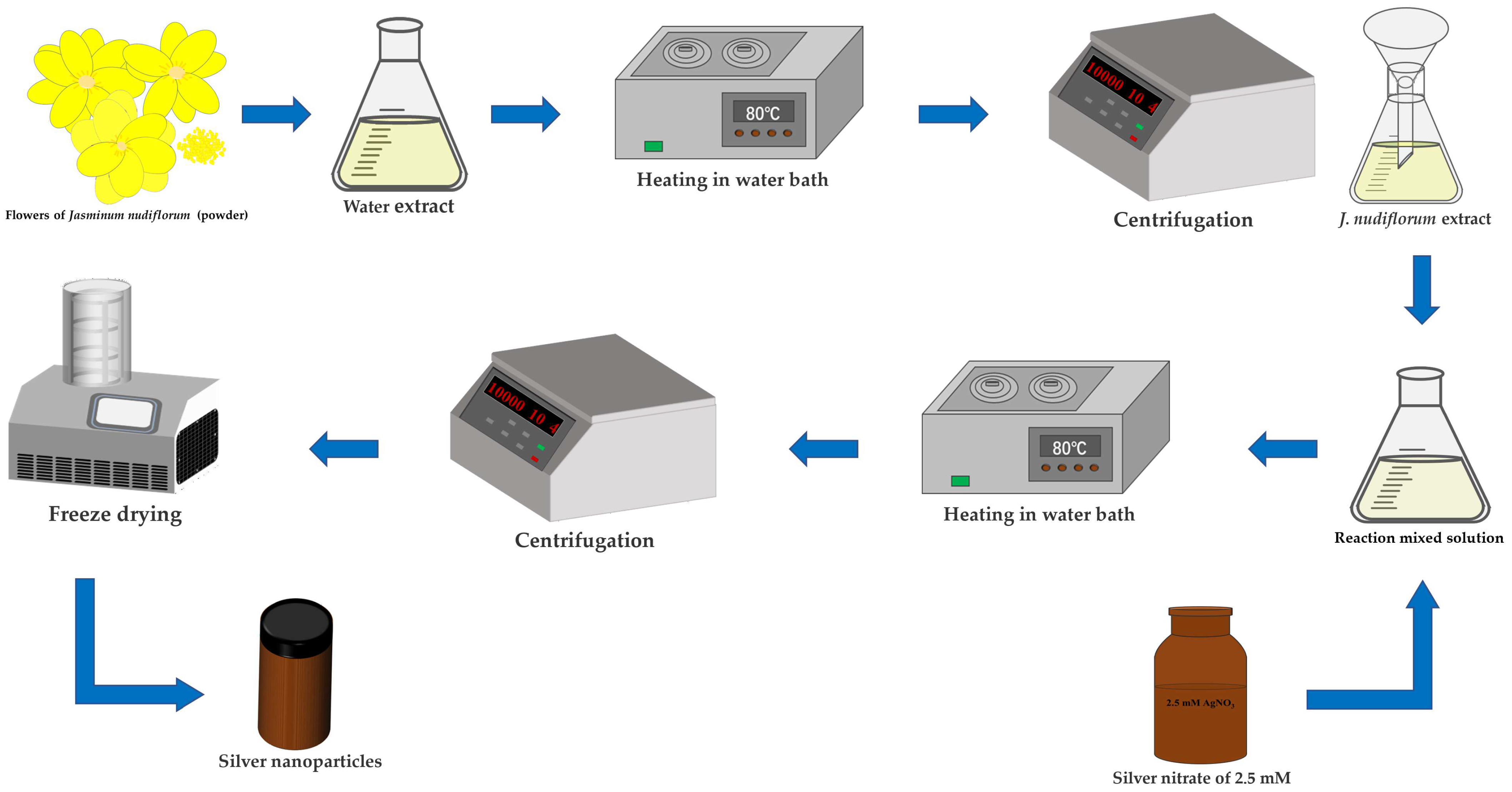
2.1. Preparation of Plant Materials and Extracts

The flowers of J. nudiflorum were collected from the campus of Guizhou University (106°66′ E, 26°45′ N) in Guiyang, Guizhou province, China. The flowers were washed with tap water, rinsed with ultrapure water to removes the dust particles, air-dried, and ground into fine powder with an electric grinder. The powder was passed through a 40-mesh sieve to remove large impurities. The extract was prepared by putting 1.5 g of powder together with 100 mL of ultrapure water in a 250 mL conical flask and heating it in a water bath at 80 °C for 60 min. The crude water extract was centrifuged at 10,000 rpm for 10 min and filtered with filter paper. Then, the extract was stored in a refrigerator at 4 °C for future research.

2.2. Preparation of AgNPs

The green-synthesis method is referred to by Mehata et al. [30]. To synthesize AgNPs, an orthogonal test was conducted to determine the optimal volume ratio of reaction mixture (A) and reaction time (B) (Table 1). Three different volume ratios of J. nudiflorum flower extract to a 2.5 mM AgNO3 (SCRC, Shanghai, China) solution were tested: 1 mL:1 mL for A1, 1.25 mL:1 mL for A2, and 1.5 mL:1 mL for A3. The reaction was carried out in a water bath at 80 °C for seven different durations: B1 for 1 h; B2 for 2 h; B3 for 3 h; B4 for 4 h; B5 for 5 h; B6 for 6 h; and B7 for 7 h, respectively. The mixtures were then centrifuged at 10,000 rpm for 10 min, and the precipitate was washed with ultrapure water to remove any unreacted reagents. Subsequently, the resulting AgNPs were freeze-dried into a powder and stored in a brown bottle. Figure 1 illustrates the process of synthesizing AgNPs from the flower extract of J. nudiflorum.

2.3. Characterization of AgNPs

To identify the structure, properties, and reaction mechanism of the synthesized nanoparticles, various characterization techniques were utilized [17]. The liquid mixture after reaction in the water bath was subjected to analysis via an Ultraviolet-Visible (UV-Vis) spectrophotometer (Analytic Jena AG, Specord 200 Plus, Jena, Germany). The silver nanoparticles (AgNPs), after freeze-drying, were examined using Fourier-transform infrared (FTIR) spectroscopy (Thermo Scientific Nicolet iS20, Waltham, MA, USA), X-ray diffraction (XRD) (Bruker D8 Advance, Mannheim, Germany) pattern analysis, and scanning electron microscopy (SEM) (ZEISS Sigma 300, Jena, Germany). The absorption spectra of the biosynthesized silver nanoparticles were measured using a UV-Vis spectrophotometer with a wavelength range of 280–700 nm. The FTIR spectroscopy was employed to identify the functional groups present in the silver nanoparticles within the range of 4000–400 cm−1. The XRD diffraction pattern was obtained using Cu Kα radiation at 40 kV and 40 mA. The SEM was utilized to assess the morphology and particle size distribution of the nanoparticles.

2.4. Antifungal Activity of AgNPs

The strain of A. longipes was isolated from tobacco leaves infected with brown spot disease in Meitan County, Guizhou Province and stored in the Guizhou Provincial Key Laboratory for Tobacco Quality Research.

2.4.1. Effect of AgNPs on A. longipes Mycelium

The effect of AgNPs on the mycelium of A. longipes was evaluated by inoculating 6 mm diameter pathogenic bacteria cake in a PCA medium containing different concentrations of nanoparticles, taking 0 μg/mL nanoparticles as the control.
The effect of silver nanoparticles on A. longipes mycelium was evaluated by inoculating 6 mm diameter pathogenic cake in a PCA medium containing different concentrations of AgNPs, with 0 μg/mL NPs serving as the control. The mycelium dimension was measured at 3, 6, and 9 days, and the inhibition rates were calculated. Each treatment was replicated three times [22]. The inhibition rate was calculated as follows:
Inhibition   rate   % = C o n t r o l   g r o u p   c o l o n y   g r o w t h   d i a m e t e r T r e a t m e n t   g r o u p   c o l o n y   g r o w t h   d i a m e t e r C o n t r o l   g r o u p   c o l o n y   g r o w t h   d i a m e t e r × 100

2.4.2. Effect of AgNPs on Membrane Permeability and Malondialdehyde Content of A. longipes

A. longipes was cultured in PDB medium following inoculation and incubated in a rotating shaker at 150 rpm and 28 °C for a duration of 10 days. The mycelia were collected by centrifugation at 8000× g for 5 min using a filter centrifuge tube. Subsequently, 0.2 g of mycelium was placed in a PDB culture medium containing different concentrations of nanoparticles. The mixture was cultured in a rotating shaker at 150 rpm and 28 °C for 0, 2, 4, and 8 h to assess the selected parameters. This experimental process was repeated three times [31].
To determine cell membrane permeability, the mycelia were removed through centrifugation at 8000× g for 15 min, and the resulting supernatant was collected for subsequent measurement of cell leakage. Nucleic acid leakage was quantified at 260 nm using a spectrophotometer [31]. The formula for the quantity of DNA was as follows [32]:
Quantity   of   DNA   μ g mL = OD   at   260   nm × 50
The measurement of MDA content involved adding 0.2 g of mycelium to 2 mL of a 10% trichloroacetic acid (TCA) solution, which was then homogenized by grinding. After centrifuging at 8000× g and 4 °C for 15 min, the supernatant was collected. Subsequently, 1 mL of the supernatant was mixed with an equal volume of a 0.6% thiobarbituric acid (TBA) solution. The resulting mixture was boiled for 30 min, rapidly cooled to room temperature using tap water, and measured at 450 nm, 532 nm, and 600 nm. The MDA content was expressed as μmol/g [31].
M D A   μ mol . g 1 = 6.452 × A 532 A 600 0.559 × A 450 × V t V s × m
In this formula, Vt represents the total volume (mL) of the extract, Vs represents the volume (mL) of the extract for measurement, and m represents the sample weight (m).

2.4.3. Determining the Minimum Antifungal Concentration of AgNPs

The minimum inhibitory concentration (MIC) of silver nanoparticles was deter-mined by visually observing the growth of fungi in continuously diluted suspensions of nanoparticles. Initially, PDB and nanoparticles were mixed in the first conical flask and subsequently used to dilute the ensuing conical flasks. Next, a spore suspension containing 2 × 10⁵ spores/mL was inoculated into each conical flask, and the cultures were incubated at 28 °C for a period of 3 days. Visual observation of fungal growth was con-ducted to ascertain the value of the MIC. This experiment was repeated three times [31].

2.5. Antioxidant Activity of AgNPs

The antioxidant activity of silver nanoparticles was evaluated using two different assays. The first assay, based on the 1,1-diphenyl-2-trinitrophenylhydrazine (DPPH) radical scavenging method, was conducted with a slight modification of the procedure outlined by Dridi et al. [33]. In this assay, a 1 mL sample solution was mixed with an equal volume (1 mL) of DPPH solution. After vigorous shaking, the mixture was allowed to react in darkness for 30 min. The absorbance of the resulting solution was measured at 517 nm, and the percentage of inhibition was then calculated using the following formula:
DPPH   clearance   rate   % = A 0 A i A j A 0 × 100
In this formula, A0 represents the absorbance of a solution containing 1 mL anhydrous ethanol and 2 mL DPPH, Ai represents the absorbance of a solution containing 1 mL sample and 2 mL DPPH, and Aj represents the absorbance of a solution containing 1 mL sample and 2 mL anhydrous ethanol.
For the second assay, the hydroxyl radical scavenging activity of the AgNPs was determined following the procedure described by Ansar et al. [34]. A reaction mixture consisting of approximately 3 mL was prepared by adding 1 mM salicylic acid, 9 mM ferrous sulfate, and 1 mL hydrogen peroxide to 2 mL of synthesized AgNPs. The mixture was then subjected to a reaction at 60 °C in a water bath for 37 min. After incubation, the absorbance of the resulting solution was measured at 510 nm. The percentage of hydroxyl radical scavenging activity was calculated using the following formula:
Hydroxyl   radical   scavenging   rate   % = A 0 A i A j A 0 × 100
In this formula, A0 represents the absorbance of a solution containing 1 mL salicylic acid, 1 mL ferrous sulfate, 1 mL hydrogen peroxide, and 2 mL ultrapure water; Ai represents the absorbance of a solution containing 1 mL salicylic acid, 1 mL ferrous sulfate, 1 mL hydrogen peroxide, and 2 mL sample; and Aj represents the absorbance of a solution containing 1 mL salicylic acid, 1 mL ferrous sulfate, 1 mL ultrapure water, and 2 mL sample.

2.6. Data Processing

The data were presented as the standard error of the mean, based on three replicates. To compare the means between treatments, analysis of variance (ANOVA) and least significant difference (LSD) tests were conducted using SPSS 26.0 software. A significance level of p < 0.05 was used to determine statistical significance.

3. Results and Discussion

3.1. Production of Silver Nanoparticles

To determine the optimal synthesis ratios of the flower extract of J. nudiflorum and AgNO3 solution and reaction times for synthesizing AgNPs, three different ratios of plant extracts to AgNO3 solution (1 mL:1 mL, 1.25 mL:1 mL, and 1.5 mL:1 mL) and seven different reaction durations (1 h, 2 h, 3 h, 4 h, 5 h, 6 h, and 7 h) were prepared, resulting in a total of 21 experimental conditions. The reaction between the flower extracts of J. nudiflorum and AgNO3 resulted in a visible color transition from yellowish to reddish-brown, indicating the successful green synthesis of AgNPs (Figure 2). This color alteration was attributed to the surface plasmon resonance (SPR) of silver ions, which were reduced to stable AgNPs by active molecules in the extract [30,35]. At 1 h, the red-brown color was clearly observed in A1B1, A2B1, and A3B1, indicating the initiation of AgNP synthesis. However, as the reaction time was extended to 2–7 h, no significant visual changes were observed among A1, A2, and A3. Nevertheless, over time, the color of the reaction mixture with the same ratio of plant extract to AgNO3 solution became slightly lighter, and the solution became gradually denser (Figure 2). Additionally, the 21 treated reaction mixtures were characterized using UV-Vis spectroscopy (Figure 3a). At 1 h, the reddish-brown color of A1B1 was lighter and the solution was thicker compared to A2B1 and A3B1 (Figure 2). The UV-Vis spectrum showed an increase in absorbance for A1B1(Figure 3a), indicating the presence of a greater number of silver nanoparticles [36]. As time progressed, each reaction solution became denser and the absorbance increased (Figure 2 and Figure 3a), suggesting that more AgNPs were synthesized.

3.2. Analysis of UV-Vis Absorption Spectra of Green-Synthesized AgNPs

The UV-Vis spectrum of the AgNP liquid, as depicted in Figure 2, revealed an absorption peak within the 280~700 nm range, with a prominent peak at approximately 435 nm when compared to the control (CK, ultrapure water) as shown in Figure 3a. The absorption peak around 330 nm might be attributed to the plant extract (Figure 3b). This UV-Vis analysis confirmed the formation of AgNPs. Typically, the UV-Vis spectrum of AgNPs presents a strong visible light band around 435 nm (Figure 3a), and the 350–550 nm range is identified as the typical SPR absorption band of AgNPs. The absorption peak for spherical AgNPs is usually between 410 and 450 nm [37,38]. It was observed that with an increasing reaction time (1–7 h), and a volume ratio of J. nudiflorum flower extract to AgNO3 solution at 1 mL:1 mL, AgNPs exhibited a blue shift, indicating the formation of smaller AgNPs [30,39], and an increase in absorption spectral intensity, suggesting that more AgNPs were synthesized [30,40]. When the volume ratio of flower extract to AgNO3 solution was increased to 1.25 mL:1 mL and 1.5 mL:1 mL, a redshift phenomenon was observed in AgNPs, and the absorption spectral intensity increased with extended reaction time (1–7 h). In the ultraviolet absorption spectrum, the absorption peak of A2B7 (435 nm, 1.0561) is positioned between A1B7 (433 nm, 1.0009) and A3B7 (438 nm, 1.0418), with A2B7 exhibiting a higher peak. As a result, AgNPs synthesized under A2B7 condition were selected for further investigation.
In this study, the AgNPs synthesized in test A2B7 by the flower extract of J. nudiflorum exhibited a maximum absorption peak at 435 nm, corresponding to surface plasmon resonance [41]. By comparing the UV-Vis spectra of the flower extract and AgNO3 (Figure 3b), at 435 nm, there was no absorption peak in the flower extract of J. nudiflorum and AgNO3 solution, the formation of AgNPs was confirmed (results that are similar to the study of Takcı et al. [28]), and there was no absorption peak in the plant extract at the maximum absorption peak of AgNPs. Further experiments were then conducted on AgNPs synthesized under A2B7 conditions.

3.3. Fourier Infrared Spectroscopy of Green-Synthesized AgNPs

The FTIR analysis was employed to identify the functional groups potentially involved in the synthesis of AgNPs. In the case of the green synthesis of NPs, plant extracts are utilized as both reducing and stabilizing agents, presumably due to the presence of various metabolites, including flavonoids, alkaloids, amino acids, proteins, vitamins, and enzymes [8,42,43]. The FTIR spectra of the synthetic AgNPs derived from J. nudiflorum flower are presented, which display prominent peaks at 3431 cm−1, 2918 cm−1, 1622 cm−1, and 1022 cm−1 (Figure 4). These peaks can be attributed to the vibrations of O-H, C-H, C=C stretching, and C-O stretching, respectively. Notably, the substantial absorption peaks observed at 1029 cm−1, 2922 cm−1, and 3414 cm−1 primarily correspond to the polyphenols extracted from the plants [28]. Additionally, peaks at 3795 cm−1, 3452 cm−1, 3439 cm−1, 3430 cm−1,3422 cm−1, 3418 cm−1, 3360 cm−1, and 3255 cm−1 can be ascribed to the stretching vibrations of the O-H or N-H groups present in alcohol or phenol compounds that bind to the surface of AgNPs [44,45,46,47,48].
Moreover, peaks at 2930 cm−1, 2923 cm−1, 2922 cm−1,2921 cm−1, 2900 cm−1, and 2880 cm−1 are associated with the C-H stretching vibrations of alkanes [23,28,35,46,47]. The presence of aldehyde or ketone functional groups (C=O) from flavonoid and tannin derivatives can be inferred from the peaks at 1617 cm−1,1626 cm−1, 1650 cm−1, and 1653 cm−1 [44,47,49]. While the peak at 1633 cm−1 and 1631 cm−1 signifies C=C stretching vibrations of the hydrocarbon portion of biological double-bond molecules, the band at 1052 cm−1 and 1031 cm−1 may be attributed to C-O stretching [35,46,50]. And the peaks at 1607 cm−1 and 1396 cm−1 correspond to the asymmetric stretching of NO2 [50]. Flavonoids, monoterpenes, iridoids, mites, and glycosides constitute the major compounds in the Oleaceae Hoffmans. & Link family of J. nudiflorum [51].

3.4. Analysis of Green-Synthesized AgNPs through X-ray Spectroscopy

To ascertain the crystallinity of AgNPs, powder XRD analysis was conducted on the AgNPs. The XRD spectrum of AgNPs green-synthesized using the flower extract of J. nudiflorum demonstrates that the diffraction peaks align with the standard silver planes (PDF: 04-003-7118) at angles of 37.88°, 44.18°, 64.17°, 77.09°, and 81.17°, respectively, corresponding to the (111), (200), (220), (311), and (222) planes (Figure 5). This confirms the face-centered cubic (fcc) crystal structure of AgNPs. Compared with the standard silver characteristic peaks 37.98°, 44.14°, 64.20°, 77.09°, and 81.21°, there are slight differences in peak positions, which may be related to the residual plant extract on the surface of the synthesized silver nanoparticles [52]. In addition, 27.69°, 32.06°, 46.00°, 54.57°, and 57.08°corresponded to the (111), (200), (220), (311), and (222) planes of the standard AgClNPs (PDF:97-006-4734), respectively (Figure 5). This was similar to the XRD spectra of Kohan Baghkheirati [53] and Kumkoon et al. [54]. The formation of AgClNPs might be the result of the reaction of Ag in AgNO3 with the chemical compound Cl in the plant extract [54]. Additionally, the XRD spectrum showcased additional unmatched peaks, possibly attributable to the crystallization of bio-organic compounds on the surface of AgNPs. The noise is related to the phytochemical components in the possible extract of the impurity peak [24,49,54,55]. The size of AgNPs was calculated using Scherrer’s formula: D = K × λ β × c o s θ . The size of the green-synthesized silver nanoparticles from the flower extract of J. nudiflorum was about 13 nm.

3.5. Analysis of the Morphology and Size Range of Green-Synthesized AgNPs Using Scanning Electron Microscopy

The morphology and size of AgNPs synthesized from J. nudiflorum flower were examined using SEM images. The images revealed that the synthesized AgNPs were predominantly spherical in shape and exhibited good dispersion (Figure 6). The size of the synthesized AgNPs was measured using Nano Measurer, yielding a range of 11 to 78 nm, with an average size of approximately 25 nm (Figure 6b). Furthermore, these AgNPs displayed a predominantly spherical shape, which corresponds well with the characteristic SPR peaks observed in the UV-Vis spectrum [28]. It is worth noting that the green-synthesis method resulted in some agglomeration of the nanoparticles, as high surface activity and the absence of coordinating atoms on their surface tend to cause their aggregation [5].

3.6. Inhibitory Effect of Green-Synthesized AgNPs on A. longipes

The effect of AgNPs on the growth of A. longipes mycelium was investigated. Mycelium dimension was measured on days 3, 6, and 9 and compared to a control group (0 μg/mL AgNPs). The results showed that the growth of A. longipes mycelium was significantly inhibited by AgNPs in concentrations ranging from 16 to 128 μg/mL. The inhibition rate increased as the concentration of AgNPs increased, with ranges of 6.25–79.60%, 5.06–75.72%, and 3.33–70.64% observed at 3, 6, and 9 days, respectively (Figure 7a and Figure 7b). This finding supports the study by Malandrakis et al. [56], which demonstrated the inhibitory effect of NPs on the in vitro mycelial growth of fungal strains.
To further investigate the impact of AgNPs on the culture of A. longipes, the strain was cultured on PCA medium with varying concentrations of AgNPs, and the colony morphology was observed throughout the cultivation period (Figure 7c). The results showed that the radial growth of A. longipes treated with AgNPs was slower compared to the control group, and the growth rate decreased with increasing concentrations of AgNPs. Similar results were found by Xiang et al. [22] in their study on the inhibitory effect of AgNPs on the growth of Alternaria alternata. However, Xiang et al. [22] noted that the inhibitory effect was more prominent at lower concentrations of AgNPs, which could be attributed to variations in strain sensitivity and the smaller particle size of their AgNPs. When the particle size is smaller, nanoparticles can easily penetrate the cell wall of microorganisms and enhance their absorption by microbial cells [57]. The presence of nanoscale micropores in biofilms, such as cell membranes (0.4–1 nm) and nuclear membranes (50–70 nm), as well as the cell wall micropores (5–20 nm), facilitate the direct entry of metal-based nanoparticles into cells and even organelles. Moreover, smaller particle sizes of metal-based nanoparticles promote their accumulation and potential toxicity in organisms [58]. The presence of negative surface charges on nanoparticles, which are closely associated with bacterial cell wall charges, can also reduce efficacy by causing repulsion and hindering absorption by bacteria [59]. Based on these findings, the concentration of 128 μg/mL AgNPs was selected to further investigate its antifungal mechanism.

3.7. Assessment of Cell Membrane Permeability and Malondialdehyde Content

Cell membrane permeability serves as a critical indicator of cell membrane impairment. AgNP treatment resulted in nucleic acid leakage in A. longipes. When A. longipes was treated with 0 μg/mL AgNPs, there was no notable change in nucleic acid leakage. However, nucleic acid leakage was detected after 4 h in A. longipes treated with 128 μg/mL AgNPs, and the leakage increased gradually with longer incubation times. After incubation with 128 μg/mL AgNPs for 8 h, the quantity of DNA 94.32 ± 1.48 μg/mL was significantly higher than the measured value of 47.48 ± 0.72 μg/mL at 0 h (Figure 8a). Xiang et al. [22]. observed that AgNP treatment disrupted the cell membrane permeability of A. alternata.
The MDA content showed no significant change in A. longipes treated with 0 μg/mL AgNPs over time. However, there was a significant change in the MDA content of A. longipes treated with 128 μg/mL AgNPs. After incubation for 8 h, the MDA content in A. longipes treated with 128 μg/mL AgNPs was 4.82 ± 0.14 μmol/g, which was significantly higher than 3.37 ± 0.17 μmol/g at 0 h (Figure 8b). This indicates that AgNPs induced lipid peroxidation in A. longipes. Excessive accumulation of MDA alters the structure and functionality of biomolecules on the cell membrane, including lipids, proteins, and nucleic acids, ultimately disrupting cell metabolism and potentially leading to cell death [60]. Hirpara et al. [61] discovered that the antifungal effect of green-synthesized AgNPs on MIC at day 3 involved mycelial cell membrane leakage (sugars and proteins), lipid peroxidation, inhibition of respiratory chain dehydrogenase activity, and disruption of the mycelium nucleus disc structure, resulting in the death of the plant pathogen. Furthermore, Zhu et al. [31] found that ZnO NPs induced excessive MDA accumulation in A. alternata, causing cell membrane damage and resulting in protein and nucleic acid leakage. Therefore, we propose that the green-synthesized AgNPs from the flower extract of J. nudiflorum inhibit the activity of A. longipes by inducing the excessive accumulation of MDA, which leads to increased membrane permeability. This mechanism may contribute to the antifungal properties of AgNPs.

3.8. Minimum Inhibitory Concentration of AgNPs

The minimum inhibitory concentration (MIC), which denotes the concentration at which A. longipes is completely inhibited, was evaluated. As demonstrated (Figure 9), the MIC value for AgNPs synthesized from spring green was 32 μg/mL. It is important to note that there existed a disparity in the inhibitory concentration of AgNPs against spores and mycelium, a finding aligned with Malandrakis et al.’s [56] study. Their research indicated that AgNPs, CuNPs, CuONPs, and ZnO2NPs generally exhibit toxicity levels towards spores that are 10 to 100 times higher than that to mycelium. These results suggest that AgNPs synthesized from the flower extracts of J. nudiflorum possess antifungal potential.

3.9. Antioxidant Capacity of Green Synthetic AgNPs

The antioxidant capacity of AgNPs was assessed through DPPH and hydroxyl radical scavenging experiments. Ascorbic acid (AsA) served as the standard antioxidant. The scavenging capacity of the sample for free radicals increased with higher concentrations of AgNPs (1–128 μg/mL), indicating its antioxidant properties (Figure 10). Consistent with the findings of Padalia et al. [41], the antioxidant activity also increased with increasing concentration. The DPPH (Figure 10a) and hydroxyl radical scavenging activities (Figure 10b) of AgNPs ranged from 3.80 ± 0.32% to 25.46 ± 0.90% and from 2.50 ± 0.22% to 28.62 ± 0.59%, respectively. The IC50 values for DPPH and hydroxyl radical scavenging with AgNPs were 16.07 and 222.53, respectively. Correspondingly, the IC50 values for AsA in DPPH and hydroxyl radical scavenging were 4.83 and 104.42, respectively. The scavenging potential of AgNPs was relatively lower compared to the significant antioxidant activity of the standard AgNPs, as supported by Ansar et al. [33]. Thus, the synthesized AgNPs demonstrated potential antioxidant properties, as evidenced by the aforementioned experiments.

4. Conclusions

This study presents a straightforward and eco-friendly approach to synthesizing AgNPs by employing J. nudiflorum flower extract for green synthesis. The utilization of J. nudiflorum flower extract serves dual purposes as both a reducing agent and a capping agent, thereby facilitating the formation of stable AgNPs. The UV-Vis spectrum peak, occurring at approximately 435 nm, and the X-ray diffraction patterns, specifically the 2θ peaks at 37.88°, 44.18°, 64.17°, 77.09°, and 81.17°, confirm the face-centered cubic (fcc) crystal structure of the AgNPs, and the particle size is about 13 nm. SEM examinations indicate that the particles exhibit predominantly spherical morphology, and FTIR spectroscopy analysis reveals prominent O-H, C-H, C=O, and C-O stretching vibrations. Furthermore, the synthesis of AgNPs exhibits notable antifungal effects, exhibiting an antifungal rate of 79.60% at 3 days, 75.72% at 6 days, and 70.64% at 9 days for AgNPs at a concentration of 128 g/mL. The minimum inhibitory concentration is determined to be 32 μg/mL, which induces an increase in MDA levels, damage to the cell membrane, and leakage of nucleic acids in A. longipes. Moreover, the green synthesized AgNPs show considerable antioxidant activity, with DPPH inhibition rates ranging from 3.80 ± 0.32% to 25.46 ± 0.90%, and hydroxyl radical scavenging capacities ranging from 2.50 ± 0.22% to 28.62 ± 0.59%. It seems that the green synthesis of AgNPs from the flower extract of J. nudiflorum provides a feasible alternative method for chemical synthesis, and the green-synthesized AgNPs has antifungal and antioxidant potential and is expected to be developed into an antifungal agent and an antioxidant.

Author Contributions

Conceptualization, Q.Y. and J.P.; Methodology, Q.Y., G.L. and J.P.; Validation, Q.Y., J.G., X.L. and C.P.; Formal analysis, Q.Y., J.G., X.L. and C.P.; Writing—original draft, Q.Y.; Resources, J.P.; Writing—review and editing, J.P.; Supervision, J.P.; Funding acquisition, G.L. and J.P. All authors have read and agreed to the published version of the manuscript.

Funding

This work was supported by the National Natural Science Foundation of China, funding number: 32060420; the Science and Technology Project of Guizhou Company of China Tobacco Corporation, funding number: 201913.

Data Availability Statement

Data produced in this study can be made available upon reasonable request.

Acknowledgments

We are grateful to Lijuan Peng for the donation of the experimental material.

Conflicts of Interest

The authors declare no conflict of interest.

References

  1. Zhang, F.; Jia, J.; Yao, X. Allium ampeloprasum leaf aqueous extract green-formulated Ag nanoparticles: Determination of anti-human lung cancer and antioxidant effects. J. Eng. Res. 2023, 11, 100091. [Google Scholar] [CrossRef]
  2. Raduwan, N.F.; Shaari, N.; Kamarudin, S.K.; Masdar, M.S.; Yunus, R.M. An overview of nanomaterials in fuel cells: Synthesis method and application. Int. J. Hydrogen Energy 2022, 47, 18468–18495. [Google Scholar] [CrossRef]
  3. Chaudhary, J.; Tailor, G.; Yadav, M.; Mehta, C. Green route synthesis of metallic nanoparticles using various herbal extracts: A review. Biocatal. Agric. Biotechnol. 2023, 50, 102692. [Google Scholar] [CrossRef]
  4. Hossain, N.; Mobarak, M.H.; Hossain, A.; Khan, F.; Mim, J.J.; Chowdhury, M.A. Advances of plant and biomass extracted zirconium nanoparticles in dental implant application. Heliyon 2023, 9, e15973. [Google Scholar] [CrossRef] [PubMed]
  5. Yao, H.; Li, L.; Li, W.; Qi, D.; Fu, W.; Wang, N. Application of nanomaterials in waterborne coatings: A review. Resour. Chem. Mater. 2022, 1, 184–200. [Google Scholar] [CrossRef]
  6. Saratale, R.; Benelli, G.; Kumar, G.; Kim, D.; Saratale, G. Bio-fabrication of silver nanoparticles using the leaf extract of an ancient herbal medicine, dandelion (Taraxacum officinale), evaluation of their antioxidant, anticancer potential, and antimicrobial activity against phytopathogens. Environ. Sci. Pollut. Res. 2018, 25, 10392–10406. [Google Scholar] [CrossRef]
  7. Vanlalveni, C.; Lallianrawna, S.; Biswas, A.; Selvaraj, M.; Changmai, B.; Rokhum, S.L. Green synthesis of silver nanoparticles using plant extracts and their antimicrobial activities: A review of recent literature. RSC Adv. 2021, 11, 2804–2837. [Google Scholar] [CrossRef]
  8. Selmani, A.; Kovačević, D.; Bohinc, K. Nanoparticles: From synthesis to applications and beyond. Adv. Colloid Interface Sci. 2022, 303, 102640. [Google Scholar] [CrossRef]
  9. Muñoz, J.; Cervantes, J.; Esparza, R.; Rosas, G. Iron nanoparticles produced by high-energy ball milling. J. Nanopart. Res. 2007, 9, 945–950. [Google Scholar] [CrossRef]
  10. Zhan, L.; Xiang, X.; Xie, B.; Gao, B. Preparing lead oxide nanoparticles from waste electric and electronic equipment by high temperature oxidation-evaporation and condensation. Powder Technol. 2017, 308, 30–36. [Google Scholar] [CrossRef]
  11. Car, J.; Blažeka, D.; Bajan, T.; Krce, L.; Aviani, I.; Krstulovic, N. A quantitative analysis of colloidal solution of metal nanoparticles produced by laser ablation in liquids. Appl. Phys. A 2021, 127, 1–14. [Google Scholar] [CrossRef]
  12. Petrikaitė, V.; Skapas, M.; Stankevičius, E. Generation of gold and silver nanoparticles using laser ablation of thin bimetallic films and bulk targets in water. Opt. Mater. 2023, 137, 113535. [Google Scholar] [CrossRef]
  13. Habibi, M.H.; Karimi, B. Effect of the annealing temperature on crystalline phase of copper oxide nanoparticle by copper acetate precursor and sol–gel method. J. Therm. Anal. Calorim. 2014, 115, 419–423. [Google Scholar] [CrossRef]
  14. Dargahi, M.; Masteri-Farahani, M.; Shahsavarifar, S.; Feizi, M. Microemulsion-mediated preparation of Ce2(MoO4)3 nanoparticles for photocatalytic degradation of crystal violet in aqueous solution. Environ. Sci. Pollut. Res. 2020, 27, 12047–12054. [Google Scholar] [CrossRef] [PubMed]
  15. Pei, K.; Li, D.; Qi, W.; Wu, D. Ultrarapid Microwave-Assisted Synthesis of Fluorescent Silver Coordination Polymer Nanoparticles and Its Application in Detecting Alkaline Phosphatase Activity. Molecules 2023, 28, 1892. [Google Scholar] [CrossRef] [PubMed]
  16. Barbhuiya, R.I.; Singha, P.; Asaithambi, N.; Singh, S.K. Ultrasound-assisted rapid biological synthesis and characterization of silver nanoparticles using pomelo peel waste. Food Chem. 2022, 385, 132602. [Google Scholar] [CrossRef] [PubMed]
  17. Shabbir Awan, S.; Taj Khan, R.; Mehmood, A.; Hafeez, M.; Rizwan Abass, S.; Nazir, M.; Raffi, M. Ailanthus altissima leaf extract mediated green production of zinc oxide (ZnO) nanoparticles for antibacterial and antioxidant activity. Saudi J. Biol. Sci. 2023, 30, 103487. [Google Scholar] [CrossRef] [PubMed]
  18. Mondéjar-López, M.; López-Jimenez, A.J.; Ahrazem, O.; Gómez-Gómez, L.; Niza, E. Chitosan coated—Biogenic silver nanoparticles from wheat residues as green antifungal and nanoprimig in wheat seeds. Int. J. Biol. Macromol. 2023, 225, 964–973. [Google Scholar] [CrossRef]
  19. Borah, D.; Das, N.; Sarmah, P.; Ghosh, K.; Chandel, M.; Rout, J.; Pandey, P.; Ghosh, N.N.; Bhattacharjee, C.R. A facile green synthesis route to silver nanoparticles using cyanobacterium Nostoc carneum and its photocatalytic, antibacterial and anticoagulative activity. Mater. Today Commun. 2023, 34, 105110. [Google Scholar] [CrossRef]
  20. Bapat, M.S.; Singh, H.; Shukla, S.K.; Singh, P.P.; Vo, D.N.; Yadav, A.; Goyal, A.; Sharma, A.; Kumar, D. Evaluating green silver nanoparticles as prospective biopesticides: An environmental standpoint. Chemosphere 2022, 286, 131761. [Google Scholar] [CrossRef]
  21. Zhou, X.; Jia, X.; Zhang, Z.; Chen, K.; Wang, L.; Chen, H.; Yang, Z.; Li, C.; Zhao, L. AgNPs seed priming accelerated germination speed and altered nutritional profile of Chinese cabbage. Sci. Total Environ. 2022, 808, 151896. [Google Scholar] [CrossRef]
  22. Xiang, S.; Wang, J.; Zhongyu, X.; Huan, S.; Zhe, C.; Long, J.; Ma, X.; Daibin, W.; Shuai, Z.; Huang, J.; et al. Preparation of A Novel Silver Nanoparticle and Its Antifungal Mechanism Against Alternaria alternata. Sci. Agric. Sin. 2020, 53, 2885–2896. [Google Scholar] [CrossRef]
  23. Tarighi, S.; Soltani Nejad, M. Ecofriendly fabrication of silver nanoparticles using quince petal extract and its antibacterial properties against fire blight disease. J. Nat. Pestic. Res. 2023, 4, 100026. [Google Scholar] [CrossRef]
  24. Sahayaraj, K.; Balasubramanyam, G.; Chavali, M. Green synthesis of silver nanoparticles using dry leaf aqueous extract of Pongamia glabra Vent (Fab.), Characterization and phytofungicidal activity. Environ. Nanotechnol. Monit. Manag. 2020, 14, 100349. [Google Scholar] [CrossRef]
  25. Kora, A.J.; Mounika, J.; Jagadeeshwar, R. Rice leaf extract synthesized silver nanoparticles: An in vitro fungicidal evaluation against Rhizoctonia solani, the causative agent of sheath blight disease in rice. Fungal Biol. 2020, 124, 671–681. [Google Scholar] [CrossRef]
  26. Dutta, T.; Ghosh, N.N.; Das, M.; Adhikary, R.; Mandal, V.; Chattopadhyay, A.P. Green synthesis of antibacterial and antifungal silver nanoparticles using Citrus limetta peel extract: Experimental and theoretical studies. J. Environ. Chem. Eng. 2020, 8, 104019. [Google Scholar] [CrossRef]
  27. Yarrappagaari, S.; Gutha, R.; Narayanaswamy, L.; Thopireddy, L.; Benne, L.; Mohiyuddin, S.S.; Vijayakumar, V.; Saddala, R.R. Eco-friendly synthesis of silver nanoparticles from the whole plant of Cleome viscosa and evaluation of their characterization, antibacterial, antioxidant and antidiabetic properties. Saudi J. Biol. Sci. 2020, 27, 3601–3614. [Google Scholar] [CrossRef] [PubMed]
  28. Takcı, D.K.; Ozdenefe, M.S.; Genc, S. Green synthesis of silver nanoparticles with an antibacterial activity using Salvia officinalis aqueous extract. J. Cryst. Growth 2023, 614, 127239. [Google Scholar] [CrossRef]
  29. Gu, H.; Foong, S.Y.; Lam, S.S.; Yue, X.; Yang, J.; Peng, W. Characterization and potential utilization of extracts and pyrolyzates from Jasminum nudiflorum Lindl. Bark. J. Anal. Appl. Pyrolysis 2021, 155, 105092. [Google Scholar] [CrossRef]
  30. Mehata, M.S. Green route synthesis of silver nanoparticles using plants/ginger extracts with enhanced surface plasmon resonance and degradation of textile dye. Mater. Sci. Eng. B 2021, 273, 115418. [Google Scholar] [CrossRef]
  31. Zhu, W.; Hu, C.; Ren, Y.; Lu, Y.; Song, Y.; Ji, Y.; Han, C.; He, J. Green synthesis of zinc oxide nanoparticles using Cinnamomum camphora (L.) Presl leaf extracts and its antifungal activity. J. Environ. Chem. Eng. 2021, 9, 106659. [Google Scholar] [CrossRef]
  32. Saroch, G.; Paul, M.P.R. A comparative study on UV spectrophotometric quantification of DNA extracted from human saliva. Egypt. J. Forensic Sci. 2012, 2, 123–125. [Google Scholar] [CrossRef]
  33. Dridi, R.; Essghaier, B.; Hannachi, H.; Khedher, G.B.; Chaffei, C.; Zid, M.F. Biosynthesized silver nanoparticles using Anagallis monelli: Evaluation of antioxidant activity, antibacterial and antifungal effects. J. Mol. Struct. 2022, 1251, 132076. [Google Scholar] [CrossRef]
  34. Ansar, S.; Tabassum, H.; Aladwan, N.; Ali, M.N.; Almaarik, B.; AlMahrouqi, S.; Abudawood, M.; Banu, N.; Alsubki, R. Eco friendly silver nanoparticles synthesis by Brassica oleracea and its antibacterial, anticancer and antioxidant properties. Sci. Rep. 2020, 10, 18564. [Google Scholar] [CrossRef] [PubMed]
  35. Ganaie, S.A.; Zahoor, I.; Singh, R. Prunella vulgaris leaf extract assisted green synthesis of silver nanoparticles: Antimicrobial activity. Mater. Today Proc. 2023, 79, 107–112. [Google Scholar] [CrossRef]
  36. Ibrahim, H.M.M. Green synthesis and characterization of silver nanoparticles using banana peel extract and their antimicrobial activity against representative microorganisms. J. Radiat. Res. Appl. Sci. 2015, 8, 265–275. [Google Scholar] [CrossRef]
  37. Zuas, O.; Hamim, N.; Sampora, Y. Bio-synthesis of silver nanoparticles using water extract of Myrmecodia pendan (Sarang Semut plant). Mater. Lett. 2014, 123, 156–159. [Google Scholar] [CrossRef]
  38. Zahran, M.; El-Kemary, M.; Khalifa, S.; El-Seedi, H. Spectral studies of silver nanoparticles biosynthesized by Origanum majorana. Green Process. Synth. 2018, 7, 100–105. [Google Scholar] [CrossRef]
  39. Urnukhsaikhan, E.; Bold, B.; Gunbileg, A.; Sukhbaatar, N.; Mishig-Ochir, T. Antibacterial activity and characteristics of silver nanoparticles biosynthesized from Carduus crispus. Sci. Rep. 2021, 11, 21047. [Google Scholar] [CrossRef]
  40. Singla, S.; Jana, A.; Thakur, R.; Kumari, C.; Goyal, S.; Pradhan, J. Green synthesis of silver nanoparticles using Oxalis griffithii extract and assessing their antimicrobial activity. OpenNano 2022, 7, 100047. [Google Scholar] [CrossRef]
  41. Padalia, H.; Chanda, S. Synthesis of silver nanoparticles using Ziziphus nummularia leaf extract and evaluation of their antimicrobial, antioxidant, cytotoxic and genotoxic potential (4-in-1 system). Artif. Cells Nanomed. Biotechnol. 2021, 49, 354–366. [Google Scholar] [CrossRef] [PubMed]
  42. Soni, V.; Raizada, P.; Singh, P.; Cuong, H.N.; S, R.; Saini, A.; Saini, R.V.; Le, Q.V.; Nadda, A.K.; Le, T.; et al. Sustainable and green trends in using plant extracts for the synthesis of biogenic metal nanoparticles toward environmental and pharmaceutical advances: A review. Environ. Res. 2021, 202, 111622. [Google Scholar] [CrossRef] [PubMed]
  43. Moond, M.; Singh, S.; Sangwan, S.; Devi, P.; Beniwal, A.; Rani, J.; Kumari, A.; Rani, S. Biosynthesis of Silver Nanoparticles Utilizing Leaf Extract of Trigonella foenum-graecum L. for Catalytic Dyes Degradation and Colorimetric Sensing of Fe3+/Hg2+. Molecules 2023, 28, 951. [Google Scholar] [CrossRef] [PubMed]
  44. Essghaier, B.; Dridi, R.; Mottola, F.; Rocco, L.; Zid, M.F.; Hannachi, H. Biosynthesis and Characterization of Silver Nanoparticles from the Extremophile Plant Aeonium haworthii and Their Antioxidant, Antimicrobial and Anti-Diabetic Capacities. Nanomaterials 2023, 13, 100. [Google Scholar] [CrossRef] [PubMed]
  45. George, I.E.; Cherian, T.; Ragavendran, C.; Mohanraju, R.; Dailah, H.G.; Hassani, R.; Alhazmi, H.A.; Khalid, A.; Mohan, S. One-pot green synthesis of silver nanoparticles using brittle star Ophiocoma scolopendrina: Assessing biological potentialities of antibacterial, antioxidant, anti-diabetic and catalytic degradation of organic dyes. Heliyon 2023, 9, e14538. [Google Scholar] [CrossRef] [PubMed]
  46. Rahman, A.; Rehman, G.; Shah, N.; Hamayun, M.; Ali, D.; Ali, A.; Shah, S.; Khan, W.; Shah, M.I.A.; Alrefaei, A. Biosynthesis and Characterization of Silver Nanoparticles Using Tribulus terrestris Seeds: Revealed Promising Antidiabetic Potentials. Molecules 2023, 28, 4203. [Google Scholar] [CrossRef]
  47. Rama, P.; Mariselvi, P.; Sundaram, R.; Karuppiah, M. Eco-friendly green synthesis of silver nanoparticles from Aegle marmelos leaf extract and their antimicrobial, antioxidant, anticancer and photocatalytic degradation activity. Heliyon 2023, 9, e16277. [Google Scholar] [CrossRef]
  48. Younas, M.; Rasool, M.H.; Khurshid, M.; Khan, A.; Nawaz, M.Z.; Ahmad, I.; Lakhan, M.N. Moringa oleifera leaf extract mediated green synthesis of silver nanoparticles and their antibacterial effect against selected gram-negative strains. Biochem. Syst. Ecol. 2023, 107, 104605. [Google Scholar] [CrossRef]
  49. Bharali, A.; Sarma, H.; Biswas, N.; Kalita, J.M.; Das, B.; Sahu, B.P.; Prasad, S.K.; Laloo, D. Green synthesis of silver nanoparticles using hydroalcoholic root extract of Potentilla fulgens and evaluation of its cutaneous wound healing potential. Mater. Today Commun. 2023, 35, 106050. [Google Scholar] [CrossRef]
  50. Essghaier, B.; Hannachi, H.; Nouir, R.; Mottola, F.; Rocco, L. Green Synthesis and Characterization of Novel Silver Nanoparticles Using Achillea maritima subsp. maritima Aqueous Extract: Antioxidant and Antidiabetic Potential and Effect on Virulence Mechanisms of Bacterial and Fungal Pathogens. Nanomaterials 2023, 13, 1964. [Google Scholar] [CrossRef]
  51. Huang, Y.; Oppong, M.B.; Guo, Y.; Wang, L.; Fang, S.; Deng, Y.; Gao, X. The Oleaceae family: A source of secoiridoids with multiple biological activities. Fitoterapia 2019, 136, 104155. [Google Scholar] [CrossRef] [PubMed]
  52. Ajlouni, A.; Hamdan, E.; Alshalawi, R.; Shaik, M.R.; Khan, M.; Kuniyil, M.; Alwarthan, A.; Ansari, M.; Khan, M.; Alkhathlan, H.; et al. Green Synthesis of Silver Nanoparticles Using Aerial Part Extract of the Anthemis pseudocotula Boiss. Plant and Their Biological Activity. Molecules 2022, 28, 246. [Google Scholar] [CrossRef] [PubMed]
  53. Kohan Baghkheirati, E.; Bagherieh-Najjar, M.B.; Khandan Fadafan, H.; Abdolzadeh, A. Synthesis and antibacterial activity of stable bio-conjugated nanoparticles mediated by walnut (Juglans regia) green husk extract. J. Exp. Nanosci. 2016, 11, 512–517. [Google Scholar] [CrossRef]
  54. Kumkoon, T.; Srisaisap, M.; Boonserm, P. Biosynthesized Silver Nanoparticles Using Morus alba (White Mulberry) Leaf Extract as Potential Antibacterial and Anticancer Agents. Molecules 2023, 28, 1213. [Google Scholar] [CrossRef]
  55. Rehman, I.; Gondal, H.Y.; Zamir, R.; Al-Hussain, S.A.; Batool, F.; Irfan, A.; Noreen, S.; Roheen, T.; Nisar, M.; Zaki, M.E.A. Green Synthesis: The Antibacterial and Photocatalytic Potential of Silver Nanoparticles Using Extract of Teucrium stocksianum. Nanomaterials 2023, 13, 1343. [Google Scholar] [CrossRef]
  56. Malandrakis, A.A.; Kavroulakis, N.; Chrysikopoulos, C.V. Use of copper, silver and zinc nanoparticles against foliar and soil-borne plant pathogens. Sci. Total Environ. 2019, 670, 292–299. [Google Scholar] [CrossRef] [PubMed]
  57. Liu, M.; Ogunyemi, S.; Ahmed, T.; Yan, C.; Yang, Y.; Chen, J.; Li, B.; Abdallah, Y.; Abdelazez, A.; Fouad, H. Bioinspired Green Synthesis of Chitosan and Zinc Oxide Nanoparticles with Strong Antibacterial Activity against Rice Pathogen Xanthomonas oryzae pv. oryzae. Molecules 2020, 25, 4795. [Google Scholar] [CrossRef]
  58. Tan, C.; Liu, Y.; He, Y.; Li, Y.; Li, B.; Qiu, H. Relationships between the Properties of Metal-based Nanoparticles with Different Particle Sizes and Their Environmental Behaviors and Biological Responses. Mater. Rep. 2021, 35, 7121–7126. [Google Scholar] [CrossRef]
  59. Haris, M.; Kumar, A.; Ahmad, A.; Abuzinadah, M.; Basheikh, M.; Khan, S.; Mujeeb, M. Microwave-assisted green synthesis and antimicrobial activity of silver nanoparticles derived from a supercritical carbon dioxide extract of the fresh aerial parts of Phyllanthus niruri L. Trop. J. Pharm. Res. 2018, 16, 2976. [Google Scholar] [CrossRef]
  60. Yan, J.; Wu, H.; Shi, F.; Wang, H.; Chen, K.; Feng, J.; Jia, W. Antifungal activity screening for mint and thyme essential oils against Rhizopus stolonifer and their application in postharvest preservation of strawberry and peach fruits. J. Appl. Microbiol. 2021, 130, 1993–2007. [Google Scholar] [CrossRef]
  61. Hirpara, D.G.; Gajera, H.P. Green synthesis and antifungal mechanism of silver nanoparticles derived from chitin- induced exometabolites of Trichoderma interfusant. Appl. Organomet. Chem. 2020, 34, e5407. [Google Scholar] [CrossRef]
Figure 1. The process of green synthesis of AgNPs from J. nudiflorum flower extract.
Nanomaterials 13 02558 g001
Figure 2. Photographs of the process change of AgNPs synthesized using the flower extract of J. nudiflorum. A1–A3 represents the volume ratios of the extract to the AgNO3 solution of 1 mL:1 mL, 1.25 mL:1 mL, and 1.5 mL:1 mL, respectively; B0–B7 represents reaction times of 0 h, 1 h, 2 h, 3 h, 4 h, 5 h, 6 h, and 7 h, respectively.
Nanomaterials 13 02558 g002
Figure 3. UV-Vis absorption spectra of AgNPs green-synthesized by J. nudiflorum flower extract. (a) The UV-Vis absorption spectra of AgNPs synthesized using the flower extract. CK represents ultrapure water; A1–A3 represents the volume ratios of the extract to the AgNO3 solution of 1 mL:1 mL, 1.25 mL:1 mL, and 1.5 mL:1 mL, respectively; B0–B7 represents reaction times of 0 h, 1 h, 2 h, 3 h, 4 h, 5 h, 6 h, and 7 h, respectively. (b) The UV-Vis absorption spectra of A2B7; CB7, which involved a ratio of 1.25 mL of ultrapure water to 1 mL of AgNO3, and heated for 7 h; and DB7, which involved a ratio of 1.25 mL of the flower extraction solution to 1 mL of ultrapure water and was also heated for 7 h.
Nanomaterials 13 02558 g003aNanomaterials 13 02558 g003b
Figure 4. FTIR spectra of AgNPs synthesized from the flower extracts of J. nudiflorum.
Nanomaterials 13 02558 g004
Figure 5. XRD patterns of AgNPs synthesized from the flower extract of J. nudiflorum.
Nanomaterials 13 02558 g005
Figure 6. SEM of AgNPs synthesized using the flower extract of J. nudiflorum. (a) The SEM of AgNPs; (b) the particle size distribution of AgNPs.
Nanomaterials 13 02558 g006
Figure 7. Effect of green synthesis of AgNPs from the flower extract of J. nudiflorum on A. longipes. (a) The effect of using green synthetic AgNPs on the mycelium dimension of A. longipes cultured in PCA medium for 3, 6, and 9 days. (b) The inhibition rate of mycelial growth of A. Longipes cultured in PCA medium containing AgNPs for 3, 6, and 9 days. (c) Images of mycelial growth of A. Longipes on PCA medium containing AgNPs after 3, 6, and 9 days of incubation. Values labeled with different letters between the control and AgNP treatment at the same time point indicate significant differences according to the LSD test (p  <  0.05).
Nanomaterials 13 02558 g007aNanomaterials 13 02558 g007b
Figure 8. The effect of green-synthesized AgNPs on cell membrane permeability and malondialdehyde content of A. longipes. (a) Effect of green synthetic AgNPs on nucleic acid leakage of A. longipes; (b) the effect of green synthetic silver nanoparticles on malondialdehyde content of A. longipes. Values labeled with different letters at different time points of the same treatment were significantly different according to the LSD test at p  <  0.05.
Nanomaterials 13 02558 g008
Figure 9. The inhibition of green-synthetic AgNPs on A. longipes in PDB medium, incubated for 3 days.
Nanomaterials 13 02558 g009
Figure 10. Antioxidant capacity of green synthesis AgNPs from the flower extract of J. nudiflorum. (a) The scavenging rate of AgNPs to DPPH, and AsA is the standard control; (b) the scavenging rate of AgNPs to hydroxyl radical, and AsA is the standard control. The same antioxidant is represented by the values labeled with different letters between different concentrations, and there is significant difference according to the LSD test at p < 0.05.
Nanomaterials 13 02558 g010
Table 1. Orthogonal experiment for determination of reaction mixture volume ratio (A) and reaction time (B) in the synthesis of AgNPs.
Factor A (Volume Ratio of J. nudiflorum Flower
Extract to AgNO3 Solution)
Factor B (Reaction Time)
B1
(1 h)
B2
(2 h)
B3
(3 h)
B4
(4 h)
B5
(5 h)
B6
(6 h)
B7
(7 h)
A1 (1 mL:1 mL)A1B1A1B2A1B3A1B4A1B5A1B6A1B7
A2 (1.25 mL:1 mL)A2B1A2B2A2B3A2B4A2B5A2B6A2B7
A3 (1.5 mL:1 mL)A3B1A3B2A3B3A3B4A3B5A3B6A3B7
Disclaimer/Publisher’s Note: The statements, opinions and data contained in all publications are solely those of the individual author(s) and contributor(s) and not of MDPI and/or the editor(s). MDPI and/or the editor(s) disclaim responsibility for any injury to people or property resulting from any ideas, methods, instructions or products referred to in the content.

