Article

A Comparative Study of the Biodurability and Persistence of Gold, Silver and Titanium Dioxide Nanoparticles Using the Continuous Flow through System

1
Molecular Sciences Institute, School of Chemistry, University of Witwatersrand, Private Bag X3, Johannesburg 2050, South Africa
2
Water Research Group, Unit for Environmental Sciences and Management, Northwest University, Private Bag X6001, Potchefstroom 2520, South Africa
*
Author to whom correspondence should be addressed.
Nanomaterials 2023, 13(10), 1653; https://doi.org/10.3390/nano13101653
Submission received: 18 April 2023 / Revised: 12 May 2023 / Accepted: 12 May 2023 / Published: 16 May 2023
(This article belongs to the Section Environmental Nanoscience and Nanotechnology)

Abstract

:
The potential for nanoparticles to cause harm to human health and the environment is correlated with their biodurability in the human body and persistence in the environment. Dissolution testing serves to predict biodurability and nanoparticle environmental persistence. In this study, dissolution testing using the continuous flow through system was used to investigate the biodurability and persistence of gold nanoparticles (AuNPs), silver nanoparticles (AgNPs) and titanium dioxide nanoparticles (TiO2 NPs) in five different simulated biological fluids and two synthetic environmental media to predict their behaviour in real life situations. This study examined the physicochemical properties and agglomeration state of gold, silver and titanium dioxide nanoparticles before and after dissolution tests using three different techniques (UV-vis, XRD and TEM). The UV-vis spectra revealed that all three nanoparticles shifted to higher wavelengths after being exposed to simulated fluids. The titanium powder was found to be mixed with both rutile and anatase, according to XRD examination. The average diameter of gold nanoparticles was 14 nm, silver nanoparticles were 10 nm and titanium dioxide nanoparticles were 25 nm, according to TEM images. The gold and silver nanoparticles were observed to be spherical, but the titanium dioxide nanoparticles were irregular in shape, with some being spherical. The level of dissolved nanoparticles in simulated acidic media was higher in magnitude compared to that dissolved in simulated alkaline media. The results obtained via the continuous flow through dissolution system also displayed very significant dissolution rates. For TiO2 NPs the calculated half-times were in the range of 13–14 days, followed by AuNPs ranging between 4–12 days, significantly longer if compared to the half-times of AgNPs ranging between 2–7 days. AuNPs and TiO2 NPs were characterized by low dissolution rates therefore are expected to be (bio)durable in physiological surroundings and persistent in the environment thus, they might impose long-term effects on humans and the environment. In contrast, AgNPs have high dissolution rates and not (bio)durable and hence may cause short-term effects. The results suggest a hierarchy of biodurability and persistence of TiO2 NPs > AuNPs > AgNPs. It is recommended that nanoparticle product developers should follow the test guidelines stipulated by the OECD to ensure product safety for use before it is taken to the market.

1. Introduction

The manufacturing, production and application of nanoparticles is ever increasing and making a profound impact [1,2]. For example, gold nanoparticles (AuNPs) are used in the medical field as drug delivery agents since they are biocompatible, easy to manipulate in size and shape and are chemically stable [3,4,5,6]. Whereas silver nanoparticles (AgNPs) owing to their antimicrobial properties are used in the food and cosmetics industries [7,8]. Titanium dioxide nanoparticles (TiO2 NPs) are extensively used as food colourants, nutritional supplements and for food packaging materials [9]. This is due to their ability to filter UV radiation, have antimicrobial properties and are excellent inhibitors of corrosion [10]. Their extensive use in consumer products has resulted in humans being increasingly exposed and they are also released to the environment in many ways including waste disposal [11]. However, much is still unknown about the effects of nanoparticles on human health and the environment. Many discussions are currently ongoing as to whether exposure of NPs to the ecosystem (i.e., plants and animals, humans and the environment) may be conceived as harmful or not [1].
The application of nanoparticles offers a wide range of benefits; however, unlocking this potential requires a responsible and co-ordinated approach to ensure that potential challenges are being addressed in parallel with the development and use of nanotechnology [12]. The traditional testing and assessment methods used to determine the safety of traditional chemicals are not necessarily applicable to NPs [13,14]. The concept of safe by design has been used in a variety of industries to identify potential risks and minimize those risks early in the technological development process. Biotechnology, crop breeding and drug design are examples of industries [15]. To ensure that safety and sustainable usage of nanoparticles is a key priority, safe by design concepts and methodologies used in these industries should also be used in nanotechnology and the development of advanced and smart materials [15]. In this study dissolution was used to assess the biodurability and persistence of AuNPs, AgNPs and TiO2 NPs to gain a better understanding of their effects on human health and the environment. This is because many studies are concerned mostly with the assessment of toxicity, a challenging but yet unaddressed issue of nanoparticles is their biodurability, which is the tendency to resist dissolution and biodegradation within biological and environmental surroundings [16]. Whereas persistence is the capacity of a substance, particle or fibre to remain unchanged in the environment for a very long time [17,18]. Dissolution tests provide a measure of nanoparticles biodurability and persistence, which can provide useful information about their acute and long-term toxicity as well as the particles’ pathogenicity [16]. For example, if a particle dissolves rapidly, it is more likely to cause short-term health effects and its impact on the environment can manifest faster [19]. However, particles that dissolves slow are biodurable and hence may cause both short-term and long-term health effects and show high environmental persistency [12]. For metal-containing nanomaterials, the release of metal ions is thought to be the primary cause of any induced toxicity [12,16,18]. Therefore, it is of utmost importance to study dissolution to better understand the behaviour of nanoparticles in real life situations.
A proper understanding of the safety of nanoparticles requires information on their biodurability in physiological surroundings and persistence in the environment. Currently, several research studies have been conducted on the risk assessment and safety of nanoparticles. For example, a study conducted by Avellan et al. [20] predicted the fate of AuNPs in mesocosms freshwater wetland to simulate aquatic environments and found that some plants can oxidize AuNPs thereby releasing Au+ ions. Other data in the literature have reported on the biodistribution and accumulation of AuNPs in several cell lines and models and the factors identified to influence their toxicity are surface charge and functionalization, size and shape of AuNPs [3,21,22,23]. Furthermore, long-term and short-term dissolution studies of AgNPs have been conducted by numerous researchers [1,24,25,26,27]. Factors which influence dissolution include agglomeration state of nanoparticles, ionic strength of the media and particle surface functionalization [24,28,29,30,31]. Even though TiO2 NPs are considered insoluble therefor undergo negligible dissolution in biological and environmental media, there is sizable research that has been conducted which elucidates their dissolution in media [32,33,34,35].
However, a lot of these studies do not thoroughly elucidate the dissolution kinetics of particles. Little is known about how long it would take for nanoparticles to disintegrate in the body and the environment, and how fast that process occurs. This current research study is concerned with addressing these issues. Therefore, there is a need to elucidate the biodurability and persistence of nanoparticles to gain a better understanding of their safety and predict their behaviour in real life situations. In this work we predicted the biodurability and persistence of AuNPs, AgNPs and TiO2 NPs in a wide range of in five different simulated biological fluids and two synthetic environmental media to predict their behaviour in real life situations. These parameters were predicted by studying the dissolution kinetics, including the dissolution rates, rate constants, order of reaction and half-times of AuNPs, AgNPs and TiO2 NPs to predict their behaviour in physiological and environmental conditions.
It is hypothesized that since nanoparticles are utilised in a wide variety of consumer products, there is concern regarding potential exposure. If these nanoparticles are released into the environment, they may cause negative effects on both the environment and biological organisms. It is expected that when subjected to simulated acidic fluids, the nanoparticles will release ions, whereas in neutral simulated fluid, the nanoparticles will be stable. Short-term toxicity could be due to either the particles or the ions released by them. Longer half-time nanoparticles, on the other hand, will have more severe long-term consequences.

2. Materials and Methods

2.1. Characterization of Gold, Silver and Titanium Dioxide Nanoparticles

The three different types of nanoparticles namely AuNPs, AgNPs and TiO2 NPs were tested for their biodurability and persistence through investigating their dissolution behaviour and dissolution kinetics. The dissolution tests were conducted using the continuous flow-through system in simulated biological fluids and synthetic environmental media to mimic body fluids and environmental media. The 14 nm in diameter AuNPs were obtained in three different types and provided by MINTEK (Randburg, South Africa). The first type was the citrate stabilized gold nanoparticle (AuNPs-cit) with the concentration of about 3.8 nM, followed by PEGylated carboxyl functionalized gold nanoparticle (AuNPs-COOH) whose concentration was 4.0 nM and the last one was the PEGylated amine functionalized gold nanoparticles (AuNPs-NH2) whose stock solution had a concentration of 3.0 nM. AgNPs were purchased from (Sigma Aldrich Johannesburg, South Africa) in the size of 10 nm in diameter with the concentration of 0.02 mg mL×1 suspended in a 1% sodium citrate solution as a stabilizer. For TiO2 NPs, a unit of standard reference material (SRM) 1898 was purchased from the National Institute of Standards and Technology (NIST, Gaithersburg, MD, USA). All the nanoparticle suspensions were prepared under sterile conditions. Transmission electron microscope (TEM) (JOEL Ltd. JEM-2100) (Lireweg, The Netherlands) analyses were performed before and after dissolution studies to monitor the morphological changes in the nanoparticles upon exposure to simulated fluids. The Specord 50 Analytik Jena Ultraviolet-Visible spectrophotometer (UV-is) (Analytik Jena GmbH+Co. KG, Jena/Germany) was used to determine the agglomeration and aggregation state of NPs in simulated media at various wavelengths before and after dissolution experiments. Titanium dioxide nanoparticles were further characterized with an X-ray diffractometer the PANayltical X’Pert Pro powder diffractometer instrument (Malvern, United Kingdom) was used to determine their crystalline structure and to confirm whether they existed in the anatase or rutile crystal phase. This instrument was fitted with 1D X’Celerator detector, 10 mm programmable divergence slit and sample spinner (Spinner PW3064) with a rotation time of 1 s. The X-ray radiation source was Cu Kα (λ = 0.15405 nm) tube, operating at 40 kV and 40 mA conditions. The measurement was carried out under Gonio scan axis with continuous scan type, step size, scan step time and 2θ range of 0.0170°, 2θ, 87 s and (5 to 90°), respectively. The P-XRD sample was transferred onto the low background silicon sample holder. After the X-ray measurements, raw data were interpreted by using High Score (Plus) software with ICDD PDF-4+ 2019 database. The concentrations of dissolved Au, Ag and Ti ions were obtained using inductively coupled mass spectrometer (ICP-MS) (Agilent Technologies, 7700 series ICP-MS, Santa Clara, CA, USA).

2.2. Preparation of Simulated Fluids

Nanoparticles can enter the human body via various routes, the focus of this present research study was therefore exposure via inhalation, ingestion, intravenous and environmental exposure through waste disposal. Subsequently, simulated phagolysosomal fluid (PSF) and Gamble’s fluid (GF) were chosen to represent lung fluids found in cellular lysosomes and deep within the lungs at pH 4.5 and pH 7.4, respectively. Whereas gastric fluid (GIF) and intestinal fluid (IF) were representative of stomach fluids at pH 2.0 and pH 7.5, respectively. Lastly, blood plasma (BP) at pH 7.2 which is a fluid that carries blood components throughout the body. The synthetic environmental media of choice were freshwater (FW) and seawater (SW). The preparation of all the simulated fluids was adopted from the procedure presented by Innes et al. [16] and Marques et al. [36] using the reagents listed in Table 1. Synthetic environmental media were prepared following the procedure recommended by the United States (U.S) Environmental Protection Agency (EPA). These reagents were dissolved in 5 L of ultrapure milli-Q water with a resistivity of 18.2 MΩ·cm in the order given in Table 1, and the pH was adjusted with either 1 M hydrochloric acid or 1 M sodium hydroxide. A 25 µL alkylbenzyldimethylammonium chloride (ABDC) the anti-fungal agent was added to each 5 L container to preserve the simulated biological fluids and synthetic environmental media.

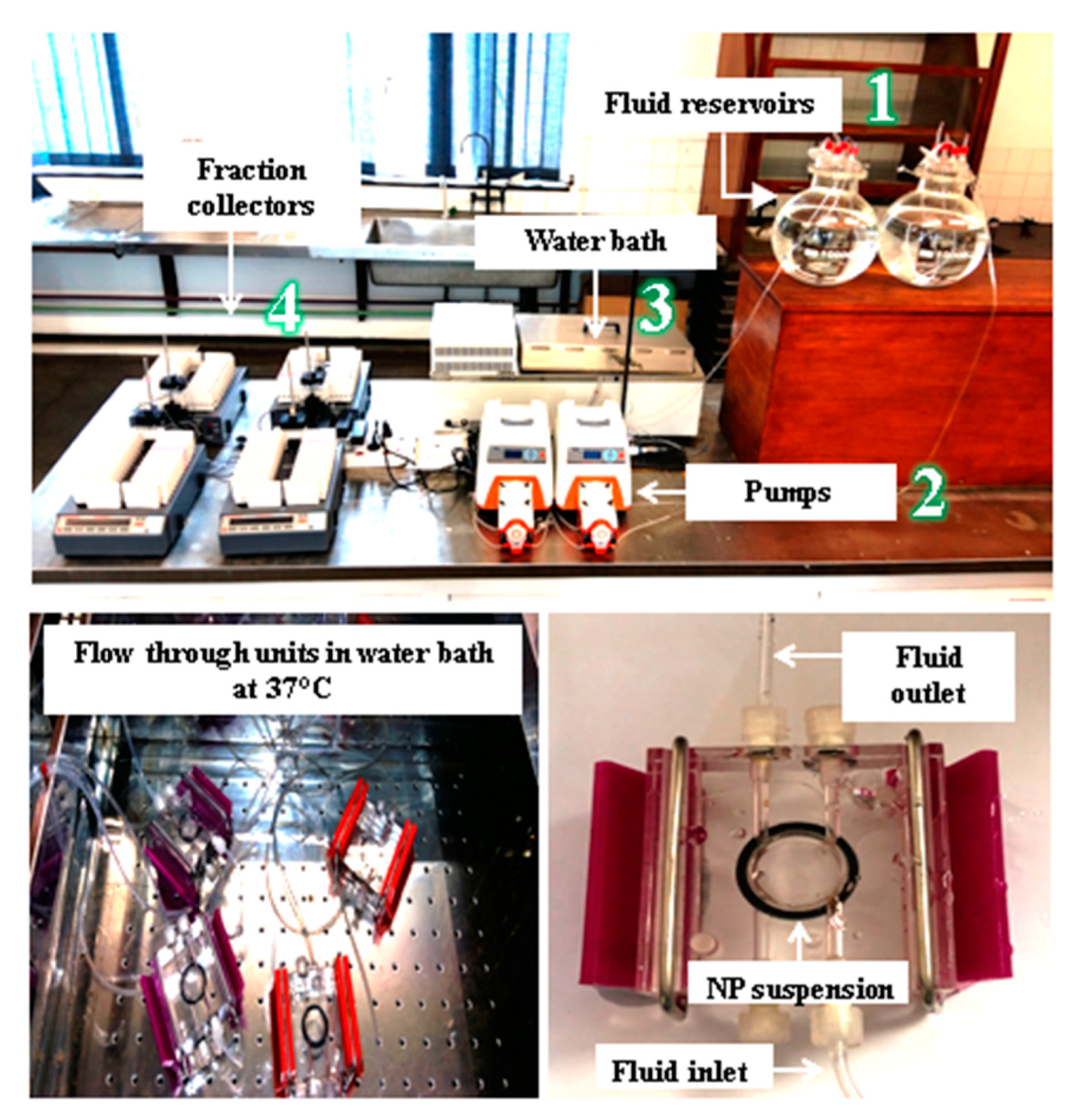
2.3. Continuous Flow-Through Dissolution Procedure

The continuous flow-through dynamic method of dissolution testing protocol shown in Figure 1 used in this study was adapted from Keller et al. [37] and Koltermann-Jülly et al. [38] with minor changes to match the specifications of nanoparticles. This dissolution protocol was specifically selected because it is regarded to be more reflective of dissolution occurring in biological and environmental surroundings. It is therefore recommended to avoid achieving an equilibrium that would restrict dissolution. A volume of 2 mL of gold and silver nanoparticles were drawn from the nanoparticle stock solutions and transferred into small centrifuge tubes. These were centrifuged at 13,000 times gravity for 30 min to pre-concentrate the samples which formed pellets. The pellets were transferred separately into the lower chamber of the flow through units. The flow through units containing the pellets were then filled with simulated fluids to create a nanoparticle suspension. TiO2 NPs were in a powder form therefore, a mass of 1 mg of titanium dioxide nanoparticle powder was weighed onto a membrane and was also transferred into the lower chamber of the flow through unit which was also filled with simulated fluid forming a nanoparticle suspension. An o-ring membrane holder was placed on top of the flow through unit containing the NP suspensions which was then sealed with the 3.5 kD membrane. The three separate flow through units containing AuNP, AgNP and TiO2 NP suspensions were then closed with a membrane (Spectrum/Por 3—Standard RC Discs—MWCO: 3.5 kD–33 mm2) pore size to only permit the movement of dissolved ions. A small pore size membrane was carefully selected to ensure that all the nanoparticles were kept inside the flow through units whilst only permitting the dissolved ions to diffuse into the bulk fluid. A second flow through unit (upper chamber) was placed on top of the membrane sealed lower chamber and tightly closed to only permit the movement of dissolved ions into the fraction collectors. The three separate flow through units were simultaneously submerged in a water bath maintained at 37 °C to mimic physiological conditions and room temperature 25 °C for synthetic environmental fluids. The simulated fluids from the fluid reservoir were pumped through the flow through units using the peristaltic pump at 8 mL/h and the eluate containing dissolved ions were continuously collected by the fraction collectors. The concentration of released ions from the eluate were analysed by ICP-MS to determine the level of dissolved ions of gold, silver and titanium. The programmable sampler drew 8 mL/h of the eluate. The dissolution experiments were conducted over a period of 10 days and triplicate samples were taken and measured. Samples were collected in 30 min interval for the first 4 h and once a day for the next 10 days. Sampling times were 0 h, 0.5 h, 1 h, 1.5 h, 2 h, 2.5 h, 3 h, 3.5 h and 4 h. From day 2 to day 10 samples were collected at 24 h, 48 h, 72 h, 96 h, 120 h, 144 h, 168 h, 192 h and 2016 h. Reported in the results section is an average of the three measurements.

2.4. Determination of the Kinetic Parameters

The dissolution of nanoparticles follows the first order reaction kinetics and involves the mass transfer rate process whereby the solute is transported from the nanoparticle surface to the bulk fluid surrounding the nanoparticles. The rate of solute liberation and transport from the nanoparticle surface is calculated using the dissolution kinetics model described below. To determine the dissolution rate constant and half-time of nanoparticles in the current research, the dissolution data were fit to a first order kinetic model previously described by Keller et al. [39] and Koltermann-Jülly et al. [38] using the following equation:
M d i s s o l v e d T = m ( E N M ) m ( m e t a l   i o n ) × i = 0 T c i i o n × V i × t i
where M d i s s o l v e d T is the mass of the dissolved nanoparticles, m E N M is the initial mass of the nanoparticles weighed before the commencement of the dissolution experiments, m ( m e t a l   i o n ) is the mass of dissolved nanoparticles obtained at different sampling time points, c i i o n is the concentration of dissolved ions obtained at a specific sampling time point, V i is the volume sampled at different times and t i is the different time interval where samples were collected to determine the amount of dissolved ions. Equation (1) gives us the rate of mass removal and the dissolution rate k is obtained using Equation (2) which is calculated as follows:
M s o l i d s T = M 0 M d i s s o l v e d ( T )
From Equation (2) the mass of nanoparticles remaining can be determined where M s o l i d s T is the mass remaining after dissolution has occurred, M 0 represents the initial mass of the before dissolution and M d i s s o l v e d T is the mass dissolved at different sampling time points. Equation (2) allows us to determine the dissolution rate k which is calculated using the following equation:
k = l n { M 0 / M s o l i d s ( T ) } / ( SSA ( T ) × T )
where k is the dissolution rate, SSA represents the initial surface area of the nanoparticles before dissolution and T is the time taken for the duration of the dissolution experiments. The initial surface area provided in Table 2 was used to calculate the half-time of nanoparticles to predict their duration in biological and environmental surroundings using the following equation:
t 1 / 2 = ln 2 k S S A
To calculate the mass of dissolved ions and account for the molar masses of the nanoparticles and detectable metal ions, we multiplied the measured ion concentration of each eluate by the eluted volume. This allowed us to calculate the percentage mass of remaining nanoparticles during the sampling time intervals.

2.5. Statistical Analysis

The data on dissolution are presented as the mean standard deviation of at least three independent measurements. To determine significant differences in the dissolution kinetics of AuNPs, AgNPs and TiO2 NPs in various simulated body fluids and synthetic environmental media, a multiple variable ANOVA analysis was performed using RStudio version 1.2 software. p < 0.05 was considered statistically significant.

3. Results

3.1. Physichichemical Properties of AuNPs, AgNPs and TiO2NPs

Investigating the biodurability and persistence of nanoparticles requires a thorough and accurate characterization of the particles’ physicochemical properties which can in turn be linked to their dissolution behaviour. In the present study, UV-vis, XRD and TEM were used to characterize, assess and monitor morphological changes and agglomeration states of AuNPs, AgNPs and TiO2 NPs before and after the dissolution experiments. Table 2 shows the physicochemical characterization of AuNPs, AgNPs and TiO2 NPs.
Generally, the UV-vis spectra of gold, silver and titanium dioxide nanoparticles have a localized surface plasmon resonance peaks at 520 nm, 400 nm and 300 nm, respectively [40,41,42,43]. This was confirmed by the UV-vis characterization of these nanoparticles before the dissolution experiments as shown in Table 2. After exposure to simulated fluids there was an observable shift to higher wavelengths for all the three nanoparticles. Interestingly this red shift for AuNPs was functional group specific. For example, -AuNPs-cit shifted to 547 nm followed by AuNPs-COOH at 540 nm then lastly AuNPs-NH2 shifted to 540 nm. These subtle differences are likely due to that the functionalized AuNPs are coated with polyethylene glycol (PEG) then functionalized with the carboxyl and amine functional groups. Consequently, PEG provides electro steric stabilizing thereby preventing the particles from combining to form agglomerates as a result, they remain monodispersed [44,45]. However, the citrate on the citrate stabilized AuNPs is just a stabilizing agent which can be easily displaced from the NP surface as a result it is easier to from agglomerates once the stabilizing agent is removed. For AgNPs there was an observable shift to higher wavelength (450 nm) for particles in contact with neutral media such as blood plasma, intestinal fluid, Gamble’s fluid and freshwater. This indicates that after a prolonged exposure of silver nanoparticles to these simulated fluids the particles physically coalesce to form larger particles. Generally larger particles absorb light at higher wavelength than smaller particles hence there was an observable shift to higher wavelengths for the agglomerates. TiO2 NPs exhibited a similar trend whereby there was a shift to higher wavelengths (320 nm) after exposure to simulated fluids indicative of particle aggregation as the time of exposure to simulated fluids increased. For AuNPs and AgNPs this red shift in wavelength was due to particle agglomeration whereas, for TiO2 NPs the major cause of the shift was formation of particle aggregates. Particle aggregation leads to a reduced surface area because particles combine to form a union of larger particles through weak Van der Waal forces [35,46,47]. As a result, the nanoparticle absorbs UV-light at a much higher wavelength as shown in Table 2 after dissolution experiments.

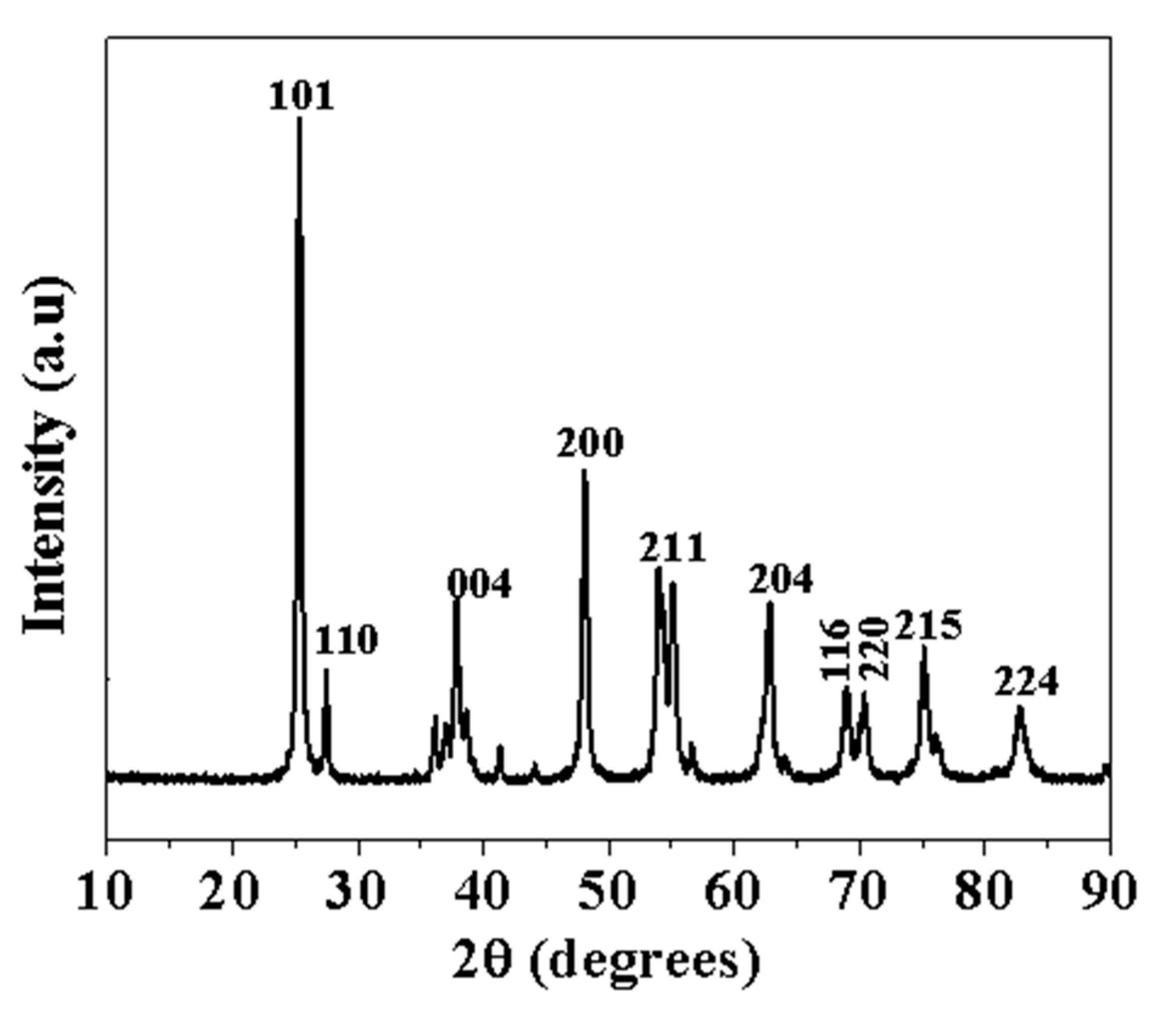
3.2. XRD Characterization of TiO2 NP Powder

The TiO2 nanoparticle powder was examined using XRD to assess its crystallographic phase, whether it was rutile, anatase, or a combination of both. The XRD pattern of the nanoparticles can be seen in Figure 2, with the peak positions at 2θ and their Miller indices. The TiO2 XRD data demonstrated very sharp peaks. The strong diffraction peaks exhibited by the XRD pattern at angles 25°, 37°, 47°, 55°, 62°, 68°, 70°, 75° and 82° correspond to Miller indices of (101), (004), (200), (211), (204), (116), (220), (215) and (224) plane, respectively. The major component of the TiO2 NPs sample was confirmed to be anatase. However, there was a minor presence of rutile which is represented by the peaks corresponding to (110) and (211) planes.

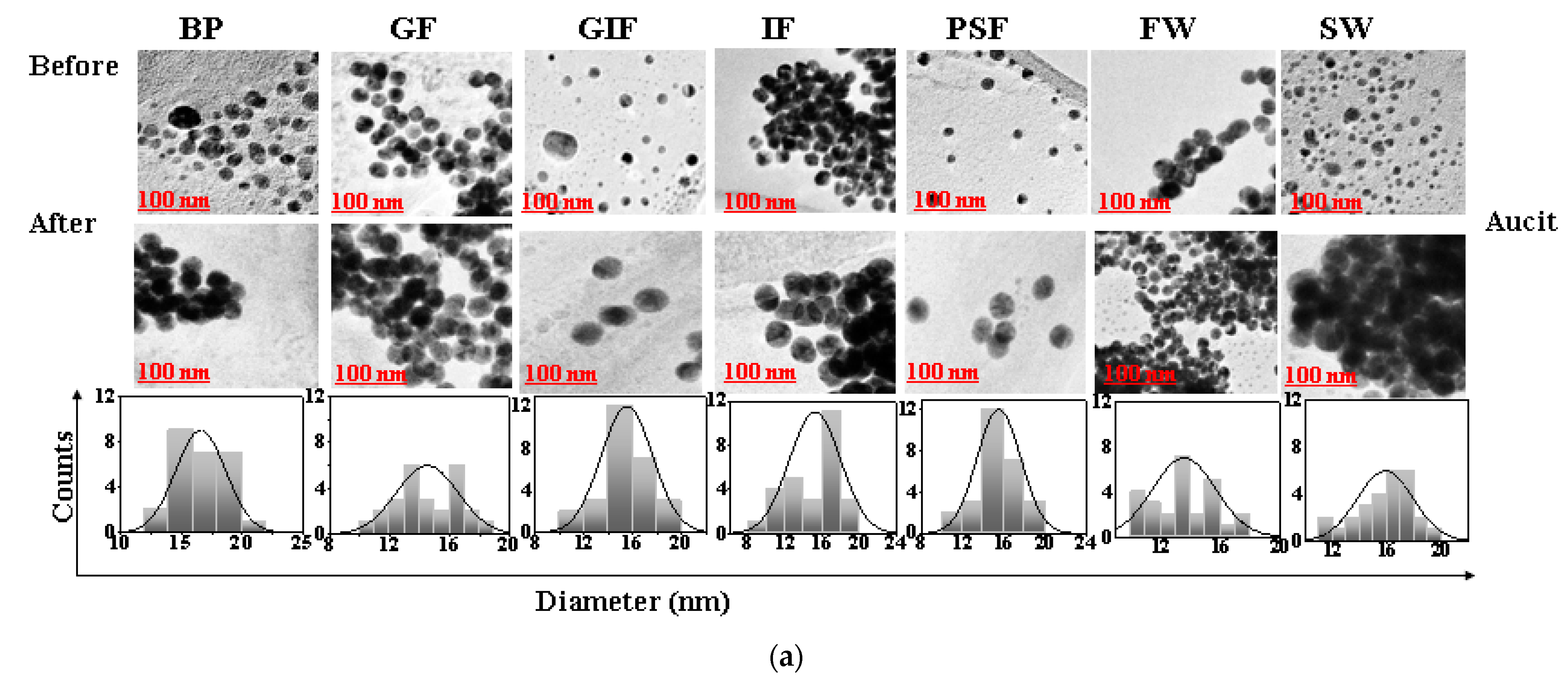
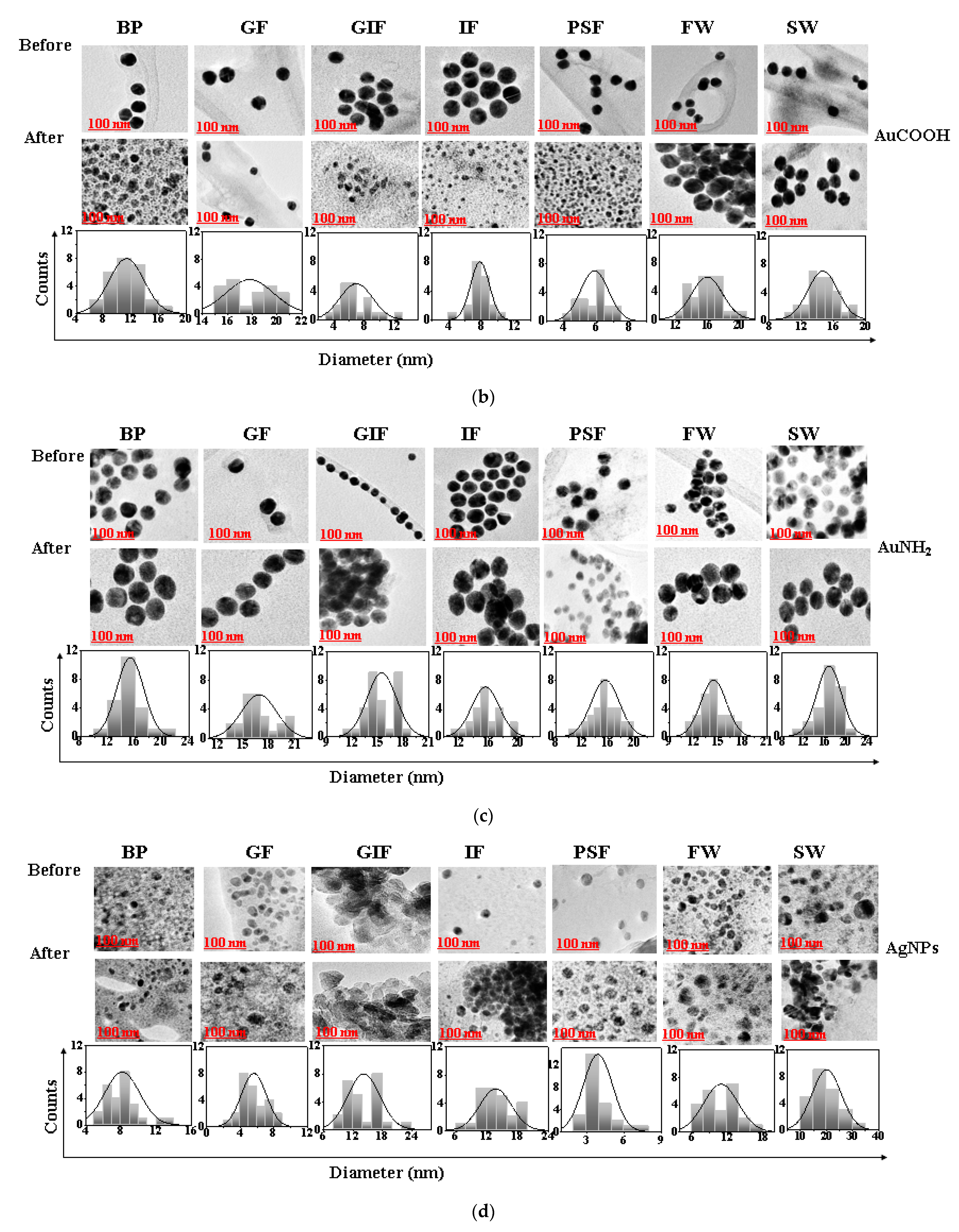
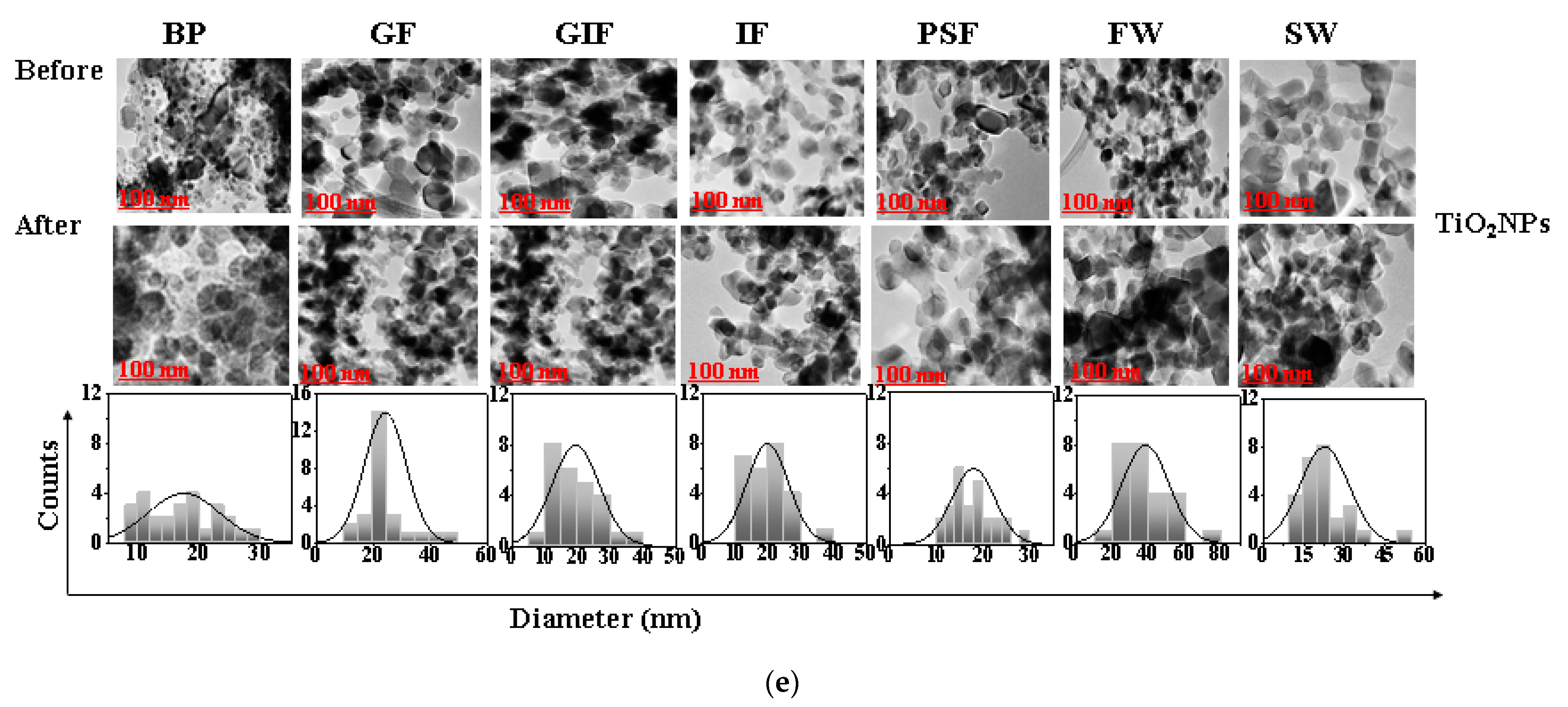
3.3. TEM Characterization of AuNPs, AgNPs and TiO2 NPs

TEM was used to investigate the morphological changes in AuNPs, AgNPs and TiO2 NPs in simulated fluids before and after the dissolution experiments. The TEM images of AuNPs-cit, AuNPs-COOH, AuNPs-NH2, AgNPs and TiO2 NPs in simulated fluids are shown in Figure 3a–e respectively.
The average nanoparticle diameter measured using Image J software (National Institute of Health, version no Java1.8.0_172) were 14 ± 2.8 nm for citrate stabilized AuNPs, 14 ± 2.3 nm for COOH-AuNPs and 14 ± 1.7 nm for NH2-AuNPs. Whereas the AgNPs had a size diameter of about 10 ± 0.8 nm which was smaller than that of TiO2 NPs with the size of 25 ± 3.1 nm. The gold and silver nanoparticles were spherical in shape as shown in Figure 3a–d. However, TiO2 NPs were irregular in morphology, and some were spherical as seen in Figure 3e. Among the AuNPs the citrate stabilized AuNPs tended to form multiple single particle clusters after exposure to simulated fluids. In contract, the pegylated carboxyl and amine functionalized gold nanoparticles (AuCOOH) and (AuNH2) remained monodispersed throughout the duration of the dissolution experiments. The Peg polymer coating present on the surface of functionalized gold nanoparticles provides steric stability which prevents the particles from colliding together to form clusters. Additionally, polymers are always present in the suspension system for steric stabilization, and they adsorb onto the particle surface, resulting in an additional steric repulsive force. Silver nanoparticles were spherical in shape and monodispersed. The morphological analysis of the TiO2 nanopowder by TEM (Figure 3e) showed high degrees of particle aggregation in all simulated fluids despite the differences in chemical composition, pH and ionic strength of the simulated fluids [48,49]. The formation of nanoparticle aggregates is due to Van der Waals interactions on the nanoparticle surface. During particle–particle interactions, if the force of attraction far exceeds the repulsive forces, then particles will tend to stick together to form aggregates [29,50]. The formation of nanoparticle aggregates can hinder the dissolution process by reducing the exposed surface area of the particle [51]. Additionally, particle aggregation can introduce a kinetic hindrance effect to the diffusion process thereby significantly reducing chances of dissolution [52].

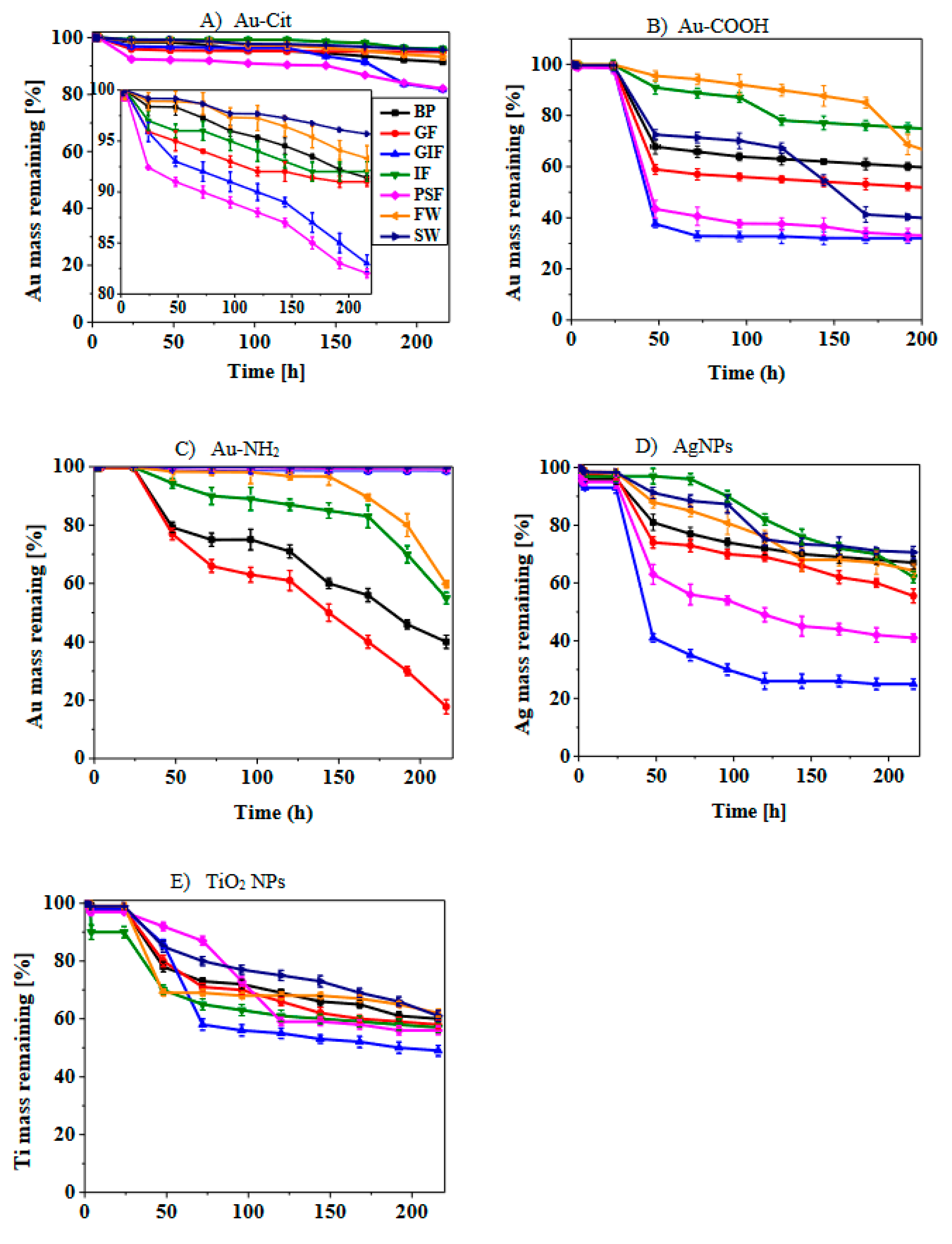
3.4. Dissolution Curves of AuNPs, AgNPs and TiO2 NPs

Figure 4 presents the dissolution curves of AuNPs, AgNPs and TiO2 NPs in various simulated biological fluids and synthetic environmental media. The dissolution curves are reported as a percentage mass of nanoparticles that remained undissolved in the reaction vessel over a period of 10 days expressed as time in hours. This method was adopted from these researchers Koltermann-Jülly et al. [38] and Keller et al. [39].
There was no complete dissolution of all the nanoparticles in all the simulated fluids. Additionally, dissolution was gradual in all cases meaning the release of ions from all the nanoparticle surfaces commenced after 24 h. Of the three types of AuNPs, COOH-AuNPs showed the highest amount of dissolved Au+ ions with the maximum found in acidic media such as gastric fluid and phagolysosomal fluid. For example, from the starting mass of 1 mg, only 32% and 33% mass remained undissolved in simulated gastric fluid and phagolysosomal fluid, respectively. Over the period of 10 days, cit-AuNPs exhibited the lowest dissolution whereby the maximum dissolution occurred in simulated phagolysosomal fluid. In addition, 81% mass of the particles remained undissolved. However, for NH2-AuNPs, the dissolution was higher in alkaline media such as simulated blood plasma and Gamble’s fluid. The reason for high dissolution of these nanoparticles could be due to the presence of this compound in the simulated fluid which acts as a solubilizing agent and encourages formation of more soluble complexes [53]. This occurs via the complexation of the nanoparticles with these compounds thereby facilitating the liberation of these nanoparticle ions which ultimately diffuse into the bulk fluid [53]. Interestingly, cit-AuNPs showed limited dissolution compared to functionalized COOH-AuNPs and NH2-AuNPs. This demonstrates the effect of surface functionalization on dissolution.
AgNPs showed significantly higher dissolution compared to both AuNPs and TiO2 NPs. TEM images of AgNPs revealed a decrease in size diameter after the end of the dissolution experiments and this is evidence of dissolution. The nanoparticles started releasing ions after 24 h of exposure to simulated fluids. AgNPs dissolved in acidic simulated gastric fluid and phagolysosomal fluid within 48 h but took longer to dissolve in alkaline media such as Gamble’s fluid, intestinal fluid and blood plasma. Generally, when particles were exposed to simulated fluids, the dissolution was significantly lower in alkaline media than in acidic media. There was no observable plateau reached during dissolution because under continuous flow through conditions the equilibrium is not reached therefore, the particles keep releasing ions till the end of the dissolution experiments. These results are in agreement with those obtained by Keller et al. [39] where barium sulphate dissolved in phagolysosomal fluid after two years.
For TiO2 NPs the amount of dissolved Ti ions did not even reach 50% of the initial mass in all simulated fluids regardless of the differences in chemical composition and pH of simulated fluids. The dissolution profile of TiO2 NPs showed low dissolution in both alkaline and acidic media. From the physicochemical properties data provided and the TEM images in Figure 3, it can be observed that the surface area of TiO2 NPs is larger compared to that of AuNPs and AgNPs. Research has shown that particles with a larger surface area are less reactive than those with smaller surface area [54,55].
When comparing the dissolution profiles of the synthetic environmental fluids, synthetic seawater had lower dissolution of all three nanoparticles compared to freshwater. However, COOH-AuNPs submerged in seawater dissolved faster than those exposed to freshwater. Even though COOH-AuNPs dissolved faster in seawater, the dissolution of particles in seawater and freshwater was not statistically significant except AgNPs. Particles showed degrees of agglomeration in both waters, but in seawater this was enhanced by the high ionic strength of the media. This could be attributed to the high ionic strength of seawater and the presence of divalent cations in high concentrations [30,56]. The influence of ionic strength is further explained in the discussion section.

3.5. Dissolution Kinetics of AuNPs, AgNPs and TiO2 NPs

Dissolution kinetics are a crucial factor in determining the safety of nanoparticles, which affect the biodurability and persistence of particles in biological and environmental surroundings. The kinetic model presented in the materials and methodology section was used to determine the dissolution kinetics. Additionally, the biodurability and persistence of AuNPs, AgNPs and TiO2 NPs were estimated using the dissolution rates and half-times. The data are presented in Table 3. The dissolution rates were determined over a period of 10 days in different simulated biological fluids and synthetic environmental media. From Table 3 it was observed that the dissolution rates of AgNPs were significantly higher (p < 0.05) and half-times were shorter in all media compared to those of AuNPs and TiO2 NPs. For example, the half-times of AgNPs range between the period of 2–7 days, whereas the half-times of AuNPs and TiO2 NPs fall within the range of 4–12.5 days and 13.5–14.4 days, respectively. TiO2 NPs had longer half-times regardless of the pH and chemical composition of simulated fluids. Generally, dissolution rates of particles in simulated gastric fluid and phagolysosomal fluid were higher than those of alkaline media such as blood plasma and intestinal fluid with the exception of NH2-AuNPs. The dissolution rate constants in Table 3 show that the dissolution rates of AuNPs increase in the rate of COOH > NH2 > citrate. Accordingly, the aggregation follows the inverse order and is influence by the protection of the gold core by the polyethylene-glycol in the case of COOH and NH2 functionalized nanoparticles. The high dissolution rate of amine functionalized nanoparticles in simulated blood plasma, Gamble’s fluid and intestinal fluid could be due to the interaction of the amine group and components of simulated fluids leading to the formation of more soluble complexes. In the case of synthetic environmental fluids, dissolution in simulated freshwater was faster than in seawater for all the nanoparticles. This is due to the ionic strength of seawater. Furthermore, the dissolution rates data indicate that the rate and extent of dissolution depends on the pH of simulated fluids, chemical composition of the simulated fluids, nanoparticle surface area and aggregation state and are nanoparticle specific. The observed dissolution rates of the nanoparticles in this study followed the order AgNPs > AuNPs > TiO2 NPs. These results were corroborated by those obtained by Koltermann-Jülly et al. [38]; Keller et al. [39]; Braun et al. [54]; Shinohara et al. [57].

4. Discussion

The high dissolution rates of silver nanoparticles can be attributed to the nanoparticle surface exposed in the simulated fluids. From the TEM images it is evident that these silver nanoparticles are monodispersed and not agglomerated meaning there is a larger particle surface area exposed and this results in enhanced interaction between the components of the simulated fluids and silver nanoparticles thereby encouraging dissolution. However, this is not the case for titanium dioxide nanoparticles, as most of the particles are highly aggregated thereby minimizing the exposed surface area. The effect of poor dissolution because of particle aggregation is dramatically enhanced for poorly soluble particles such as TiO2 NPs [53,57]. This is because smaller particles have many reactive atoms on the nanoparticle surface and ready to interact with the components of the simulated fluids. In addition, for this reason, TiO2 NPs take longer to release Ti ions, therefore have low dissolution. Additionally, the TEM images in Figure 2e show high degrees of TiO2 particle aggregation. In addition, particle aggregation has been shown to slow dissolution [17,58]. This is because as the particles combine to form clusters, this significantly reduces the particle surface area available for dissolution. As a result, the diffusion of Ti ions from the surface is inhibited thereby limiting dissolution.
In addition to the surface area, particle size also affects the dissolution of nanoparticles. From the physicochemical properties outlined in Table 1, it can be observed that AgNPs have the smallest size diameter followed by AuNPs and TiO2 NPs have the largest particle diameter at 10 nm, 14 nm and 25 nm, respectively. The smaller the particle size the greater the availability of the surface area and this leads to increased chances of ion diffusion from the surface to the bulk fluid. It is for this reason that about 70% mass of Ag+ ions dissolved in simulated gastric fluid. Whereas the highest dissolution of Ti+ ions could only reach a maximum of 55% in simulated gastric fluid. These results are corroborated by those obtained by Hedberg et al. [50] and Murugadoss et al. [58].
Similar to particle size, particle surface functionalization is another factor that influences the dissolution kinetics of nanoparticles. It could be inferred that the addition of functional groups to the gold nanoparticles surface enhanced dissolution because the amine and carboxyl functional groups have better solubilizing properties compared to citrate stabilized AuNPs. Additionally, citrate was present on the AuNP surface as a stabilizing agent therefore can be easily displaced thereby encouraging nanoparticle agglomeration. These authors also concluded that the rate of dissolution depends on the type of functional group attached to the nanoparticle surface [59,60,61].
Of all the simulated fluids, synthetic seawater had the highest ionic strength and highest concentration of divalent cations such as Ca2+ and Mg2+ These divalent cations are known to induce particle aggregation by suppressing the electrostatic repulsive forces between the particle–particle interactions [30,56]. This leads to the reduction in the surface area to volume ratio thereby inhibiting dissolution from occurring. This would explain the low dissolution rates of nanoparticles exposed to synthetic seawater. In so far as particle functionalization is concerned, the presence of Polyethylene glycol (PEG) as a coating agent on the surface of functionalized gold nanoparticles (COOH-AuNPs) and (NH2-AuNPs) reduces particle agglomeration and this is attributed to the steric repulsive forces imparted by PEG on the nanoparticle surface. These results were corroborated by Botha et al. [44] and Breitner et al. [45].
Generally, when particles were exposed to simulated fluids, the dissolution was significantly lower in alkaline media than in acidic media. This is because acidic conditions, as evidenced by low pH values in gastric and phagolysosomal fluids, enable the oxidation of nanoparticles into ions, increasing their solubility and thus the likelihood of dissolution [24,62]. As a result, nanoparticles exposed to acidic media would be less stable and dissolve more readily than in alkaline media. This could explain why citrate stabilized gold nanoparticles, carboxyl functionalized gold nanoparticles and silver nanoparticles dissolved faster in highly acidic simulated gastric fluid and phagolysosomal fluid. Other researchers corroborate these results and have observed that particles exposed to simulated fluids characterized by alkaline conditions reach a point of zero charge and generally low amounts of ions get released under these circumstances [53,63].

5. Conclusions

This study investigated the biodurability and persistence of gold, silver and titanium dioxide nanoparticles using the continuous flow-through system. The dissolution kinetics of the nanoparticles were affected by pH, ionic strength, particle aggregation and agglomeration state, as well as surface functionalization. Results showed that all three types of nanoparticles had varying levels of biodurability/persistence; however, silver nanoparticles had the highest rate of dissolution in all simulated biological fluids and synthetic environmental media. This suggests that silver nanoparticles are more likely to have short-term health and environmental effects, which could be similar to those caused by dissolved Ag ions. Gold nanoparticles on the other hand may have the potential to cause both short-term and long-term health and environmental effects depending on their surface functionalization. PEGylated gold nanoparticles are more resistant to agglomeration than citrate-stabilized gold nanoparticles due to ligand-promoted processes that increase dissolution rates. Citrate-stabilized gold nanoparticles have low dissolution rates and can cause long-term health effects as they are more stable and persistent. Titanium dioxide nanoparticles have low dissolution rates, high stability and form agglomerates, making them particularly biodurable and biopersistent in aquatic environments and likely to cause long-term toxicity. To ensure the safety of workers, consumers and the environment, it is critical to study the biodurability and persistence of nanoparticles.

Author Contributions

O.M.: Conceptualization, Methodology, Validation, Formal analysis, Investigation, Writing—Original draft E.C.: Resources, Writing—Review and Editing, Visualization M.G.: Resources, Writing—Review and Editing, Visualization, Supervision, Funding. All authors have read and agreed to the published version of the manuscript.

Funding

This research was funded by the European Union’s Horizon 2020 research and innovation program grant number 814401 (Gov4Nano) and the Department of Science and Innovation South Africa.

Data Availability Statement

Data will be made freely available on request, it can be requested from mary.gulumian@NWU.ac.za.

Acknowledgments

We gratefully acknowledge funding from the European Union’s Horizon 2020 research and innovation program grant number 814401 (Gov4Nano). We also would like to acknowledge the South African Department of Science and Innovation (DSI) and the University of Northwest for their assistance.

Conflicts of Interest

The authors declare no conflict of interest.

References

  1. Bove, P.; Malvindi, M.A.; Kote, S.S.; Bertorelli, R.; Summa, M.; Sabella, S. Dissolution test for risk assessment of nanoparticles: A pilot study. Nanoscale 2017, 9, 6315–6326. [Google Scholar] [CrossRef] [PubMed]
  2. Landvik, N.E.; Skaug, V.; Mohr, B.; Verbeek, J.; Zienolddiny, S. Criteria for grouping of manufactured nanomaterials to facilitate hazard and risk assessment, a systematic review of expert opinions. Regul. Toxicol. Pharmacol. 2018, 95, 270–279. [Google Scholar] [CrossRef] [PubMed]
  3. Adewale, O.B.; Davids, H.; Cairncross, L.; Roux, S. Toxicological Behavior of Gold Nanoparticles on Various Models: Influence of Physicochemical Properties and Other Factors. Int. J. Toxicol. 2019, 38, 357–384. [Google Scholar] [CrossRef] [PubMed]
  4. Elahi, N.; Kamali, M.; Baghersad, M.H. Recent biomedical applications of gold nanoparticles: A review. Talanta 2018, 184, 537–556. [Google Scholar] [CrossRef]
  5. Fan, J.; Cheng, Y.; Sun, M. Functionalized Gold Nanoparticles: Synthesis, Properties and Biomedical Applications. Chem. Rec. 2020, 20, 1474–1504. [Google Scholar] [CrossRef]
  6. Kalimuthu, K.; Cha, B.S.; Kim, S.; Park, K.S. Eco-friendly synthesis and biomedical applications of gold nanoparticles: A review. Microchem. J. 2019, 152, 104296. [Google Scholar] [CrossRef]
  7. Bapat, R.A.; Chaubal, T.V.; Joshi, C.P.; Bapat, P.R.; Choudhury, H.; Pandey, M.; Gorain, B.; Kesharwani, P. An overview of application of silver nanoparticles for biomaterials in dentistry. Mater. Sci. Eng. C 2018, 91, 881–898. [Google Scholar] [CrossRef]
  8. Burdușel, A.C.; Gherasim, O.; Grumezescu, A.M.; Mogoantă, L.; Ficai, A.; Andronescu, E. Biomedical applications of silver nanopar-ticles: An up-to-date overview. Nanomaterials 2018, 8, 681. [Google Scholar] [CrossRef]
  9. Irshad, M.A.; Nawaz, R.; Rehman, M.Z.U.; Adrees, M.; Rizwan, M.; Ali, S.; Ahmad, S.; Tasleem, S. Synthesis, characterization and advanced sustainable applications of titanium dioxide nanoparticles: A review. Ecotoxicol. Environ. Saf. 2021, 212, 111978. [Google Scholar] [CrossRef]
  10. Mbanga, O.; Cukrowska, E.; Gulumian, M. Dissolution of titanium dioxide nanoparticles in synthetic biological and environmental media to predict their biodurability and persistence. Toxicol. Vitr. 2022, 84, 105457. [Google Scholar] [CrossRef]
  11. Kansara, K.; Bolan, S.; Radhakrishnan, D.; Palanisami, T.; Al-Muhtaseb, A.H.; Bolan, N.; Vinu, A.; Kumar, A.; Karakoti, A. A critical review on the role of abiotic factors on the transformation, environmental identity and toxicity of engineered nanomaterials in aquatic environment. Environ. Pollut. 2022, 296, 118726. [Google Scholar] [CrossRef] [PubMed]
  12. OECD. Joint Meeting of the Chemicals Committee and the Working Party on Chemicals, Pesticides and Biotechnology; OECD: Paris, France, 2018; Volume 86.
  13. Klaessig, F.C. Dissolution as a paradigm in regulating nanomaterials. Environ. Sci. Nano 2018, 5, 1070–1077. [Google Scholar] [CrossRef]
  14. Sauer, U.G.; Werle, K.; Waindok, H.; Hirth, S.; Hachmöller, O.; Wohlleben, W. Critical Choices in Predicting Stone Wool Biodurability: Lysosomal Fluid Compositions and Binder Effects. Chem. Res. Toxicol. 2021, 34, 780–792. [Google Scholar] [CrossRef] [PubMed]
  15. Gulumian, M.; Cassee, F.R. Safe by design (SbD) and nanotechnology: A much-discussed topic with a prudence? Part. Fibre Toxicol. 2021, 18, 32. [Google Scholar] [CrossRef]
  16. Innes, E.; Yiu, H.H.P.; McLean, P.; Brown, W.; Boyles, M. Simulated biological fluids—A systematic review of their biological relevance and use in relation to inhalation toxicology of particles and fibres. Crit. Rev. Toxicol. 2021, 51, 217–248. [Google Scholar] [CrossRef]
  17. Sohal, I.S.; Cho, Y.K.; O’fallon, K.S.; Gaines, P.; Demokritou, P.; Bello, D. Dissolution Behavior and Biodurability of Ingested Engineered Nanomaterials in the Gastrointestinal Environment. ACS Nano 2018, 12, 8115–8128. [Google Scholar] [CrossRef]
  18. Utembe, W.; Potgieter, K.; Stefaniak, A.B.; Gulumian, M. Dissolution and biodurability: Important parameters needed for risk as-sessment of nanomaterials. Part. Fibre Toxicol. 2015, 12, 11. [Google Scholar] [CrossRef]
  19. Laux, P.; Riebeling, C.; Booth, A.M.; Brain, J.D.; Brunner, J.; Cerrillo, C.; Creutzenberg, O.; Estrela-Lopis, I.; Gebel, T.; Johanson, G.; et al. Biokinetics of nanomaterials: The role of biopersistence. Nanoimpact 2017, 6, 69–80. [Google Scholar] [CrossRef]
  20. Avellan, A.; Simonin, M.; McGivney, E.; Bossa, N.; Spielman-Sun, E.; Rocca, J.D.; Bernhardt, E.S.; Geitner, N.K.; Unrine, J.M.; Wiesner, M.R.; et al. Gold nanoparticle biodissolution by a freshwater macrophyte and its associated microbiome. Nat. Nanotechnol. 2018, 13, 1072–1077. [Google Scholar] [CrossRef]
  21. Boldeiu, A.; Simion, M.; Mihalache, I.; Radoi, A.; Banu, M.; Varasteanu, P.; Nadejde, P.; Vasile, E.; Acasandrei, A.; Popescu, R.C.; et al. Comparative analysis of honey and citrate stabilized gold nanoparticles: In vitro interaction with proteins and toxicity studies. J. Photochem. Photobiol. B Biol. 2019, 197, 111519. [Google Scholar] [CrossRef]
  22. John, T.; Gladytz, A.; Kubeil, C.; Martin, L.L.; Risselada, H.J.; Abel, B. Impact of nanoparticles on amyloid peptide and protein ag-gregation: A review with a focus on gold nanoparticles. Nanoscale 2018, 10, 20894–20913. [Google Scholar] [CrossRef] [PubMed]
  23. Nambiar, S.; Osei, E.; Fleck, A.; Darko, J.; Mutsaers, A.J.; Wettig, S. Synthesis of curcumin-functionalized gold nanoparticles and cytotoxicity studies in human prostate cancer cell line. Appl. Nanosci. 2018, 8, 347–357. [Google Scholar] [CrossRef]
  24. Fernando, I.; Zhou, Y. Impact of pH on the stability, dissolution and aggregation kinetics of silver nanoparticles. Chemosphere 2019, 216, 297–305. [Google Scholar] [CrossRef] [PubMed]
  25. Loza, K.; Diendorf, J.; Sengstock, C.; Ruiz-Gonzalez, L.; Gonzalez-Calbet, J.M.; Vallet-Regi, M.; Köller, M.; Epple, M. The dissolution and biological effects of silver nanoparticles in biological media. J. Mater. Chem. B 2014, 2, 1634–1643. [Google Scholar] [CrossRef]
  26. Mbanga, O.; Cukrowska, E.; Gulumian, M. Dissolution kinetics of silver nanoparticles: Behaviour in simulated biological fluids and synthetic environmental media. Toxicol. Rep. 2022, 9, 788–796. [Google Scholar] [CrossRef] [PubMed]
  27. Zienkiewicz-Strzałka, M.; Deryło-Marczewska, A.; Skorik, Y.A.; Petrova, V.A.; Choma, A.; Komaniecka, I. Silver Nanoparticles on Chitosan/Silica Nanofibers: Characterization and Antibacterial Activity. Int. J. Mol. Sci. 2020, 21, 166. [Google Scholar] [CrossRef] [PubMed]
  28. Baccaro, M.; Undas, A.K.; De Vriendt, J.; Van Den Berg, J.H.J.; Peters, R.J.B.; Van Den Brink, N.W. Ageing, dissolution and biogenic formation of nanoparticles: How do these factors affect the uptake kinetics of silver nanoparticles in earthworms? Environ. Sci. Nano 2018, 5, 1107–1116. [Google Scholar] [CrossRef]
  29. Jiang, X.; Wu, Y.; Gray, P.; Zheng, J.; Cao, G.; Zhang, H.; Zhang, X.; Boudreau, M.; Croley, T.R.; Chen, C.; et al. Influence of gastrointestinal environment on free radical generation of silver nanoparticles and implications for their cytotoxicity. Nanoimpact 2018, 10, 144–152. [Google Scholar] [CrossRef]
  30. de Souza, T.A.J.; Rosa Souza, L.R.; Franchi, L.P. Silver nanoparticles: An integrated view of green synthesis methods, transformation in the environment, and toxicity. Ecotoxicol. Environ. Saf. 2019, 171, 691–700. [Google Scholar] [CrossRef]
  31. Zhong, L.; Hu, X.; Cao, Z.; Wang, H.; Chen, Y.; Lian, H. Aggregation and dissolution of engineering nano Ag and ZnO pre-treated with natural organic matters in the simulated lung biological fluids. Chemosphere 2019, 225, 668–677. [Google Scholar] [CrossRef]
  32. Schmidt, J.; Vogelsberger, W. Dissolution Kinetics of Titanium Dioxide Nanoparticles: The Observation of an Unusual Kinetic Size Effect. J. Phys. Chem. B 2006, 110, 3955–3963. [Google Scholar] [CrossRef] [PubMed]
  33. Schmidt, J.; Vogelsberger, W. Aqueous Long-Term Solubility of Titania Nanoparticles and Titanium (IV) Hydrolysis in a Sodium Chloride System Studied by Adsorptive Stripping Voltammetry. J. Solut. Chem. 2009, 38, 1267–1282. [Google Scholar] [CrossRef]
  34. Shkol’nikov, E.V. Thermodynamics of the dissolution of amorphous and polymorphic TiO2 modifications in acid and alkaline media. Russ. J. Phys. Chem. A 2016, 90, 567–571. [Google Scholar] [CrossRef]
  35. Wang, H.; Burgess, R.M.; Cantwell, M.G.; Portis, L.M.; Perron, M.M.; Wu, F.; Ho, K.T. Stability and aggregation of silver and titanium dioxide nanoparticles in seawater: Role of salinity and dissolved organic carbon. Environ. Toxicol. Chem. 2014, 33, 1023–1029. [Google Scholar] [CrossRef] [PubMed]
  36. Marques, M.R.C.; Loebenberg, R.; Almukainzi, M. Simulated Biological Fluids with Possible Application in Dissolution Testing. Dissolution Technol. 2011, 18, 15–28. [Google Scholar] [CrossRef]
  37. Keller, J.G.; Peijnenburg, W.; Werle, K.; Landsiedel, R.; Wohlleben, W. Understanding Dissolution Rates via Continuous Flow Systems with Physiologically Relevant Metal Ion Saturation in Lysosome. Nanomaterials 2020, 10, 311. [Google Scholar] [CrossRef] [PubMed]
  38. Koltermann-Jülly, J.; Keller, J.G.; Vennemann, A.; Werle, K.; Müller, P.; Ma-Hock, L.; Landsiedel, R.; Wiemann, M.; Wohlleben, W. Abiotic dissolution rates of 24 (nano)forms of 6 substances compared to macrophage-assisted dissolution and in vivo pulmonary clearance: Grouping by biodissolution and transformation. NanoImpact 2018, 12, 29–41. [Google Scholar] [CrossRef]
  39. Keller, J.G.; Graham, U.M.; Koltermann-Jülly, J.; Gelein, R.; Ma-Hock, L.; Landsiedel, R.; Wiemann, M.; Oberdörster, G.; Elder, A.; Wohlleben, W. Predicting dissolution and transformation of inhaled nanoparticles in the lung using abiotic flow cells: The case of barium sulfate. Sci. Rep. 2020, 10, 458, Correction in Sci. Rep. 2021, 11, 8813. [Google Scholar] [CrossRef]
  40. Badiah, H.I.; Seedeh, F.; Supriyanto, G.; Zaidan, A.H. Synthesis of Silver Nanoparticles and the Development in Analysis Method. IOP Conf. Series: Earth Environ. Sci. 2019, 217, 012005. [Google Scholar] [CrossRef]
  41. Dobrucka, R. Synthesis of Titanium Dioxide Nanoparticles Using Echinacea purpurea Herba. Iran. J. Pharm. Res. IJPR 2017, 16, 756–762. [Google Scholar]
  42. Monfared, A.H.; Jamshidi, M. Synthesis of polyaniline/titanium dioxide nanocomposite (PAni/TiO2) and its application as pho-tocatalyst in acrylic pseudo paint for benzene removal under UV/VIS lights. Prog. Org. Coat. 2019, 136, 105257. [Google Scholar] [CrossRef]
  43. Pashkov, D.M.; Guda, A.A.; Kirichkov, M.V.; Martini, A.; Soldatov, S.A.; Soldatov, A.V. Quantitative Analysis of the UV–Vis Spectra for Gold Nanoparticles Powered by Supervised Machine Learning. J. Phys. Chem. C 2021, 125, 8656–8666. [Google Scholar] [CrossRef]
  44. Botha, T.L.; James, T.E.; Wepener, V. Comparative Aquatic Toxicity of Gold Nanoparticles and Ionic Gold Using a Species Sensi-tivity Distribution Approach. J. Nanomater. 2015, 2015, 986902. [Google Scholar] [CrossRef]
  45. Breitner, E.K.; Hussain, S.M.; Comfort, K.K. The role of biological fluid and dynamic flow in the behavior and cellular interactions of gold nanoparticles. J. Nanobiotechnology 2015, 13, 2–10. [Google Scholar] [CrossRef] [PubMed]
  46. De Matteis, V.; Cascione, M.; Brunetti, V.; Toma, C.C.; Rinaldi, R. Toxicity assessment of anatase and rutile titanium dioxide nano-particles: The role of degradation in different pH conditions and light exposure. Toxicol. Vitr. 2016, 37, 201–210. [Google Scholar] [CrossRef]
  47. Zhong, L.; Yu, Y.; Lian, H.; Hu, X.; Fu, H.; Chen, Y. Solubility of nano-sized metal oxides evaluated by using in vitro sim-ulated lung and gastrointestinal fluids: Implication for health risks. J. Nanoparticle Res. 2017, 19, 375. [Google Scholar] [CrossRef]
  48. Klonos, P.; Dapei, G.; Sulym, I.Y.; Zidropoulos, S.; Sternik, D.; Deryło-Marczewska, A.; Borysenko, M.V.; Gun’ko, V.M.; Kyritsis, A.; Pissis, P. Morphology and molecular dynamics investigation of PDMS adsorbed on titania nanoparticles: Effects of polymer molecular weight. Eur. Polym. J. 2016, 74, 64–80. [Google Scholar] [CrossRef]
  49. Lin, X.; Li, J.; Ma, S.; Liu, G.; Yang, K.; Tong, M.; Lin, D. Toxicity of TiO2 Nanoparticles to Escherichia coli: Effects of Particle Size, Crystal Phase and Water Chemistry. PLoS ONE 2014, 9, e110247. [Google Scholar] [CrossRef]
  50. Hedberg, J.; Blomberg, E.; Wallinder, I.O. In the Search for Nanospecific Effects of Dissolution of Metallic Nanoparticles at Freshwater-Like Conditions: A Critical Review. Environ. Sci. Technol. 2019, 53, 4030–4044. [Google Scholar] [CrossRef]
  51. Pujalté, I.; Dieme, D.; Haddad, S.; Serventi, A.M.; Bouchard, M. Toxicokinetics of titanium dioxide (TiO2) nanoparticles after inha-lation in rats. Toxicol. Lett. 2017, 265, 77–85. [Google Scholar] [CrossRef]
  52. Borm, P.; Klaessig, F.C.; Landry, T.D.; Moudgil, B.; Pauluhn, J.; Thomas, K.; Trottier, R.; Wood, S. Research Strategies for Safety Evaluation of Nanomaterials, Part V: Role of Dissolution in Biological Fate and Effects of Nanoscale Particles. Toxicol. Sci. 2006, 90, 23–32. [Google Scholar] [CrossRef] [PubMed]
  53. Avramescu, M.L.; Rasmussen, P.E.; Chénier, M.; Gardner, H.D. Influence of pH, particle size and crystal form on dissolution be-haviour of engineered nanomaterials. Environ. Sci. Pollut. Res. 2017, 24, 1553–1564. [Google Scholar] [CrossRef] [PubMed]
  54. Braun, K.; Pochert, A.; Beck, M.; Fiedler, R.; Gruber, J.; Lindén, M. Dissolution kinetics of mesoporous silica nanoparticles in different simulated body fluids. J. Sol-Gel Sci. Technol. 2016, 79, 319–327. [Google Scholar] [CrossRef]
  55. Donovan, A.R.; Adams, C.D.; Ma, Y.; Stephan, C.; Eichholz, T.; Shi, H. Single particle ICP-MS characterization of titanium dioxide, silver, and gold nanoparticles during drinking water treatment. Chemosphere 2016, 144, 148–153. [Google Scholar] [CrossRef]
  56. Cupi, D.; Hartmann, N.B.; Baun, A. Influence of pH and media composition on suspension stability of silver, zinc oxide, and titanium dioxide nanoparticles and immobilization of Daphnia magna under guideline testing conditions. Ecotoxicol. Environ. Saf. 2016, 127, 144–152. [Google Scholar] [CrossRef]
  57. Shinohara, N.; Zhang, G.; Oshima, Y.; Kobayashi, T.; Imatanaka, N.; Nakai, M.; Sasaki, T.; Kawaguchi, K.; Gamo, M. Kinetics and dissolution of intratracheally administered nickel oxide nanomaterials in rats. Part. Fibre Toxicol. 2017, 14, 48. [Google Scholar] [CrossRef]
  58. Murugadoss, S.; Brassinne, F.; Sebaihi, N.; Petry, J.; Cokic, S.M.; Van Landuyt, K.L.; Godderis, L.; Mast, J.; Lison, D.; Hoet, P.H.; et al. Agglomeration of titanium dioxide nano-particles increases toxicological responses in vitro and in vivo. Part. Fibre Toxicol. 2020, 17, 10. [Google Scholar] [CrossRef]
  59. Bozich, J.S.; Lohse, S.E.; Torelli, M.D.; Murphy, C.J.; Hamers, R.J.; Klaper, R.D. Surface chemistry, charge and ligand type impact the toxicity of gold nanoparticles to Daphnia magna. Environ. Sci. Nano 2014, 1, 260–270. [Google Scholar] [CrossRef]
  60. Li, Y.; Zhang, W.; Niu, J.; Chen, Y. Surface-coating-dependent dissolution, aggregation, and reactive oxygen species (ROS) gener-ation of silver nanoparticles under different irradiation conditions. Environ. Sci. Technol. 2013, 47, 10293–10301. [Google Scholar]
  61. Tejamaya, M.; Römer, I.; Merrifield, R.C.; Lead, J.R. Stability of Citrate, PVP, and PEG Coated Silver Nanoparticles in Ecotoxicology Media. Environ. Sci. Technol. 2012, 46, 7011–7017. [Google Scholar] [CrossRef]
  62. Chambers, B.A.; Afrooz, A.R.M.N.; Bae, S.; Aich, N.; Katz, L.; Saleh, N.B.; Kirisits, M.J. Effects of Chloride and Ionic Strength on Physical Morphology, Dissolution, and Bacterial Toxicity of Silver Nanoparticles. Environ. Sci. Technol. 2014, 48, 761–769. [Google Scholar] [CrossRef] [PubMed]
  63. Xu, N.; Cheng, X.; Wang, D.; Xu, X.; Huangfu, X.; Li, Z. Effects of Escherichia coli and phosphate on the transport of titanium dioxide nanoparticles in heterogeneous porous media. Water Res. 2018, 146, 264–274. [Google Scholar] [CrossRef] [PubMed]
Figure 1. Continuous flow-through dissolution system protocol.
Nanomaterials 13 01653 g001
Figure 2. X-ray diffraction pattern of TiO2 NPs powder.
Nanomaterials 13 01653 g002
Figure 3. TEM images of cit-stabilized AuNPs (a); COOH-AuNPs (b) NH2-AuNPs (c), AgNPs (d) and TiO2 NPs (e) in simulated biological and environmental fluids before and after dissolution tests. BP, GF, GIF, IF and PSF are simulated biological fluids for blood plasma, Gamble’s fluid, gastric fluid, intestinal fluid and phagolysosomal fluid, respectively. FW and SW are synthetic environmental fluids for freshwater and seawater, respectively.
Nanomaterials 13 01653 g003aNanomaterials 13 01653 g003bNanomaterials 13 01653 g003c
Figure 4. Dissolution profiles for cit-AuNPs (A); COOH-AuNPs (B); NH2-AuNPs (C); AgNPs (D) and TiO2 NPs (E) in simulated fluids. Simulated biological fluids are BP—Blood plasma, GF—Gamble’s fluid, GIF—Gastric fluid, IF—Intestinal fluid and PSF—Phagolysosomal fluid. Synthetic environmental media are FW—Freshwater and SW—Seawater.
Nanomaterials 13 01653 g004
Table 1. Chemical composition, pH and ionic strength of simulated fluids (Marques et al., 2011).
Chemical Composition (g 5 L−1)BPGFGIFIFPSFFWSW
Bile salts---45 mL---
Borax------0.17
Calcium chloride1.46--2.49---
Calcium chloride anhydrous------1.320
Calcium chloride dihydrate-1.84--0.14--
Calcium sulphate anhydrous-----0.37-
Glycine----2.25--
Magnesium chloride-1.015 0.95 47.5
Magnesium chloride hexahydrate1.65 -----
Magnesium sulphate anhydrous-----0.037-
Mucin--15 mg----
Pancreatin---45 mL---
Pepsin--5 mL ----
Potassium bromide------0.44
Potassium chloride1.121.49351.49 0.00253.05
Potassium hydrogen phthalate--1.215-20.43--
Potassium phosphate dibasic trihydrate1.15------
Sodium acetate-4.76-----
Sodium chloride40.1730.0914.61 33.25 105.1
Sodium hydrogen carbonate1.7713.02---0.060.85
Sodium hydrogen phosphate-0.71--0.171--
Sodium sulphate0.36 0.36 17.6
Sodium sulphate anhydrous-0.085-----
Strontium chloride------0.1
Tris(hydroxymethyl) aminomethane30.59------
Trisodium citrate dihydrate-0.485-----
Urea---1.5---
1 M HCl195 mL---- -
Ionic strength
(mol L−1)
0.150.170.160.160.340.053.5
pH7.27.42.06.84.56.88.0
BP—Blood plasma; GF—Gamble’s fluid; GIF—Gastric fluid; IF—Intestinal fluid; PSF—Phagolysosomal fluid; FW—Freshwater; SW—Seawater.
Table 2. Physical–chemical description of AuNPs, AgNPs and TiO2 NPs.
NanoparticlesSimulated FluidsUV-Vis Absorption WavelengthSurface Area Particle
Size Diameter
Crystallinity (XRD)
[nm][m2/g][nm][%]
BeforeAfter
Citrate-AuNPsBP5205492514None
Citrate-AuNPsGF5205492114None
Citrate-AuNPsGIF5205492314None
Citrate-AuNPsIF5205492114None
Citrate-AuNPsPSF5205492014None
Citrate-AuNPsFW5205492214None
Citrate-AuNPsSW5205492014None
COOH-AuNPsBP5205472414None
COOH-AuNPsGF5205472314None
COOH-AuNPsGIF5205472414None
COOH-AuNPsIF5205472614None
COOH-AuNPsPSF5205472614None
COOH-AuNPsFW5205472414None
COOH-AuNPsSW5205472514None
NH2-AuNPsBP5205402214None
NH2-AuNPsGF5205402314None
NH2-AuNPsGIF5205402214None
NH2-AuNPsIF5205042214None
NH2-AuNPsPSF5205042014None
NH2-AuNPsFW5205401814None
NH2-AuNPsSW5205402014None
AgNPsBP4004502210None
AgNPsGF4004502210None
AgNPsGIF4004001810None
AgNPsIF4004501510None
AgNPsPSF4004001510None
AgNPsFW4004502610None
AgNPsSW4004002010None
TiO2 NPsBP3003205725Mix rutile/anatase
TiO2 NPsGF3003205825Mix rutile/anatase
TiO2 NPsGIF3003205625Mix rutile/anatase
TiO2 NPsIF3003205525Mix rutile/anatase
TiO2 NPsPSF3003205525Mix rutile/anatase
TiO2 NPsFW3003205925Mix rutile/anatase
TiO2 NPsSW3003205825Mix rutile/anatase
Table 3. Comparison of the dissolution rates and half-times of AuNPs, AgNPs and TiO2 NPs in simulated fluids.
NanoparticlesSimulated FluidsDissolution Rate
k
Half-Time
t 1 / 2
p-Value
[ng/cm2/h][days]
Citrate-AuNPsBP0.09100.0621
Citrate-AuNPsGF0.088.60.1138
Citrate-AuNPsGIF0.088.60.2144
Citrate-AuNPsIF0.0612.50.0720
Citrate-AuNPsPSF0.107.30.0591
Citrate-AuNPsFW0.0611.50.0820
Citrate-AuNPsSW0.0512.50.0931
COOH-AuNPsBP0.086.50.0656
COOH-AuNPsGF0.0870.0809
COOH-AuNPsGIF0.1050.0633
COOH-AuNPsIF0.0690.0744
COOH-AuNPsPSF0.105.70.0537
COOH-AuNPsFW0.06100.0644
COOH-AuNPsSW0.097.50.0937
NH2-AuNPsBP0.1340.1151
NH2-AuNPsGF0.1160.2413
NH2-AuNPsGIF0.06100.0655
NH2-AuNPsIF0.0970.0594
NH2-AuNPsPSF0.06100.0742
NH2-AuNPsFW0.1340.0894
NH2-AuNPsSW0.06100.0942
AgNPsBP0.154 0.0021
AgNPsGF0.1540.0008
AgNPsGIF0.1820.0144
AgNPsIF0.1070.0420
AgNPsPSF0.220.0231
AgNPsFW0.1260.0320
AgNPsSW0.1070.0231
TiO2NPsBP3.70 × 19−0513.6 0.0778
TiO2NPsGF3.47 × 10−0514.30.0898
TiO2NPsGIF3.63 × 10−0514.10.0755
TiO2NPsIF3.67 × 10−0514.20.0894
TiO2NPsPSF3.65 × 10−0514.30.0842
TiO2NPsFW3.40 × 10−0514.30.2329
TiO2NPsSW3.43 × 10−0514.40.1142
Disclaimer/Publisher’s Note: The statements, opinions and data contained in all publications are solely those of the individual author(s) and contributor(s) and not of MDPI and/or the editor(s). MDPI and/or the editor(s) disclaim responsibility for any injury to people or property resulting from any ideas, methods, instructions or products referred to in the content.

