Article

Biogenic Synthesis of Fluorescent Carbon Dots (CDs) and Their Application in Bioimaging of Agricultural Crops

1
Nanobiotechnology Laboratory, Department of Biotechnology, Sant Gadge Baba Amravati University, Amravati 444602, Maharashtra, India
2
Department of Botany, The Institute of Science, 15, Madame Cama Road, Mumbai 400032, Maharashtra, India
3
Academy of Biology and Biotechnology, Southern Federal University, 344090 Rostov-on-Don, Russia
4
Department of Microbiology, Nicolaus Copernicus University, 87-100 Torun, Poland
5
All-Russian Research Institute of Phytopathology, 143050 Moscow, Russia
6
Department of Biological Sciences and Biotechnology, Institute of Chemical Technology, Mumbai 400019, Maharashtra, India
*
Authors to whom correspondence should be addressed.
Nanomaterials 2023, 13(1), 209; https://doi.org/10.3390/nano13010209
Submission received: 15 December 2022 / Revised: 28 December 2022 / Accepted: 28 December 2022 / Published: 3 January 2023

Abstract

:
Fluorescent nanoparticles have a transformative potential for advanced sensors and devices for point-of-need diagnostics and bioimaging, bypassing the technical burden of meeting the assay performance requirements. Carbon dots (CDs) are rapidly emerging carbon-based nanomaterials. Regardless of their fate, they will find increasing applications. In this study, a simple approach for synthesizing CDs from fruit peels was developed. The CDs were fabricated from Annona squamosa (L.) peels using a carbonization technique through microwave-assisted hydrothermal digestion at temperatures around 200 °C. Synthesized CDs were detected using a UV transilluminator for the preliminary confirmation of the presence of fluorescence. UV–Vis spectrophotometry (absorbance at 505 nm) analysis, zeta potential measurement (−20.8 mV), nanoparticles tracking analysis (NTA) (average size: 15.4 nm and mode size: 9.26 nm), photoluminescence, and Fourier transform infrared (FT-IR) analysis were used to identify the capping functional groups on the CDs. The total quantum yield exhibited was 8.93%, and the field emission scanning electron microscopy (FESEM) showed the size range up to 40 nm. The germinating mung bean (Vigna radiata (L.)) seeds were incubated with biogenically synthesized CDs to check the absorption of CDs by them. The fluorescence was observed under a UV-transilluminator in the growing parts of seeds, indicating the absorption of CDs during the germination, development, and growth. These fluorescent CDs could be used as a bioimaging agent. This novel method of synthesizing CDs was found to be eco-friendly, rapid, and cost-effective.

1. Introduction

In recent years, carbon nanoparticles (CNPs) have been continuously studied for their photoluminescence properties. Though CNPs cause blue or green photoluminescence, their levels are lower than those of fluorescent carbon quantum dots. Carbon nanomaterials (CNMs) have a peculiar property that enables them to enter a living cell without any further modification or functionalization. This allows their potential application in cell imaging. CNMs showed new, unique, and improved properties like fluorescence, photoluminescence, photon absorption, and scattering because of their higher surface area-to-volume ratio with a decreasing particle size [1,2,3,4]. Nanotechnology has promising applications in diagnostic biomarkers, cell labeling, contrast agents for biological imaging, microbiocidal agents, and a vehicle for drug delivery systems for the treatment of various diseases [5,6,7]. There are several fluorescent nanomaterials (NMs) such as fluorescently doped silica and sol-gels [8], hydrophilic polymers (hydrogels) [9,10], semiconducting polymer dots [11], quantum dots, carbon dots [12], other carbonaceous nanomaterials, hydrophobic organic polymers [13,14], up-conversion NPs [15], various other nanomaterials like noble metal NPs (mainly gold and silver) [16,17,18], and dendrimers [19] that have been reported for their application in bioimaging [20,21]. Other applications of CDs include heavy metal sensing and antimicrobials [22,23]; antibiotic carriers; and fluorescent probes [24]. Despite all of their advantages, they are not without limitations. As there is a limitation in the introduction of specific functional groups, it becomes difficult for an additional coating, water solubility, and fluorescence quenching due to high chromophore loading [20].
Carbon-based materials like carbon black, until recently, were generally considered to be water-soluble [22] and weakly fluorescent. However, organic carbon compounds are sparingly soluble in water [25]. Animal and plant wastes, industrial wastes, etc., were reported for the synthesis of CDs, where the fabrication was based on various transformation processes [26]. Natural organic materials like hair, fruit peels, plastic, etc., are rich in various biomolecules [27,28,29,30]. The higher production of wheat crops can be achieved using water-soluble CDs, which might be an attribute of their growth promotion activity and easy assimilation [31]. Carbon dots (CDs) with excellent optical properties and cytocompatibility are an ideal class of nanomaterials applied in the field of biomedicine. These tiny CDs have recently attracted wide attention globally because of their strong fluorescence, for which they are referred to as fluorescent CDs [32]. CDs are a novel class of zero-dimensional nanoparticles with sizes below 10 nm [33,34,35], first obtained during the purification of single-walled carbon nanotubes (SWCNTs) through electrophoresis in 2004. CDs have gradually become the center of attraction in the carbon family, due to their benign, abundant, and inexpensive nature and their unique properties that reveal a strong fluorescence.
Until recently, a wide range of CDs synthesis and fabrication methods has been reported [36]. The physical methods involve laser ablation, arc discharge, passivation, and a plasma treatment. The biological methods include the synthesis of CDs using bacterial, fungal, plant, and animal extracts as well as by-products. Biological methods have gained more importance because they are rapid, less toxic, cost-effective, and, most importantly, eco-friendly. CDs have inspired intensive research interests in the scientific community in recent times because of their unique properties like facile functionalization, fluorescence, and resistance to photobleaching. Therefore, CDs provide a variety of promising applications in photocatalysis, bioimaging, and optoelectronic devices [37]. The carbon nanotube enhances the roots in onions and cucumbers [38]. The increased reactive oxygen species (ROS) and decreased cell viability were observed in rice plants [39], as well as the growth stimulation of Arabidopsis mesophyll cells [40]. Despite their in vitro toxicity in the biological systems and environment, most CNMs have proven to have a huge range of applications in biological systems [12,41]. CDs synthesized and fabricated from various precursors have shown their solubility in polar solvents like water, their assimilation by actively growing plant cells, and the enhancement of the plant growth by an accumulation inside plant tissues and thereby improving the photosynthesis rate [42]. Thus, there is a need to develop an environmentally benign approach for the synthesis of carbon nanoparticles with high photoluminescence properties, and this is still a great challenge. From the available literature, it is observed that little work has been carried out on the biological synthesis of fluorescent CDs and their applications in various fields.
The present study accounts for the fruit peels-based synthesis of CDs, the optimization of the synthesis process, and their application for in-planta photo-imaging in germinating Vigna radiata (L.), i.e., mung bean seeds, which could be a novel approach and application in the bioimaging of agricultural crops.

2. Materials and Methods

2.1. Collection and Preparation of Fruit Peels Powder

Initially, all the fruit peels (Musa acuminata (L.), Citrus limetta (L.), Citrus indica (L.), and Annona squamosa (L.)) were collected from local fruit markets in Amravati city, Maharashtra, India. The fruit peels were cleaned by washing them under running tap water separately to remove any dirt residue and other possible sources of contamination. The cleaned peels were washed with distilled water three times to remove any remaining surface impurities. The procedure for the fruit peel primary carbonization was adopted from the reports of Sheth and Patel [43] to obtain the activated carbon powder. The cleaned peels were dried in a hot air oven at 200 °C for 2 h for carbonization [43,44]. The carbonized fruit peels were collected and ground individually with the help of a mixer grinder to make a fine powder, which was then sieved through a muslin cloth to remove any coarse particles. The fine carbonized powders were stored separately in an airtight bag to avoid any contact with the air and humidity.

2.2. Optimization of CDs Synthesis and Observation under UV-Transilluminator

The CDs were synthesized from various fruit peels by microwave-assisted hydrothermal digestion. The process parameters were optimized to develop a reproducible method for the CD synthesis, as mentioned. For the optimization of the CDs synthesis, pre-carbonized fruit peel powders were dissolved in distilled water separately (100 mg/25 mL). The CDs were synthesized by the microwave pyrolysis method. Then, they were baked in a microwave oven (LG MC- 7148MS) at various temperatures (160 °C, 320 °C, 480 °C, 640 °C, and 800 °C) and for various time intervals (10, 20, 30, 60, and 150 min), respectively. These baked solutions were filtered through a 0.2 µ filter (TRANSONS ROCKYVAC 400) and centrifuged (Beckman Coulter 64R) at 20,000 rpm for 20 min to remove of any impurities. These solutions were then observed under a UV-transilluminator (Clever Scientific Ltd., Kingston Upon Thames, UK) at 365 nm for the preliminary confirmation of the synthesis of the CDs. The synthesized CDs were primarily detected by observing the visual color change and exposing them under a UV-transilluminator at 365 nm.

2.3. Quantum Yield of A. squamosa-Mediated CDs

The quantum yield (QY) of the samples was estimated according to Equation (1), where r refers to the reference and I, A, and n represent the emission intensity, absorbance intensity, and refractive index of the solvent, respectively. Quinine sulfate in 0.1 M of H2SO4 aqueous solution was taken as the reference. To avoid the inner filter effect, the concentration of the sample aqueous solutions was set at 0.005 mg mL−1 [45,46].
Q Y s a m p l e =   Q Y r × [ ( I   s a m p l e × A   r × n s a m p l e 2 ) / ( I r × A   s a m p l e × n r 2 ) ]

2.4. Characterization of Carbon Dots (CDs)

2.4.1. UV–Vis Spectrophotometric Analysis

The biosynthesized CDs were detected by a double-beam UV–visible spectrophotometer (Shimadzu UV-1700, Shimadzu Precision Instruments, Inc., Kyoto, Japan). It is one of the well-known and well-established facts that there is a relationship between UV–visible absorbance patterns and the size, shape, and morphology of nanoparticles [3,47,48]. Nanoparticles synthesized by the biological approach showed a broad peak, which indicates the polydisperse nature of the NPs [49].

2.4.2. Nanoparticle Tracking and Analysis (NTA) and Field Emission Scanning Electron Microscopy (FESEM) Studies

The NTA LM 20 is a laser-based light scattering system in which the average particle size and particle size distribution are determined under the LM viewing unit. NTA utilizes both the light scattering and Brownian motion of CDs [50]. Field Emission Scanning Electron Microscopy (FESEM) was performed to measure the exact size of the CDs and visualize their shape by surface scanning under an electron beam of 18.00 kV for 10 µs.

2.4.3. Zeta Potential Analysis

Malvern’s Zeta Sizer (Nano ZS-90, Malvern Panalytical, Malvern, UK) is a high-performance molecular size analyzer used for the measurement of the zeta potential, which is a measure of the charge and stability of the nanoparticles at a pH value of 7. Nanoparticles with a zeta potential more positive than +30 mV or more negative than −30 mV are considered to be stable [51]. The stability of the colloidal systems is indicated by the magnitude of the zeta potential.

2.4.4. Fourier Transform Infrared (FT-IR) Spectrophotometric Analysis

The FT-IR analysis is performed to reveal the presence of various functional groups on a CDs’ surface as a capping agent for the stability of the CDs. The FTIR spectra were recorded using a BRUKER Optics Alpha ATR (Germany) unit for synthesized CDs and peel powder as a control. The scans recorded were an average of 40 scans, and the contribution of the background was accounted for. Each sample was measured in transmission mode at a resolution of 4 cm−1 [52,53,54].

2.4.5. XRD and EDX Analysis of Biogenically Synthesized Carbon Dots (CDs)

X-ray diffraction analysis was performed to elucidate the purity and physical nature of the CDs and to determine their purity [55]. Energy Dispersive X-Ray Analysis (EDX), also called Energy Dispersive Spectroscopy (EDS), was made to confirm the elemental composition and presence of any elemental impurities or doped elemental components in the CDs [53,56].

2.4.6. Photo Luminance Analysis

A photo luminance study is performed by a fluorescence spectrophotometer (F-7000), in which the fluorescence emission and excitation were studied. A compact system capable of performing many new functions such as sensitivity (S/N 800: RMS) and ultra-high speed (60,000 nm/min) at the highest level of its class [53,57,58,59].

2.5. Evaluation of Effect of CDs on the Germination of Mung Bean (Vigna radiata (L.)) Seeds

Mung bean (Vigna radiata (L.)) seeds were purchased from the local market and kept in a dry condition at room temperature before their use. The synthesized CDs were dissolved in water at different concentrations in a 1:1 proportion and were used to evaluate their effect on the germination of mung bean seeds [60,61].

2.5.1. Germination Experiment

The mung bean seeds were first washed with tap water to remove any dust particles or other residues, then with distilled water. Then, the seeds were washed with a 2% Tween 80 solution for 3 min followed by rinsing three times in distilled water. Then, the seeds were sterilized with 0.5% mercuric chloride for 6 min to ensure the surface’s sterility. Again, the seeds were washed with sterilized water three times and later soaked in distilled water (the control) and the CDs’ solution for 6 h, tied in a muslin cloth and placed in moist conditions overnight for germination. Different concentrations of CDs were prepared in Petri plates, and 5 seeds were transferred into each plate and kept in moist conditions at room temperature for 7 days. After the incubation period, the shoot length, root length, and fluorescent emission were measured. Each treatment was conducted in triplicate and repeated several times. The results obtained, i.e., the root length, shoot length, and germination percentage were calculated as the mean ± SD (standard deviation) [60,61].

2.5.2. In-Planta Photoluminescence Detection in Germinating Mung bean (V. radiata (L.)) Seeds

The germinating mung bean (V. radiata (L.)) seeds were examined for the presence of in-planta photoluminescence after the treatment with CDs in the aqueous phase by observing the seeds under UV light [62].

3. Results

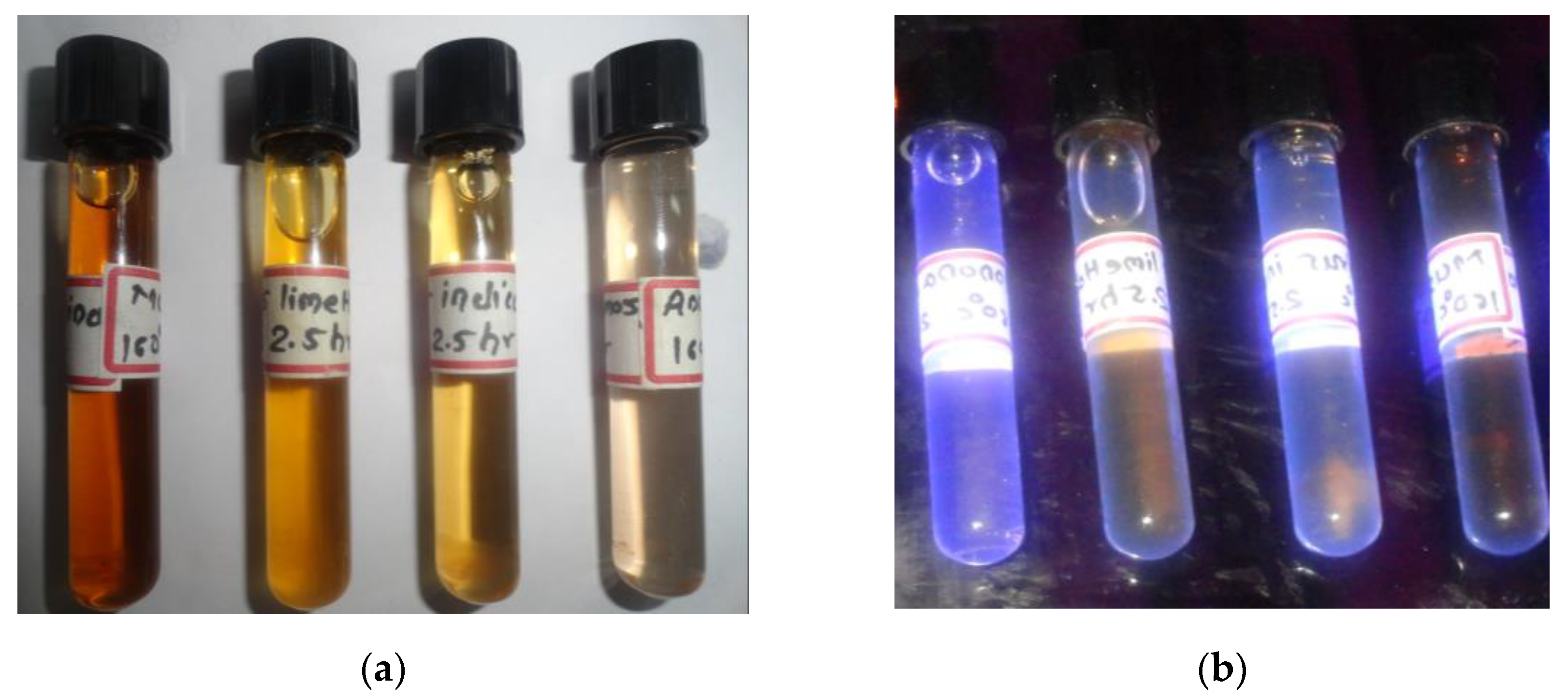
3.1. Optimization of Carbon Dots (CDs) Synthesis and Detection under UV-Transilluminator

The hydrothermal digestion of the carbonized solution from all of the selected fruit peel was used to create CDs, which were then placed in transparent screw cap bottles for the detection of their fluorescence under a UV-transilluminator (Figure 1a,b). Among the selected filtrates, A. squamosa (L.) digested at 160 °C for 150 min showed a better fluorescence compared to the others. Therefore, the CDs prepared from A. squamosa (L.) peels were selected, characterized, and used for a further testing. As a medium for the CDs synthesis, water results in less yield (83 mg per 1 g of digested fruit peel carbonized powder; nearly 8%).

3.2. Characterization of Carbon Dots (CDs)

3.2.1. Quantification of Carbon Dots (CDs)

The quantum yield (QY) of the CDs synthesized for the hydrothermal carbonization of A. squamosa peel was calculated based on the PL intensity and absorbance. In the present study, A. squamosa-mediated carbon dots exhibited a QY of 8.93%, which was probably due to the presence of nitrogen containing functional groups in the CDs.

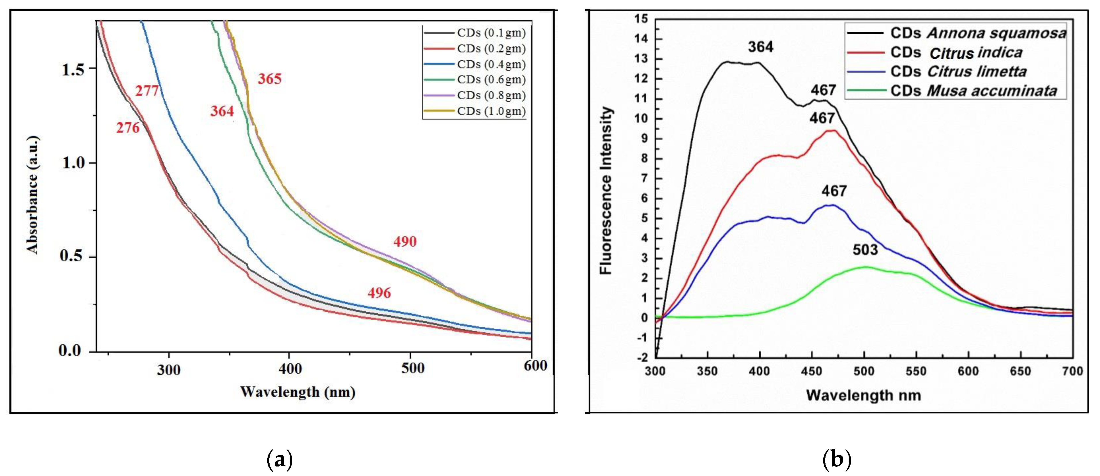
3.2.2. UV–Vis Spectrophotometric and Photo Luminance Analysis

A spectrophotometric and photoluminescence analysis was conducted to determine the absorbance maxima in the UV–Vis range, as well as their emission and excitation wavelengths. Various concentrations of A. squamosa peel powder (0.1, 0.2, 0.4, 0.6, 0.8, and 1.0 g per 25 mL of distilled water) showed different absorbance maxima at 276 nm, 277 nm, 496 nm, and 490 nm, respectively (Figure 2a). The observed deflections in the spectrum are likely caused by electron π−π* transitions. For A. squamosa, the aqueous CDs solution exhibited the strongest photoluminescence at 467 nm (Figure 2b). The CDs synthesized by A. squamosa peels were selected for a further study after the UV–Vis spectrum and fluorescence emission of different plant samples. It was found that the excitation wavelength for the CDs was between 270 nm and 370 nm, and the fluorescence peak for the CDs was between 420 nm and 500 nm.

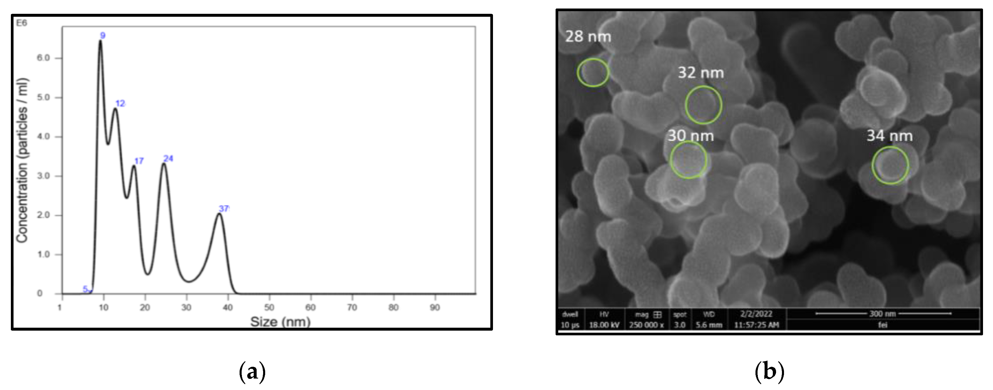
3.2.3. Nanoparticles Tracking and Analysis (NTA)

To determine the size and concentration of synthesized CDs, NTA was conducted. Based on the NTA, the average CD diameter was 15.4 nm, with a mode of 9.26 nm and a standard deviation of 11.4 nm. Figure 3a shows the synthesis of A. squamosa CDs with diameters ranging from 5 to 37 nm. In the sample examined, the CDs accounted for 6.39 × 10⁸ particles mL−1. The FESEM depicted the spherical shape of CDs, with their sizes ranging below 40 nm. The FESEM image of the CDs at 18 kV and the 300 nm scale with a resolution of 250,000× is shown in Figure 3b.

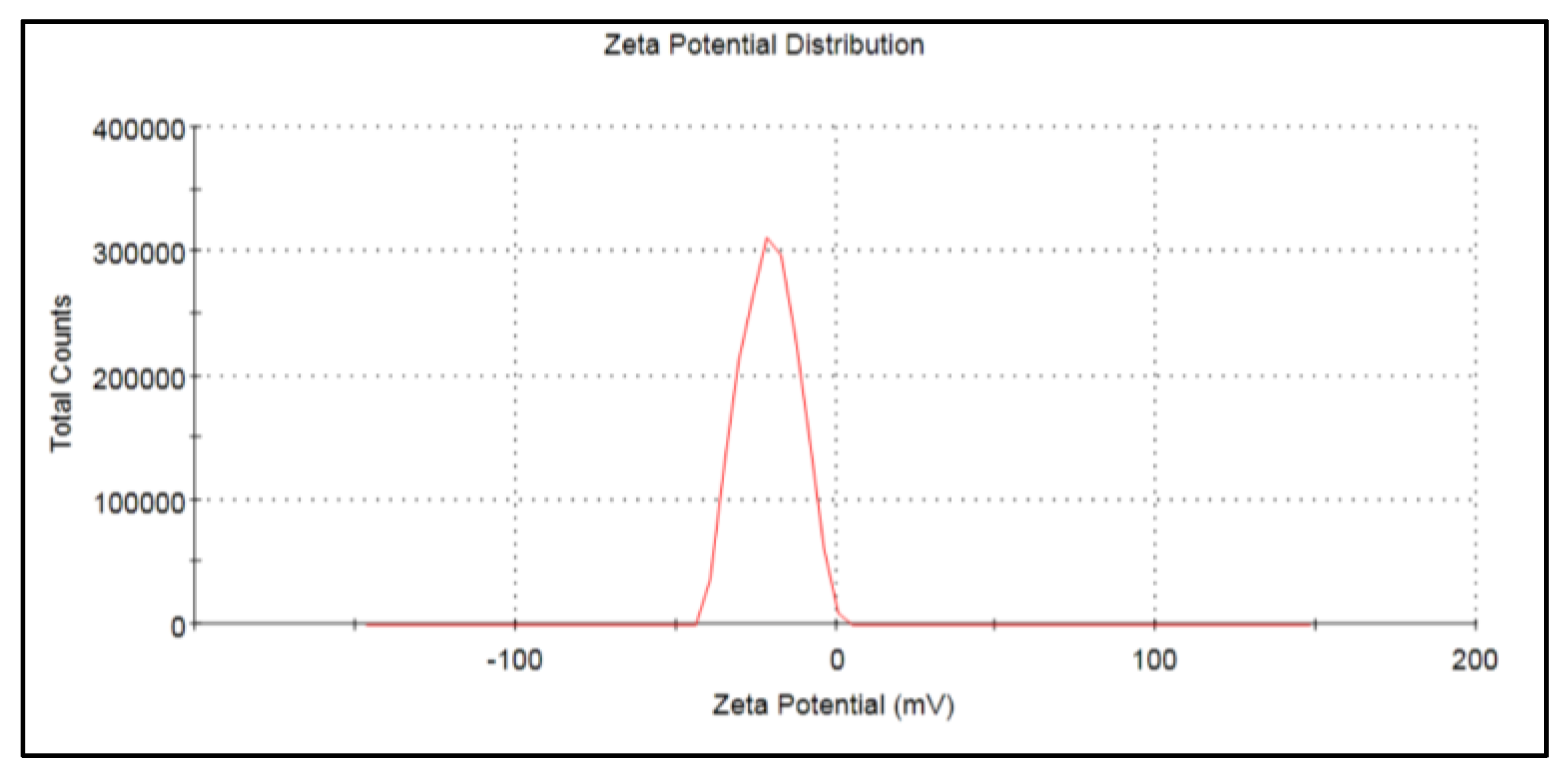
3.2.4. Zeta Potential Analysis

The zeta potential measurements were carried out to determine the stability of CDs in a colloidal or soluble form. The zeta potential was found to be −20.8 mV (Figure 4), which indicates that the nanoparticles were moderately stable.

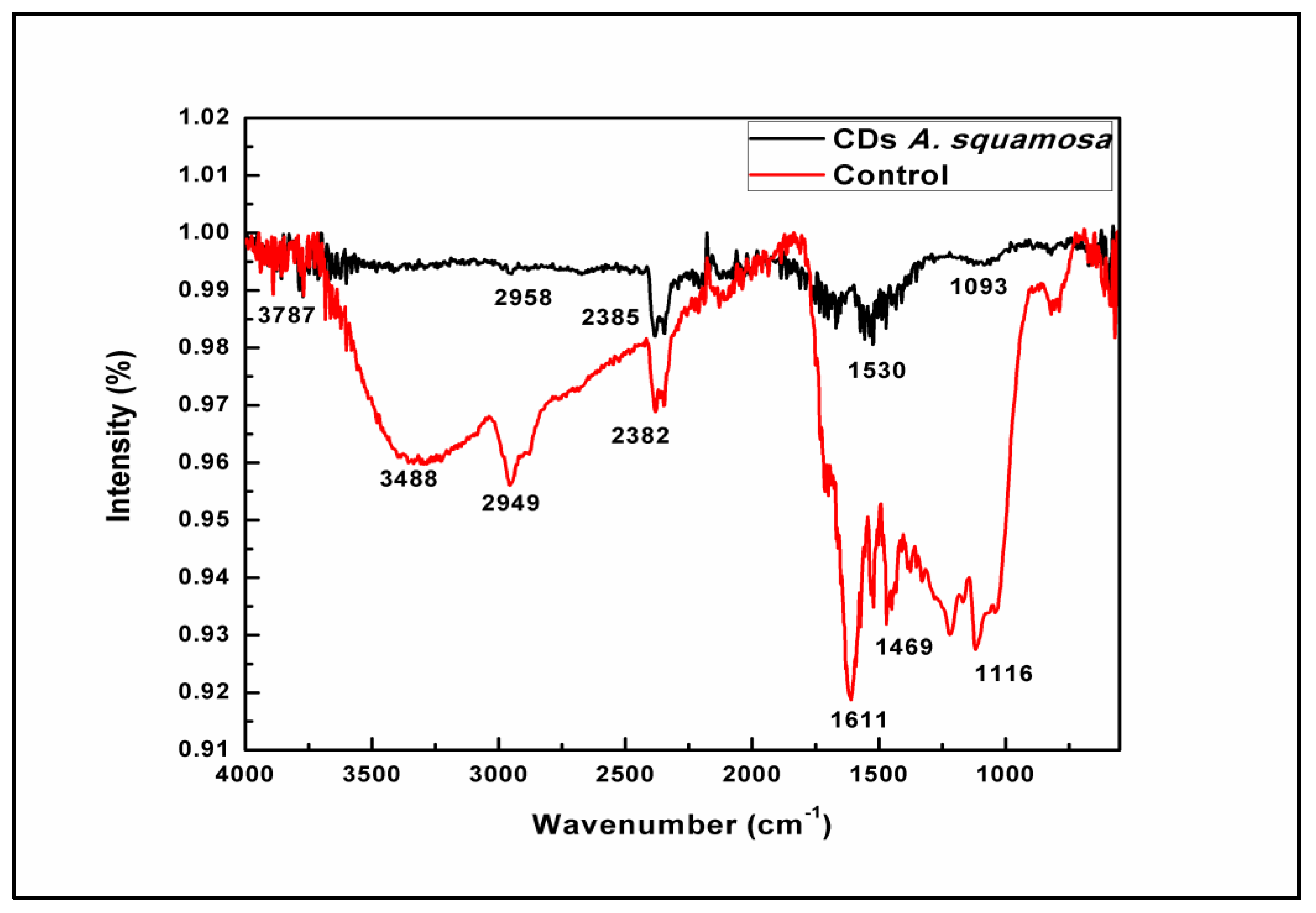
3.2.5. Fourier Transform Infrared (FT-IR) Spectrophotometric Analysis

The FT-IR analysis was performed to determine the biochemical composition and exposed functional groups present in the CDs. A main characteristic absorption band of the O-H and N-H stretching vibration modes at 3787 cm−1 was observed, while 2958 cm−1 gives a C-H and S-H stretching vibration mode. The band at 2385 cm−1 corresponds to polycyclic aromatic hydrocarbons stretching into C=C molecules. N-H bending vibrations at 1643 cm−1 and the band at 1093 cm−1 correspond to C-O-C and C-H bending vibrations of the pyranose ring in fruit peels, which may have assisted in the capping and stabilization of CDs (Figure 5).

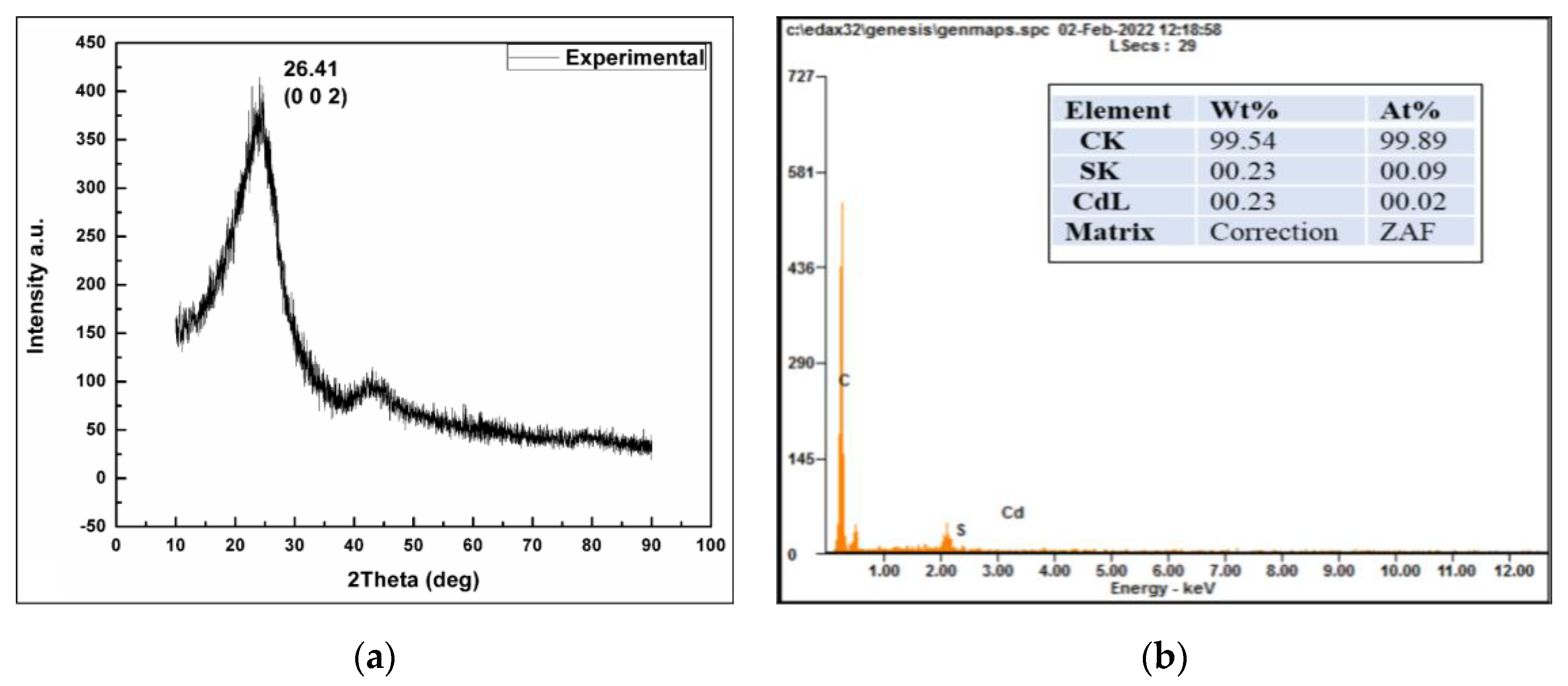
3.2.6. XRD and EDX Analysis of Synthesized CDs

The XRD pattern of the CDs shows a broad peak at 26.41° corresponding to the (002) hkl plane (JCPDS card no. 26-1076) as per Figure 6a. Energy-dispersive X-ray (EDX) spectroscopy depicted 99.54 % of the carbon content with minor signs of cadmium and sulfur impurities, as shown in Figure 6b.

3.3. Evaluation of the Effect of CDs on Germination of Mung Bean (Vigna radiata (L.)) Seeds

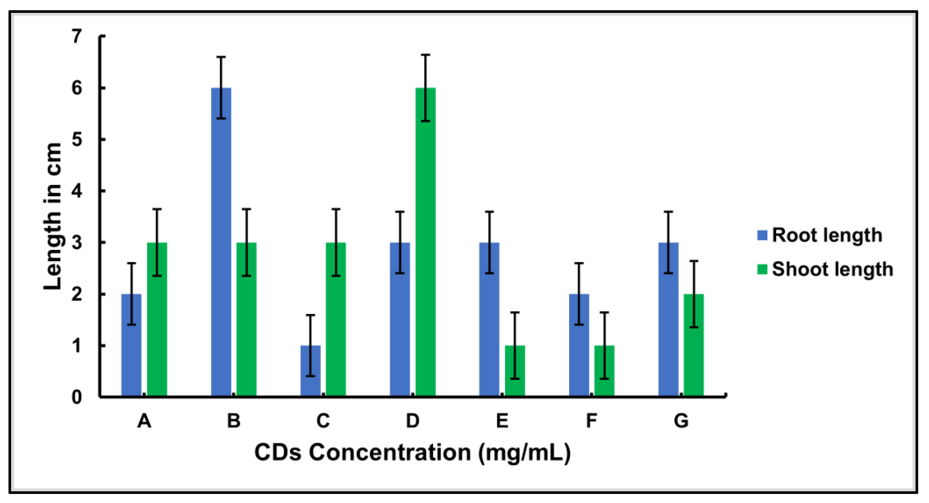
3.3.1. Germination Experiment

The CDs were tested for their ability as growth enhancers or inhibitors on sprouted seeds, their in-planta fluorescence in germinating seeds, and their phytotoxicity. After surface cleaning, the mung bean seeds were treated with various concentrations of CDs prepared from A. squamosa (L.). The germination of the seeds was monitored for the growth of the root and shoot at room temperature. Figure 7 shows the effect of CDs on both seeds after 8 days of incubation. During the 8 days of incubation, it was observed that the root length in the mung bean seedlings reached its maximum at a concentration of 4 mg/mL and the shoot length was highest at a concentration of 16 mg/mL.

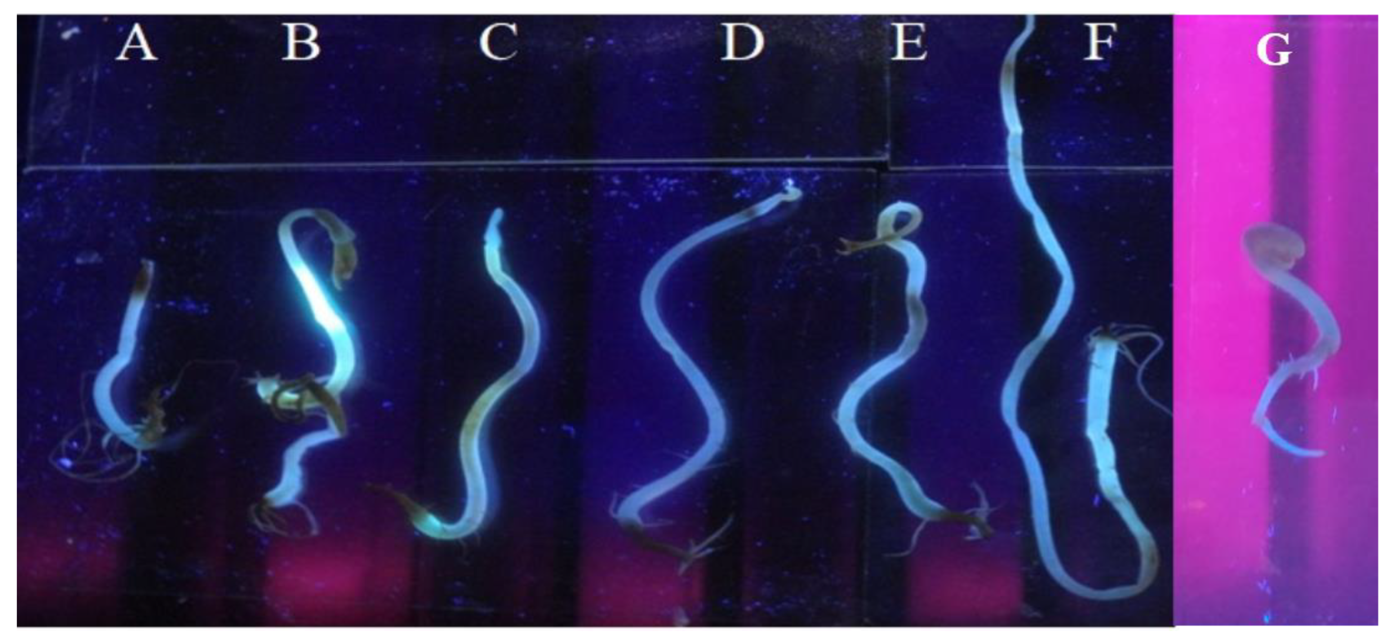
3.3.2. In-planta Photoluminescence Detection

The assimilation of CDs by the plant cells was determined by detecting the photoluminescence in the treated germinating seeds. When observed under UV-light (302/312 nm (UV-B)), the germinated mung bean seeds showed a higher level of light emissions, and the effect was not dose-dependent (Figure 8). Further, no functionalization of the synthesized CDs was required to be absorbed in the shoot cells of V. radiata (L.). The above excitation and fluorescence analysis (Figure 2) led us to conclude that CDs emit a green or blue fluorescence when exposed to UV light. Figure 8 shows the blue-green fluorescence caused by the CDs-treated seeds and growing parts, which were confirmed to have absorbed CDs without being functionalized. In the absence of CDs, no fluorescence was observed in the control seeds (Figure 8G).

4. Discussion

Carbon Dots (CDs) offer a great potential for a broad range of applications such as energy-saving, bioimaging, biosensors, lasers, and light-emitting diodes [4,35,63,64]. They may also be applied as a promising new type of fluorescence marker as well as a high-efficiency catalyst design for applications in bioscience and energy technology [4,65,66]. In a study, Das and colleagues reported a one-step synthesis of fluorescent CDs for biolabeling [67]. A simple and facile one-pot synthesis of the fluorescent CDs from orange waste peels was performed using the hydrothermal carbonization method at a mild temperature (180 °C). Likewise, in the present study, the biogenic synthesis of the CDs in the aqueous system results in a lesser yield, as mentioned in the results Section 3.1, i.e., 83 mg per 1 g equating to nearly 8%, however, is more economical than other organic solvents like PEG (polyethylene glycol) used in the synthesis of CDs reported earlier in several studies [23,32,35,61,68]. The QY analysis demonstrated a lesser QY, that might be an attribute of larger CDs and a broad size distribution. The presence of nitrogenous compounds contributing towards the stability of CDs was confirmed with the FT-IR analysis in Section 2.4.4. The presence of N-H stretches, and bends was confirmed in the FT-IR spectrum. Other reported studies on the quantum yield of the CDs derived from various precursors showed a vast range of QY from 7.8% [45] to 85% [23,46,58].
A spectrophotometric and photoluminescence analysis showed a different absorbance maxima at 276 nm, 277 nm, 496 nm, and 490 nm, which are in line with the previous studies that also described the deflections in the spectrum caused due to the electron transitions at π−π* [69,70]. The morphological features and chemical composition of the obtained CDs were evaluated using various spectroscopies and transmission electron microscopy (TEM). The carbonization and functionalization occur through the dehydration of the orange peels, leading to the formation of nano-sized fluorescent carbon particles [71]. It was also found that the excitation wavelength for the CDs was between 270 nm and 370 nm, and the fluorescence peak for the CDs was between 420 nm and 500 nm. This indicated the emission of a blue or green fluorescence [23,72,73]. In the NTA, we observed individual nanoparticles in motion and their size distribution, which could be used to estimate the number of particles mL−1 in the solution. The NTA results corroborate the TEM analysis of the C-dots synthesized from orange waste peels [63].
The negative zeta potential value of the nanoparticles may be attributed to the negatively charged functional groups present on the surface of the CDs created from fruit peel [51,74,75,76]. In the study of Wu et al. [77], the observed zeta potential values were consistent with the reports of a stable CD synthesis [35]. The FT-IR analysis showed the peaks depicting the functional groups capped and stabilized the CDs. Prasannan and Imae [63] reported that CDs exhibit a main characteristic absorption band of O-H and N-H stretching vibration modes at 3787 cm−1, while 2958 cm−1 gives a C-H and S-H stretching vibration mode. The band at 2385 cm−1 corresponds to polycyclic aromatic hydrocarbons stretching into C=C molecules. The N-H bending vibrations at 1643 cm−1 and band at 1093 cm−1 correspond to C-O-C and C-H bending vibrations of the pyranose ring in fruit peels, which may have assisted in the capping and stabilization of the CDs, as shown in Figure 5 [45,57,63].
The XRD pattern revealed a broad peak at 26.41° when compared with JCPDS card no. 26-1076; it was analogous to the plane (hkl), corresponding to the Miller index (002) for the CDs. This indicates that the space between the layers is larger than the graphitic spaces (0.33 nm), as is the width. As such, C-dots have a poor crystalline structure [55]. The EDX spectroscopy showed 99.54% of the carbon content with minor signs of cadmium and sulfur impurities that corroborated the report by Hu et al. [56].
After the characterization as an application part for the bioimaging, the CDs were tested for their ability as growth enhancers or inhibitors of sprouted seeds, their in-planta fluorescence in germinating seeds, and their phytotoxicity. The mung bean seeds were treated with the CDs synthesized by A. squamosa (L.). A significant effect of the CDs on the root and shoot growth and development was observed after 8 days. At a concentration of 4 mg/mL CDs, the root growth was at the maximum while the shoot length was highest at the concentration of 16 mg/mL. It was found that other types of carbon nanomaterials are toxic to plants, such as multi-walled CNTs and fullerenes demonstrated toxic effects on corn (Zea mays (L.)) and soybean (Glycine max (L.)), which reduced the biomass [78]. The above results indicated that CDs produce a dose-dependent effect, depending upon the growth stage. In plants, the growth promotion may depend on the ability of cells to assimilate the nutrients, which may vary with the developmental stages [61,70,79]. The CDs’ specific optical and chemical properties make them reasonably hydrophilic and ease their transport across plant membranes [80]. The present study indicates the promotion of seed germination and growth due to a certain concentration of CDs. Higher concentrations might be responsible for having no further effect on the germinating seeds. This indicates the dose dependency of the developing seeds on the CDs during the crucial growth stages of seed germination in the case of onions and watermelons [81,82]. The plant physiology is altered to a certain extent to obtain the desired variation in the plant system [42,83].
Chemical and physical methods are more effective. Wang and Hu [84] summarize the advantages and disadvantages of various approaches to CDs synthesis. There has been some difficulty in the synthesis and application of major physical and chemical methods. The method reported here is facile, reproducible, quick, and generates CDs of a range of sizes and can also be used to design bioassays based on their fluorescence properties. Additionally, the in-planta results reported here support the application of A. squamosa-based CDs in plant bioassays and in vivo biolabeling [68,78]. These biocompatible CDs can thus be assessed for their fate in vivo and be applied in biolabeling studies to understand the various biomolecular pathways and predict the desired spatial distribution of the nanomaterials [4,35,85,86].

5. Conclusions

The current use of bioimaging dyes and agents are mostly chemicals, which are hazardous to humans and the environment. Chemical dyes damage the cell wall, are more expensive, and can only stain dead cells. In the present study, a green, eco-friendly, and economically viable approach has been developed for the synthesis of CDs. CDs synthesized from plant-based materials are eco-friendly and cost-effective compared to chemical agents. CDs were assimilated by the cells of germinating mung bean (Vigna radiata (L.)) seeds without the further functionalization for their entry into plant tissues. CDs also showed a significant effect on the seed germination in mung beans. It is suggested that the fluorescent CDs synthesized from fruit peels by the microwave-assisted method could be used as a bioimaging agent because there were no signs of any visible phytotoxic effect on the germinating seeds, as indicated by zero mortality at any concentration used for the assessment; thus, indicating their biocompatibility with the plant systems for the development of biolabeling assays.
Moreover, the findings of the present study would be useful in understanding and addressing the problem of toxicity associated with chemical bioimaging dyes. Further, thorough studies on the conjugation of CDs with the desired biocatalyst for specific functions in-planta would be helpful to reveal the applicability of CDs in various fields. The present findings open a new avenue for the commercialization of CDs as a viable alternative to synthetic organic dyes as multi-color emitting probes for cell labeling in bioimaging.

Author Contributions

Conceptualization, A.K.G.; data curation, A.M.P., P.U.I. and S.S.S.; formal analysis, S.S.S. and V.D.R.; funding acquisition, T.M.M., V.D.R., S.S.S. and V.P.K.; investigation, A.M.P.; methodology, A.M.P., S.S.S. and A.K.G.; resources, V.D.R.; software, A.K.G.; supervision, M.R. and A.K.G.; validation, P.U.I., R.W.R., M.R. and A.K.G.; visualization, T.M.M. and V.P.K.; writing—original draft, A.M.P. and P.U.I.; writing—review and editing, R.W.R., S.S.S., M.R., T.M.M. and A.K.G. All authors have read and agreed to the published version of the manuscript.

Funding

The study was supported by the grant from the Russian Science Foundation (Project No. 21-77-20089).

Data Availability Statement

Not applicable.

Acknowledgments

The authors are grateful to the Scientific research center “Kurchatov Institute”.

Conflicts of Interest

The authors declare no conflict of interest.

References

  1. Dreaden, E.C.; Alkilany, A.M.; Huang, X.; Murphy, C.J.; El-Sayed, M.A. The Golden Age: Gold Nanoparticles for Biomedicine. Chem. Soc. Rev. 2012, 41, 2740–2779. [Google Scholar] [CrossRef] [PubMed] [Green Version]
  2. Boschi, F.; de Sanctis, F. Overview of the Optical Properties of Fluorescent Nanoparticles for Optical Imaging. Eur. J. Histochem. 2017, 61, 2830. [Google Scholar] [CrossRef] [PubMed]
  3. Khan, I.; Saeed, K.; Khan, I. Nanoparticles: Properties, Applications and Toxicities. Arab. J. Chem. 2019, 12, 908–931. [Google Scholar] [CrossRef]
  4. Manikandan, V.; Lee, N.Y. Green Synthesis of Carbon Quantum Dots and Their Environmental Applications. Environ. Res. 2022, 212, 113283. [Google Scholar] [CrossRef] [PubMed]
  5. Marcato, P.D.; Durán, N. New Aspects of Nanopharmaceutical Delivery Systems. J. Nanosci. Nanotechnol. 2008, 8, 2216–2229. [Google Scholar] [CrossRef] [Green Version]
  6. Laurent, S.; Forge, D.; Port, M.; Roch, A.; Robic, C.; vander Elst, L.; Muller, R.N. Magnetic Iron Oxide Nanoparticles: Synthesis, Stabilization, Vectorization, Physicochemical Characterizations, and Biological Applications. Chem. Rev. 2010, 110, 2574. [Google Scholar] [CrossRef]
  7. Rai, M.; Ingle, A. Role of Nanotechnology in Agriculture with Special Reference to Management of Insect Pests. Appl. Microbiol. Biotechnol. 2012, 94, 287–293. [Google Scholar] [CrossRef]
  8. Gubala, V.; Giovannini, G.; Kunc, F.; Monopoli, M.P.; Moore, C.J. Dye-Doped Silica Nanoparticles: Synthesis, Surface Chemistry and Bioapplications. Cancer Nanotechnol. 2020, 11, 1. [Google Scholar] [CrossRef]
  9. PekaÅTM, M. Hydrogels with Micellar Hydrophobic (Nano) Domains. Front. Mater. 2015, 1, 35. [Google Scholar] [CrossRef] [Green Version]
  10. Ahmed, E.M. Hydrogel: Preparation, Characterization, and Applications: A Review. J. Adv. Res. 2015, 6, 105–121. [Google Scholar] [CrossRef]
  11. Gupta, R.; Peveler, W.J.; Lix, K.; Algar, W.R. Comparison of Semiconducting Polymer Dots and Semiconductor Quantum Dots for Smartphone-Based Fluorescence Assays. Anal. Chem. 2019, 91, 10955–10960. [Google Scholar] [CrossRef] [PubMed]
  12. Liu, J.; Li, R.; Yang, B. Carbon Dots: A New Type of Carbon-Based Nanomaterial with Wide Applications. ACS Cent. Sci. 2020, 6, 2179–2195. [Google Scholar] [CrossRef] [PubMed]
  13. Akagi, T.; Baba, M.; Akashi, M. Preparation of Nanoparticles by the Self-Organization of Polymers Consisting of Hydrophobic and Hydrophilic Segments: Potential Applications. Polymer 2007, 48, 6729–6747. [Google Scholar] [CrossRef] [Green Version]
  14. Liu, P.; Chen, Y.; Yu, Z. Organic-Inorganic Hydrophobic Nanocomposite Film with a Core-Shell Structure. Materials 2016, 9, 1021. [Google Scholar] [CrossRef] [PubMed] [Green Version]
  15. Liang, G.; Wang, H.; Shi, H.; Wang, H.; Zhu, M.; Jing, A.; Li, J.; Li, G. Recent Progress in the Development of Upconversion Nanomaterials in Bioimaging and Disease Treatment. J. Nanobiotechnol. 2020, 18, 154. [Google Scholar] [CrossRef]
  16. Kravets, V.; Almemar, Z.; Jiang, K.; Culhane, K.; Machado, R.; Hagen, G.; Kotko, A.; Dmytruk, I.; Spendier, K.; Pinchuk, A. Imaging of Biological Cells Using Luminescent Silver Nanoparticles. Nanoscale Res. Lett. 2016, 11, 30. [Google Scholar] [CrossRef] [Green Version]
  17. Yaqoob, S.B.; Adnan, R.; Rameez Khan, R.M.; Rashid, M. Gold, Silver, and Palladium Nanoparticles: A Chemical Tool for Biomedical Applications. Front. Chem. 2020, 8, 376. [Google Scholar] [CrossRef]
  18. Si, P.; Razmi, N.; Nur, O.; Solanki, S.; Pandey, C.M.; Gupta, R.K.; Malhotra, B.D.; Willander, M.; de la Zerda, A. Gold Nanomaterials for Optical Biosensing and Bioimaging. Nanoscale Adv. 2021, 3, 2679–2698. [Google Scholar] [CrossRef]
  19. Trzepiński, P.; Klajnert-Maculewicz, B. Dendrimers for Fluorescence-Based Bioimaging. J. Chem. Technol. Biotechnol. 2017, 92, 1157–1166. [Google Scholar] [CrossRef]
  20. Wolfbeis, O.S. An Overview of Nanoparticles Commonly Used in Fluorescent Bioimaging. Chem. Soc. Rev. 2015, 44, 4743–4768. [Google Scholar] [CrossRef]
  21. Zhang, H.; Wang, G.; Zhang, Z.; Lei, J.H.; Liu, T.-M.; Xing, G.; Deng, C.-X.; Tang, Z.; Qu, S. One Step Synthesis of Efficient Red Emissive Carbon Dots and Their Bovine Serum Albumin Composites with Enhanced Multi-Photon Fluorescence for in Vivo Bioimaging. Light Sci. Appl. 2022, 11, 113. [Google Scholar] [CrossRef] [PubMed]
  22. Das, P.; Maruthapandi, M.; Saravanan, A.; Natan, M.; Jacobi, G.; Banin, E.; Gedanken, A. Carbon Dots for Heavy-Metal Sensing, PH-Sensitive Cargo Delivery, and Antibacterial Applications. ACS Appl. Nano Mater. 2020, 3, 11777–11790. [Google Scholar] [CrossRef]
  23. Qie, X.; Zan, M.; Gui, P.; Chen, H.; Wang, J.; Lin, K.; Mei, Q.; Ge, M.; Zhang, Z.; Tang, Y.; et al. Design, Synthesis, and Application of Carbon Dots With Synergistic Antibacterial Activity. Front. Bioeng. Biotechnol. 2022, 10, 894100. [Google Scholar] [CrossRef] [PubMed]
  24. Saravanan, A.; Maruthapandi, M.; Das, P.; Ganguly, S.; Margel, S.; Luong, J.H.T.; Gedanken, A. Applications of N-Doped Carbon Dots as Antimicrobial Agents, Antibiotic Carriers, and Selective Fluorescent Probes for Nitro Explosives. ACS Appl. Bio Mater. 2020, 3, 8023–8031. [Google Scholar] [CrossRef]
  25. Zhang, C.; Chen, M.; Kang, S.; Yan, F.; Han, X.; Gautam, S.; Hu, Z.; Zheng, H.; Chen, P.; Gao, S.; et al. Light Absorption and Fluorescence Characteristics of Water-Soluble Organic Compounds in Carbonaceous Particles at a Typical Remote Site in the Southeastern Himalayas and Tibetan Plateau. Environ. Pollut. 2021, 272, 116000. [Google Scholar] [CrossRef]
  26. de Oliveira, B.P.; da Silva Abreu, F.O.M. Carbon Quantum Dots Synthesis from Waste and By-Products: Perspectives and Challenges. Mater. Lett. 2021, 282, 128764. [Google Scholar] [CrossRef]
  27. Barbosa, C.D.d.E.S.; Corrêa, J.R.; Medeiros, G.A.; Barreto, G.; Magalhães, K.G.; de Oliveira, A.L.; Spencer, J.; Rodrigues, M.O.; Neto, B.A.D. Carbon Dots (C-Dots) from Cow Manure with Impressive Subcellular Selectivity Tuned by Simple Chemical Modification. Chem. A Eur. J. 2015, 21, 5055–5060. [Google Scholar] [CrossRef]
  28. Guo, Y.; Zhang, L.; Cao, F.; Leng, Y. Thermal Treatment of Hair for the Synthesis of Sustainable Carbon Quantum Dots and the Applications for Sensing Hg2+. Sci. Rep. 2016, 6, 35795. [Google Scholar] [CrossRef] [Green Version]
  29. Tyagi, A.; Tripathi, K.M.; Singh, N.; Choudhary, S.; Gupta, R.K. Green Synthesis of Carbon Quantum Dots from Lemon Peel Waste: Applications in Sensing and Photocatalysis. RSC Adv. 2016, 6, 72432. [Google Scholar] [CrossRef]
  30. Kumari, A.; Kumar, A.; Sahu, S.K.; Kumar, S. Synthesis of Green Fluorescent Carbon Quantum Dots Using Waste Polyolefins Residue for Cu2+ Ion Sensing and Live Cell Imaging. Sens. Actuators B Chem. 2018, 254, 197–205. [Google Scholar] [CrossRef]
  31. Tripathi, S.; Sarkar, S. Influence of Water Soluble Carbon Dots on the Growth of Wheat Plant. Appl. Nanosci. 2015, 5, 609–616. [Google Scholar] [CrossRef] [Green Version]
  32. Kong, W.; Liu, R.; Li, H.; Liu, J.; Huang, H.; Liu, Y.; Kang, Z. High-Bright Fluorescent Carbon Dots and Their Application in Selective Nucleoli Staining. J. Mater. Chem. B 2014, 2, 5077. [Google Scholar] [CrossRef]
  33. Anwar, S.; Ding, H.; Xu, M.; Hu, X.; Li, Z.; Wang, J.; Liu, L.; Jiang, L.; Wang, D.; Dong, C.; et al. Recent Advances in Synthesis, Optical Properties, and Biomedical Applications of Carbon Dots. ACS Appl. Bio Mater. 2019, 2, 2317–2338. [Google Scholar] [CrossRef] [PubMed]
  34. Semeniuk, M.; Yi, Z.; Poursorkhabi, V.; Tjong, J.; Jaffer, S.; Lu, Z.-H.; Sain, M. Future Perspectives and Review on Organic Carbon Dots in Electronic Applications. ACS Nano 2019, 13, 6224–6255. [Google Scholar] [CrossRef] [PubMed]
  35. Magesh, V.; Sundramoorthy, A.K.; Ganapathy, D. Recent Advances on Synthesis and Potential Applications of Carbon Quantum Dots. Front. Mater. 2022, 9, 906838. [Google Scholar] [CrossRef]
  36. Liu, M.L.; Chen, B.B.; Li, C.M.; Huang, C.Z. Carbon Dots: Synthesis, Formation Mechanism, Fluorescence Origin and Sensing Applications. Green Chem. 2019, 21, 449–471. [Google Scholar] [CrossRef]
  37. Li, H.; Kang, Z.; Liu, Y.; Lee, S.-T. Carbon Nanodots: Synthesis, Properties and Applications. J. Mater. Chem. 2012, 22, 24230. [Google Scholar] [CrossRef]
  38. Cañas, J.E.; Long, M.; Nations, S.; Vadan, R.; Dai, L.; Luo, M.; Ambikapathi, R.; Lee, E.H.; Olszyk, D. Effects of functionalized and nonfunctionalized single-walled carbon nanotubes on root elongation of select crop species. Environ. Toxicol. Chem. 2008, 27, 1922–1931. [Google Scholar] [CrossRef]
  39. Tan, X.; Lin, C.; Fugetsu, B. Studies on Toxicity of Multi-Walled Carbon Nanotubes on Suspension Rice Cells. Carbon N. Y. 2009, 47, 3479–3487. [Google Scholar] [CrossRef]
  40. Yuan, H.; Hu, S.; Huang, P.; Song, H.; Wang, K.; Ruan, J.; He, R.; Cui, D. Single Walled Carbon Nanotubes Exhibit Dual-Phase Regulation to Exposed Arabidopsis Mesophyll Cells. Nanoscale Res. Lett. 2011, 6, 44. [Google Scholar] [CrossRef]
  41. Yuan, X.; Zhang, X.; Sun, L.; Wei, Y.; Wei, X. Cellular Toxicity and Immunological Effects of Carbon-Based Nanomaterials. Part. Fibre Toxicol. 2019, 16, 18. [Google Scholar] [CrossRef] [PubMed]
  42. Li, Y.; Xu, X.; Wu, Y.; Zhuang, J.; Zhang, X.; Zhang, H.; Lei, B.; Hu, C.; Liu, Y. A Review on the Effects of Carbon Dots in Plant Systems. Mater. Chem. Front. 2020, 4, 437–448. [Google Scholar] [CrossRef]
  43. Sheth, K.N.; Patel, R.V. Removal of Cadmium (II) and Chromium (IV) Using OPAC and PAC. Environ. Poll. Cont. J. 2005, 9, 49–51. [Google Scholar]
  44. Kang, K.-H.; Kam, S.-K.; Lee, M.-G. Preparation of activated carbon from waste citrus peels by ZnCl2. J. Environ. Sci. 2007, 16, 1091–1098. [Google Scholar]
  45. Yang, Y.; Cui, J.; Zheng, M.; Hu, C.; Tan, S.; Xiao, Y.; Yang, Q.; Liu, Y. One-Step Synthesis of Amino-Functionalized Fluorescent Carbon Nanoparticles by Hydrothermal Carbonization of Chitosan. Chem. Commun. 2012, 48, 380–382. [Google Scholar] [CrossRef] [PubMed]
  46. Feng, B.; Xu, Z.; Wang, J.; Gai, L. Water-Soluble Organic Polymer/Silica Composite Nanofilms with Improved Fluorescence Quantum Yield. J. Lumin. 2019, 211, 347–354. [Google Scholar] [CrossRef]
  47. Talam, S.; Karumuri, S.R.; Gunnam, N. Synthesis, Characterization, and Spectroscopic Properties of ZnO Nanoparticles. ISRN Nanotechnol. 2012, 2012, 372505. [Google Scholar] [CrossRef] [Green Version]
  48. Mourdikoudis, S.; Pallares, R.M.; Thanh, N.T.K. Characterization Techniques for Nanoparticles: Comparison and Complementarity upon Studying Nanoparticle Properties. Nanoscale 2018, 10, 12871–12934. [Google Scholar] [CrossRef] [Green Version]
  49. Ashraf, J.M.; Ansari, M.A.; Khan, H.M.; Alzohairy, M.A.; Choi, I. Green Synthesis of Silver Nanoparticles and Characterization of Their Inhibitory Effects on AGEs Formation Using Biophysical Techniques. Sci. Rep. 2016, 6, 20414. [Google Scholar] [CrossRef] [Green Version]
  50. Gallego-Urrea, J.A.; Tuoriniemi, J.; Pallander, T.; Hassellöv, M. Measurements of Nanoparticle Number Concentrations and Size Distributions in Contrasting Aquatic Environments Using Nanoparticle Tracking Analysis. Environ. Chem. 2010, 7, 67. [Google Scholar] [CrossRef]
  51. Bramhanwade, K.; Shende, S.; Bonde, S.; Gade, A.; Rai, M. Fungicidal Activity of Cu Nanoparticles against Fusarium Causing Crop Diseases. Environ. Chem. Lett. 2016, 14, 229–235. [Google Scholar] [CrossRef]
  52. Andrade, P.F.; Nakazato, G.; Durán, N. Additive Interaction of Carbon Dots Extracted from Soluble Coffee and Biogenic Silver Nanoparticles against Bacteria. J. Phys. Conf. Ser. 2017, 838, 012028. [Google Scholar] [CrossRef]
  53. Dager, A.; Uchida, T.; Maekawa, T.; Tachibana, M. Synthesis and Characterization of Mono-Disperse Carbon Quantum Dots from Fennel Seeds: Photoluminescence Analysis Using Machine Learning. Sci. Rep. 2019, 9, 14004. [Google Scholar] [CrossRef] [PubMed] [Green Version]
  54. Pudza, M.Y.; Abidin, Z.Z.; Abdul-Rashid, S.; Yassin, F.M.; Noor, A.S.M.; Abdullah, M. Synthesis and Characterization of Fluorescent Carbon Dots from Tapioca. ChemistrySelect 2019, 4, 4140–4146. [Google Scholar] [CrossRef]
  55. Shaikh, A.F.; Tamboli, M.S.; Patil, R.H.; Bhan, A.; Ambekar, J.D.; Kale, B.B. Bioinspired Carbon Quantum Dots: An Antibiofilm Agents. J. Nanosci. Nanotechnol. 2019, 19, 2339–2345. [Google Scholar] [CrossRef]
  56. Hu, Q.; Gong, X.; Liu, L.; Choi, M.M.F. Characterization and Analytical Separation of Fluorescent Carbon Nanodots. J. Nanomater. 2017, 2017, 1804178. [Google Scholar] [CrossRef] [Green Version]
  57. Li, X.; Zhang, S.; Kulinich, S.A.; Liu, Y.; Zeng, H. Engineering Surface States of Carbon Dots to Achieve Controllable Luminescence for Solid-Luminescent Composites and Sensitive Be2+ Detection. Sci. Rep. 2015, 4, 4976. [Google Scholar] [CrossRef] [Green Version]
  58. Lin, H.; Huang, J.; Ding, L. Preparation of Carbon Dots with High-Fluorescence Quantum Yield and Their Application in Dopamine Fluorescence Probe and Cellular Imaging. J. Nanomater. 2019, 2019, 5037243. [Google Scholar] [CrossRef]
  59. Ding, H.; Li, X.-H.; Chen, X.-B.; Wei, J.-S.; Li, X.-B.; Xiong, H.-M. Surface States of Carbon Dots and Their Influences on Luminescence. J. Appl. Phys. 2020, 127, 231101. [Google Scholar] [CrossRef]
  60. Pandey, K.; Lahiani, M.H.; Hicks, V.K.; Hudson, M.K.; Green, M.J.; Khodakovskaya, M. Effects of Carbon-Based Nanomaterials on Seed Germination, Biomass Accumulation and Salt Stress Response of Bioenergy Crops. PLoS ONE 2018, 13, e0202274. [Google Scholar] [CrossRef] [Green Version]
  61. Li, H.; Huang, J.; Lu, F.; Liu, Y.; Song, Y.; Sun, Y.; Zhong, J.; Huang, H.; Wang, Y.; Li, S.; et al. Impacts of Carbon Dots on Rice Plants: Boosting the Growth and Improving the Disease Resistance. ACS Appl. Bio Mater. 2018, 1, 663–672. [Google Scholar] [CrossRef] [PubMed] [Green Version]
  62. Costanzo, E.; Gulino, M.; Lanzanò, L.; Musumeci, F.; Scordino, A.; Tudisco, S.; Sui, L. Single Seed Viability Checked by Delayed Luminescence. Eur. Biophys. J. 2008, 37, 235–238. [Google Scholar] [CrossRef] [PubMed]
  63. Wang, B.; Li, J.; Tang, Z.; Yang, B.; Lu, S. Near-Infrared Emissive Carbon Dots with 33.96% Emission in Aqueous Solution for Cellular Sensing and Light-Emitting Diodes. Sci. Bull. 2019, 64, 1285–1292. [Google Scholar] [CrossRef] [Green Version]
  64. Yang, X.; Sui, L.; Wang, B.; Zhang, Y.; Tang, Z.; Yang, B.; Lu, S. Red-Emitting, Self-Oxidizing Carbon Dots for the Preparation of White LEDs with Super-High Color Rendering Index. Sci. China Chem. 2021, 64, 1547–1553. [Google Scholar] [CrossRef]
  65. Zhu, S.; Meng, Q.; Wang, L.; Zhang, J.; Song, Y.; Jin, H.; Zhang, K.; Sun, H.; Wang, H.; Yang, B. Highly Photoluminescent Carbon Dots for Multicolor Patterning, Sensors, and Bioimaging. Angew. Chem. Int. Ed. 2013, 52, 3953–3957. [Google Scholar] [CrossRef] [PubMed]
  66. Rai, S.; Singh, B.K.; Bhartiya, P.; Singh, A.; Kumar, H.; Dutta, P.K.; Mehrotra, G.K. Lignin Derived Reduced Fluorescence Carbon Dots with Theranostic Approaches: Nano-Drug-Carrier and Bioimaging. J. Lumin. 2017, 190, 492–503. [Google Scholar] [CrossRef]
  67. Das, P.; Bose, M.; Das, A.K.; Banerjee, S.; Das, N.C. One-Step Synthesis of Fluorescent Carbon Dots for Bio-Labeling Assay. Macromol. Symp. 2018, 382, 1800077. [Google Scholar] [CrossRef]
  68. Kianpour, E.; Azizian, S. Optimization of One-Step and One-Substrate Synthesis of Carbon Nanodots by Microwave Pyrolysis. RSC Adv. 2014, 4, 40907–40911. [Google Scholar] [CrossRef]
  69. Yan, D.; Easwaran, V.; Chau, V.; Okamoto, M.; Ierullo, M.; Kimura, M.; Endo, A.; Yano, R.; Pasha, A.; Gong, Y.; et al. NIN-like Protein 8 Is a Master Regulator of Nitrate-Promoted Seed Germination in Arabidopsis. Nat. Commun. 2016, 7, 13179. [Google Scholar] [CrossRef] [Green Version]
  70. Zheng, X.T.; Than, A.; Ananthanaraya, A.; Kim, D.-H.; Chen, P. Graphene Quantum Dots as Universal Fluorophores and Their Use in Revealing Regulated Trafficking of Insulin Receptors in Adipocytes. ACS Nano 2013, 7, 6278–6286. [Google Scholar] [CrossRef]
  71. Prasannan, A.; Imae, T. One-Pot Synthesis of Fluorescent Carbon Dots from Orange Waste Peels. Ind. Eng. Chem. Res. 2013, 52, 15673–15678. [Google Scholar] [CrossRef]
  72. Lu, S.; Sui, L.; Liu, J.; Zhu, S.; Chen, A.; Jin, M.; Yang, B. Near-Infrared Photoluminescent Polymer-Carbon Nanodots with Two-Photon Fluorescence. Adv. Mater. 2017, 29, 1603443. [Google Scholar] [CrossRef] [PubMed]
  73. Qian, K.; Guo, H.; Chen, G.; Ma, C.; Xing, B. Distribution of Different Surface Modified Carbon Dots in Pumpkin Seedlings. Sci. Rep. 2018, 8, 7991. [Google Scholar] [CrossRef] [PubMed] [Green Version]
  74. Shende, S.; Gade, A.; Rai, M. Large-Scale Synthesis and Antibacterial Activity of Fungal-Derived Silver Nanoparticles. Environ. Chem. Lett. 2017, 15, 427–434. [Google Scholar] [CrossRef]
  75. Bayati, M.; Dai, J.; Zambrana, A.; Rees, C.; Fidalgo de Cortalezzi, M. Effect of Water Chemistry on the Aggregation and Photoluminescence Behavior of Carbon Dots. J. Environ. Sci. 2018, 65, 223–235. [Google Scholar] [CrossRef] [PubMed]
  76. Shende, S.S.; Rajput, V.D.; Gorovtsov, A.V.; Harish; Saxena, P.; Minkina, T.M.; Chokheli, V.A.; Jatav, H.S.; Sushkova, S.N.; Kaur, P.; et al. Interaction of Nanoparticles with Microbes. In Advances in Science, Technology and Innovation; Springer Nature: Cham, Switzerland, 2021; pp. 175–188. [Google Scholar]
  77. Wu, Y.-N.; Li, Y.; Cao, M.-J.; Dai, C.-L.; He, L.; Yang, Y.-P. Preparation and Stabilization Mechanism of Carbon Dots Nanofluids for Drag Reduction. Pet. Sci. 2020, 17, 1717–1725. [Google Scholar] [CrossRef]
  78. de La Torre-Roche, R.; Hawthorne, J.; Deng, Y.; Xing, B.; Cai, W.; Newman, L.A.; Wang, Q.; Ma, X.; Hamdi, H.; White, J.C. Multiwalled Carbon Nanotubes and C60 Fullerenes Differentially Impact the Accumulation of Weathered Pesticides in Four Agricultural Plants. Environ. Sci. Technol. 2013, 47, 12539–12547. [Google Scholar] [CrossRef]
  79. Sharma, A.; Das, J. Small Molecules Derived Carbon Dots: Synthesis and Applications in Sensing, Catalysis, Imaging, and Biomedicine. J. Nanobiotechnol. 2019, 17, 92. [Google Scholar] [CrossRef] [Green Version]
  80. Guo, B.; Liu, G.; Li, W.; Hu, C.; Lei, B.; Zhuang, J.; Zheng, M.; Liu, Y. The role of carbon dots in the life cycle of crops. Indust. Crops Prod. 2022, 187, 115427. [Google Scholar] [CrossRef]
  81. Acharya, P.; Jayaprakasha, G.K.; Crosby, K.M.; Jifon, J.L.; Patil, B.S. Green-Synthesized Nanoparticles Enhanced Seedling Growth, Yield, and Quality of Onion (Allium cepa L.). ACS Sustain. Chem. Eng. 2019, 7, 14580–14590. [Google Scholar] [CrossRef]
  82. Acharya, P.; Jayaprakasha, G.K.; Crosby, K.M.; Jifon, J.L.; Patil, B.S. Nanoparticle-Mediated Seed Priming Improves Germination, Growth, Yield, and Quality of Watermelons (Citrullus lanatus) at multi-locations in Texas. Sci. Rep. 2020, 10, 5037. [Google Scholar] [CrossRef] [PubMed] [Green Version]
  83. Szőllősi, R.; Molnár, Á.; Kondak, S.; Kolbert, Z. Dual Effect of Nanomaterials on Germination and Seedling Growth: Stimulation vs. Phytotoxicity. Plants 2020, 9, 1745. [Google Scholar] [CrossRef] [PubMed]
  84. Wang, Y.; Hu, A. Carbon Quantum Dots: Synthesis, Properties and Applications. J. Mater. Chem. C Mater. 2014, 2, 6921. [Google Scholar] [CrossRef] [Green Version]
  85. Cao, X.; Pan, X.; Couvillion, S.P.; Zhang, T.; Tamez, C.; Bramer, L.M.; White, J.C.; Qian, W.-J.; Thrall, B.D.; Ng, K.W.; et al. Fate, Cytotoxicity and Cellular Metabolomic Impact of Ingested Nanoscale Carbon Dots Using Simulated Digestion and a Triculture Small Intestinal Epithelial Model. NanoImpact 2021, 23, 100349. [Google Scholar] [CrossRef] [PubMed]
  86. Fernandes, S.; da Silva, J.C.G.E.; da Silva, L.P. Comparative Life Cycle Assessment of High-Yield Synthesis Routes for Carbon Dots. NanoImpact 2021, 23, 100332. [Google Scholar] [CrossRef]
Figure 1. (a) Synthesized CDs solutions from different fruit peels from left to right – Annona squamosa (L.), Citrus indica (L.), Citrus limetta (L.), and Musa acuminata (L.); (b) CDs solutions under UV-transilluminator.
Nanomaterials 13 00209 g001
Figure 2. (a) UV–Vis spectrum of Annona squamosa-mediated CDs synthesized at various concentrations; (b) photoelectric luminance analysis (excitation wavelength 364 nm).
Nanomaterials 13 00209 g002
Figure 3. (a) NTA (Nanosight-LM-20) nanoparticle size distribution histogram showing the average size of 15.4 nm; (b) FESEM image of CDs at 250,000× resolution.
Nanomaterials 13 00209 g003
Figure 4. The zeta potential graph for biosynthesized CDs shows a zeta potential at −20.8 mV.
Nanomaterials 13 00209 g004
Figure 5. Fourier transform infrared (FTIR) spectral analysis of A. squamosa CDs.
Nanomaterials 13 00209 g005
Figure 6. (a) X-ray diffraction pattern; (b) EDX spectrum of A. squamosa CDs.
Nanomaterials 13 00209 g006
Figure 7. Effect of fluorescent CDs on Germinating seeds of Mung bean (V. radiata (L.)) after 8 Days of incubation. Error bars indicate the uncertainty or errors in the measurements reported. Where A, B, C, D, E, F, and G refer to the treatment of germinating seeds with CDs at concentrations of 1, 2, 4, 8, 12, 16 mg/mL, and untreated (the control) seeds, respectively.
Nanomaterials 13 00209 g007
Figure 8. In-planta effect of CDs on V. radiata (L.) seeds; fluorescence in germinating seeds due to absorption of CDs; where (AG) refer the treatments of CDs on germinating seeds at concentrations of 1, 2, 4, 8, 12, 16 mg/mL, and untreated (the control) seeds.
Nanomaterials 13 00209 g008
Disclaimer/Publisher’s Note: The statements, opinions and data contained in all publications are solely those of the individual author(s) and contributor(s) and not of MDPI and/or the editor(s). MDPI and/or the editor(s) disclaim responsibility for any injury to people or property resulting from any ideas, methods, instructions or products referred to in the content.

