
“Stealth Scripts”: Ultrashort Pulse Laser Luminescent Microscale Encoding of Bulk Diamonds via Ultrafast Multi-Scale Atomistic Structural Transformations
 , ,
, ,  ,
,  , , , , ,
  and
and 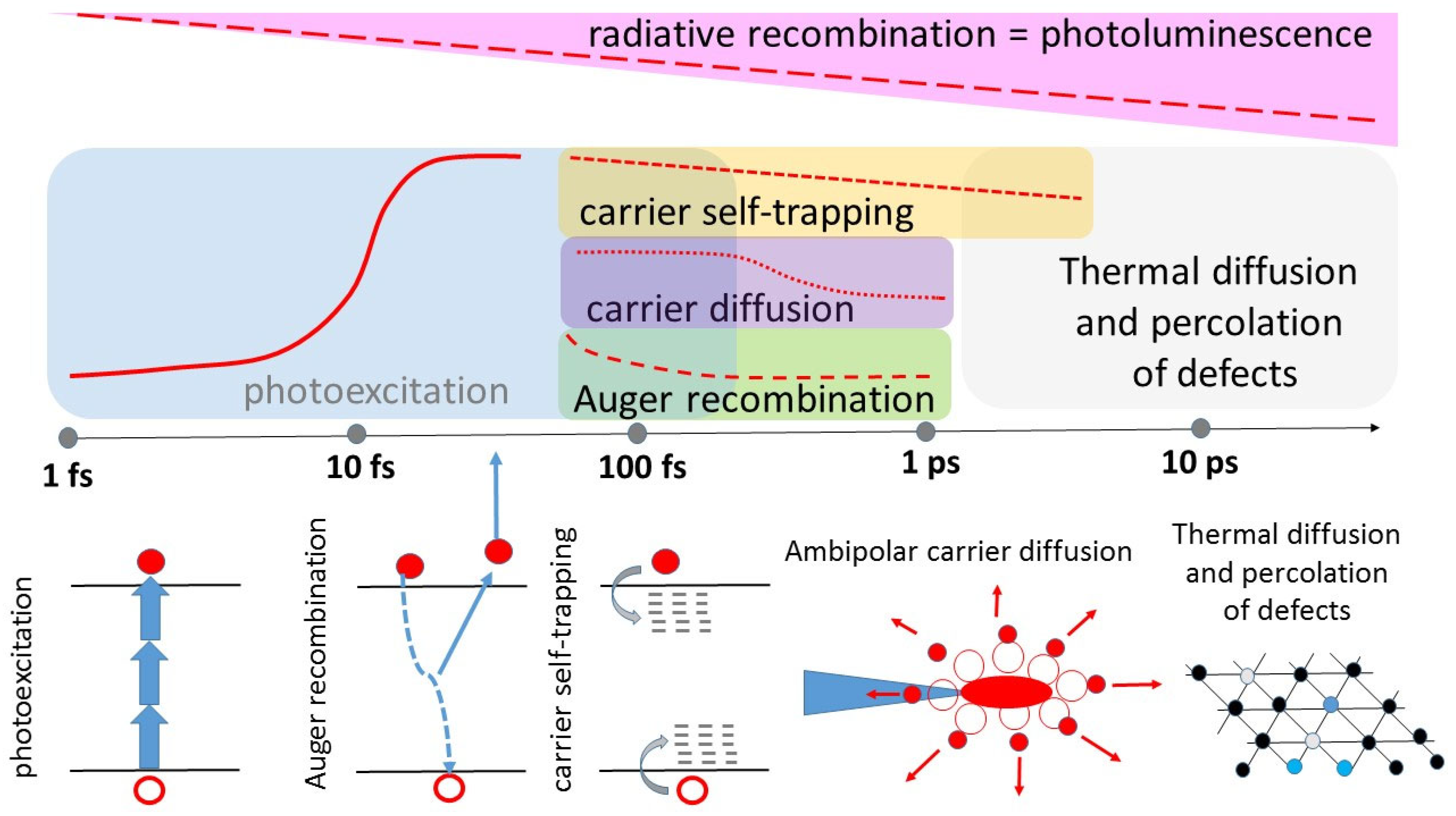
Abstract
:1. Introduction
2. Basic Physical Processes of Ultrashort Pulse Laser-Diamond Interaction
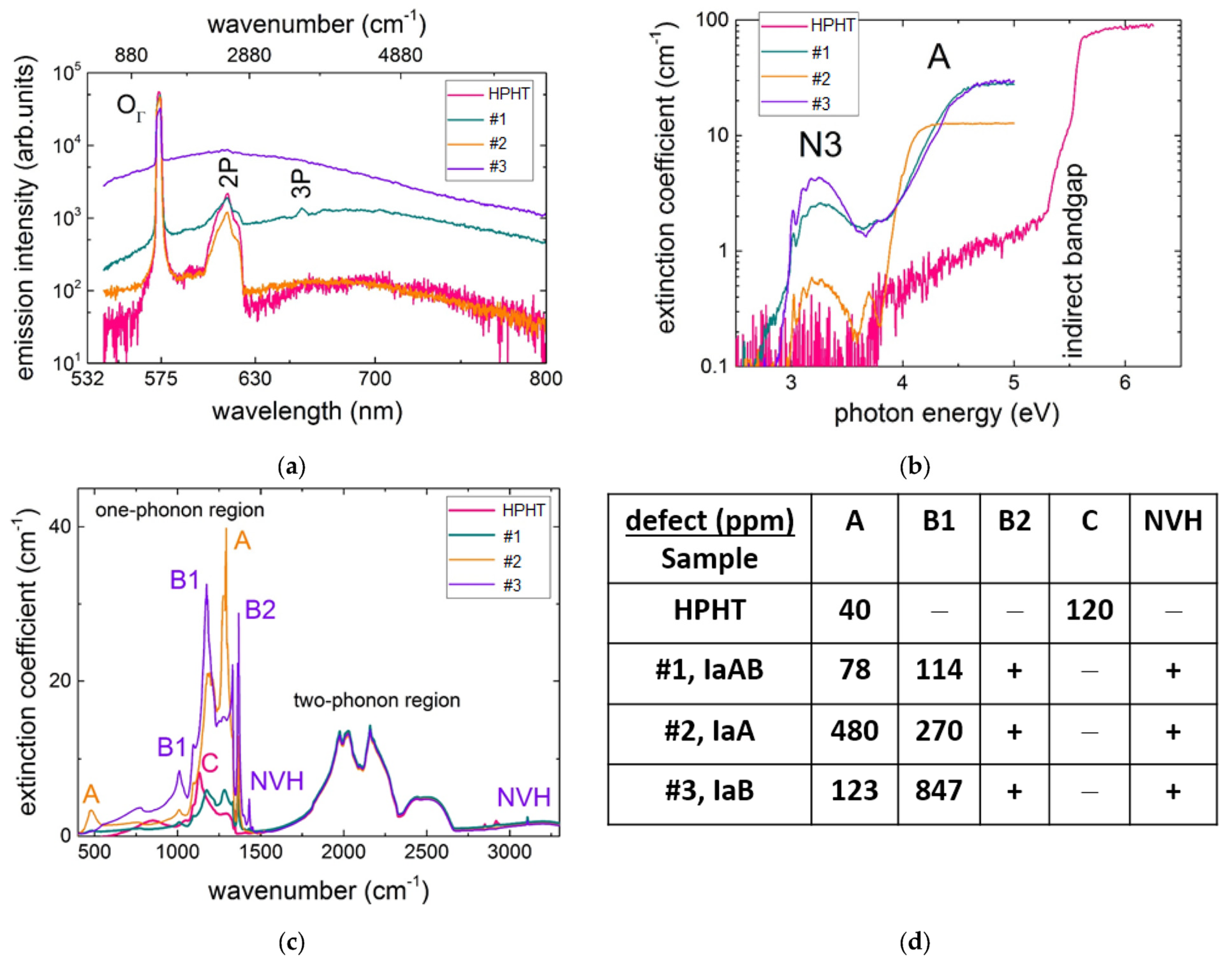
2.1. Multi-Spectral Characterization of Native Optical Centers
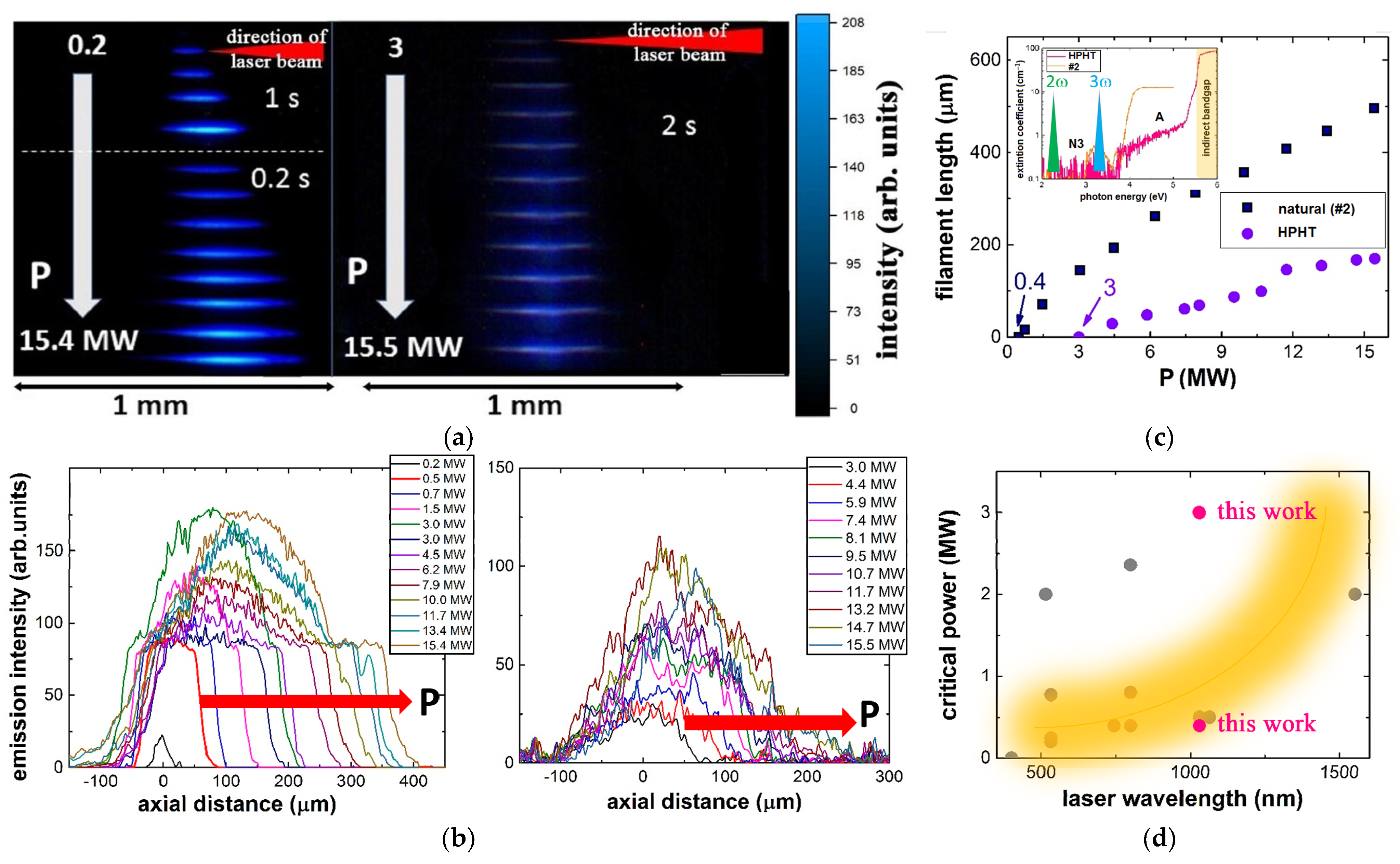
2.2. Delivery of Ultrashort Laser Pulses in Bulk Diamonds: Linear Focusing versus Non-Linear Self-Focusing and Filamentation
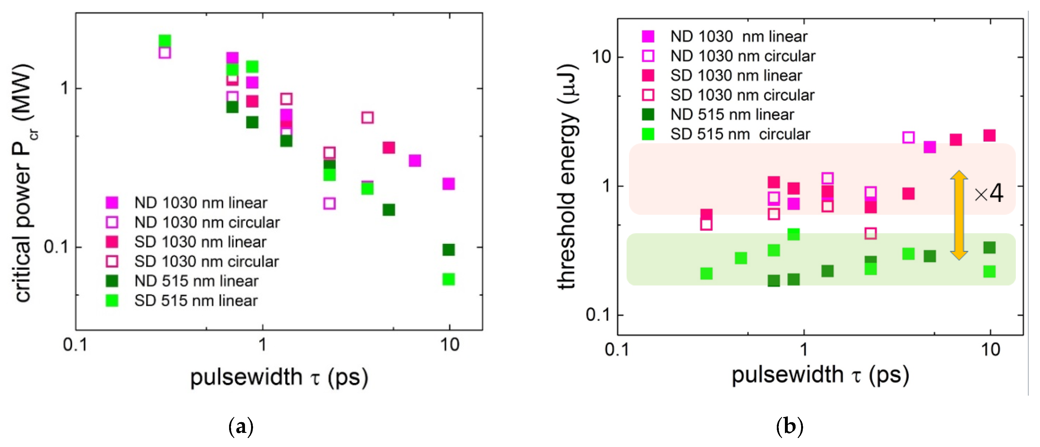
2.3. Pulsewidth- and Polarization-Dependent Ultrafast Laser Photoexcitation
2.4. Ultrafast EHP Energy Transport and Transfer into Lattice
2.5. Ultrafast Lattice Dynamics Envisioned by In Situ Raman Scattering
3. Novel Atomistic Paths in Ultrashort Pulse Laser Tailoring of Optical Nitrogen Centers in Diamonds: Making Visible Invisible and Back
3.1. Laser-Induced Generation of Blue-Red Luminous Centers in IaAB Diamonds: Vacancy-Mediated Conversion
3.2. Visible Range Laser Bleaching of HPHT Diamonds: Vacancy-Mediated Aggregation
4. Concluding Remarks
Author Contributions
Funding
Data Availability Statement
Acknowledgments
Conflicts of Interest
References
- Bradac, C.; Gao, W.; Forneris, J.; Trusheim, M.E.; Aharonovich, I. Quantum nanophotonics with group IV defects in diamond. Nat. Commun. 2019, 10, 5625, 1–13. [Google Scholar] [CrossRef] [PubMed] [Green Version]
- Blakley, S.M.; Fedotov, A.B.; Becker, J.; Altangerel, N.; Fedotov, I.V.; Hemmer, P.; Scully, M.O.; Zheltikov, A.M. Stimulated fluorescence quenching in nitrogen–vacancy centers of diamond: Temperature effects. Opt. Lett. 2016, 41, 2077–2080. [Google Scholar] [CrossRef]
- Rugar, A.E.; Lu, H.; Dory, C.; Sun, S.; McQuade, P.J.; Shen, Z.X.; Melosh, N.A.; Vučković, J. Generation of tin-vacancy centers in diamond via shallow ion implantation and subsequent diamond overgrowth. Nano Lett. 2020, 20, 1614–1619. [Google Scholar] [CrossRef] [PubMed] [Green Version]
- Czelej, K.; Zemła, M.R.; Kamińska, P.; Śpiewak, P.; Kurzydłowski, K.J. Clustering of hydrogen, phosphorus, and vacancies in diamond: A density functional theory analysis. Phys. Rev. B 2018, 98, 075208. [Google Scholar] [CrossRef]
- Bhaskar, M.K.; Sukachev, D.D.; Sipahigil, A.; Evans, R.E.; Burek, M.J.; Nguyen, C.T.; Rogers, L.J.; Siyushev, P.; Metsch, M.H.; Park, H.; et al. Quantum nonlinear optics with a germanium-vacancy color center in a nanoscale diamond waveguide. Phys. Rev. Lett. 2017, 118, 223603. [Google Scholar] [CrossRef] [Green Version]
- Ekimov, E.A.; Lyapin, S.G.; Kondrin, M.V. Tin-vacancy color centers in micro-and polycrystalline diamonds synthesized at high pressures. Diam. Relat. Mater. 2018, 87, 223–227. [Google Scholar] [CrossRef]
- Maze, J.R.; Stanwix, P.L.; Hodges, J.S.; Hong, S.; Taylor, J.M.; Cappellaro, P.; Jiang, L.; Dutt, M.; Togan, E.; Zibrov, A.; et al. Nanoscale magnetic sensing with an individual electronic spin in diamond. Nature 2008, 455, 644–647. [Google Scholar] [CrossRef]
- Taylor, J.M.; Cappellaro, P.; Childress, L.; Jiang, L.; Budker, D.; Hemmer, P.; Yacoby, A.; Walsworth, R.; Lukin, M. High-sensitivity diamond magnetometer with nanoscale resolution. Nat. Phys. 2008, 4, 810–816. [Google Scholar] [CrossRef]
- Sipahigil, A.; Evans, R.E.; Sukachev, D.D.; Burek, M.J.; Borregaard, J.; Bhaskar, M.K.; Nguyen, C.T.; Pacheco, J.L.; Atikian, H.A.; Meuwly, C.; et al. An integrated diamond nanophotonics platform for quantum-optical networks. Science 2016, 354, 847–850. [Google Scholar] [CrossRef] [Green Version]
- Blakley, S.; Liu, X.; Fedotov, I.; Cojocaru, I.; Vincent, C.; Alkahtani, M.; Becker, J.; Kieschnick, M.; Lühman, T.; Meijer, J.; et al. Fiber-optic quantum thermometry with germanium-vacancy centers in diamond. ACS Photonics 2019, 6, 1690–1693. [Google Scholar] [CrossRef]
- Zhou, Y.; Mu, Z.; Adamo, G.; Bauerdick, S.; Rudzinski, A.; Aharonovich, I.; Gao, W.-B. Direct writing of single germanium vacancy center arrays in diamond. New J. Phys. 2018, 20, 125004. [Google Scholar] [CrossRef] [Green Version]
- Hadden, J.P.; Bharadwaj, V.; Sotillo, B.; Rampini, S.; Osellame, R.; Witmer, J.D.; Jayakumar, H.; Fernandez, T.T.; Chiappini, A.; Armellini, C.; et al. Integrated waveguides and deterministically positioned nitrogen vacancy centers in diamond created by femtosecond laser writing. Opt. Lett. 2018, 43, 3586–3589. [Google Scholar] [CrossRef] [PubMed] [Green Version]
- Eaton, S.M.; Hadden, J.P.; Bharadwaj, V.; Forneris, J.; Picollo, F.; Bosia, F.; Sotillo, B.; Giakoumaki, A.N.; Jedrkiewicz, O.; Chiappini, A.; et al. Quantum micro–nano devices fabricated in diamond by femtosecond laser and ion irradiation. Adv. Quantum Technol. 2019, 2, 1900006. [Google Scholar] [CrossRef] [Green Version]
- Sotillo, B.; Bharadwaj, V.; Hadden, J.; Sakakura, M.; Chiappini, A.; Fernandez, T.T.; Longhi, S.; Jedrkiewicz, O.; Shimotsuma, Y.; Criante, L.; et al. Diamond photonics platform enabled by femtosecond laser writing. Sci. Rep. 2016, 6, 35566, 1–9. [Google Scholar] [CrossRef] [Green Version]
- Kononenko, T.V. Microstructuring of diamond bulk by IR femtosecond laser pulses. Appl. Phys. A 2008, 90, 645–651. [Google Scholar] [CrossRef]
- Shimizu, M.; Shimotsuma, Y.; Sakakura, M.; Yuasa, T.; Homma, H.; Minowa, Y.; Tanaka, K.; Miura, K.; Hirao, K. Periodic metallo-dielectric structure in diamond. Opt. Express 2009, 17, 46–54. [Google Scholar] [CrossRef] [PubMed]
- Bharadwaj, V.; Courvoisier, A.; Fernandez, T.T.; Ramponi, R.; Galzerano, G.; Nunn, J.; Booth, M.J.; Osellame, R.; Eaton, S.M.; Salter, P.S. Femtosecond laser inscription of Bragg grating waveguides in bulk diamond. Opt. Lett. 2017, 42, 3451–3453. [Google Scholar] [CrossRef] [Green Version]
- Bharadwaj, V.; Jedrkiewicz, O.; Hadden, J.P.; Sotillo, B.; Vázquez, M.R.; Dentella, P.; Fernandez, T.T.; Chiappini, A.; Giakoumaki, A.N.; Phu, T.L.; et al. Femtosecond laser written photonic and microfluidic circuits in diamond. J. Phys. Photonics 2019, 1, 022001. [Google Scholar] [CrossRef]
- Glezer, E.N.; Mazur, E. Ultrafast-laser driven micro-explosions in transparent materials. Appl. Phys. Lett. 1997, 71, 882–884. [Google Scholar] [CrossRef]
- Ionin, A.A.; Kudryashov, S.I.; Mikhin, K.E.; Seleznev, L.V.; Sinitsyn, D.V. Bulk femtosecond laser marking of natural diamonds. Laser Phys. 2010, 20, 1778–1782. [Google Scholar] [CrossRef]
- Chen, Y.C.; Salter, P.S.; Knauer, S.; Weng, L.; Frangeskou, A.C.; Stephen, C.J.; Ishmael, S.N.; Dolan, P.R.; Johnson, S.; Green, B.L.; et al. Laser writing of coherent colour centres in diamond. Nat. Photonics 2017, 11, 77–80. [Google Scholar] [CrossRef] [Green Version]
- Kurita, T.; Shimotsuma, Y.; Fujiwara, M.; Fujie, M.; Mizuochi, N.; Shimizu, M.; Miura, K. Direct writing of high-density nitrogen-vacancy centers inside diamond by femtosecond laser irradiation. Appl. Phys. Lett. 2021, 118, 214001. [Google Scholar] [CrossRef]
- Yurgens, V.; Zuber, J.A.; Flågan, S.; De Luca, M.; Shields, B.J.; Zardo, I.; Maletinsky, P.; Warburton, R.J.; Jakubczyk, T. Low-charge-noise nitrogen-vacancy centers in diamond created using laser writing with a solid-immersion lens. ACS Photonics 2021, 8, 1726–1734. [Google Scholar] [CrossRef]
- Motojima, M.; Suzuki, T.; Shigekawa, H.; Kainuma, Y.; An, T.; Hase, M. Giant nonlinear optical effects induced by nitrogen-vacancy centers in diamond crystals. Opt. Express 2019, 27, 32217–32227. [Google Scholar] [CrossRef] [PubMed] [Green Version]
- Abulikemu, A.; Kainuma, Y.; An, T.; Hase, M. Second-harmonic generation in bulk diamond based on inversion symmetry breaking by color centers. ACS Photonics 2021, 8, 988–993. [Google Scholar] [CrossRef]
- Ando, T.; Asai, K.; Macpherson, J.; Einaga, Y.; Fukuma, T.; Takahashi, Y. Nanoscale reactivity mapping of a single-crystal boron-doped diamond particle. Anal. Chem. 2021, 93, 5831–5838. [Google Scholar] [CrossRef]
- Kempkes, M.; Zier, T.; Singer, K.; Garcia, M.E. Ultrafast nonthermal NV center formation in diamond. Carbon 2021, 174, 524–530. [Google Scholar] [CrossRef]
- Griffiths, B.; Kirkpatrick, A.; Nicley, S.S.; Patel, R.L.; Zajac, J.M.; Morley, G.W.; Booth, M.J.; Salter, P.S.; Smith, J.M. Microscopic processes during ultra-fast laser generation of Frenkel defects in diamond. Phys. Rev. B 2021, 104, 174303. [Google Scholar] [CrossRef]
- Sokolowski-Tinten, K.; Bialkowski, J.; Boing, M.; Cavalleri, A.; von der Linde, D. Thermal and nonthermal melting of gallium arsenide after femtosecond laser excitation. Phys. Rev. B 1998, 58, R11805. [Google Scholar] [CrossRef]
- Rousse, A.; Rischel, C.; Fourmaux, S.; Uschmann, I.; Sebban, S.; Grillon, G.; Balcou, P.; Förster, E.; Geindre, J.P.; Audebert, P.; et al. Non-thermal melting in semiconductors measured at femtosecond resolution. Nature 2001, 410, 65–68. [Google Scholar] [CrossRef]
- Medvedev, N.; Fang, Z.; Xia, C.; Li, Z. Thermal and nonthermal melting of III-V compound semiconductors. Phys. Rev. B 2019, 99, 144101. [Google Scholar] [CrossRef] [Green Version]
- Medvedev, N.; Kopecky, M.; Chalupsky, J.; Juha, L. Femtosecond x-ray diffraction can discern nonthermal from thermal melting. Phys. Rev. B 2019, 99, 100303. [Google Scholar] [CrossRef]
- Akhmanov, S.A.; Emel’yanov, V.I.; Koroteev, N.I.; Seminogov, V.N. Interaction of powerful laser radiation with the surfaces of semiconductors and metals: Nonlinear optical effects and nonlinear optical diagnostics. Sov. Phys. Uspekhi 1985, 28, 1084. [Google Scholar] [CrossRef]
- Sundaram, S.K.; Mazur, E. Inducing and probing non-thermal transitions in semiconductors using femtosecond laser pulses. Nat. Mater. 2002, 1, 217–224. [Google Scholar] [CrossRef] [PubMed]
- Ekimov, E.A.; Kondrin, M.V. Vacancy–impurity centers in diamond: Prospects for synthesis and applications. Phys.-Uspekhi 2017, 60, 539. [Google Scholar] [CrossRef]
- Couairon, A.; Mysyrowicz, A. Femtosecond filamentation in transparent media. Phys. Rep. 2007, 441, 47–189. [Google Scholar] [CrossRef]
- Krasin, G.K.; Kovalev, M.S.; Kudryashov, S.I.; Danilov, P.A.; Martovitskii, V.P.; Gritsenko, I.V.; Podlesnykh, I.M.; Khmelnitskii, R.A.; Kuzmin, E.V.; Gulina, Y.S.; et al. Polarization-dependent near-IR ultrashort-pulse laser ablation of natural diamond surfaces. Appl. Surf. Sci. 2022, 595, 153549. [Google Scholar] [CrossRef]
- Kudryashov, S.I.; Danilov, P.A.; Kuzmin, E.V.; Gulina, Y.S.; Rupasov, A.E.; Krasin, G.K.; Zubarev, I.G.; Levchenko, A.O.; Kovalev, M.S.; Pakholchuk, P.P.; et al. Pulse-width-dependent critical power for self-focusing of ultrashort laser pulses in bulk dielectrics. Opt. Lett. 2022, 47, 3487–3490. [Google Scholar] [CrossRef]
- Young, J.F.; Van Driel, H.M. Ambipolar diffusion of high-density electrons and holes in Ge, Si, and GaAs: Many-body effects. Phys. Rev. B 1982, 26, 2147. [Google Scholar] [CrossRef]
- Li, C.-M.; Sjodin, T.; Dai, H.-L. Photoexcited carrier diffusion near a Si(111) surface: Non-negligible consequence of carrier-carrier scattering. Phys. Rev. B 1997, 56, 15252–15255. [Google Scholar] [CrossRef]
- Danilov, P.; Ionin, A.; Khmelnitskii, R.; Kiseleva, I.; Kudryashov, S.; Mel’nik, N.; Rudenko, A.; Smirnov, N.; Zayarny, D. Electron-ion coupling and ambipolar diffusion in dense electron-hole plasma in thin amorphous Si films studied by single-shot, pulse-width dependent ultrafast laser ablation. Appl. Surf. Sci. 2017, 425, 170–175. [Google Scholar] [CrossRef]
- Smirnov, N.A.; Kudryashov, S.I.; Rudenko, A.A.; Zayarny, D.A.; Ionin, A.A. Pulsewidth and ambient medium effects during ultrashort-pulse laser ablation of silicon in air and water. Appl. Surf. Sci. 2021, 562, 150243. [Google Scholar] [CrossRef]
- Quoix, C.; Grillon, G.; Antonetti, A.; Geindre, J.-P.; Audebert, P.; Gauthier, J.-C. Time-resolved studies of short pulse laser-produced plasmas in silicon dioxide near breakdown threshold. Eur. Phys. J. Appl. Phys. 1999, 5, 163–169. [Google Scholar] [CrossRef]
- Temnov, V.V.; Sokolowski-Tinten, K.; Zhou, P.; El-Khamhawy, A.; von der Linde, D. Multiphoton ionization in dielectrics: Comparison of circular and linear polarization. Phys. Rev. Lett. 2006, 97, 237403. [Google Scholar] [CrossRef] [PubMed]
- Sarpe, C.; Köhler, J.; Winkler, T.; Wollenhaupt, M.; Baumert, T. Real-time observation of transient electron density in water irradiated with tailored femtosecond laser pulses. New J. Phys. 2012, 14, 075021. [Google Scholar] [CrossRef] [Green Version]
- Kudryashov, S.; Samokhvalov, A.; Shelygina, S.; Karabutov, A.; Tsibidis, G.D.; Pankin, D.; Veiko, V. Electronic and vibrational processes in absorbing liquids in femtosecond laser sub-and filamentation regimes: Ultrasonic and optical characterization. Laser Phys. Lett. 2020, 17, 105302. [Google Scholar] [CrossRef]
- Spataru, C.D.; Benedict, L.X.; Louie, S.G. Ab initio calculation of band-gap renormalization in highly excited GaAs. Phys. Rev. B 2004, 69, 205204. [Google Scholar] [CrossRef] [Green Version]
- Glezer, E.N.; Siegal, Y.; Huang, L.; Mazur, E. Laser-induced band-gap collapse in GaAs. Phys. Rev. B 1995, 51, 6959. [Google Scholar] [CrossRef]
- Kudryashov, S.I.; Kandyla, M.; Roeser, C.A.; Mazur, E. Intraband and interband optical deformation potentials in femtosecond-laser-excited α−Te. Phys. Rev. B 2007, 75, 085207. [Google Scholar] [CrossRef]
- Song, K.; Williams, R.T. Self-Trapped Excitons; Springer-Verlag Berlin: Heidelberg, Germany, 1993. [Google Scholar]
- Ionin, A.A.; Kudryashov, S.I.; Makarov, S.V.; Saltuganov, P.N.; Seleznev, L.V.; Sinitsyn, D.V.; Sharipov, A. Ultrafast electron dynamics on the silicon surface excited by an intense femtosecond laser pulse. JETP Lett. 2012, 96, 375–379. [Google Scholar] [CrossRef]
- Garcia, M.E.; Jeschke, H.O. Theoretical approach to the laser-induced melting of graphite under different pressure conditions. Appl. Surf. Sci. 2003, 208, 61–70. [Google Scholar] [CrossRef]
- Govorkov, S.; Emel’yanov, V.; Koroteev, N.; Shumay, I. Femtosecond dynamics of laser-induced phase transition of the GaAs surface layer to a centrosymmetric phase. J. Lumin. 1992, 53, 153–158. [Google Scholar] [CrossRef]
- Emel’yanov, V.; Babak, D. Ultrafast vibronic phase transitions induced in semiconductors by femtosecond laser pulses. Phys. Solid State 1999, 41, 1338–1342. [Google Scholar] [CrossRef]
- Stampfli, P.; Bennemann, K. Dynamical theory of the laser-induced lattice instability of silicon. Phys. Rev. B 1992, 46, 10686. [Google Scholar] [CrossRef] [PubMed]
- Klett, I.; Zier, T.; Rethfeld, B.; Garcia, M.E.; Zijlstra, E.S. Isostructural elemental crystals in the presence of hot carriers. Phys. Rev. B 2015, 91, 144303. [Google Scholar] [CrossRef]
- Jeschke, H.O.; Garcia, M.E. Theoretical description of the ultrafast ablation of diamond and graphite: Dependence of thresholds on pulse duration. Appl. Surf. Sci. 2002, 197, 107–113. [Google Scholar] [CrossRef]
- Romero, A.H.; Jeschke, H.O.; Garcia, M.E. Laser manipulation of nanodiamonds. Comput. Mater. Sci. 2006, 35, 179–182. [Google Scholar] [CrossRef]
- Hayes, W.; Stoneham, A.M. Defects and Defect Processes in Nonmetallic Solids; Dover: New York, NY, USA, 1985. [Google Scholar]
- Zaitsev, A.M. Optical Properties of Diamond: A Data Handbook; Springer-Verlag: Berlin/Heidelberg, Germany, 2001. [Google Scholar]
- Dobrinets, I.A.; Vins, V.G.; Zaitsev, A.M. HPHT-Treated Diamonds; Springer-Verlag Berlin: Heidelberg, Germany, 2016. [Google Scholar]
- Behrens, H.; Janecke, J.; Schopper, H.; Landolt, H.; Bornstein, R. Landolt-Bornstein: Numerical Data and Functional Relationships in Science and Technology; Springer-Verlag Berlin: Heidelberg, Germany, 1969. [Google Scholar]
- Zaitsev, A.M. Vibronic spectra of impurity-related optical centers in diamond. Phys. Rev. B 2000, 61, 12909. [Google Scholar] [CrossRef]
- Ashfold, M.N.; Goss, J.P.; Green, B.L.; May, P.W.; Newton, M.E.; Peaker, C.V. Nitrogen in diamond. Chem. Rev. 2020, 120, 5745–5794. [Google Scholar] [CrossRef]
- Lu, H.C.; Lin, M.Y.; Chou, S.L.; Peng, Y.C.; Lo, J.I.; Cheng, B.M. Identification of nitrogen defects in diamond with photoluminescence excited in the 160–240 nm region. Anal. Chem. 2012, 84, 9596–9600. [Google Scholar] [CrossRef]
- Teofilov, N.; Schliesing, R.; Thonke, K.; Zacharias, H.; Sauer, R.; Kanda, H. Optical high excitation of diamond: Phase diagram of excitons, electron–hole liquid and electron–hole plasma. Diam. Relat. Mater. 2003, 12, 636–641. [Google Scholar] [CrossRef]
- Manfredotti, C.; Wang, F.; Polesello, P.; Vittone, E.; Fizzotti, F.; Scacco, A. Blue-violet electroluminescence and photocurrent spectra from polycrystalline chemical vapor deposited diamond film. Appl. Phys. Lett. 1995, 67, 3376–3378. [Google Scholar] [CrossRef]
- Davies, G. The Jahn-Teller effect and vibronic coupling at deep levels in diamond. Rep. Prog. Phys. 1981, 44, 787. [Google Scholar] [CrossRef]
- Levshin, L.; Saletskii, A. Optical Methods for Studying Molecular Systems. Molecular Spectroscopy; MGU: Moscow, Russia, 1994. [Google Scholar]
- Nadolinny, V.; Komarovskikh, A.; Palyanov, Y. Incorporation of large impurity atoms into the diamond crystal lattice: EPR of split-vacancy defects in diamond. Crystals 2017, 7, 237. [Google Scholar] [CrossRef] [Green Version]
- Zukerstein, M.; Kozák, M.; Trojánek, F.; Malý, P. Experimental observation of anharmonic effects in coherent phonon dynamics in diamond. Diam. Relat. Mater. 2018, 90, 202–206. [Google Scholar] [CrossRef]
- Schubert, M.; Wilhelmi, B. Nonlinear Optics and Quantum Electronics; Wiley: New York, NY, USA, 1986. [Google Scholar]
- Palik, E.D. Handbook of Optical Constants of Solids; Academic Press: Orlando, FL, USA, 1998. [Google Scholar]
- Kudryashov, S.I.; Levchenko, A.O.; Danilov, P.A.; Smirnov, N.A.; Ionin, A.A. IR femtosecond laser micro-filaments in diamond visualized by inter-band UV photoluminescence. Opt. Lett. 2020, 45, 2026–2029. [Google Scholar] [CrossRef]
- Liu, P.; Yen, R.; Bloembergen, N. Dielectric breakdown threshold, two-photon absorption, and other optical damage mechanisms in diamond. IEEE J. Quantum Electron. 1978, 14, 574–576. [Google Scholar] [CrossRef]
- Klein, C.A.; DeSalvo, R. Thresholds for dielectric breakdown in laser-irradiated diamond. Appl. Phys. Lett. 1993, 63, 1895–1897. [Google Scholar] [CrossRef] [Green Version]
- Pimenov, S.M.; Vlasov, I.I.; Khomich, A.A.; Neuenschwander, B.; Muralt, M.; Romano, V. Picosecond-laser-induced structural modifications in the bulk of single-crystal diamond. Appl. Phys. A 2011, 105, 673–677. [Google Scholar] [CrossRef]
- Kononenko, T.V.; Zavedeev, E.V.; Kononenko, V.V.; Ashikkalieva, K.K.; Konov, V.I. Graphitization wave in diamond bulk induced by ultrashort laser pulses. Appl. Phys. A 2015, 119, 405–414. [Google Scholar] [CrossRef]
- Batista, A.J.; Vianna, P.G.; Ribeiro, H.B.; Matos, C.J.S.D.; Gomes, A.S.L. QR code micro-certified gemstones: Femtosecond writing and Raman characterization in Diamond, Ruby and Sapphire. Sci. Rep. 2019, 9, 8927, 1–7. [Google Scholar] [CrossRef] [PubMed] [Green Version]
- Marburger, J.H. Self-focusing: Theory. Prog. Quantum Electron. 1975, 4, 35–110. [Google Scholar] [CrossRef]
- Boyd, R.W.; Lukishova, S.G.; Shen, Y.R. Self-Focusing: Past and Present: Fundamentals and Prospects; Springer-Verlag: New York, NY, USA, 2009. [Google Scholar]
- Fedorov, V.Y.; Kandidov, V. Interaction/laser radiation with matter filamentation of laser pulses with different wavelengths in air. Laser Phys. 2008, 18, 1530–1538. [Google Scholar] [CrossRef]
- Zharova, N.A.; Litvak, A.G.; Mironov, V.A. On the collapse of wave packets in a medium with normal group velocity dispersion. JETP Lett. 2002, 75, 539–542. [Google Scholar] [CrossRef]
- Fibich, G.; Papanicolaou, G. Self-focusing in the perturbed and unperturbed nonlinear Schrödinger equation in critical dimension. SIAM J. Appl. Math. 1999, 60, 183–240. [Google Scholar] [CrossRef] [Green Version]
- Kudryashov, S.I.; Levchenko, A.O.; Danilov, P.A.; Smirnov, N.A.; Rupasov, A.E.; Khmel’nitskii, R.A.; Koval’chuk, O.E.; Ionin, A.A. Fine structure of the photoluminescence spectrum of diamond under the multiple emission of an optical phonon during the autolocalization of photoexcited electrons. JETP Lett. 2020, 112, 533–536. [Google Scholar] [CrossRef]
- Kudryashov, S.; Stsepuro, N.; Danilov, P.; Smirnov, N.; Levchenko, A.; Kovalev, M. Cumulative defocusing of sub-MHz-rate femtosecond-laser pulses in bulk diamond envisioned by transient A-band photoluminescence. Opt. Mater. Express 2021, 11, 2234–2241. [Google Scholar] [CrossRef]
- Kudryashov, S.; Danilov, P.; Smirnov, N.; Levchenko, A.; Kovalev, M.; Gulina, Y.; Kovalchuk, O.; Ionin, A. Femtosecond-laser-excited luminescence of the A-band in natural diamond and its thermal control. Opt. Mater. Express 2021, 11, 2505–2513. [Google Scholar] [CrossRef]
- Kudryashov, S.; Khmelnitskii, R.; Danilov, P.; Smirnov, N.; Levchenko, A.; Kovalchuk, O.; Uspenskaya, M.; Oleynichuk, E.; Kovalev, M. Broadband and fine-structured luminescence in diamond facilitated by femtosecond laser driven electron impact and injection of “vacancy-interstitial” pairs. Opt. Lett. 2021, 46, 1438–1441. [Google Scholar] [CrossRef]
- Kozák, M.; Otobe, T.; Zukerstein, M.; Trojánek, F.; Malý, P. Anisotropy and polarization dependence of multiphoton charge carrier generation rate in diamond. Phys. Rev. B 2019, 99, 104305. [Google Scholar] [CrossRef]
- Zukerstein, M.; Trojánek, F.; Kozák, M.; Malý, P. Multiphoton-excited exciton molecules in diamond. J. Lumin. 2021, 231, 117774. [Google Scholar] [CrossRef]
- Kudryashov, S.I.; Danilov, P.A.; Smirnov, N.A.; Stsepuro, N.G.; Rupasov, A.E.; Khmelnitskii, R.A.; Oleynichuk, E.A.; Kuzmin, E.V.; Levchenko, A.O.; Gulina, Y.S.; et al. Signatures of ultrafast electronic and atomistic dynamics in bulk photoluminescence of CVD and natural diamonds excited by ultrashort laser pulses of variable pulsewidth. Appl. Surf. Sci. 2021, 575, 151736. [Google Scholar] [CrossRef]
- Ionin, A.A.; Kudryashov, S.I.; Makarov, S.V.; Seleznev, L.V.; Sinitsyn, D.V. Multiple filamentation of intense femtosecond laser pulses in air. JETP Lett. 2009, 90, 423–427. [Google Scholar] [CrossRef]
- Bulgakova, N.M.; Stoian, R.; Rosenfeld, A.; Hertel, I.V.; Campbell, E.E.B. Electronic transport and consequences for material removal in ultrafast pulsed laser ablation of materials. Phys. Rev. B 2004, 69, 054102. [Google Scholar] [CrossRef]
- Keldysh, L.V. Ionization in the field of a strong electromagnetic wave. Sov. Phys. JETP 1965, 20, 1307–1314. [Google Scholar]
- Kudryashov, S.I. Microscopic model of electronic Kerr effect in strong electric fields of intense femtosecond laser pulses. In Proceedings of the Conference on Quantum Electronics and Laser Science, Baltimore, MD, USA, 22–27 May 2005. [Google Scholar]
- Apostolova, T.; Obreshkov, B.; Ionin, A.; Kudryashov, S.; Makarov, S.; Mel’nik, N.; Rudenko, A. Ultrafast photoionization and excitation of surface-plasmon-polaritons on diamond surfaces. Appl. Surf. Sci. 2018, 427, 334–343. [Google Scholar] [CrossRef] [Green Version]
- Shirk, M.; Molian, P.; Malshe, A. Ultrashort pulsed laser ablation of diamond. J. Laser Appl. 1998, 10, 64–70. [Google Scholar] [CrossRef]
- Boerner, P.; Hajri, M.; Ackerl, N.; Wegener, K. Experimental and theoretical investigation of ultrashort pulsed laser ablation of diamond. J. Laser Appl. 2019, 31, 022202. [Google Scholar] [CrossRef]
- Yoffa, E.J. Screening of hot-carrier relaxation in highly photoexcited semiconductors. Phys. Rev. B 1981, 23, 1909. [Google Scholar] [CrossRef]
- Shank, C.V.; Yen, R.; Hirlimann, C. Time-resolved reflectivity measurements of femtosecond-optical-pulse-induced phase transitions in silicon. Phys. Rev. Lett. 1983, 50, 454. [Google Scholar] [CrossRef]
- Dziewior, J.; Schmid, W. Auger coefficients for highly doped and highly excited silicon. Appl. Phys. Lett. 1977, 31, 346–348. [Google Scholar] [CrossRef]
- Strauss, U.; Rühle, W.W.; Köhler, K. Auger recombination in intrinsic GaAs. Appl. Phys. Lett. 1993, 62, 55–57. [Google Scholar] [CrossRef]
- Kozák, M.; Trojánek, F.; Malý, P. Hot-carrier transport in diamond controlled by femtosecond laser pulses. New J. Phys. 2015, 17, 053027. [Google Scholar] [CrossRef]
- Grigoriev, I.S.; Meĭlikhov, E.Z.; Radzig, A.A. Handbook of Physical Quantities; CRC: Boca Raton, FL, USA, 1996. [Google Scholar]
- Liu, J.M. Simple technique for measurements of pulsed Gaussian-beam spot sizes. Opt. Lett. 1982, 7, 196–198. [Google Scholar] [CrossRef] [PubMed]
- Reitze, D.H.; Ahn, H.; Downer, M.C. Optical properties of liquid carbon measured by femtosecond spectroscopy. Phys. Rev. B 1992, 45, 2677. [Google Scholar] [CrossRef]
- Yin, J.; Chen, G.; Zhu, Z.; Jin, M.; Hu, B. Ablation mechanism investigation and ablation threshold prediction of single crystal diamond irradiated by femtosecond laser. Diam. Relat. Mater. 2021, 111, 108173. [Google Scholar] [CrossRef]
- Li, Q.Z.; Wang, J.; Sun, T. Atomistic simulations of ultrashort pulsed laser ablation of polycrystalline diamond. Curr. Nanosci. 2013, 9, 804–811. [Google Scholar] [CrossRef]
- Gamaly, E.G.; Juodkazis, S.; Nishimura, K.; Misawa, H.; Luther-Davies, B.; Hallo, L.; Nicolai, P.; Tikhonchuk, V.T. Laser-matter interaction in the bulk of a transparent solid: Confined microexplosion and void formation. Phys. Rev. B 2006, 73, 214101. [Google Scholar] [CrossRef]
- Thomsen, C.; Grahn, H.T.; Maris, H.J.; Tauc, J. Surface generation and detection of phonons by picosecond light pulses. Phys. Rev. B 1986, 34, 4129. [Google Scholar] [CrossRef]
- Kudryashov, S.I. Some effects affecting laser-induced damage of solid dielectrics excited by ultrashort laser pulses. In Nonlinear Frequency Generation and Conversion: Materials, Devices, and Applications IV, V. 5710, Proceedings of the Lasers and Applications in Science and Engineering, San Jose, California, USA; SPIE: Bellingham, WA, USA, 2005; pp. 120–129. [Google Scholar]
- Kudryashov, S.I.; Danilov, P.A.; Sdvizhenskii, P.A.; Lednev, V.N.; Chen, J.; Ostrikov, S.A.; Kuzmin, E.V.; Kovalev, M.S.; Levchenko, A.O. Transformations of the Spectrum of an Optical Phonon Excited in Raman Scattering in the Bulk of Diamond by Ultrashort Laser Pulses with a Variable Duration. JETP Lett. 2022, 115, 251–255. [Google Scholar] [CrossRef]
- Grimsditch, M.H.; Anastassakis, E.; Cardona, M. Effect of uniaxial stress on the zone-center optical phonon of diamond. Phys. Rev. B 1978, 18, 901. [Google Scholar] [CrossRef]
- Cardona, M.; Peter, Y.Y. Fundamentals of Semiconductors, 3rd ed.; Springer-Verlag: Berlin/Heidelberg, Germany, 2005. [Google Scholar]
- Cavalleri, A.; Siders, C.W.; Brown, F.L.H.; Leitner, D.M.; Tóth, C.; Squier, J.A.; Barty, C.P.J.; Wilson, K.R.; Sokolowski-Tinten, K.; Horn von Hoegen, M.; et al. Anharmonic lattice dynamics in germanium measured with ultrafast x-ray diffraction. Phys. Rev. Lett. 2000, 85, 586. [Google Scholar] [CrossRef] [Green Version]
- Zhao, J.; Zhang, C.; Liu, F.; Cheng, G.J. Understanding femtosecond laser internal scribing of diamond by atomic simulation: Phase transition, structure and property. Carbon 2021, 175, 352–363. [Google Scholar] [CrossRef]
- Cardona, M. Effects of pressure on the phonon–phonon and electron–phonon interactions in semiconductors. Phys. Status Solidi B 2004, 241, 3128–3137. [Google Scholar] [CrossRef]
- Pu, M.; Zhang, F.; Liu, S.; Irifune, T.; Lei, L. Tensile-strain induced phonon splitting in diamond. Chin. Phys. B 2019, 28, 053102. [Google Scholar] [CrossRef]
- Klemens, P.G. Anharmonic decay of optical phonon in diamond. Phys. Rev. B 1975, 11, 3206. [Google Scholar] [CrossRef]
- Kudryashov, S.I.; Vins, V.G.; Danilov, P.A.; Kuzmin, E.V.; Muratov, A.V.; Kriulina, G.Y.; Chen, J.; Kirichenko, A.N.; Gulina, Y.S.; Ostrikov, S.A.; et al. Permanent optical bleaching in HPHT-diamond via aggregation of C-and NV-centers excited by visible-range femtosecond laser pulses. Carbon 2023, 201, 399–407. [Google Scholar] [CrossRef]
- Danilov, P.; Kuzmin, E.; Rimskaya, E.; Chen, J.; Khmelnitskii, R.; Kirichenko, A.; Rodionov, N.; Kudryashov, S. Up/Down-Scaling Photoluminescent Micromarks Written in Diamond by Ultrashort Laser Pulses: Optical Photoluminescent and Structural Raman Imaging. Micromachines 2022, 13, 1883. [Google Scholar] [CrossRef]
- Fedotov, I.V.; Zheltikov, A.M. Background-free two-photon fluorescence readout via a three-photon charge-state modulation of nitrogen-vacancy centers in diamond. Opt. Lett. 2019, 44, 3737–3740. [Google Scholar] [CrossRef]
- Krasin, G.; Kudryashov, S.; Danilov, P.; Smirnov, N.; Levchenko, A.; Kovalev, M. Ultrashort-laser electron–hole plasma and intragap states in diamond. Eur. Phys. J. D 2021, 75, 221. [Google Scholar] [CrossRef]
- ALROSA. Identification of Diamonds Using Laser Nanomarks. Available online: https://youtu.be/X3Z_jcWowks (accessed on 11 December 2022).
- Vasil’ev, V.; Velichansky, V.; Zibrov, S.; Ionin, A.; Kudryashov, S.; Levchenko, A.; Seleznev, L.; Sinitsyn, D. Method of Forming Optically Permeable Image Inside Diamond, Apparatus for Realising Said Method (Versions) and Apparatus for Detecting Said Image. Russian Patent RU 2465377 C1, 30 June 2011. [Google Scholar]
- Vasil’ev, V.; Velichansky, V.; Zibrov, S.; Ionin, A.; Kudryashov, S.; Levchenko, A.; Seleznev, L.; Sinitsyn, D. Device for Creating an Optically Permeable Image Inside a Diamond (Options) and Device for Detecting the Indicated Image. Russian Patent RU 109877 U1, 4 July 2011. [Google Scholar]
- Ionin, A.A.; Kudryashov, S.I.; Smirnov, N.A.; Danilov, P.A.; Levchenko, A.O. Method for Creating and Detecting an Optically Permeable Image Inside a Diamond. US Patent App. 17/267,588, 14 October 2021. [Google Scholar]
- Ionin, A.A.; Kudryashov, S.I.; Smirnov, N.A.; Danilov, P.A.; Levchenko, A.O. Optically Transparent Mark for Marking Gemstones. US Patent App. 17/267,609, 14 October 2021. [Google Scholar]
- Sobolev, E.; Eliseev, A. Thermostimulated luminescence and phosphorescence of natural diamonds at low temperatures. J. Struct. Chem. 1976, 17, 799–801. [Google Scholar] [CrossRef]
- Kudryashov, S.I.; Kriulina, G.Y.; Danilov, P.A.; Kuzmin, E.V.; Kirichenko, A.N.; Rodionov, N.B.; Khmelnitskii, R.A.; Chen, J.; Rimskaya, E.N.; Shur, V.Y. Nanoscale vacancy-mediated aggregation, dissociation and splitting of nitrogen centers in natural diamond excited by visible-range femtosecond laser pulses. Nanomaterials 2022. submitted. [Google Scholar]
- Rong, Y.; Cheng, K.; Ju, Z.; Pan, C.; Ma, Q.; Liu, S.; Shen, S.; Wu, B.; Jia, T.; Zeng, H.; et al. Bright near-surface silicon vacancy centers in diamond fabricated by femtosecond laser ablation. Opt. Lett. 2019, 44, 3793–3796. [Google Scholar] [CrossRef] [PubMed]
- Komlenok, M.S.; Kudryavtsev, O.S.; Pasternak, D.G.; Vlasov, I.I.; Konov, V.I. Laser printing of diamond nanoparticles with luminescent SiV center. Comput. Opt. 2021, 45, 860–864. [Google Scholar] [CrossRef]
- Castelletto, S.; Maksimovic, J.; Katkus, T.; Ohshima, T.; Johnson, B.C.; Juodkazis, S. Color centers enabled by direct femtosecond laser writing in wide bandgap semiconductors. Nanomaterials 2020, 11, 72. [Google Scholar] [CrossRef] [PubMed]
- Gao, S.; Duan, Y.Z.; Tian, Z.N.; Zhang, Y.L.; Chen, Q.D.; Gao, B.R.; Sun, H.B. Laser-induced color centers in crystals. Opt. Laser Technol. 2022, 146, 107527. [Google Scholar] [CrossRef]
- Wang, X.J.; Fang, H.H.; Sun, F.W.; Sun, H.B. Laser writing of color centers. Laser Photonics Rev. 2022, 16, 2100029. [Google Scholar] [CrossRef]
- Gan, L.; Zhang, D.; Zhang, R.; Zhang, Q.; Sun, H.; Li, Y.; Ning, C.Z. Large-Scale, High-Yield Laser Fabrication of Bright and Pure Single-Photon Emitters at Room Temperature in Hexagonal Boron Nitride. ACS Nano 2022, 16, 14254–14261. [Google Scholar] [CrossRef]
- Gao, S.; Yin, S.Y.; Liu, Z.X.; Zhang, Z.D.; Tian, Z.N.; Chen, Q.D.; Chen, N.-K.; Sun, H.B. Narrow-linewidth diamond single-photon sources prepared via femtosecond laser. Appl. Phys. Lett. 2022, 120, 023104. [Google Scholar] [CrossRef]
- Wang, H.; Lei, Y.; Wang, L.; Sakakura, M.; Yu, Y.; Shayeganrad, G.; Kazansky, P.G. 100-Layer Error-Free 5D Optical Data Storage by Ultrafast Laser Nanostructuring in Glass. Laser Photonics Rev. 2022, 16, 2100563. [Google Scholar] [CrossRef]
- Li, L.; Kong, W.; Chen, F. Femtosecond laser-inscribed optical waveguides in dielectric crystals: A concise review and recent advances. Adv. Photonics 2022, 4, 024002. [Google Scholar] [CrossRef]
- Yu, J.; Xu, J.; Zhang, A.; Song, Y.; Qi, J.; Dong, Q.; Chen, J.; Liu, Z.; Chen, W.; Cheng, Y. Manufacture of Three-Dimensional Optofluidic Spot-Size Converters in Fused Silica Using Hybrid Laser Microfabrication. Sensors 2022, 22, 9449. [Google Scholar] [CrossRef] [PubMed]


Disclaimer/Publisher’s Note: The statements, opinions and data contained in all publications are solely those of the individual author(s) and contributor(s) and not of MDPI and/or the editor(s). MDPI and/or the editor(s) disclaim responsibility for any injury to people or property resulting from any ideas, methods, instructions or products referred to in the content. |
© 2023 by the authors. Licensee MDPI, Basel, Switzerland. This article is an open access article distributed under the terms and conditions of the Creative Commons Attribution (CC BY) license (https://creativecommons.org/licenses/by/4.0/).
Share and Cite
Kudryashov, S.; Danilov, P.; Smirnov, N.; Krasin, G.; Khmelnitskii, R.; Kovalchuk, O.; Kriulina, G.; Martovitskiy, V.; Lednev, V.; Sdvizhenskii, P.; et al. “Stealth Scripts”: Ultrashort Pulse Laser Luminescent Microscale Encoding of Bulk Diamonds via Ultrafast Multi-Scale Atomistic Structural Transformations. Nanomaterials 2023, 13, 192. https://doi.org/10.3390/nano13010192
Kudryashov S, Danilov P, Smirnov N, Krasin G, Khmelnitskii R, Kovalchuk O, Kriulina G, Martovitskiy V, Lednev V, Sdvizhenskii P, et al. “Stealth Scripts”: Ultrashort Pulse Laser Luminescent Microscale Encoding of Bulk Diamonds via Ultrafast Multi-Scale Atomistic Structural Transformations. Nanomaterials. 2023; 13(1):192. https://doi.org/10.3390/nano13010192
Chicago/Turabian StyleKudryashov, Sergey, Pavel Danilov, Nikita Smirnov, George Krasin, Roman Khmelnitskii, Oleg Kovalchuk, Galina Kriulina, Victor Martovitskiy, Vasily Lednev, Pavel Sdvizhenskii, and et al. 2023. "“Stealth Scripts”: Ultrashort Pulse Laser Luminescent Microscale Encoding of Bulk Diamonds via Ultrafast Multi-Scale Atomistic Structural Transformations" Nanomaterials 13, no. 1: 192. https://doi.org/10.3390/nano13010192