
Metal and Metal Halogenide-Filled Single-Walled Carbon Nanotubes: Kinetics, Electronic Properties, Engineering the Fermi Level


Abstract
:1. Introduction
2. Overview of Substances Filled Inside SWCNTs
3. Kinetics
4. Electronic Properties
4.1. Filling of SWCNTs with Metals
4.2. Filling of SWCNTs with Metal Halogenides
4.3. Doping and Hybridization Effects
4.3.1. Optical Absorption Spectroscopy
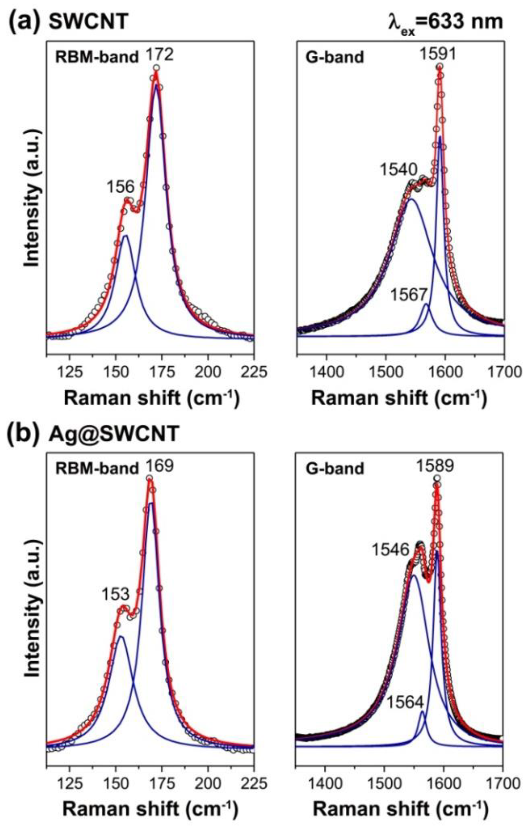
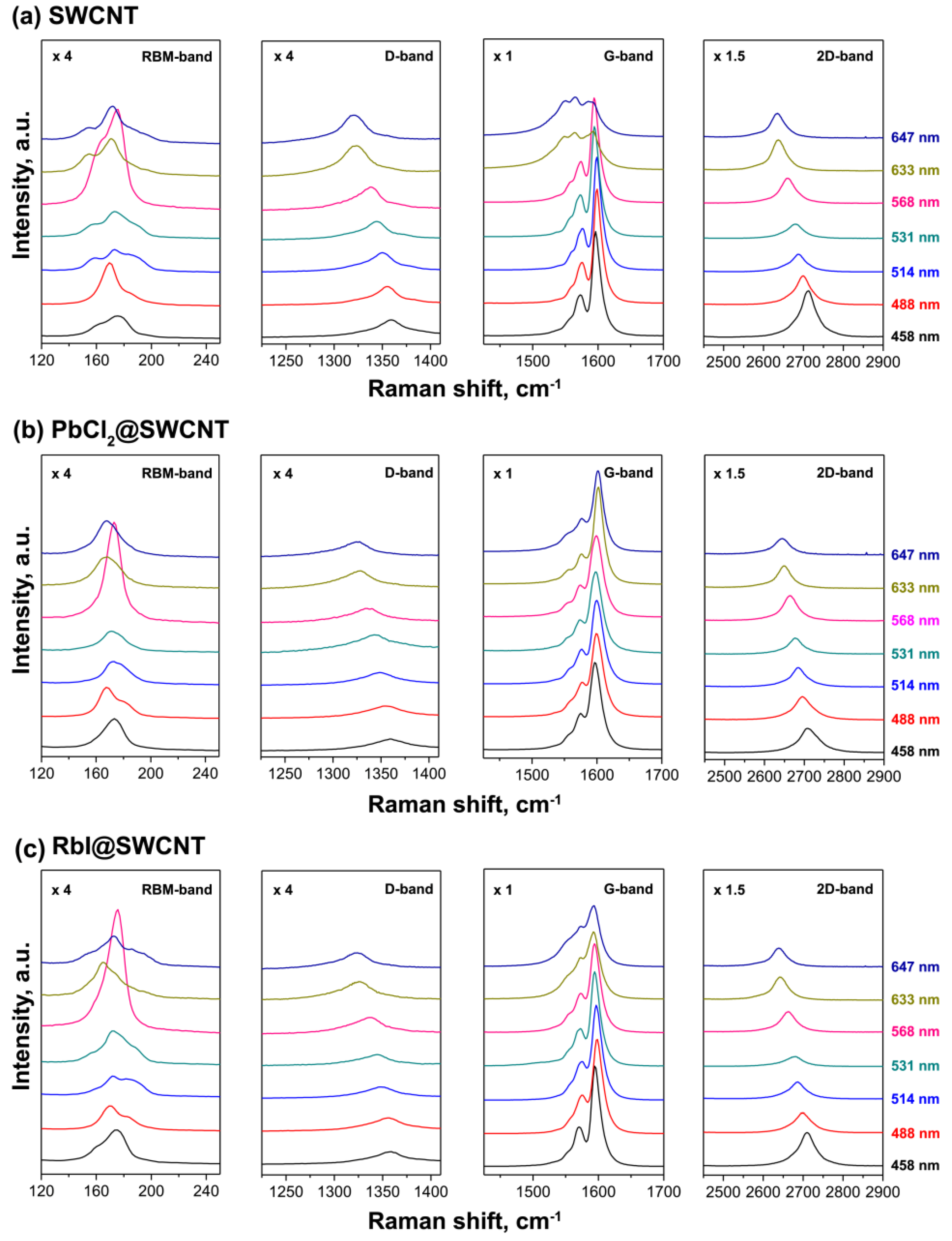
4.3.2. Raman Spectroscopy
4.3.3. Near Edge X-ray Absorption Fine Structure Spectroscopy
4.3.4. Photoemission Spectroscopy
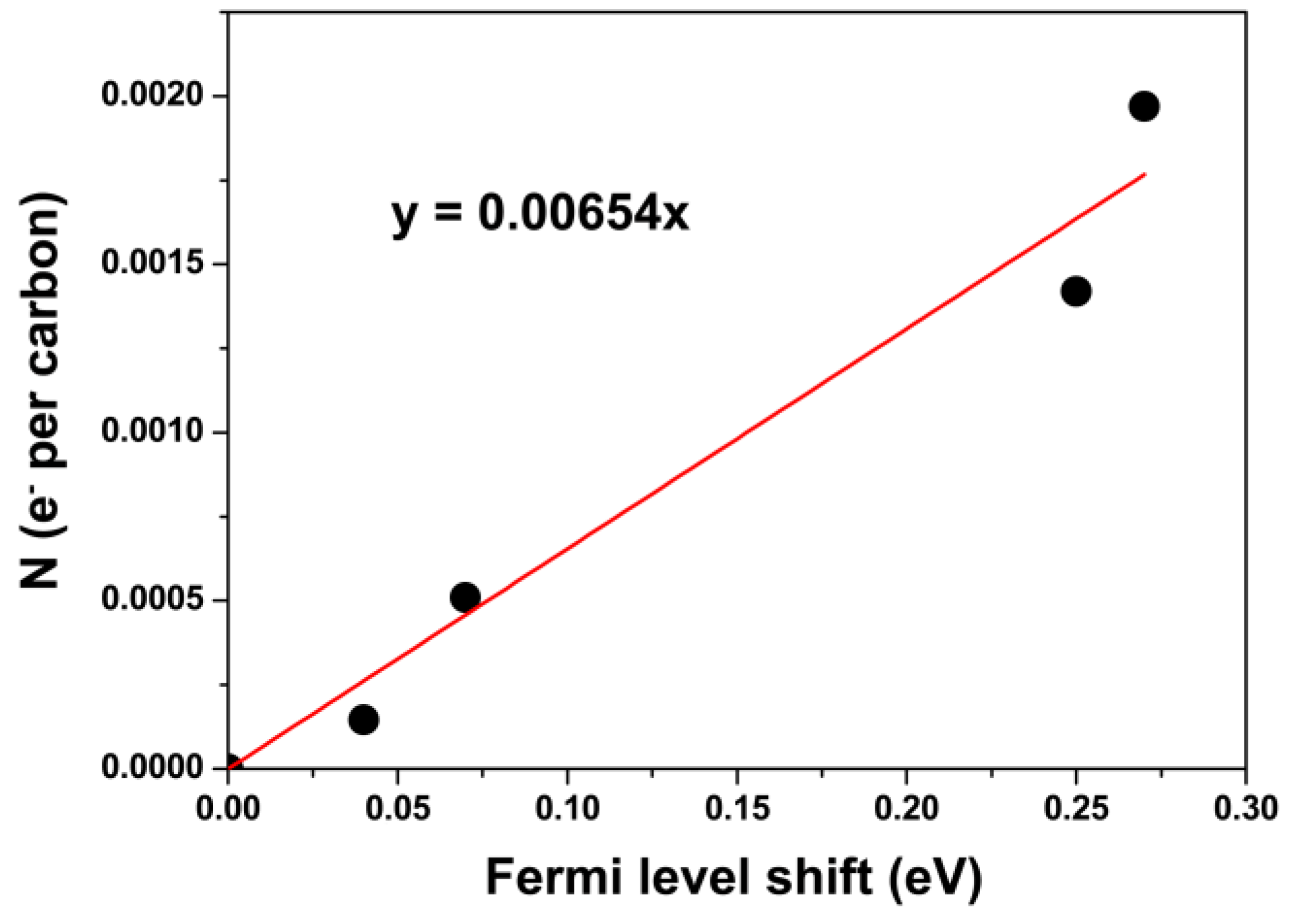
5. Quantification of Charge Transfer in SWCNTs Filled with Inorganic Compounds
6. Conclusions
Author Contributions
Funding
Data Availability Statement
Acknowledgments
Conflicts of Interest
References
- Jeon, I.; Xiang, R.; Shawky, A.; Matsuo, Y.; Maruyama, S. Single-Walled Carbon Nanotubes in Emerging Solar Cells: Synthesis and Electrode Applications. Adv. Energy Mater. 2019, 9, 1801312. [Google Scholar] [CrossRef]
- Ferguson, V.; Silva, S.R.P.; Zhang, W. Carbon Materials in Perovskite Solar Cells: Prospects and Future Challenges. Energy Environ. Mater. 2019, 2, 107–118. [Google Scholar] [CrossRef] [Green Version]
- Fukumaru, T.; Fujigaya, T.; Nakashima, N. Development of n-type cobaltocene-encapsulated carbon nanotubes with remarkable thermoelectric property. Sci. Rep. 2015, 5, 7951. [Google Scholar] [CrossRef] [PubMed] [Green Version]
- Lee, J.W.; Jeon, I.; Lin, H.S.; Seo, S.; Han, T.H.; Anisimov, A.; Kauppinen, E.I.; Matsuo, Y.; Maruyama, S.; Yang, Y. Vapor-Assisted Ex-Situ Doping of Carbon Nanotube toward Efficient and Stable Perovskite Solar Cells. Nano Lett. 2019, 19, 2223–2230. [Google Scholar] [CrossRef]
- Jordan, J.W.; Lowe, G.A.; McSweeney, R.L.; Stoppiello, C.T.; Lodge, R.W.; Skowron, S.T.; Biskupek, J.; Rance, G.A.; Kaiser, U.; Walsh, D.A.; et al. Host-Guest Hybrid Redox Materials Self-Assembled from Polyoxometalates and Single-Walled Carbon Nanotubes. Adv. Mater. 2019, 31, 1904182. [Google Scholar] [CrossRef]
- Martincic, M.; Tobias, G. Filled carbon nanotubes in biomedical imaging and drug delivery. Expert Opin. Drug Deliv. 2015, 12, 563–581. [Google Scholar] [CrossRef]
- Liu, B.L.; Wu, F.Q.; Gui, H.; Zheng, M.; Zhou, C.W. Chirality-Controlled Synthesis and Applications of Single-Wall Carbon Nanotubes. ACS Nano 2017, 11, 31–53. [Google Scholar] [CrossRef]
- Bati, A.S.R.; Yu, L.P.; Batmunkh, M.; Shapter, J.G. Recent Advances in Applications of Sorted Single-Walled Carbon Nanotubes. Adv. Funct. Mater. 2019, 29, 1902273. [Google Scholar] [CrossRef]
- Moore, K.E.; Tune, D.D.; Flavel, B.S. Double-Walled Carbon Nanotube Processing. Adv. Mater. 2015, 27, 3105–3137. [Google Scholar] [CrossRef]
- Kharlamova, M.V. Advances in tailoring the electronic properties of single-walled carbon nanotubes. Prog. Mater. Sci. 2016, 77, 125–211. [Google Scholar] [CrossRef]
- Tkachenko, N.V. Optical Spectroscopy: Methods and Instrumentations; Elsevier: Amsterdam, The Netherlands, 2006. [Google Scholar]
- Kharlamova, M.V.; Kramberger, C.; Sauer, M.; Yanagi, K.; Pichler, T. Comprehensive spectroscopic characterization of high purity metallicity-sorted single-walled carbon nanotubes. Phys. Status Solidi B Basic Solid State Phys. 2015, 252, 2512–2518. [Google Scholar] [CrossRef]
- Ayala, P.; Miyata, Y.; De Blauwe, K.; Shiozawa, H.; Feng, Y.; Yanagi, K.; Kramberger, C.; Silva, S.R.P.; Follath, R.; Kataura, H.; et al. Disentanglement of the electronic properties of metallicity-selected single-walled carbon nanotubes. Phys. Rev. B 2009, 80, 205427. [Google Scholar] [CrossRef]
- Kuzmany, H. Solid-State Spectroscopy: An Introduction, 2nd ed.; Springer-Verlag: Berlin/Heidelberg, Germany, 2009. [Google Scholar]
- Atalla, R.H.; Agarwal, U.P.; Bond, J.L. Raman Spectroscopy; Springer Series in Wood Science; Methods in Lignin Chemistry; Springer: Berlin/Heidelberg, Germany, 1992; pp. 162–176. [Google Scholar]
- Smekal, A. The quantum theory of dispersion. Naturwissenschaften 1923, 11, 873–875. [Google Scholar] [CrossRef]
- Raman, C.V.; Krishnan, K.S. The optical analog of the Compton eect. Nature 1928, 121, 711. [Google Scholar] [CrossRef]
- Lamdsberg, G.; Mandelstam, L. A novel efect of light scattering in crystals. Naturwissenschaften 1928, 16, 5757–5758. [Google Scholar]
- Maiman, T.H. Stimulated optical radiation in ruby. Nature 1960, 187, 493–494. [Google Scholar] [CrossRef]
- Porto, S.P.S.; Wood, D.L. Ruby optical maser as a raman source. J. Opt. Soc. Am. 1962, 52, 251–252. [Google Scholar] [CrossRef]
- Smith, E.; Dent, G. Modern Raman Spectroscopy: A Practical Approach; John Wiley & Sons Ltd.: Hoboken, NJ, USA, 2005. [Google Scholar]
- Kharlamova, M.V.; Mochalin, V.N.; Lukatskaya, M.R.; Niu, J.; Presser, V.; Mikhalovsky, S.; Gogotsi, Y. Adsorption of proteins in channels of carbon nanotubes: Effect of surface chemistry. Mater. Express 2013, 3, 1–10. [Google Scholar] [CrossRef]
- Burdanova, M.G.; Tsapenko, A.P.; Kharlamova, M.V.; Kauppinen, E.I.; Gorshunov, B.P.; Kono, J.; Lloyd-Hughes, J. A review of the terahertz conductivity and photoconductivity of carbon nanotubes and heteronanotubes. Adv. Opt. Mater. 2021, 9, 2101042. [Google Scholar] [CrossRef]
- Fouquet, M.; Telg, H.; Maultzsch, J.; Wu, Y.; Chandra, B.; Hone, J.; Heinz, T.F.; Thomsen, C. Longitudinal Optical Phonons in Metallic and Semiconducting Carbon Nanotubes. Phys. Rev. Lett. 2009, 102, 075501. [Google Scholar] [CrossRef] [Green Version]
- Kharlamova, M.V.; Burdanova, M.G.; Paukov, M.I.; Kramberger, C. Synthesis, Sorting, and Applications of Single-Chirality Single-Walled Carbon Nanotubes. Materials 2022, 15, 5898. [Google Scholar] [CrossRef] [PubMed]
- Das, A.; Sood, A.K. Renormalization of the phonon spectrum in semiconducting single-walled carbon nanotubes studied by Raman spectroscopy. Phys. Rev. B 2009, 79, 235429. [Google Scholar] [CrossRef]
- Grimm, S.; Schiessl, S.P.; Zakharko, Y.; Rother, M.; Brohmann, M.; Zaumseil, J. Doping-dependent G-mode shifts of small diameter semiconducting single-walled carbon nanotubes. Carbon 2017, 118, 261–267. [Google Scholar] [CrossRef]
- Tsang, J.C.; Freitag, M.; Perebeinos, V.; Liu, J.; Avouris, P. Doping and phonon renormalization in carbon nanotubes. Nat. Nanotechnol. 2007, 2, 725–730. [Google Scholar] [CrossRef]
- Das, A.; Sood, A.K.; Govindaraj, A.; Saitta, A.M.; Lazzeri, M.; Mauri, F.; Rao, C.N.R. Doping in carbon nanotubes probed by Raman and transport measurements. Phys. Rev. Lett. 2007, 99, 136803. [Google Scholar] [CrossRef] [Green Version]
- Kalbac, M.; Farhat, H.; Kavan, L.; Kong, J.; Dresselhaus, M.S. Competition between the Spring Force Constant and the Phonon Energy Renormalization in Electrochemically Doped Semiconducting Single-Walled Carbon Nanotubes. Nano Lett. 2008, 8, 3532–3537. [Google Scholar] [CrossRef]
- Zhang, L.; Liao, V.; Yu, Z.H. Raman spectroelectrochemistry of a single-wall carbon nanotube bundle. Carbon 2010, 48, 2582–2589. [Google Scholar] [CrossRef]
- Kharlamova, M.V.; Paukov, M.; Burdanova, M.G. Nanotube Functionalization: Investigation, Methods and Demonstrated Applications. Materials 2022, 15, 5386. [Google Scholar] [CrossRef]
- Lazzeri, M.; Piscanec, S.; Mauri, F.; Ferrari, A.C.; Robertson, J. Phonon linewidths and electron-phonon coupling in graphite and nanotubes. Phys. Rev. B 2006, 73, 155426. [Google Scholar] [CrossRef] [Green Version]
- Caudal, N.; Saitta, A.M.; Lazzeri, M.; Mauri, F. Kohn anomalies and nonadiabaticity in doped carbon nanotubes. Phys. Rev. B 2007, 75, 115423. [Google Scholar] [CrossRef] [Green Version]
- Nguyen, K.T.; Gaur, A.; Shim, M. Fano lineshape and phonon softening in single isolated metallic carbon nanotubes. Phys. Rev. Lett. 2007, 98, 145504. [Google Scholar] [CrossRef]
- Farhat, H.; Son, H.; Samsonidze, G.G.; Reich, S.; Dresselhaus, M.S.; Kong, J. Phonon softening in individual metallic carbon nanotubes due to the Kohn anomaly. Phys. Rev. Lett. 2007, 99, 145506. [Google Scholar] [CrossRef] [PubMed] [Green Version]
- Watts, J.F.; Wolstenholme, J. An Introduction to Surface Analysis by XPS and AES; John Wiley & Sons, Ltd.: Hoboken, NJ, USA, 2003. [Google Scholar]
- Leckrey, R. Ultraviolet Photoelectron Spectroscopy of Solids. Surface Analysis Methods in Materials Science; Springer Series in Surface Sciences; O’Connor, D.J., Sexton, B.A., Smart, R.S.C., Eds.; Springer: Berlin/Heidelberg, Germany, 1992; Volume 23. [Google Scholar]
- Hertz, H. Uber einen einuss des ultravioletten lichtes auf die electrische entladung. Ann. Phys. 1887, 267, 983–1000. [Google Scholar] [CrossRef] [Green Version]
- Einstein, A. Uber einen die erzeugung und verwandlung des lichtes betreenden heuristischen gesichtspunkt. Ann. Phys. 1905, 322, 132–148. [Google Scholar] [CrossRef]
- Innes, P.D. On the velocity of the cathode particles emitted by various metals under the inuence of Röntgen rays, and its bearing on the theory of atomic disintegration. Proc. R. Soc. Lond. Ser. A 1907, 79, 442–462. [Google Scholar]
- Briggs, D.; Grant, G.T. Perspectives on XPS and AES. Surface Analysis by Auger and X-ray Photoelectron Spectroscopy; IM Publications and SurfaceSpectra Limited: Lancashire, UK, 2003; pp. 1–30. [Google Scholar]
- Steinhardt, R.G.; Serfass, E.J. Surface analysis with X-ray photoelectron spectrometer. Anal. Chem. 1953, 25, 697–700. [Google Scholar] [CrossRef]
- Nordling, C.; Sokolowski, E.; Siegbahn, K. Precision method for obtaining absolute values of atomic binding energies. Rhys. Rev. 1957, 105, 1676–1677. [Google Scholar] [CrossRef]
- Siegbahn, K.; Nordling, C.; Fahlman, A.; Nordberg, L.; Hamrin, K.; Hedman, J.; Johansson, J.; Bergmark, T.; Karlsson, S.E.; Lindgren, I.; et al. Electron Spectroscopy for Chemical Analysis. Atomic, Molecular and Solid State Structure Studies by Means of Electron Spectroscopy; Almquist and Wiksells: Uppsala, Sweden, 1967. [Google Scholar]
- Briggs, D. XPS: Basic Principles, Spectral Features and Qualitative Analysis. Surface Analysis by Auger and X-ray Photoelectron Spectroscopy; IM Publications and SurfaceSpectra Limited: Lancashire, UK, 2003; pp. 31–56. [Google Scholar]
- Vilesov, F.I.; Kurbatov, B.L.; Terenin, A.N. Electron Distribution Over Energies In Photoionization Of Aromatic Amines in Gaseous Phase. Sov. Phys. Dokl. 1961, 6, 490. [Google Scholar]
- Spicer, W.E.; Beglund, C.N. d-Band of copper. Phys. Rev. Lett. 1964, 12, 9–11. [Google Scholar] [CrossRef]
- Berglund, C.N.; Spicer, W.E. Photoemission studies of copper and silver. Phys. Rev. 1964, 136, 1030. [Google Scholar] [CrossRef]
- Turner, D.W.; Baker, C.; Baker, A.D.; Brundle, C.R. Molecular Photoelectron Spectroscopy; Wiley-Interscience: Hoboken, NJ, USA, 1970. [Google Scholar]
- Turner, D.W.; Al-Joboury, M.I. Determination of ionization potentials by photoelectron energy measurement. J. Chem. Phys. 1962, 37, 3007–3008. [Google Scholar] [CrossRef]
- Heber, M.; Grunert, W. Application of ultraviolet photoelectron spectroscopy (UPS) in the surface characterisation of polycrystalline oxide catalysts. I. Methodics and analytical potential. Top. Catal. 2001, 15, 3–11. [Google Scholar] [CrossRef]
- Park, Y.; Choong, V.; Gao, Y. Work function of indium tin oxide transparent conductor measured by photoelectron spectroscopy. Appl. Phys. Lett. 2021, 68, 2699–2701. [Google Scholar] [CrossRef]
- Kramberger, C.; Rauf, H.; Shiozawa, H.; Knupfer, M.; Buchner, B.; Pichler, T.; Batchelor, D.; Kataura, H. Unraveling van Hove singularities in x-ray absorption response of single-wall carbon nanotubes. Phys. Rev. B 2007, 75, 235437. [Google Scholar] [CrossRef]
- Su, W.S.; Leung, T.C.; Chan, C.T. Work function of single-walled and multiwalled carbon nanotubes: First-principles study. Phys. Rev. B 2007, 76, 235413. [Google Scholar] [CrossRef] [Green Version]
- Doniach, S.; Sunjic, M. Many-Electron Singularity in X-ray Photoemission and X-ray Line Spectra from Metals. J. Phys. Part C Solid State Phys. 1970, 3, 285–291. [Google Scholar] [CrossRef]
- Ishii, H.; Kataura, H.; Shiozawa, H.; Yoshioka, H.; Otsubo, H.; Takayama, Y.; Miyahara, T.; Suzuki, S.; Achiba, Y.; Nakatake, M.; et al. Direct observation of Tomonaga-Luttinger-liquid state in carbon nanotubes at low temperatures. Nature 2003, 426, 540–544. [Google Scholar] [CrossRef]
- Kharlamova, M.V.; Kramberger, C. Spectroscopy of Filled Single-Walled Carbon Nanotubes. Nanomaterials 2021, 12, 42. [Google Scholar] [CrossRef]
- Schnohr, C.S.; Ridgway, M. (Eds.) X-ray Absorption Spectroscopy of Semiconductors; Springer: Berlin/Heidelberg, Germany, 2015. [Google Scholar]
- Stohr, J. NEXAFS Spectroscopy; Springer Series in Surface Sciences; Springer: Berlin/Heidelberg, Germany, 1992; Volume 25. [Google Scholar]
- Wang, M.Y.; Arnadottir, L.; Xu, Z.C.J.; Feng, Z.X. In Situ X-ray Absorption Spectroscopy Studies of Nanoscale Electrocatalysts. Nano-Micro Lett. 2019, 11, 1–18. [Google Scholar] [CrossRef] [Green Version]
- Bianconi, A. Surface X-ray Absorption-Spectroscopy—Surface Exafs and Surface Xanes. Appl. Surf. Sci. 1980, 6, 392–418. [Google Scholar] [CrossRef]
- De Broglie, M. Sur un nouveau procédé permettant d’obtenir la photographie des spectres de raies des rayons Röntgen. C. R. Acad. Sci. 1913, 157, 924–926. [Google Scholar]
- Fricke, H. The K-Characteristic Absorption Frequencies for the Chemical Elements Magnesium to Chromium. Phys. Rev. 1920, 16, 202–215. [Google Scholar] [CrossRef]
- Hertz, G. Uber die Absorptionsgrenzen in der L-Serie. Z. Phys. 1920, 3, 19–25. [Google Scholar] [CrossRef] [Green Version]
- Hanawalt, J.D. The Dependence of X-ray Absorption Spectra upon Chemical and Physical State. Phys. Rev. 1931, 37, 715–726. [Google Scholar] [CrossRef]
- Kronig, R.d.L. Zur Theorie der Feinstruktur in den Rontgenabsorptionsspektren. Z. Phys. 1931, 70, 317–323. [Google Scholar] [CrossRef]
- Kronig, R.d.L. Zur Theorie der Feinstruktur in den R¡§ontgenabsorptionsspektren. II. Z. Phys. 1932, 75, 191–210. [Google Scholar] [CrossRef]
- Kronig, R.d.L. Zur Theorie der Feinstruktur in den R¡§ontgenabsorptionsspektren. III. Z. Phys. 1932, 75, 468–475. [Google Scholar] [CrossRef]
- Petersen, H. Zur Theorie der R¡§ontgenabsorption molekularer Gase. Z. Phys. 1932, 76, 768–776. [Google Scholar] [CrossRef]
- Petersen, H. Zur Theorie der R¡§ontgenabsorption molekularer Gase. II. Z. Phys. 1933, 80, 258–266. [Google Scholar] [CrossRef]
- Petersen, H. Zur Theorie der Rontgenabsorption molekularer Gase. III. Z. Phys. 1936, 98, 569–575. [Google Scholar] [CrossRef]
- Cauchois, Y.; Mott, N.F. The Interpretation of X-ray Absorption Spectra of Solids. Phil. Mag. 1949, 40, 1260–1269. [Google Scholar] [CrossRef]
- Lytle, F.W. X-ray Absorption Fine-Structure Investigations at Cryogenic Temperatures. Developments in Applied Spectroscopy; Ferraro, J.R., Ziomek, J.S., Eds.; Plenum: New York, NY, USA, 1963; Volume 2, pp. 285–296. [Google Scholar]
- Sawada, M.; Tsutsumi, K.; Shiraiwa, T.; Obashi, M. On the Fine Structures of X-ray Absorption Spectra of Amorphous Substances: The Amorphous State of the Binary System of Nickel-Sulfur. II. J. Phys. Soc. Jpn. 1955, 10, 464–468. [Google Scholar] [CrossRef]
- Shiraiwa, T. The Theory of the Fine Structure of the X-ray Absorption Spectrum, II. J. Phys. Soc. Jpn. 1960, 15, 240–250. [Google Scholar] [CrossRef]
- Snyder, T.M.; Shaw, C.H. The Fine Structure of the X-ray Absorption Limits of Bromine and Chlorine. Phys. Rev. 1940, 57, 881–886. [Google Scholar] [CrossRef]
- Lytle, F. Determination of Interatomic Distances from X-ray Absorption Fine Structure. Adv. X-ray Anal. 1966, 9, 398–409. [Google Scholar]
- Van Nordstand, R.A. The Use of X-ray K-Absorption Edges in the Study of Catalytically Active Solids. Adv. Catal. 1960, 12, 149–187. [Google Scholar]
- Van Nordstrand, R. Handbook of X-Rays; Kaelble, E., Ed.; McGraw-Hill: New York, NY, USA, 1967; pp. 41–48. [Google Scholar]
- Sayers, D.E.; Stern, E.A.; Lytle, F.W. New Technique for Investigating Noncrystalline Structures: Fourier Analysis of the Extended X-ray Absorption Fine Structure. Phys. Rev. Lett. 1971, 27, 1204–1207. [Google Scholar] [CrossRef]
- Lytle, F.W.; Sayers, D.E.; Stern, E.A. Extended X-ray-Absorption Fine-Structure Technique. II. Experimental Practice and Selected Results. Phys. Rev. B 1975, 11, 4825–4835. [Google Scholar] [CrossRef]
- Rehr, J.J.; Stern, E.A.; Martin, E.L.; Davidson, E.R. Extended X-ray-Absorption Fine-Structure Amplitudes, Wave-Function Relaxation and Chemical Effects. Phys. Rev. B 1978, 17, 560–565. [Google Scholar] [CrossRef]
- Sandstrom, D.R.; Lytle, F.W. Developments in Extended X-ray Absorption Fine Structure Applied to Chemical Systems. Ann. Rev. Phys. Chem. 1979, 30, 215–238. [Google Scholar] [CrossRef]
- Stern, E.A. Theory of the Extended X-ray-Absorption Fine Structure. Phys. Rev. B 1974, 10, 3027–3037. [Google Scholar] [CrossRef]
- Stern, E.A.; Sayers, D.E.; Lytle, F.W. Extended X-ray-Absorption Fine-Structure Technique. III. Determination of Physical Parameters. Phys. Rev. B 1975, 11, 4836–4846. [Google Scholar] [CrossRef]
- Ashley, C.A.; Doniach, S. Theory of Extended X-ray Absorption Edge Fine Structure (EXAFS) in Crystalline Solids. Phys. Rev. B 1975, 11, 1279–1288. [Google Scholar] [CrossRef]
- Beni, G.; Platzman, P.L. Temperature and Polarization Dependence of Extended X-ray Absorption Fine-Structure Spectra. Phys. Rev. B 1976, 14, 1514–1518. [Google Scholar] [CrossRef]
- Lee, P.A.; Pendry, J.B. Theory of the Extended X-ray Absorption Fine Structure. Phys. Rev. B 1975, 11, 2795–2811. [Google Scholar] [CrossRef]
- Schaich, W.L. Comment on the Theory of Extended X-ray-Absorption Fine Structure. Phys. Rev. B 1973, 8, 4028–4032. [Google Scholar] [CrossRef]
- Sevillano, G.; Meuth, H.; Rehr, J. Extended X-ray Absorption Fine Structure Debye-Waller Factors. I. Monatomic Crystals. Phys. Rev. B 1979, 20, 4908–4911. [Google Scholar] [CrossRef]
- Lamberti, C.; van Bockhoven, J.A. Introduction: Historical Perspective on XAS. X-ray Absorption and X-ray Emission Spectroscopy: Theory and Applications, 1st ed.; van Bockhoven, J.A., Lamberti, C., Eds.; John Wiley & Sons, Ltd.: Hoboken, NJ, USA, 2016. [Google Scholar]
- Yano, J.; Yachandra, V.K. X-ray absorption spectroscopy. Photosynth. Res. 2009, 102, 241–254. [Google Scholar] [CrossRef] [Green Version]
- Smith, B.W.; Luzzi, D.E. Formation mechanism of fullerene peapods and coaxial tubes: A path to large scale synthesis. Chem. Phys. Lett. 2000, 321, 169–174. [Google Scholar] [CrossRef]
- Yudasaka, M.; Ajima, K.; Suenaga, K.; Ichihashi, T.; Hashimoto, A.; Iijima, S. Nano-extraction and nano-condensation for C-60 incorporation into single-wall carbon nanotubes in liquid phases. Chem. Phys. Lett. 2003, 380, 42–46. [Google Scholar] [CrossRef]
- Zhang, Y.; Iijima, S.; Shi, Z.; Gu, Z. Defects in arc-discharge-produced single-walled carbon nanotubes. Philos. Mag. Lett. 1999, 79, 473–479. [Google Scholar] [CrossRef]
- Simon, F.; Kramberger, C.; Pfeiffer, R.; Kuzmany, H.; Zolyomi, V.; Kurti, J.; Singer, P.M.; Alloul, H. Isotope engineering of carbon nanotube systems. Phys. Rev. Lett. 2005, 95, 017401. [Google Scholar] [CrossRef] [Green Version]
- Kharlamova, M.V.; Kramberger, C.; Sato, Y.; Saito, T.; Suenaga, K.; Pichler, T.; Shiozawa, H. Chiral vector and metal catalyst-dependent growth kinetics of single-walled carbon nanotube. Carbon 2018, 133, 283–292. [Google Scholar] [CrossRef]
- Kharlamova, M.V.; Kramberger, C.; Saito, T.; Sato, T.; Suenaga, K.; Pichler, T.; Shizawa, H. Chirality-dependent growth of single-walled carbon nanotubes as revealed insie nano-test tubes. Nanoscale 2017, 7, 7998–8006. [Google Scholar] [CrossRef] [PubMed] [Green Version]
- Wang, Z.Y.; Shi, Z.J.; Gu, Z.N. Synthesis of single-walled carbon nanotube/metal nanoparticle hybrid materials from potassium-filled nanotubes. Carbon 2010, 48, 443–446. [Google Scholar] [CrossRef]
- Kiang, C.H.; Choi, J.S.; Tran, T.T.; Bacher, A.D. Molecular nanowires of 1 nm diameter from capillary filling of single-walled carbon nanotubes. J. Phys. Chem. B 1999, 103, 7449–7451. [Google Scholar] [CrossRef]
- Borowiak-Palen, E.; Mendoza, E.; Bachmatiuk, A.; Rummeli, M.H.; Gemming, T.; Nogues, J.; Skumryev, V.; Kalenczuk, R.J.; Pichler, T.; Silva, S.R.P. Iron filled single-wall carbon nanotubes—A novel ferromagnetic medium. Chem. Phys. Lett. 2006, 421, 129–133. [Google Scholar] [CrossRef]
- Borowiak-Palen, E.; Bachmatiuk, A.; Rummeli, M.H.; Gemming, T.; Pichler, T.; Kalenczuk, R.J. Iron filled singlewalled carbon nanotubes—Synthesis and characteristic properties. Phys. Status Solidi B Basic Solid State Phys. 2006, 243, 3277–3280. [Google Scholar] [CrossRef]
- Cui, T.T.; Pan, X.L.; Dong, J.H.; Miao, S.; Miao, D.Y.; Bao, X.H. A versatile method for the encapsulation of various non-precious metal nanoparticles inside single-walled carbon nanotubes. Nano Res. 2018, 11, 3132–3144. [Google Scholar] [CrossRef]
- Li, Y.F.; Kaneko, T.; Ogawa, T.; Takahashi, M.; Hatakeyama, R. Novel properties of single-walled carbon nanotubes with encapsulated magnetic atoms. Jpn. J. Appl. Phys. 2008, 47, 2048–2055. [Google Scholar] [CrossRef]
- Domanov, O.; Weschke, E.; Saito, T.; Peterlik, H.; Pichler, T.; Eisterer, M.; Shiozawa, H. Exchange coupling in a frustrated trimetric molecular magnet reversed by a 1D nano-confinement. Nanoscale 2019, 11, 10615–10621. [Google Scholar] [CrossRef] [Green Version]
- Sloan, J.; Hammer, J.; Zwiefka-Sibley, M.; Green, M.L.H. The opening and filling of single walled carbon nanotubes (SWTs). Chem. Commun. 1998, 347–348. [Google Scholar] [CrossRef]
- Govindaraj, A.; Satishkumar, B.C.; Nath, M.; Rao, C.N.R. Metal nanowires and intercalated metal layers in single-walled carbon nanotube bundles. Chem. Mater. 2000, 12, 202–205. [Google Scholar] [CrossRef]
- Kharlamova, M.V.; Niu, J.J. Comparison of metallic silver and copper doping effects on single-walled carbon nanotubes. Appl. Phys. A 2012, 109, 25–29. [Google Scholar] [CrossRef]
- Kharlamova, M.V.; Niu, J.J. Donor doping of single-walled carbon nanotubes by filling of channels with silver. J. Exp. Theor. Phys. 2012, 115, 485–491. [Google Scholar] [CrossRef]
- Borowiak-Palen, E.; Ruemmeli, M.H.; Gemming, T.; Pichler, T.; Kalenczuk, R.J.; Silva, S.R.P. Silver filled single-wall carbon nanotubes—Synthesis, structural and electronic properties. Nanotechnology 2006, 17, 2415–2419. [Google Scholar] [CrossRef]
- Corio, P.; Santos, A.P.; Santos, P.S.; Temperini, M.L.A.; Brar, V.W.; Pimenta, M.A.; Dresselhaus, M.S. Characterization of single wall carbon nanotubes filled with silver and with chromium compounds. Chem. Phys. Lett. 2004, 383, 475–480. [Google Scholar] [CrossRef]
- Sloan, J.; Wright, D.M.; Woo, H.G.; Bailey, S.; Brown, G.; York, A.P.E.; Coleman, K.S.; Hutchison, J.L.; Green, M.L.H. Capillarity and silver nanowire formation observed in single walled carbon nanotubes. Chem. Commun. 1999, 699–700. [Google Scholar] [CrossRef]
- Zhang, Z.L.; Li, B.; Shi, Z.J.; Gu, Z.N.; Xue, Z.Q.; Peng, L.M. Filling of single-walled carbon nanotubes with silver. J. Mater. Res. 2000, 15, 2658–2661. [Google Scholar] [CrossRef]
- Kharlamova, M.V.; Niu, J.J. New method of the directional modification of the electronic structure of single-walled carbon nanotubes by filling channels with metallic copper from a liquid phase. JETP Lett. 2012, 95, 314–319. [Google Scholar] [CrossRef]
- Chamberlain, T.W.; Zoberbier, T.; Biskupek, J.; Botos, A.; Kaiser, U.; Khlobystov, A.N. Formation of uncapped nanometre-sized metal particles by decomposition of metal carbonyls in carbon nanotubes. Chem. Sci. 2012, 3, 1919–1924. [Google Scholar] [CrossRef]
- Costa, P.M.F.J.; Sloan, J.; Rutherford, T.; Green, M.L.H. Encapsulation of RexOy clusters within single-walled carbon nanotubes and their in tubulo reduction and sintering to Re metal. Chem. Mater. 2005, 17, 6579–6582. [Google Scholar] [CrossRef]
- Zoberbier, T.; Chamberlain, T.W.; Biskupek, J.; Kuganathan, N.; Eyhusen, S.; Bichoutskaia, E.; Kaiser, U.; Khlobystov, A.N. Interactions and Reactions of Transition Metal Clusters with the Interior of Single-Walled Carbon Nanotubes Imaged at the Atomic Scale. J. Am. Chem. Soc. 2012, 134, 3073–3079. [Google Scholar] [CrossRef] [PubMed] [Green Version]
- Kitaura, R.; Nakanishi, R.; Saito, T.; Yoshikawa, H.; Awaga, K.; Shinohara, H. High-Yield Synthesis of Ultrathin Metal Nanowires in Carbon Nanotubes. Aew. Chem. Int. Ed. 2009, 48, 8298–8302. [Google Scholar] [CrossRef]
- Nakanishi, R.; Kitaura, R.; Ayala, P.; Shiozawa, H.; De Blauwe, K.; Hoffmann, P.; Choi, D.; Miyata, Y.; Pichler, T.; Shinohara, H. Electronic structure of Eu atomic wires encapsulated inside single-wall carbon nanotubes. Phys. Rev. B 2012, 86, 115445. [Google Scholar] [CrossRef]
- Ayala, P.; Kitaura, R.; Nakanishi, R.; Shiozawa, H.; Ogawa, D.; Hoffmann, P.; Shinohara, H.; Pichler, T. Templating rare-earth hybridization via ultrahigh vacuum annealing of ErCl3 nanowires inside carbon nanotubes. Phys. Rev. B 2011, 83, 085407. [Google Scholar] [CrossRef]
- Zakalyukin, R.M.; Mavrin, B.N.; Dem’yanets, L.N.; Kiselev, N.A. Synthesis and characterization of single-walled carbon nanotubes filled with the superionic material SnF2. Carbon 2008, 46, 1574–1578. [Google Scholar] [CrossRef]
- Eliseev, A.A.; Yashina, L.V.; Brzhezinskaya, M.M.; Chernysheva, M.V.; Kharlamova, M.V.; Verbitsky, N.I.; Lukashin, A.V.; Kiselev, N.A.; Kumskov, A.S.; Zakalyuhin, R.M.; et al. Structure and electronic properties of AgX (X = Cl, Br, I)-intercalated single-walled carbon nanotubes. Carbon 2010, 48, 2708–2721. [Google Scholar] [CrossRef]
- Eliseev, A.A.; Yashina, L.V.; Verbitskiy, N.I.; Brzhezinskaya, M.M.; Kharlamova, M.V.; Chernysheva, M.V.; Lukashin, A.V.; Kiselev, N.A.; Kumskov, A.S.; Freitag, B.; et al. Interaction between single walled carbon nanotube and 1D crystal in CuX@SWCNT (X = Cl, Br, I) nanostructures. Carbon 2012, 50, 4021–4039. [Google Scholar] [CrossRef]
- Flahaut, E.; Sloan, J.; Coleman, K.; Green, M. Synthesis of 1D P-block halide crystals within single walled carbon nanotubes. AIP Conf. Proc. 2001, 591, 283–286. [Google Scholar]
- Monthioux, M.; Flahaut, E.; Cleuziou, J.P. Hybrid carbon nanotubes: Strategy, progress, and perspectives. J. Mater. Res. 2006, 21, 2774–2793. [Google Scholar] [CrossRef]
- Sloan, J.; Kirkland, A.I.; Hutchison, J.L.; Green, M.L.H. Integral atomic layer architectures of 1D crystals inserted into single walled carbon nanotubes. Chem. Commun. 2002, 1319–1332. [Google Scholar] [CrossRef]
- Eremina, V.A.; Fedotov, P.V.; Obraztsova, E.D. Copper chloride functionalization of semiconducting and metallic fractions of single-walled carbon nanotubes. J. Nanophotonics 2016, 10, 012515. [Google Scholar] [CrossRef]
- Fedotov, P.V.; Tonkikh, A.A.; Obraztsova, E.A.; Nasibulin, A.G.; Kauppinen, E.I.; Chuvilin, A.L.; Obraztsova, E.D. Optical properties of single-walled carbon nanotubes filled with CuCl by gas-phase technique. Phys. Status Solidi B Basic Solid State Phys. 2014, 251, 2466–2470. [Google Scholar] [CrossRef]
- Fedotov, P.V.; Eremina, V.A.; Tonkikh, A.A.; Chernov, A.I.; Obraztsova, E.D. Enhanced optical transparency of films formed from sorted metallic or semiconducting single-walled carbon nanotubes filled with CuCl. Phys. Status Solidi B Basic Solid State Phys. 2016, 253, 2400–2405. [Google Scholar] [CrossRef]
- Kharlamova, M.V.; Kramberger, C.; Mittelberger, A.; Yanagi, K.; Pichler, T.; Eder, D. Silver Chloride Encapsulation-Induced Modifications of Raman Modes of Metallicity-Sorted Semiconducting Single-Walled Carbon Nanotubes. J. Spectrosc. 2018, 2018, 5987428. [Google Scholar] [CrossRef]
- Kharlamova, M.V.; Kramberger, C.; Domanov, O.; Mittelberger, A.; Yanagi, K.; Pichler, T.; Eder, D. Fermi level engineering of metallicity-sorted metallic single-walled carbon nanotubes by encapsulation of few-atom-thick crystals of silver chloride. J. Mater. Sci. 2018, 53, 13018–13029. [Google Scholar] [CrossRef]
- Kharlamova, M.V.; Kramberger, C.; Domanov, O.; Mittelberger, A.; Saito, T.; Yanagi, K.; Pichler, T.; Eder, D. Comparison of Doping Levels of Single-Walled Carbon Nanotubes Synthesized by Arc-Discharge and Chemical Vapor Deposition Methods by Encapsulated Silver Chloride. Phys. Status Solidi B Basic Solid State Phys. 2018, 2018, 800178. [Google Scholar] [CrossRef]
- Burdanova, M.G.; Kharlamova, M.V.; Kramberger, C.; Nikitin, M.P. Applications of pristine and functionalized carbon nanotubes, graphene, and graphene nanoribbons in biomedicine. Nanomaterials 2021, 11, 3020. [Google Scholar] [CrossRef]
- Kharlamova, M.V.; Yashina, L.V.; Eliseev, A.A.; Volykhov, A.A.; Neudachina, V.S.; Brzhezinskaya, M.M.; Zyubina, T.S.; Lukashin, A.V.; Tretyakov, Y.D. Single-walled carbon nanotubes filled with nickel halogenides: Atomic structure and doping effect. Phys. Status Solidi B Basic Solid State Phys. 2012, 249, 2328–2332. [Google Scholar] [CrossRef]
- Kharlamova, M.V.; Eliseev, A.A.; Yashina, L.V.; Lukashin, A.V.; Tretyakov, Y.D. Synthesis of Nanocomposites on Basis of Single-Walled Carbon Nanotubes Intercalated by Manganese Halogenides; IOP Publishing Ltd.: Bristol, UK, 2012. [Google Scholar]
- Kharlamova, M.V.; Yashina, L.V.; Volykhov, A.A.; Niu, J.J.; Neudachina, V.S.; Brzhezinskaya, M.M.; Zyubina, T.S.; Belogorokhov, A.I.; Eliseev, A.A. Acceptor doping of single-walled carbon nanotubes by encapsulation of zinc halogenides. Eur. Phys. J. B 2012, 85, 34. [Google Scholar] [CrossRef]
- Kharlamova, M.V.; Brzhezinskaya, M.; Vinogradov, A.; Suzdalev, I.; Maksimov, Y.V.; Imshennik, V.; Novichikhin, S.V.; Krestinin, A.V.; Yashina, L.V.; Lukashin, A.V.; et al. The forming and properties of one-dimensional FeHaI 2 (HaI=Cl, Br, I) nanocrystals in channels of single-walled carbon nanotubes. Russ. Nanotechnol. 2009, 4, 77–8787. [Google Scholar] [CrossRef]
- Sloan, J.; Friedrichs, S.; Flahaut, E.; Brown, G.; Bailey, S.R.; Coleman, K.S.; Xu, C.; Green, M.L.H.; Hutchison, J.L.; Kirkland, A.I.; et al. The characterisation of sub-nanometer scale structures within single walled carbon nanotubes. AIP Conf. Proc. 2001, 591, 277–282. [Google Scholar]
- Fedoseeva, Y.V.; Orekhov, A.S.; Chekhova, G.N.; Koroteev, V.O.; Kanygin, M.A.; Seovskiy, B.V.; Chuvilin, A.; Pontiroli, D.; Ricco, M.; Bulusheva, L.G.; et al. Single-Walled Carbon Nanotube Reactor for Redox Transformation of Mercury Dichloride. ACS Nano 2017, 11, 8643–8649. [Google Scholar] [CrossRef]
- Kharlamova, M.V.; Yashina, L.V.; Lukashin, A.V. Charge transfer in single-walled carbon nanotubes filled with cadmium halogenides. J. Mater. Sci. 2013, 48, 8412–8419. [Google Scholar] [CrossRef]
- Kharlamova, M.V. Comparison of influence of incorporated 3d-, 4d- and 4f- metal chlorides on electronic properties of single-walled carbon nanotubes. Appl. Phys. A 2013, 111, 725–731. [Google Scholar] [CrossRef]
- Kharlamova, M.V. Electronic properties of single-walled carbon nanotubes filled with manganese halogenides. Appl. Phys. A 2016, 122, 791. [Google Scholar] [CrossRef]
- Kharlamova, M.V.; Kramberger, C.; Pichler, T. Semiconducting response in single-walled carbon nanotubes filled with cadmium chloride. Phys. Status Solidi B Basic Solid State Phys. 2016, 253, 2433–2439. [Google Scholar] [CrossRef]
- Kharlamova, M.V.; Kramberger, C.; Rudatis, P.; Pichler, T.; Eder, D. Revealing the doping effect of encapsulated lead halogenides on single-walled carbon nanotubes. Appl. Phys. A 2019, 125, 320. [Google Scholar] [CrossRef]
- Kitaura, R.; Ogawa, D.; Kobayashi, K.; Saito, T.; Ohshima, S.; Nakamura, T.; Yoshikawa, H.; Awaga, K.; Shinohara, H. High Yield Synthesis and Characterization of the Structural and Magnetic Properties of Crystalline ErCl3 Nanowires in Single-Walled Carbon Nanotube Templates. Nano Res. 2008, 1, 152–157. [Google Scholar] [CrossRef] [Green Version]
- Satishkumar, B.C.; Taubert, A.; Luzzi, D.E. Filling single-wall carbon nanotubes with d- and f-metal chloride and metal nanowires. J. Nanosci. Nanotech. 2003, 3, 159–163. [Google Scholar] [CrossRef] [PubMed]
- Xu, C.G.; Sloan, J.; Brown, G.; Bailey, S.; Williams, V.C.; Friedrichs, S.; Coleman, K.S.; Flahaut, E.; Hutchison, J.L.; Dunin-Borkowski, R.E.; et al. 1D lanthanide halide crystals inserted into single-walled carbon nanotubes. Chem. Commun. 2000, 2427–2428. [Google Scholar] [CrossRef]
- Kharlamova, M.V. Rare-earth metal halogenide encapsulation-induced modifications in Raman spectra of single-walled carbon nanotubes. Appl. Phys. A 2015, 118, 27–35. [Google Scholar] [CrossRef]
- Kharlamova, M.V.; Volykhov, A.A.; Yashina, L.V.; Egorov, A.V.; Lukashin, A.V. Experimental and theoretical studies on the electronic properties of praseodymium chloride-filled single-walled carbon nanotubes. J. Mater. Sci. 2015, 50, 5419–5430. [Google Scholar] [CrossRef]
- Kharlamova, M.V.; Kramberger, C.; Mittelberger, A. Raman spectroscopy study of the doping effect of the encapsulated terbium halogenides on single-walled carbon nanotubes. Appl. Phys. A 2017, 123, 653848. [Google Scholar] [CrossRef]
- Santidrian, A.; Kierkowicz, M.; Pach, E.; Darvasiova, D.; Ballesteros, B.; Tobias, G.; Kalbac, M. Charge transfer in steam purified arc discharge single walled carbon nanotubes filled with lutetium halides. Phys. Chem. Chem. Phys. 2020, 22, 10063–10075. [Google Scholar] [CrossRef]
- Brown, G.; Bailey, S.R.; Sloan, J.; Xu, C.G.; Friedrichs, S.; Flahaut, E.; Coleman, K.S.; Hutchison, J.L.; Dunin-Borkowski, R.E.; Green, M.L.H. Electron beam induced in situ clusterisation of 1D ZrCl4 chains within single-walled carbon nanotubes. Chem. Commun. 2001, 845–846. [Google Scholar] [CrossRef]
- Brown, G.; Bailey, S.R.; Novotny, M.; Carter, R.; Flahaut, E.; Coleman, K.S.; Hutchison, J.L.; Green, M.L.H.; Sloan, J. High yield incorporation and washing properties of halides incorporated into single walled carbon nanotubes. Appl. Phys. A 2003, 76, 457–462. [Google Scholar] [CrossRef]
- Kirkland, A.I.; Meyer, M.R.; Sloan, J.; Hutchison, J.L. Structure determination of atomically controlled crystal architectures grown within single wall carbon nanotubes. Microsc. Microanal. 2005, 11, 401–409. [Google Scholar] [CrossRef]
- Sloan, J.; Friedrichs, S.; Meyer, R.R.; Kirkland, A.I.; Hutchison, J.L.; Green, M.L.H. Structural changes induced in nanocrystals of binary compounds confined within single walled carbon nanotubes: A brief review. Inorg. Chim. Acta 2002, 330, 1–12. [Google Scholar] [CrossRef]
- Sloan, J.; Kirkland, A.I.; Hutchison, J.L.; Green, M.L.H. Aspects of crystal growth within carbon nanotubes. C. R. Phys. 2003, 4, 1063–1074. [Google Scholar] [CrossRef]
- Kharlamova, M.V.; Eliseev, A.A.; Yashina, L.V.; Petukhov, D.I.; Liu, C.P.; Wang, C.Y.; Semenenko, D.A.; Belogorokhov, A.I. Study of the electronic structure of single-walled carbon nanotubes filled with cobalt bromide. JETP Lett. 2010, 91, 196–200. [Google Scholar] [CrossRef]
- Kharlamova, M.V. Raman Spectroscopy Study of the Doping Effect of the Encapsulated Iron, Cobalt, and Nickel Bromides on Single-Walled Carbon Nanotubes. J. Spectrosc. 2015, 2015, 653848. [Google Scholar] [CrossRef] [Green Version]
- Bendall, J.S.; Ilie, A.; Welland, M.E.; Sloan, J.; Green, M.L.H. Thermal stability and reactivity of metal halide filled single-walled carbon nanotubes. J. Phys. Chem. B 2006, 110, 6569–6573. [Google Scholar] [CrossRef] [PubMed]
- Chernysheva, M.V.; Eliseev, A.A.; Lukashin, A.V.; Tretyakov, Y.D.; Savilov, S.V.; Kiselev, N.A.; Zhigalina, O.M.; Kumskov, A.S.; Krestinin, A.V.; Hutchison, J.L. Filling of single-walled carbon nanotubes by Cul nanocrystals via capillary technique. Phys. E 2007, 37, 62–65. [Google Scholar] [CrossRef]
- Hutchison, J.L.; Sloan, J.; Kirkland, A.I.; Green, M.L.H.; Green, M.L.H. Growing and characterizing one-dimensional crystals within single-walled carbon nanotubes. J. Electron Microsc. 2004, 53, 101–106. [Google Scholar] [CrossRef] [PubMed]
- Kiselev, N.A.; Zakalyukin, R.M.; Zhigalina, O.M.; Grobert, N.; Kumskov, A.S.; Grigoriev, Y.V.; Chernysheva, M.V.; Eliseev, A.A.; Krestinin, A.V.; Tretyakov, Y.D.; et al. The structure of 1D CuI crystals inside SWNTs. J. Microsc. 2008, 232, 335–342. [Google Scholar] [CrossRef]
- Kiselev, N.A.; Kumskov, A.S.; Zakalyukin, R.M.; Vasiliev, A.L.; Chernisheva, M.V.; Eliseev, A.A.; Krestinin, A.V.; Freitag, B.; Hutchison, J.L. The structure of nanocomposite 1D cationic conductor crystal@SWNT. J. Microsc. 2012, 246, 309–321. [Google Scholar] [CrossRef]
- Kumskov, A.S.; Zhigalina, V.G.; Chuvilin, A.L.; Verbitskiy, N.I.; Ryabenko, A.G.; Zaytsev, D.D.; Eliseev, A.A.; Kiselev, N.A. The structure of 1D and 3D CuI nanocrystals grown within 1.5-2.5 nm single wall carbon nanotubes obtained by catalyzed chemical vapor deposition. Carbon 2012, 50, 4696–4704. [Google Scholar] [CrossRef]
- Meyer, R.R.; Sloan, J.; Dunin-Borkowski, R.E.; Kirkland, A.I.; Novotny, M.C.; Bailey, S.R.; Hutchison, J.L.; Green, M.L.H. Discrete atom imaging of one-dimensional crystals formed within single-walled carbon nanotubes. Science 2000, 289, 1324–1326. [Google Scholar] [CrossRef] [Green Version]
- Sloan, J.; Novotny, M.C.; Bailey, S.R.; Brown, G.; Xu, C.; Williams, V.C.; Friedrichs, S.; Flahaut, E.; Callender, R.L.; York, A.P.E.; et al. Two layer 4:4 co-ordinated KI crystals grown within single walled carbon nanotubes. Chem. Phys. Lett. 2000, 329, 61–65. [Google Scholar] [CrossRef]
- Kharlamova, M.V.; Kramberger, C.; Rudatis, P.; Yanagi, K.; Eder, D. Characterization of the Electronic Properties of Single-Walled Carbon Nanotubes Filled with an Electron Donor-Rubidium Iodide: Multifrequency Raman and X-ray Photoelectron Spectroscopy Studies. Phys. Status Solidi B Basic Solid State Phys. 2019, 256, 1900209. [Google Scholar] [CrossRef]
- Flahaut, E.; Sloan, J.; Friedrichs, S.; Kirkland, A.I.; Coleman, K.S.; Williams, V.C.; Hanson, N.; Hutchison, J.L.; Green, M.L.H. Crystallization of 2H and 4H PbI2 in carbon nanotubes of varying diameters and morphologies. Chem. Mater. 2006, 18, 2059–2069. [Google Scholar] [CrossRef]
- Philp, E.; Sloan, J.; Kirkland, A.I.; Meyer, R.R.; Friedrichs, S.; Hutchison, J.L.; Green, M.L.H. An encapsulated helical one-dimensional cobalt iodide nanostructure. Nat. Mater. 2003, 2, 788–791. [Google Scholar] [CrossRef] [PubMed]
- Sloan, J.; Grosvenor, S.J.; Friedrichs, S.; Kirkland, A.I.; Hutchison, J.L.; Green, M.L.H. A one-dimensional BaI2 chain with five- and six-coordination, formed within a single-walled carbon nanotube. Aew. Chem. Int. Ed. 2002, 41, 1156–1159. [Google Scholar] [CrossRef]
- Friedrichs, S.; Falke, U.; Green, M.L.H. Phase separation of Lal(3) inside single-walled carbon nanotubes. Chemphyschem 2005, 6, 300–305. [Google Scholar] [CrossRef] [PubMed]
- Friedrichs, S.; Kirkland, A.I.; Meyer, R.R.; Sloan, J.; Green, M.L.H. LaI2@(18,3)SWNT: The unprecedented structure of a LaI2 “Crystal,” encapsulated within a single-walled carbon nanotube. Microsc. Microanal. 2005, 11, 421–430. [Google Scholar] [CrossRef] [PubMed]
- Sloan, J.; Terrones, M.; Nufer, S.; Friedrichs, S.; Bailey, S.R.; Woo, H.G.; Ruhle, M.; Hutchison, J.L.; Green, M.L.H. Metastable one-dimensional AgCl1-xIx solid-solution wurzite “tunnel” crystals formed within single-walled carbon nanotubes. J. Am. Chem. Soc. 2002, 124, 2116–2117. [Google Scholar] [CrossRef]
- Falaleev, N.S.; Kumskov, A.S.; Zhigalina, V.G.; Verbitskiy, I.I.; Vasiliev, A.L.; Makarova, A.A.; Vyalikh, D.V.; Kiselev, N.A.; Eliseev, A.A. Capsulate structure effect on SWNTs doping in RbxAg1-xI@SWNT composites. Crystengcomm 2017, 19, 3063–3070. [Google Scholar] [CrossRef] [Green Version]
- Kharlamova, M.V. Comparative analysis of electronic properties of tin, gallium, and bismuth chalcogenide-filled single-walled carbon nanotubes. J. Mater. Sci. 2014, 49, 8402–8411. [Google Scholar] [CrossRef]
- Eliseev, A.A.; Chernysheva, M.V.; Verbitskii, N.I.; Kiseleva, E.A.; Lukashin, A.V.; Tretyakov, Y.D.; Kiselev, N.A.; Zhigalina, O.M.; Zakalyukin, R.M.; Vasiliev, A.L.; et al. Chemical Reactions within Single-Walled Carbon Nanotube Channels. Chem. Mater. 2009, 21, 5001–5003. [Google Scholar] [CrossRef]
- Wang, Z.Y.; Li, H.; Liu, Z.; Shi, Z.J.; Lu, J.; Suenaga, K.; Joung, S.K.; Okazaki, T.; Gu, Z.N.; Zhou, J.; et al. Mixed Low-Dimensional Nanomaterial: 2D Ultranarrow MoS2 Inorganic Nanoribbons Encapsulated in Quasi-1D Carbon Nanotubes. J. Am. Chem. Soc. 2010, 132, 13840–13847. [Google Scholar] [CrossRef]
- Kharlamova, M.V.; Yashina, L.V.; Lukashin, A.V. Comparison of modification of electronic properties of single-walled carbon nanotubes filled with metal halogenide, chalcogenide, and pure metal. Appl. Phys. A 2013, 112, 297–304. [Google Scholar] [CrossRef]
- Kharlamova, M.V. Novel approach to tailoring the electronic properties of single-walled carbon nanotubes by the encapsulation of high-melting gallium selenide using a single-step process. JETP Lett. 2013, 98, 272–277. [Google Scholar] [CrossRef]
- Carter, R.; Sloan, J.; Kirkland, A.I.; Meyer, R.R.; Lindan, P.J.D.; Lin, G.; Green, M.L.H.; Vlandas, A.; Hutchison, J.L.; Harding, J. Correlation of structural and electronic properties in a new low-dimensional form of mercury telluride. Phys. Rev. Lett. 2006, 96, 215501. [Google Scholar] [CrossRef] [PubMed]
- Sloan, J.; Carter, R.; Meyer, R.R.; Vlandas, A.; Kirkland, A.I.; Lindan, P.J.D.; Lin, G.; Harding, J.; Hutchison, J.L. Structural correlation of band-gap modifications induced in mercury telluride by dimensional constraint in single walled carbon nanotubes. Phys. Status Solidi B Basic Solid State Phys. 2006, 243, 3257–3262. [Google Scholar] [CrossRef]
- Yashina, L.V.; Eliseev, A.A.; Kharlamova, M.V.; Volykhov, A.A.; Egorov, A.V.; Savilov, S.V.; Lukashin, A.V.; Puttner, R.; Belogorokhov, A.I. Growth and Characterization of One-Dimensional SnTe Crystals within the Single-Walled Carbon Nanotube Channels. J. Phys. Chem. C 2011, 115, 3578–3586. [Google Scholar] [CrossRef]
- Kumskov, A.S.; Eliseev, A.A.; Freitag, B.; Kiselev, N.A. HRTEM of 1DSnTe@SWNT nanocomposite located on thin layers of graphite. J. Microsc. 2012, 248, 117–119. [Google Scholar] [CrossRef] [PubMed]
- Li, L.J.; Lin, T.W.; Doig, J.; Mortimer, I.B.; Wiltshire, J.G.; Taylor, R.A.; Sloan, J.; Green, M.L.H.; Nicholas, R.J. Crystal-encapsulation-induced band-structure change in single-walled carbon nanotubes: Photoluminescence and Raman spectra. Phys. Rev. B 2006, 74, 245418. [Google Scholar] [CrossRef]
- Li, L.J.; Lin, T.W.; Doig, J.; Mortimer, I.B.; Wiltshire, J.G.; Taylor, R.A.; Sloan, J.; Green, M.L.H.; Nicholas, R.J. Band structure changes in carbon nanotubes caused by MnTe2 crystal encapsulation. AIP Conf. Proc. 2007, 893, 1047. [Google Scholar]
- Kanda, N.; Nakanishi, Y.; Liu, D.; Liu, Z.; Inoue, T.; Miyata, Y.; Tomanek, D.; Shinohara, H. Efficient growth and characterization of one-dimensional transition metal tellurides inside carbon nanotubes. Nanoscale 2020, 12, 17185–17190. [Google Scholar] [CrossRef] [PubMed]
- Nagata, M.; Shukla, S.; Nakanishi, Y.; Liu, Z.; Lin, Y.C.; Shiga, T.; Nakamura, Y.; Koyama, T.; Kishida, H.; Inoue, T.; et al. Isolation of Single-Wired Transition-Metal Monochalcogenides by Carbon Nanotubes. Nano Lett. 2019, 19, 4845–4851. [Google Scholar] [CrossRef] [PubMed]
- Araujo, P.T.; Maciel, I.O.; Pesce, P.B.C.; Pimenta, M.A.; Doorn, S.K.; Qian, H.; Hartschuh, A.; Steiner, M.; Grigorian, L.; Hata, K.; et al. Nature of the constant factor in the relation between radial breathing mode frequency and tube diameter for single-wall carbon nanotubes. Phys. Rev. B 2008, 77, 241403. [Google Scholar] [CrossRef] [Green Version]
- Brown, S.D.M.; Corio, P.; Marucci, A.; Dresselhaus, M.S.; Pimenta, M.A.; Kneipp, K. Anti-Stokes Raman spectra of single-walled carbon nanotubes. Phys. Rev. B 2000, 61, R5137–R5140. [Google Scholar] [CrossRef]
- Piscanec, S.; Lazzeri, M.; Robertson, J.; Ferrari, A.C.; Mauri, F. Optical phonons in carbon nanotubes: Kohn anomalies, Peierls distortions, and dynamic effects. Phys. Rev. B 2007, 75, 035427. [Google Scholar] [CrossRef] [Green Version]
- Dresselhaus, M.S.; Dresselhaus, G.; Saito, R.; Jorio, A. Raman spectroscopy of carbon nanotubes. Phys. Rep. 2005, 409, 47–99. [Google Scholar] [CrossRef]
- Dresselhaus, M.S.; Dresselhaus, G.; Jorio, A.; Souza, A.G.; Saito, R. Raman spectroscopy on isolated single wall carbon nanotubes. Carbon 2002, 40, 2043–2061. [Google Scholar] [CrossRef]
- Kataura, H.; Kumazawa, Y.; Maniwa, Y.; Umezu, I.; Suzuki, S.; Ohtsuka, Y.; Achiba, Y. Optical properties of single-wall carbon nanotubes. Synthet. Met. 1999, 103, 2555–2558. [Google Scholar] [CrossRef]
- Rauf, H.; Pichler, T.; Knupfer, M.; Fink, J.; Kataura, H. Transition from a Tomonaga-Luttinger liquid to a Fermi liquid in potassium-intercalated bundles of single-wall carbon nanotubes. Phys. Rev. Lett. 2004, 93, 096805. [Google Scholar] [CrossRef]


| Filled Substance | Tfilling, °C | Reference | Filled Substance | Tfilling, °C | Reference |
|---|---|---|---|---|---|
| manganese (II) chloride | 750 | [136,143] | cadmium (II) bromide | 669 | [141] |
| manganese (II) bromide | 798 | [136,143] | cadmium (II) iodide | 488 | [141] |
| iron (II) chloride | 774 | [138] | lead (II) chloride | 601 | [145] |
| iron (II) bromide | 784 | [138] | lead (II) bromide | 471 | [145] |
| iron (II) iodide | 687 | [138] | lead (II) iodide | 502 | [145] |
| cobalt (II) bromide | 778 | [158] | terbium (III) chloride | 688 | [142,149,151] |
| nickel (II) chloride | 1101 | [135] | terbium (III) bromide | 927 | [151] |
| nickel (II) bromide | 1063 | [135,159] | terbium (III) iodide | 1057 | [151] |
| copper (I) chloride | 530 | [124] | praseodymium(III) chloride | 886 | [149,150] |
| copper (I) bromide | 600 | [124] | erbium (III) chloride | 900 | [121] |
| copper (I) iodide | 705 | [124] | thulium (III) chloride | 924 | [149,179] |
| zinc (II) chloride | 400 | [137,142] | luthetium (III) chloride | 940 | [152] |
| zinc (II) bromide | 494 | [137] | luthetium (III) bromide | 1050 | [152] |
| zinc (II) iodide | 546 | [137] | luthetium (III) iodide | 1100 | [152] |
| rubidium (I) iodide | 756 | [168,175] | mercury (II) chloride | 290 | [140] |
| rubidium-silver iodide | 756 | [175] | silver (I) chloride | 555 | [123] |
| tin (II) fluoride | 300 | [122] | silver (I) bromide | 530 | [123] |
| cadmium (II) chloride | 668 | [141,142,144] | silver (I) iodide | 660 | [123] |
Disclaimer/Publisher’s Note: The statements, opinions and data contained in all publications are solely those of the individual author(s) and contributor(s) and not of MDPI and/or the editor(s). MDPI and/or the editor(s) disclaim responsibility for any injury to people or property resulting from any ideas, methods, instructions or products referred to in the content. |
© 2022 by the authors. Licensee MDPI, Basel, Switzerland. This article is an open access article distributed under the terms and conditions of the Creative Commons Attribution (CC BY) license (https://creativecommons.org/licenses/by/4.0/).
Share and Cite
Kharlamova, M.V.; Kramberger, C. Metal and Metal Halogenide-Filled Single-Walled Carbon Nanotubes: Kinetics, Electronic Properties, Engineering the Fermi Level. Nanomaterials 2023, 13, 180. https://doi.org/10.3390/nano13010180
Kharlamova MV, Kramberger C. Metal and Metal Halogenide-Filled Single-Walled Carbon Nanotubes: Kinetics, Electronic Properties, Engineering the Fermi Level. Nanomaterials. 2023; 13(1):180. https://doi.org/10.3390/nano13010180
Chicago/Turabian StyleKharlamova, Marianna V., and Christian Kramberger. 2023. "Metal and Metal Halogenide-Filled Single-Walled Carbon Nanotubes: Kinetics, Electronic Properties, Engineering the Fermi Level" Nanomaterials 13, no. 1: 180. https://doi.org/10.3390/nano13010180