
Facile Synthesis of Multifunctional Magnetoplasmonic Au-MnO Hybrid Nanocomposites for Cancer Theranostics

 ,
,  and
and 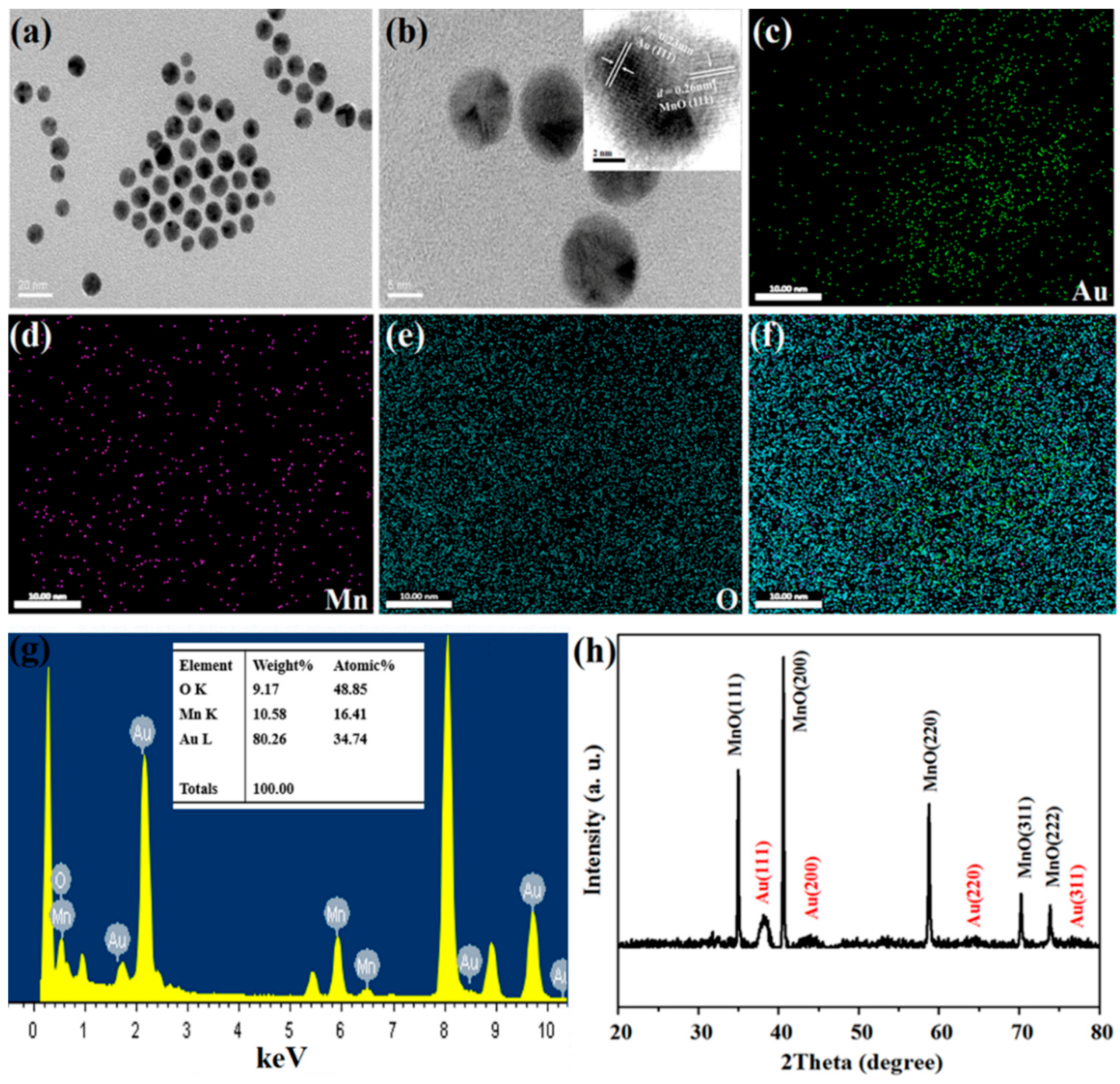
Abstract
:1. Introduction
2. Materials and Methods
2.1. Materials
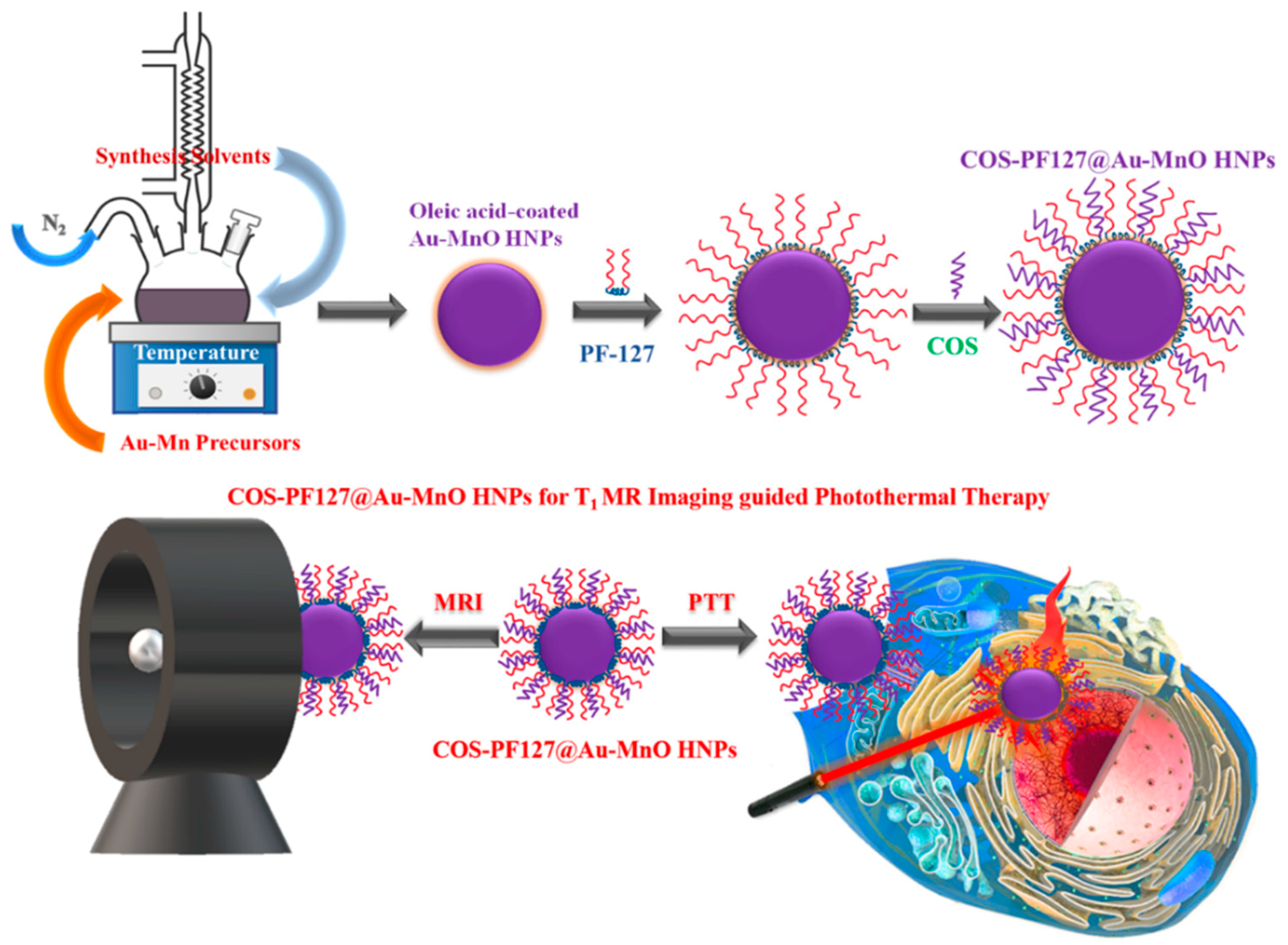
2.2. Synthesis of Au-MnO HNPs
2.3. Preparation of COS-PF127-Au-MnO HNPs
2.4. Characterizations
2.5. Cell Culture and In Vitro Cytotoxicity Assay
2.6. MR Imaging and Relaxation Properties
2.7. Cellular Uptake Studies
2.8. Photothermal Performance
2.9. In Vitro Photothermal Therapy
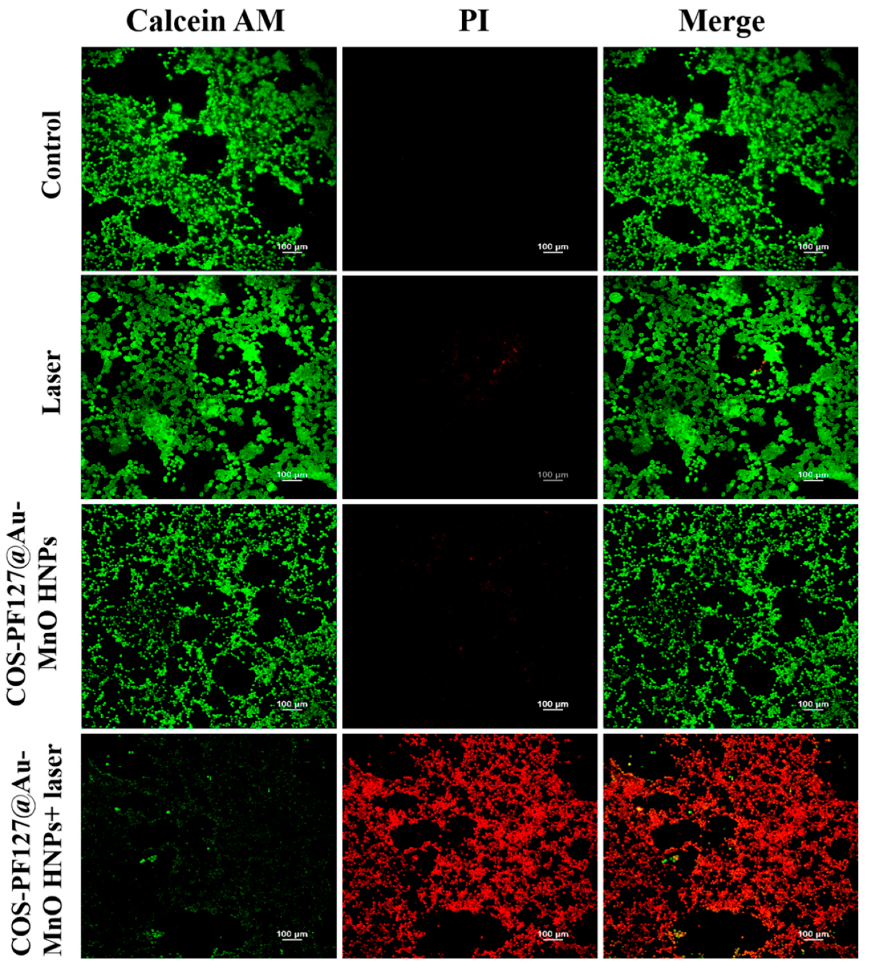
2.10. Live-Dead Cell Staining Experiments
3. Results and Discussions
3.1. Synthesis and Characterizations of Au-MnO HNPs
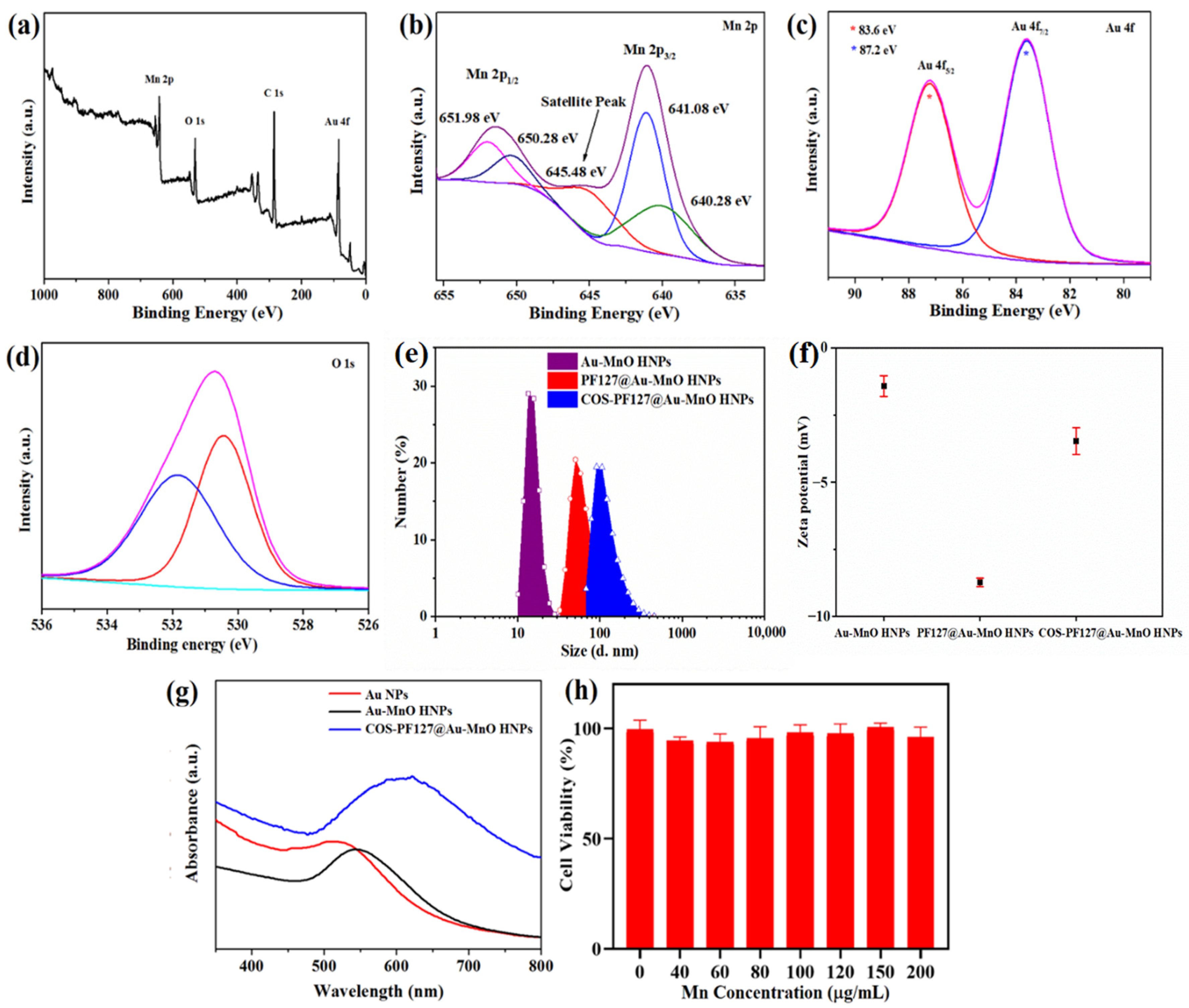
3.2. Surface Modification and Biocompatibility of Au-MnO HNPs
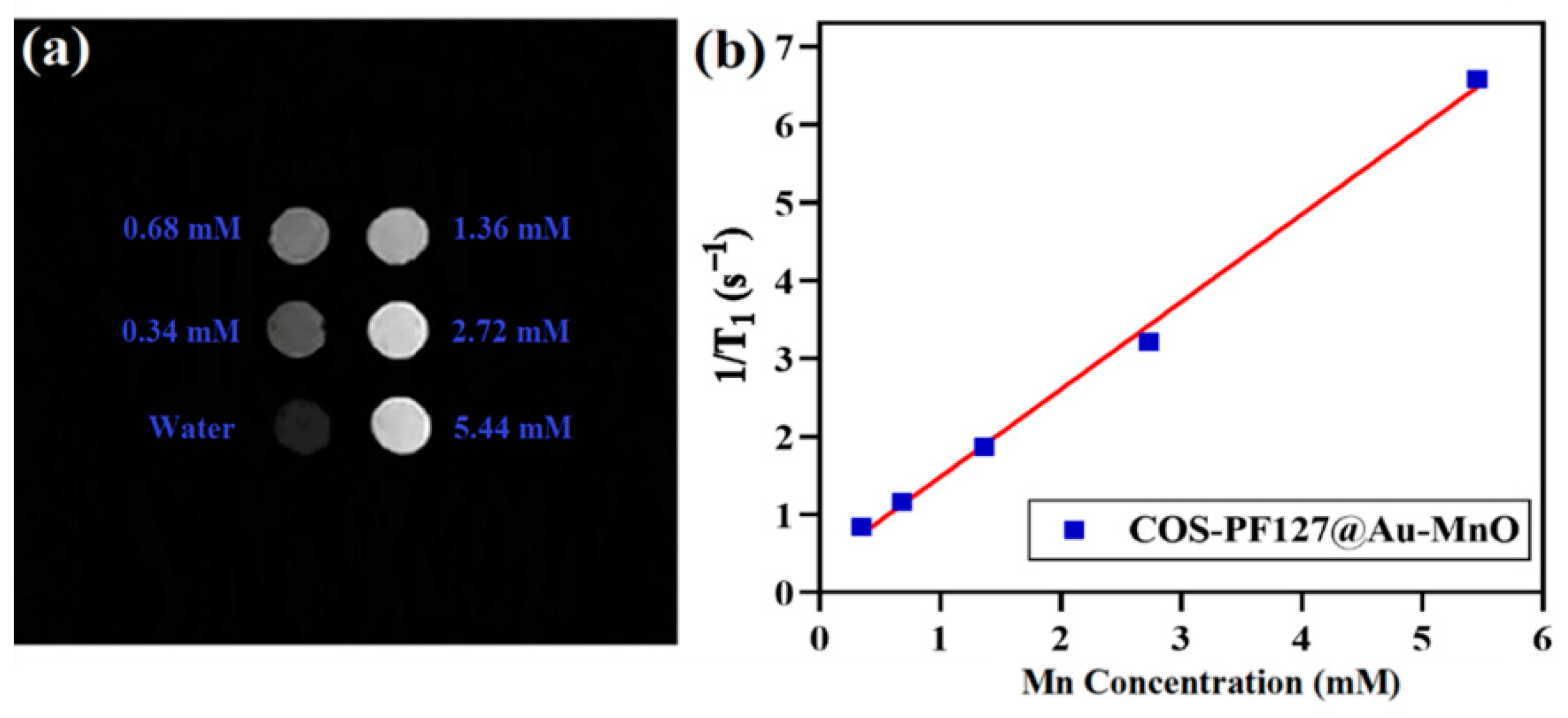
3.3. T1-MR Imaging and Relaxivity Properties of Au-MnO HNPs
3.4. Cellular Uptake of Au-MnO HNPs
3.5. Photothermal Therapy of Au-MnO HNPs
4. Conclusions
Supplementary Materials
Author Contributions
Funding
Institutional Review Board Statement
Informed Consent Statement
Data Availability Statement
Acknowledgments
Conflicts of Interest
References
- Oberdörster, G.; Oberdörster, E.; Oberdörster, J. Nanotoxicology: An emerging discipline evolving from studies of ultrafine particles. Environ. Health Perspect. 2005, 113, 823–839. [Google Scholar] [CrossRef] [PubMed]
- Satalkar, P.; Elger, B.S.; Shaw, D.M. Defining nano, nanotechnology and nanomedicine: Why should it matter? Sci. Eng. Ethics. 2016, 22, 1255–1276. [Google Scholar] [CrossRef] [PubMed]
- Oliveira, M.B.; Li, F.; Choi, J.; Mano, J.F. Nanomaterials for biomedical applications. Biotechnol. J. 2021, 16, 2170053. [Google Scholar] [CrossRef] [PubMed]
- Bououdina, M.; Rashdan, S.; Bobet, J.L.; Ichiyanagi, Y. Nanomaterials for biomedical applications: Synthesis, characterization, and applications. J. Nanomater. 2013, 2013, 962384. [Google Scholar] [CrossRef]
- Lammers, T.; Rizzo, L.Y.; Storm, G.; Kiessling, F. Personalized nanomedicine. Clin. Cancer Res. 2012, 18, 4889–4894. [Google Scholar] [CrossRef] [Green Version]
- Fornaguera, C.; García-Celma, M.J. Personalized nanomedicine: A revolution at the nanoscale. J. Pers. Med. 2017, 7, 12. [Google Scholar] [CrossRef] [Green Version]
- Iqbal, M.Z.; Ren, W.; Saeed, M.; Chen, T.; Ma, X.; Yu, X.; Zhang, J.; Zhang, L.; Li, A.; Wu, A. A facile fabrication route for binary transition metal oxide-based Janus nanoparticles for cancer theranostic applications. Nano Res. 2018, 11, 5735–5750. [Google Scholar] [CrossRef]
- Iqbal, M.Z.; Luo, D.; Akakuru, O.U.; Mushtaq, A.; Hou, Y.; Ali, I.; Ijaz, G.; Khalid, B.; Kong, X.; Wu, A. Facile synthesis of biocompatible magnetic titania nanorods for T1-magnetic resonance imaging and enhanced phototherapy of cancers. J. Mater. Chem. B 2021, 9, 6623–6633. [Google Scholar] [CrossRef]
- Mushtaq, A.; Hou, Y.; Tian, C.; Deng, T.; Xu, C.; Sun, Z.; Kong, X.; Zubair Iqbal, M. Facile synthesis of Mn doped TiO2 rhombic nanocomposites for enhanced T1-Magnetic resonance imaging and photodynamic therapy. Mater. Res. Bull. 2021, 144, 111481. [Google Scholar] [CrossRef]
- Hauner, K.; Maisch, P.; Retz, M. Nebenwirkungen der chemotherapie. Der Urol. 2017, 56, 472–479. [Google Scholar] [CrossRef]
- Mu, W.; Chu, Q.; Liu, Y.; Zhang, N. A review on nano-based drug delivery system for cancer chemoimmunotherapy. Nano-Micro Lett. 2020, 12, 142. [Google Scholar] [CrossRef] [PubMed]
- Hu, H.-T.; Park, J.-H.; Wang, Z.; Bakheet, N.; Xu, S.-J.; Lee, E.J.; Kim, D.-H.; Kim, S.H.; Song, H.-Y.; Jeon, J.Y.; et al. Localized photothermal ablation therapy of obstructive rectal cancer using a nanofunctionalized stent in a mouse model. ACS Biomater. Sci. Eng. 2021, 7, 5890–5898. [Google Scholar] [CrossRef] [PubMed]
- Tian, Q.; Jiang, F.; Zou, R.; Liu, Q.; Chen, Z.; Zhu, M.; Yang, S.; Wang, J.; Wang, J.; Hu, J. Hydrophilic Cu9S5 nanocrystals: A photothermal agent with a 25.7% heat conversion efficiency for photothermal ablation of cancer cells in vivo. ACS Nano 2011, 5, 9761–9771. [Google Scholar] [CrossRef] [PubMed]
- Long, S.; Xu, Y.; Zhou, F.; Wang, B.; Yang, Y.; Fu, Y.; Du, N.; Li, X. Characteristics of temperature changes in photothermal therapy induced by combined application of indocyanine green and laser. Oncol. Lett. 2019, 17, 3952–3959. [Google Scholar] [CrossRef] [PubMed] [Green Version]
- Yu, J.; Javier, D.; Yaseen, M.A.; Nitin, N.; Richards-Kortum, R.; Anvari, B.; Wong, M.S. Self-assembly synthesis, tumor cell targeting, and photothermal capabilities of antibody-coated indocyanine green nanocapsules. J. Am. Chem. Soc. 2010, 132, 1929–1938. [Google Scholar] [CrossRef] [Green Version]
- Zhang, Y.; Wang, Y.; Yang, X.; Yang, Q.; Li, J.; Tan, W. Polyaniline nanovesicles for photoacoustic imaging-guided photothermal-chemo synergistic therapy in the second near-infrared window. Small 2020, 16, e2001177. [Google Scholar] [CrossRef]
- Ma, X.; Lee, C.; Zhang, T.; Cai, J.; Wang, H.; Jiang, F.; Wu, Z.; Xie, J.; Jiang, G.; Li, Z. Image-guided selection of Gd@C-dots as sensitizers to improve radiotherapy of non-small cell lung cancer. J. Nanobiotechnol. 2021, 19, 284. [Google Scholar] [CrossRef]
- Li, J.; Cheng, Q.; Yue, L.; Gao, C.; Wei, J.; Ding, Y.; Wang, Y.; Zheng, Y.; Wang, R. Macrophage-hitchhiking supramolecular aggregates of CuS nanoparticles for enhanced tumor deposition and photothermal therapy. Nanoscale Horiz. 2021, 6, 907–912. [Google Scholar] [CrossRef]
- Tee, S.Y.; Ye, E.; Teng, C.P.; Tanaka, Y.; Tang, K.Y.; Win, K.Y.; Han, M.Y. Advances in photothermal nanomaterials for biomedical, environmental and energy applications. Nanoscale 2021, 13, 14268–14286. [Google Scholar] [CrossRef]
- Bao, Z.; Liu, X.; Liu, Y.; Liu, H.; Zhao, K. Near-infrared light-responsive inorganic nanomaterials for photothermal therapy. Asian J. Pharm. Sci. 2016, 11, 349–364. [Google Scholar] [CrossRef] [Green Version]
- Lin, Y.; Ren, J.; Qu, X. Nano-gold as artificial enzymes: Hidden talents. Adv. Mater. 2014, 26, 4200–4217. [Google Scholar] [CrossRef] [PubMed]
- Mapanao, A.K.; Santi, M.; Voliani, V. Combined chemo-photothermal treatment of three-dimensional head and neck squamous cell carcinomas by gold nano-architectures. J. Colloid Interface Sci. 2021, 582, 1003–1011. [Google Scholar] [CrossRef] [PubMed]
- Dewhirst, M.W.; Viglianti, B.L.; Lora-Michiels, M.; Hanson, M.; Hoopes, P.J. Basic principles of thermal dosimetry and thermal thresholds for tissue damage from hyperthermia. Int. J. Hyperth. 2003, 19, 267–294. [Google Scholar] [CrossRef] [PubMed]
- Shrestha, B.; Tang, L.; Romero, G. Nanoparticles-mediated combination therapies for cancer treatment. Adv. Ther. 2019, 2, 1900076. [Google Scholar] [CrossRef]
- Mokhtari, R.B.; Homayouni, T.S.; Baluch, N.; Morgatskaya, E.; Kumar, S.; Das, B.; Yeger, H. Combination therapy in combating cancer. Oncotarget 2017, 8, 38022. [Google Scholar] [CrossRef] [Green Version]
- Wang, Z.; Sun, X.; Huang, T.; Song, J.; Wang, Y. A sandwich nanostructure of gold nanoparticle coated reduced graphene oxide for photoacoustic imaging-guided photothermal therapy in the second NIR window. Front. Bioeng. Biotechnol. 2020, 8, 655. [Google Scholar] [CrossRef]
- Rajkumar, S.; Prabaharan, M. Theranostics based on iron oxide and gold nanoparticles for imaging- guided photothermal and photodynamic therapy of cancer. Curr. Top. Med. Chem. 2017, 17, 1858–1871. [Google Scholar] [CrossRef]
- Zhao, F.; Li, X.; Li, J.; Dou, Y.; Wang, L.; Wu, M.; Liu, Y.; Chang, J.; Zhang, X. Activatable ultrasmall gold nanorods for “off–on” fluorescence imaging-guided photothermal therapy. J. Mater. Chem. B 2017, 5, 2145–2151. [Google Scholar] [CrossRef]
- Goh, B.T.; Poon, C.Y.; Peck, R.H. The importance of routine magnetic resonance imaging in trigeminal neuralgia diagnosis. Oral Surg Oral Med Oral Pathol Oral Radiol Endod 2001, 92, 424–429. [Google Scholar] [CrossRef]
- Grover, V.P.B.; Tognarelli, J.M.; Crossey, M.M.E.; Cox, I.J.; Taylor-Robinson, S.D.; McPhail, M.J.W. Magnetic resonance imaging: Principles and techniques: Lessons for clinicians. J. Clin. Exp. Hepatol. 2015, 5, 246–255. [Google Scholar] [CrossRef] [Green Version]
- Mathur, M.; Jones, J.R.; Weinreb, J.C. Gadolinium deposition and nephrogenic systemic fibrosis: A radiologist’s primer. Radiographics 2020, 40, 153–162. [Google Scholar] [CrossRef] [PubMed]
- Cowper, S.E.; Robin, H.S.; Steinberg, S.M.; Su, L.D.; Gupta, S.; LeBoit, P.E. Scleromyxoedema-like cutaneous diseases in renal-dialysis patients. Lancet 2000, 356, 1000–1001. [Google Scholar] [CrossRef]
- Tomitaka, A.; Arami, H.; Raymond, A.; Yndart, A.; Kaushik, A.; Jayant, R.D.; Takemura, Y.; Cai, Y.; Toborek, M.; Nair, M. Development of magneto-plasmonic nanoparticles for multimodal image-guided therapy to the brain. Nanoscale 2017, 9, 764–773. [Google Scholar] [CrossRef] [PubMed] [Green Version]
- Ding, X.; Li, D.; Jiang, J. Gold-based inorganic nanohybrids for nanomedicine applications. Theranostics 2020, 10, 8061–8079. [Google Scholar] [CrossRef]
- Multari, C.; Miola, M.; Laviano, F.; Gerbaldo, R.; Pezzotti, G.; Debellis, D.; Verné, E. Magnetoplasmonic nanoparticles for photothermal therapy. Nanotechnology 2019, 30, 255705. [Google Scholar] [CrossRef]
- Nguyen, H.V.; Chen, Q.; Paletta, J.T.; Harvey, P.; Jiang, Y.; Zhang, H.; Boska, M.D.; Ottaviani, M.F.; Jasanoff, A.; Rajca, A.; et al. Nitroxide-based macromolecular contrast agents with unprecedented transverse relaxivity and stability for magnetic resonance imaging of tumors. ACS Cent. Sci. 2017, 3, 800–811. [Google Scholar] [CrossRef]
- Gale, E.M.; Atanasova, I.P.; Blasi, F.; Ay, I.; Caravan, P. A manganese alternative to gadolinium for MRI contrast. J. Am. Chem Soc. 2015, 137, 15548–15557. [Google Scholar] [CrossRef] [Green Version]
- Kohgo, Y.; Ikuta, K.; Ohtake, T.; Torimoto, Y.; Kato, J. Body iron metabolism and pathophysiology of iron overload. Int. J. Hematol. 2008, 88, 7–15. [Google Scholar] [CrossRef] [Green Version]
- Zhu, L.; Zhou, Z.; Mao, H.; Yang, L. Magnetic nanoparticles for precision oncology: Theranostic magnetic iron oxide nanoparticles for image-guided and targeted cancer therapy. Nanomedicine 2017, 12, 73–87. [Google Scholar] [CrossRef] [Green Version]
- Akakuru, O.U.; Iqbal, M.Z.; Saeed, M.; Liu, C.; Paunesku, T.; Woloschak, G.; Hosmane, N.S.; Wu, A. The transition from metal-based to metal-free contrast agents for T(1) magnetic resonance imaging enhancement. Bioconjug. Chem. 2019, 30, 2264–2286. [Google Scholar] [CrossRef]
- Yi, X.; Chen, L.; Zhong, X.; Gao, R.; Qian, Y.; Wu, F.; Song, G.; Chai, Z.; Liu, Z.; Yang, K. Core–shell Au@MnO2 nanoparticles for enhanced radiotherapy via improving the tumor oxygenation. Nano Res. 2016, 9, 3267–3278. [Google Scholar] [CrossRef]
- García-Hevia, L.; Bañobre-López, M.; Gallo, J. Recent progress on manganese-based nanostructures as responsive MRI contrast agents. Chemistry—A Eur. J. 2019, 25, 431–441. [Google Scholar] [CrossRef] [PubMed]
- Zhen, Z.; Xie, J. Development of Manganese-Based Nanoparticles as Contrast Probes for Magnetic Resonance Imaging. Theranostics 2012, 2, 45–54. [Google Scholar] [CrossRef] [PubMed]
- Na, H.B.; Lee, J.H.; An, K.; Park, Y.I.; Park, M.; Lee, I.S.; Nam, D.H.; Kim, S.T.; Kim, S.H.; Kim, S.W.; et al. Development of a T1 contrast agent for magnetic resonance imaging using MnO nanoparticles. Angew. Chem. Int. Ed. Engl. 2007, 46, 5397–5401. [Google Scholar] [CrossRef]
- Gao, M.; Song, Y.; Liu, Y.; Jiang, W.; Peng, J.; Shi, L.; Jia, R.; Muhammad, Y.; Huang, L. Controlled fabrication of Au@MnO2 core/shell assembled nanosheets by localized surface plasmon resonance. Appl. Surf. Sci. 2021, 537, 147912. [Google Scholar] [CrossRef]
- Yu, J.; Yang, W.; Xing, S.; Wang, J.; Han, H.; Zhang, P.; Xiang, C.; Zhang, B. Blended gold/MnO2@BSA nanoparticles for fluorometric and magnetic resonance determination of ascorbic acid. Mikrochim. Acta 2019, 186, 89. [Google Scholar] [CrossRef]
- Li, T.; Liu, Y.; Jia, R.; Yaseen, M.; Shi, L.; Huang, L. Irradiation regulates the size of Pt nanoparticles on Au@MnO2 nanosheets for electrocatalytic hydrogen evolution. New J. Chem. 2021, 45, 22327–22334. [Google Scholar] [CrossRef]
- Qiu, T.; Luo, B.; Giersig, M.; Akinoglu, E.M.; Hao, L.; Wang, X.; Shi, L.; Jin, M.; Zhi, L. Au@MnO2 core–shell nanomesh electrodes for transparent flexible supercapacitors. Small 2014, 10, 4136–4141. [Google Scholar] [CrossRef]
- Ijaz Dar, G.; Iqbal, M.Z.; Akakuru, O.U.; Yao, C.; Awiaz, G.; Wu, A. Facile synthesis of Au@Mn3O4 magneto-plasmonic nanoflowers for T1-weighted magnetic resonance imaging and photothermal therapy of cancer. J. Mater. Chem. B 2020, 8, 8356–8367. [Google Scholar] [CrossRef]
- Zhu, H.; Sigdel, A.; Zhang, S.; Su, D.; Xi, Z.; Li, Q.; Sun, S. Core/Shell Au/MnO nanoparticles prepared through controlled oxidation of AuMn As an electrocatalyst for sensitive H2O2 detection. Angew. Chem. Int. Ed. 2014, 53, 12508–12512. [Google Scholar] [CrossRef]
- Akash, M.S.H.; Rehman, K. Recent progress in biomedical applications of Pluronic (PF127): Pharmaceutical perspectives. J. Control. Release 2015, 209, 120–138. [Google Scholar] [CrossRef] [PubMed]
- Muanprasat, C.; Chatsudthipong, V. Chitosan oligosaccharide: Biological activities and potential therapeutic applications. Pharmacol. Ther. 2017, 170, 80–97. [Google Scholar] [CrossRef] [PubMed]
- Ma, S.; Chen, D.; Wang, W.-l. MnO nanoparticles embedded in a carbon matrix as high performance lithium-ion battery anodes: Preparation, microstructure and electrochemistry. Phys. Chem. Chem. Phys. 2016, 18, 19130–19136. [Google Scholar] [CrossRef] [PubMed]
- Wang, J.; Li, C.; Yang, Z.; Chen, D. Chemical vapor deposition-assisted fabrication of a graphene-wrapped MnO/carbon nanofibers membrane as a high-rate and long-life anode for lithium ion batteries. RSC Adv. 2017, 7, 50973–50980. [Google Scholar] [CrossRef] [Green Version]
- Sivakumar, M.; Venkatakrishnan, K.; Tan, B. Characterization of MHz pulse repetition rate femtosecond laser-irradiated gold-coated silicon surfaces. Nanoscale Res. Lett. 2011, 6, 78. [Google Scholar] [CrossRef] [Green Version]
- Tantra, R.; Schulze, P.; Quincey, P. Effect of nanoparticle concentration on zeta-potential measurement results and reproducibility. Particuology 2010, 8, 279–285. [Google Scholar] [CrossRef]
- Wang, N.; Hsu, C.; Zhu, L.; Tseng, S.; Hsu, J.-P. Influence of metal oxide nanoparticles concentration on their zeta potential. J. Colloid Interface Sci. 2013, 407, 22–28. [Google Scholar] [CrossRef]
- S. S. dos Santos, P.; M. M. M. de Almeida, J.; Pastoriza-Santos, I.; C. C. Coelho, L. Advances in plasmonic sensing at the NIR—A review. Sensors 2021, 21, 2111. [Google Scholar]
- Schladt, T.D.; Shukoor, M.I.; Schneider, K.; Tahir, M.N.; Natalio, F.; Ament, I.; Becker, J.; Jochum, F.D.; Weber, S.; Köhler, O.; et al. Au@MnO Nanoflowers: Hybrid Nanocomposites for Selective Dual Functionalization and Imaging. Angew. Chem. Int. Ed. 2010, 49, 3976–3980. [Google Scholar] [CrossRef]
- Hsu, B.Y.W.; Ng, M.; Zhang, Y.; Wong, S.Y.; Bhakoo, K.; Li, X.; Wang, J. A Hybrid Silica Nanoreactor Framework for Encapsulation of Hollow Manganese Oxide Nanoparticles of Superior T1Magnetic Resonance Relaxivity. Adv. Funct. Mater. 2015, 25, 5269–5276. [Google Scholar] [CrossRef]
- Bennewitz, M.F.; Lobo, T.L.; Nkansah, M.K.; Ulas, G.; Brudvig, G.W.; Shapiro, E.M. Biocompatible and pH-Sensitive PLGA Encapsulated MnO Nanocrystals for Molecular and Cellular MRI. ACS Nano 2011, 5, 3438–3446. [Google Scholar] [CrossRef] [PubMed] [Green Version]
- Peng, Y.-K.; Lai, C.-W.; Liu, C.-L.; Chen, H.-C.; Hsiao, Y.-H.; Liu, W.-L.; Tang, K.-C.; Chi, Y.; Hsiao, J.-K.; Lim, K.-E.; et al. A New and Facile Method To Prepare Uniform Hollow MnO/Functionalized mSiO2 Core/Shell Nanocomposites. ACS Nano 2011, 5, 4177–4187. [Google Scholar] [CrossRef] [PubMed]
- Howell, M.; Mallela, J.; Wang, C.; Ravi, S.; Dixit, S.; Garapati, U.; Mohapatra, S. Manganese-loaded lipid-micellar theranostics for simultaneous drug and gene delivery to lungs. J. Control. Release 2013, 167, 210–218. [Google Scholar] [CrossRef] [PubMed] [Green Version]
- Hu, H.; Dai, A.; Sun, J.; Li, X.; Gao, F.; Wu, L.; Fang, Y.; Yang, H.; An, L.; Wu, H.; et al. Aptamer-conjugated Mn3O4@SiO2 core–shell nanoprobes for targeted magnetic resonance imaging. Nanoscale 2013, 5, 10447–10454. [Google Scholar] [CrossRef] [PubMed]
- Yang, X.; Zhou, Z.; Wang, L.; Tang, C.; Yang, H.; Yang, S. Folate conjugated Mn3O4@SiO2 nanoparticles for targeted magnetic resonance imaging in vivo. Mater. Res. Bull. 2014, 57, 97–102. [Google Scholar] [CrossRef]
- Schladt, T.D.; Koll, K.; Prüfer, S.; Bauer, H.; Natalio, F.; Dumele, O.; Raidoo, R.; Weber, S.; Wolfrum, U.; Schreiber, L.M.; et al. Multifunctional superparamagnetic MnO@SiO2 core/shell nanoparticles and their application for optical and magnetic resonance imaging. J. Mater. Chem. 2012, 22, 9253–9262. [Google Scholar] [CrossRef]
- Zhao, R.; Han, X.; Yuliang, Z.; Wang, H.; Ji, T.; Zhao, Y.; Nie, G. Photothermal Effect Enhanced Cascade-Targeting Strategy for Improved Pancreatic Cancer Therapy by Gold Nanoshell@Mesoporous Silica Nanorod. ACS Nano 2017, 11, 8103–8113. [Google Scholar] [CrossRef]
- Ju, Y.; Zhang, H.; Yu, J.; Tong, S.; Tian, N.; Wang, Z.; Wang, X.; Su, X.; Chu, X.; Lin, J.; et al. Monodisperse Au–Fe2C Janus Nanoparticles: An Attractive Multifunctional Material for Triple-Modal Imaging-Guided Tumor Photothermal Therapy. ACS Nano 2017, 11, 9239–9248. [Google Scholar] [CrossRef]
- Wang, S.; You, Q.; Wang, J.; Song, Y.; Cheng, Y.; Wang, Y.; Yang, S.; Yang, L.; Li, P.; Lu, Q.; et al. MSOT/CT/MR imaging-guided and hypoxia-maneuvered oxygen self-supply radiotherapy based on one-pot MnO2-mSiO2@Au nanoparticles. Nanoscale 2019, 11, 6270–6284. [Google Scholar] [CrossRef]
- Hessel, C.M.; Pattani, V.P.; Rasch, M.; Panthani, M.G.; Koo, B.; Tunnell, J.W.; Korgel, B.A. Copper Selenide Nanocrystals for Photothermal Therapy. Nano Lett. 2011, 11, 2560–2566. [Google Scholar] [CrossRef] [Green Version]
- He, T.; Jiang, C.; He, J.; Zhang, Y.; He, G.; Wu, J.; Lin, J.; Zhou, X.; Huang, P. Manganese-Dioxide-Coating-Instructed Plasmonic Modulation of Gold Nanorods for Activatable Duplex-Imaging-Guided NIR-II Photothermal-Chemodynamic Therapy. Adv. Mater. 2021, 33, 2008540. [Google Scholar] [CrossRef] [PubMed]
- Poulose, A.C.; Veeranarayanan, S.; Mohamed, M.S.; Aburto, R.R.; Mitcham, T.; Bouchard, R.; Ajayan, P.M.; Sakamoto, Y.; Maekawa, T.; Kumar, D.S. Multifunctional Cu2−xTe Nanocubes Mediated Combination Therapy for Multi-Drug Resistant MDA MB 453. Sci. Rep. 2016, 6, 35961. [Google Scholar] [CrossRef] [PubMed] [Green Version]


Publisher’s Note: MDPI stays neutral with regard to jurisdictional claims in published maps and institutional affiliations. |
© 2022 by the authors. Licensee MDPI, Basel, Switzerland. This article is an open access article distributed under the terms and conditions of the Creative Commons Attribution (CC BY) license (https://creativecommons.org/licenses/by/4.0/).
Share and Cite
Tian, C.; Tang, Z.; Hou, Y.; Mushtaq, A.; Naz, S.; Yu, Z.; Farheen, J.; Iqbal, M.Z.; Kong, X. Facile Synthesis of Multifunctional Magnetoplasmonic Au-MnO Hybrid Nanocomposites for Cancer Theranostics. Nanomaterials 2022, 12, 1370. https://doi.org/10.3390/nano12081370
Tian C, Tang Z, Hou Y, Mushtaq A, Naz S, Yu Z, Farheen J, Iqbal MZ, Kong X. Facile Synthesis of Multifunctional Magnetoplasmonic Au-MnO Hybrid Nanocomposites for Cancer Theranostics. Nanomaterials. 2022; 12(8):1370. https://doi.org/10.3390/nano12081370
Chicago/Turabian StyleTian, Cong, Zhe Tang, Yike Hou, Asim Mushtaq, Shafaq Naz, Zhangsen Yu, Jabeen Farheen, Muhammad Zubair Iqbal, and Xiangdong Kong. 2022. "Facile Synthesis of Multifunctional Magnetoplasmonic Au-MnO Hybrid Nanocomposites for Cancer Theranostics" Nanomaterials 12, no. 8: 1370. https://doi.org/10.3390/nano12081370