Article

Effect of Different Solvents on Morphology and Gas-Sensitive Properties of Grinding-Assisted Liquid-Phase-Exfoliated MoS2 Nanosheets

1
Key Laboratory of New Energy and Materials Research, Xinjiang Institute of Engineering, Urumqi 830023, China
2
Xinjiang Condensed Matter Phase Transition and Microstructure Laboratory, College of Physics Science and Technology, Yili Normal University, Yining 835000, China
*
Author to whom correspondence should be addressed.
Nanomaterials 2022, 12(24), 4485; https://doi.org/10.3390/nano12244485
Submission received: 10 November 2022 / Revised: 11 December 2022 / Accepted: 15 December 2022 / Published: 18 December 2022
(This article belongs to the Special Issue Advanced Nanomaterials and Nanotechnologies for Micro/Nano-Sensors)

Abstract

:
Grinding-assisted liquid-phase exfoliation is a widely used method for the preparation of two-dimensional nanomaterials. In this study, N-methylpyrrolidone and acetonitrile, two common grinding solvents, were used during the liquid-phase exfoliation for the preparation of MoS2 nanosheets. The morphology and structure of MoS2 nanosheets were analyzed via scanning electron microscopy, X-ray diffraction, and Raman spectroscopy. The effects of grinding solvents on the gas-sensing performance of the MoS2 nanosheets were investigated for the first time. The results show that the sensitivities of MoS2 nanosheet exfoliation with N-methylpyrrolidone were 2.4-, 1.4-, 1.9-, and 2.7-fold higher than exfoliation with acetonitrile in the presence of formaldehyde, acetone, and ethanol and 98% relative humidity, respectively. MoS2 nanosheet exfoliation with N-methylpyrrolidone also has fast response and recovery characteristics to 50–1000 ppm of CH2O. Accordingly, although N-methylpyrrolidone cannot be removed completely from the surface of MoS2, it has good gas sensitivity compared with other samples. Therefore, N-methylpyrrolidone is preferred for the preparation of gas-sensitive MoS2 nanosheets in grinding-assisted liquid-phase exfoliation. The results provide an experimental basis for the preparation of two-dimensional materials and their application in gas sensors.

1. Introduction

Given the special structure and potential applications, two-dimensional (2D) materials such as graphene, boron nitride, and molybdenum disulfide (MoS2) draw plenty of concerns. Among them, MoS2 as the frontrunner in transition metal dichalcogenides (TMDCs) materials has gained the most attention [1,2,3,4] and is used in a wide variety of applications [5,6,7,8,9,10,11] due to its unique properties [12,13,14]. MoS2 is at the forefront in the race of an ideal gas-sensing material because of its large surface-to-volume ratio, enormous number of active sites, and favorable adsorption sites [15,16]. MoS2 manifests two possible crystal phases, including trigonal and hexagonal structures, with metallic and semiconducting properties, respectively [17]. The presence of weak Van der Waals force facilitates the isolation of layers from bulk MoS2. The indirect bandgap of 1.2 eV in bulk MoS2 is converted to a direct bandgap of 1.8 eV for monolayer MoS2 [3,14,18]. The absence of dangling bonds provides stability to pristine MoS2 flakes in liquid and gaseous media in the presence of oxygen, thereby facilitating its gas-sensing application [19,20]. Therefore, a reliable and low-cost technique is needed to produce 2D-MoS2 for gas-sensing applications. Currently, several methods including vapor deposition [21], mechanical exfoliation [22], lithium-ion intercalation [23], liquid-phase exfoliation [24,25], and RF sputtering [26] have been utilized to fabricate 2D-MoS2 nanomaterials. Although high-quality MoS2 nanosheets were prepared by mechanical methods for fundamental research, it is difficult to meet the need for mass production. Meanwhile, hydrothermal and solvothermal methods yield few-layer MoS2 nanosheets on a large scale. However, they often require high temperature and high pressure. Lithium intercalation into the layered structure of 2D-MoS2 is limited by long lithiation time, high temperature, and sensitivity to environmental conditions. Instead, grinding-assisted liquid-phase exfoliation is not air-sensitive, does not entail chemical reaction, and generally has acceptable yield [3,27,28]. Coleman et al. [29] evaluated multiple solvents for ultrasonic exfoliation of materials. They found that the most effective solvent was N-methylpyrrolidone (NMP), followed by acetonitrile (ACN). Yao et al. [25] reported that relatively high yields up to 26.7 mg/mL were obtained by incorporating NMP as a grinding solvent into the exfoliation procedure because the surface energy of NMP is similar to that of MoS2. As a result, NMP is the preferred solvent in liquid-phase exfoliation to obtain single or multilayered 2D nano-materials [25,27,28,29,30,31].
The studies suggest that grinding solvent plays an important role in grinding-assisted exfoliation because its physical properties, such as boiling point [32], surface tension and energy [29,33], as well as solubility parameters [24], can affect the final exfoliation materials. Emily et al. reported that the lateral size and thickness of the exfoliated flakes of MoS2 nanosheets are highly dependent on the solvent. NMP yielded flakes of the highest quality based on lateral size and flake thickness. The NMP remained on the surface of the MoS2 nanosheets when ACN was completely removed [34]. Good yields were obtained when using NMP as a grinding solvent. However, whether the NMP residue affects the performance of electronic devices is unknown. It may adversely affect the application of MoS2 nanosheets in gas sensing. To our knowledge, there is no report on the research of the effects of a grinding solvent on the gas-sensing properties of MoS2.
Therefore, we evaluated the effects of residual NMP on the morphology and gas-sensing properties of liquid-phase-exfoliated MoS2 nanosheets. We selected ACN, which has a lower boiling point and easier solvent removal compared with NMP. The morphology and structure of MoS2 nanosheets were analyzed by scanning electron microscopy (SEM), X-ray diffraction (XRD), and Raman spectroscopy. The effects of grinding solvents on the gas-sensing performance of MoS2 nanosheets were investigated for the first time.

2. Materials and Methods

2.1. Preparation of Materials

MoS2, with a purity of 99% and particle size less than 2 μm, was purchased from Sigma-Aldrich. ACN, NMP, and absolute ethanol (C2H6O) were purchased from Tianjin Zhiyuan Chemical Reagent Co. Ltd. as analytically pure reagents. The preparation of MoS2 nanosheets via grinding-assisted liquid-phase exfoliation is described as follows:
MoS2 powder (100 mg) was manually ground in a mortar for 2 h, and 0.5 mL of the chosen solvent was added during the grinding. The sample was then dried in a vacuum oven at 60 °C for 12 h. The dried sample was dispersed in 40 mL of 45 vol% absolute ethanol and sonicated for 1 h at 120 W with stirring. The dispersion was centrifuged for another 20 min (1500 r / min) to obtain the MoS2 nanosheets, and the supernatant was dried in air for further use. For convenience, the MoS2 nanosheets obtained by grinding with ACN were designated as S1, and those ground with NMP were called S2.

2.2. Characterizations

The morphology of MoS2 nanosheets was observed with a field emission scanning electron microscope (SEM, JSM-7610F Plus). The crystal structure of MoS2 nanosheets was characterized by X-ray diffraction (XRD, Bruker D8 Advance, with Cu-Kα radiation). Raman spectroscopy (Renishaw inVia, Gloucester, Britain) was used to characterize the defects and functional groups of samples. The I-t and I-V curves of the sensing chip were measured by Keithley 2636B at room temperature.

2.3. Device Fabrication and Testing

The MoS2 nanosheets were dispersed in absolute ethanol at 10 mg/mL. Dispersions (2 μL) were uniformly coated to fabricate a MoS2-based sensing chip with Ag-Pd fork-finger electrodes. The minimum width and spacing of electrodes was 0.2 mm. The interdigital electrode was dried at 25 °C and aged for 24 h at a voltage of 4 V to obtain a sensing chip with good stability. The target vapor was produced by thermal evaporation, according to our previous work [35], and a calculated amount of target liquid was dropped onto a hot plate in a 1 L container to generate target vapor in the container. Next, 98% relative humidity was obtained by saturating salt solution (potassium sulphate-K₂SO₄). Then, by transferring the sensing chip from the air to the target gas at room temperature, the Keithley 2636B recorded the change of the current signal of the sensing chip (Figure S1). The response was defined using the formula ( I G I R I R ) × 100 % , where IR and IG are the currents of the sensor in the reference gas and target gas, respectively. The response time and recovery time were defined as the response values of 90% and 10% of the current of the sensor in contact with the target gas, respectively.

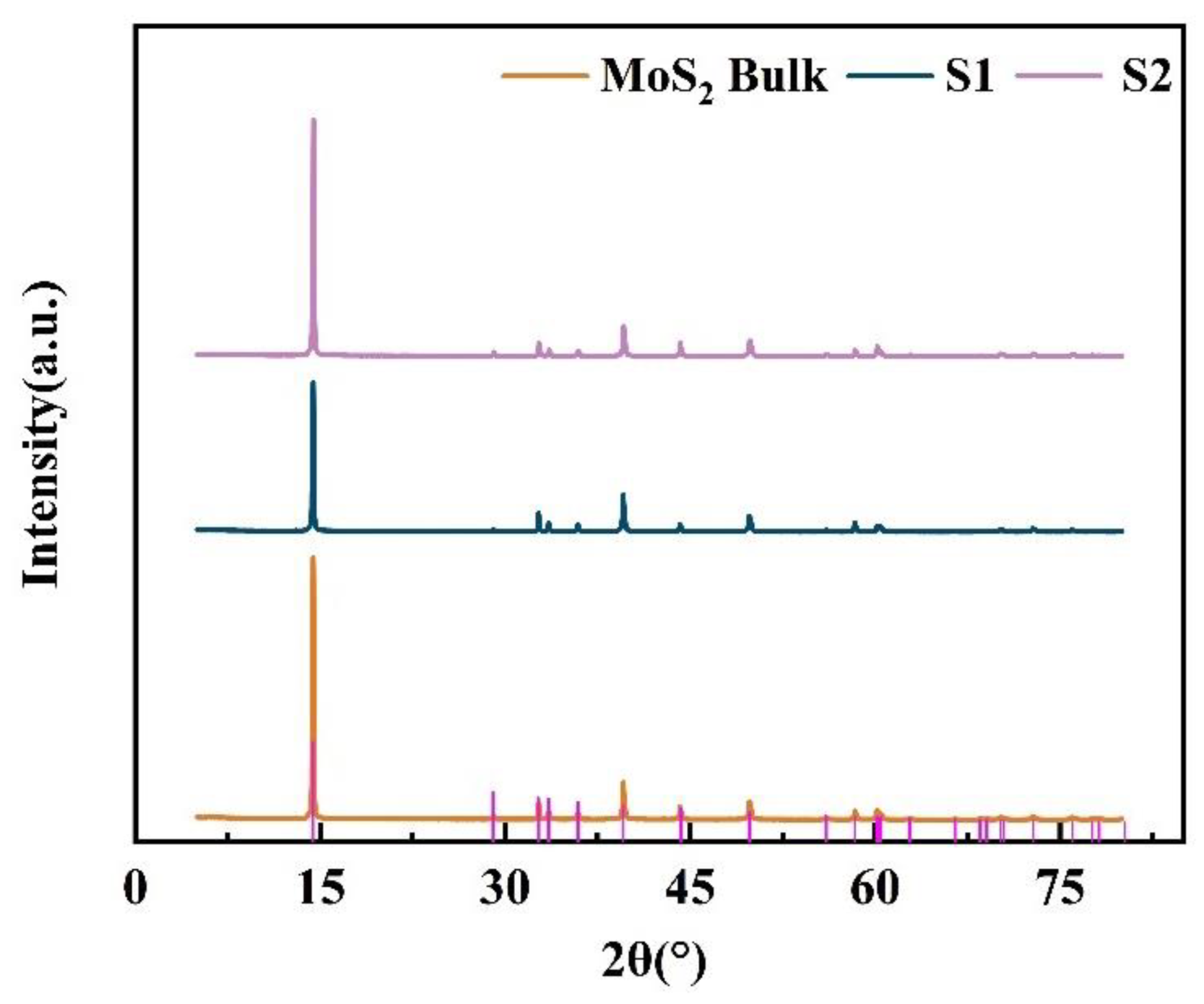
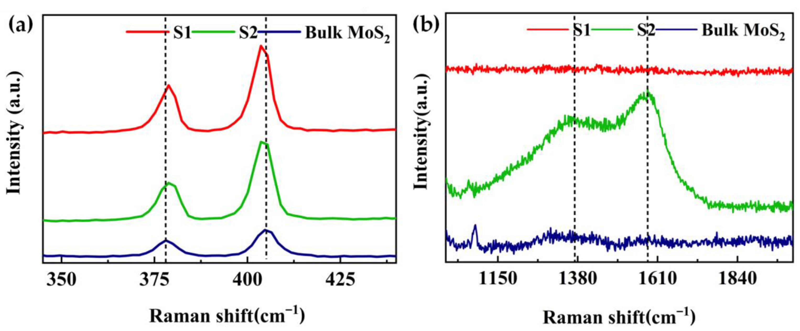
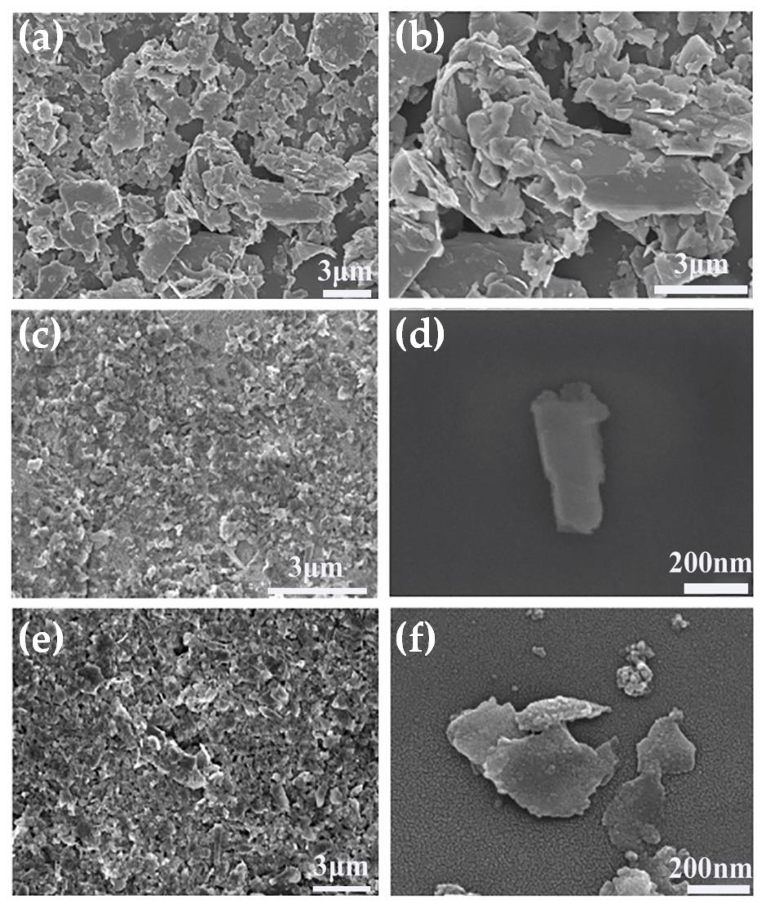
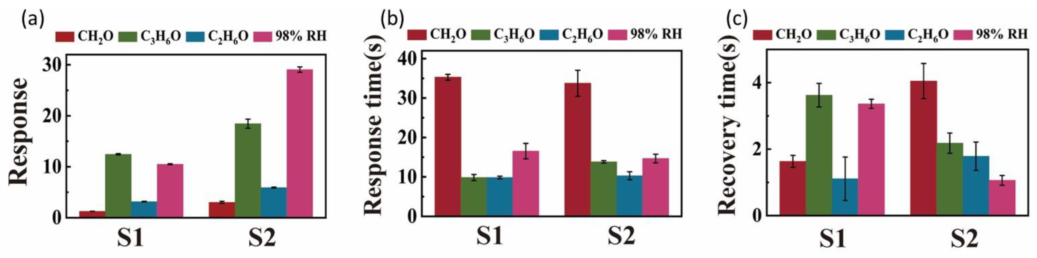
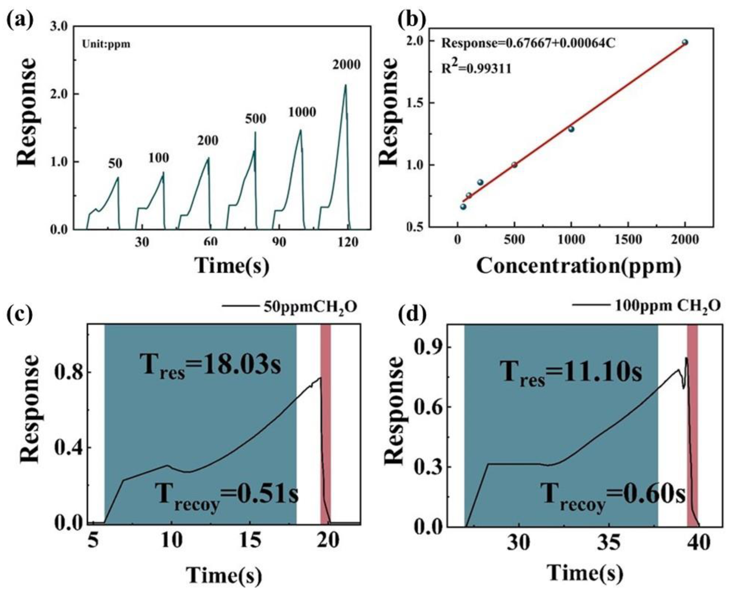
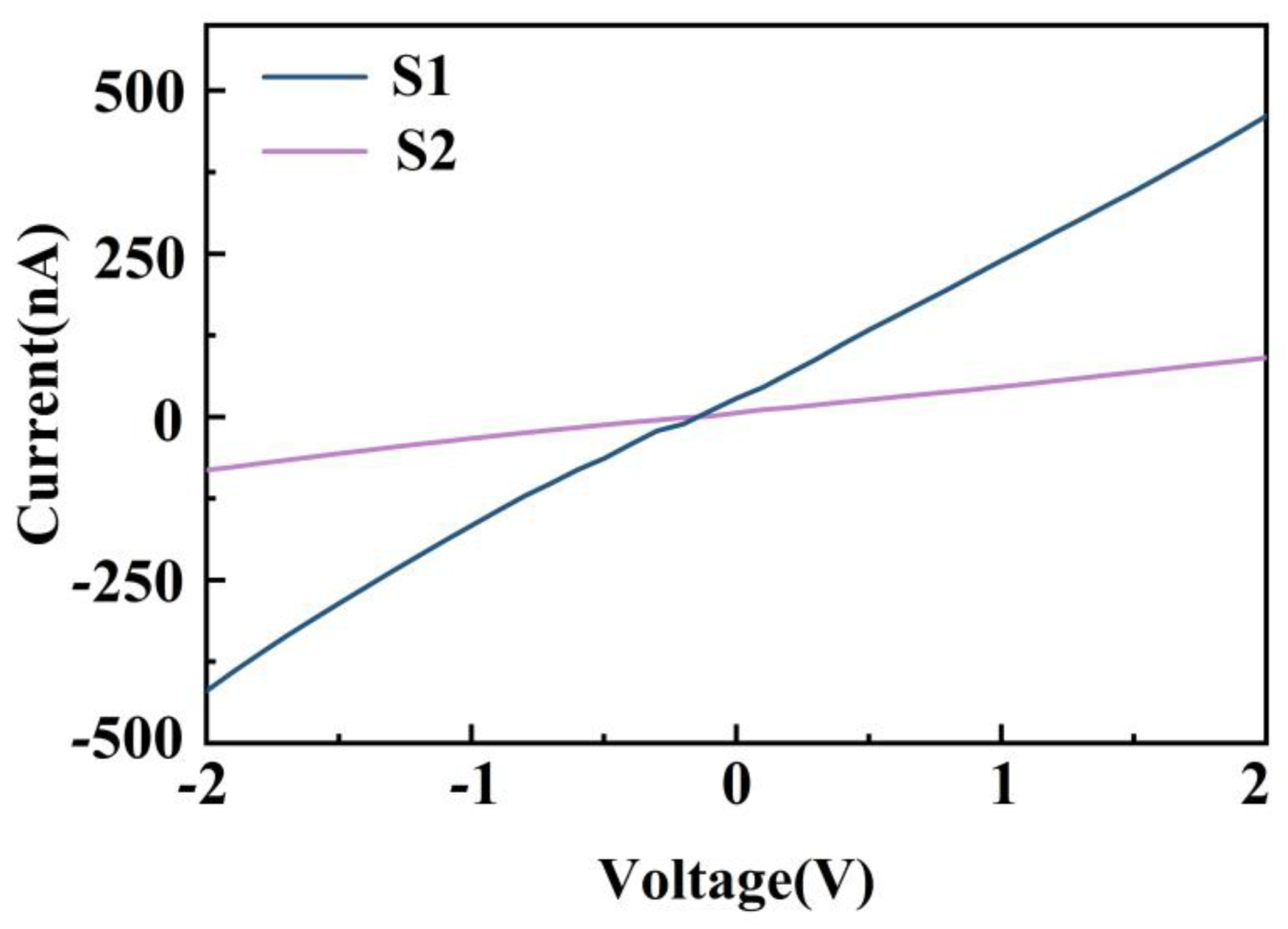
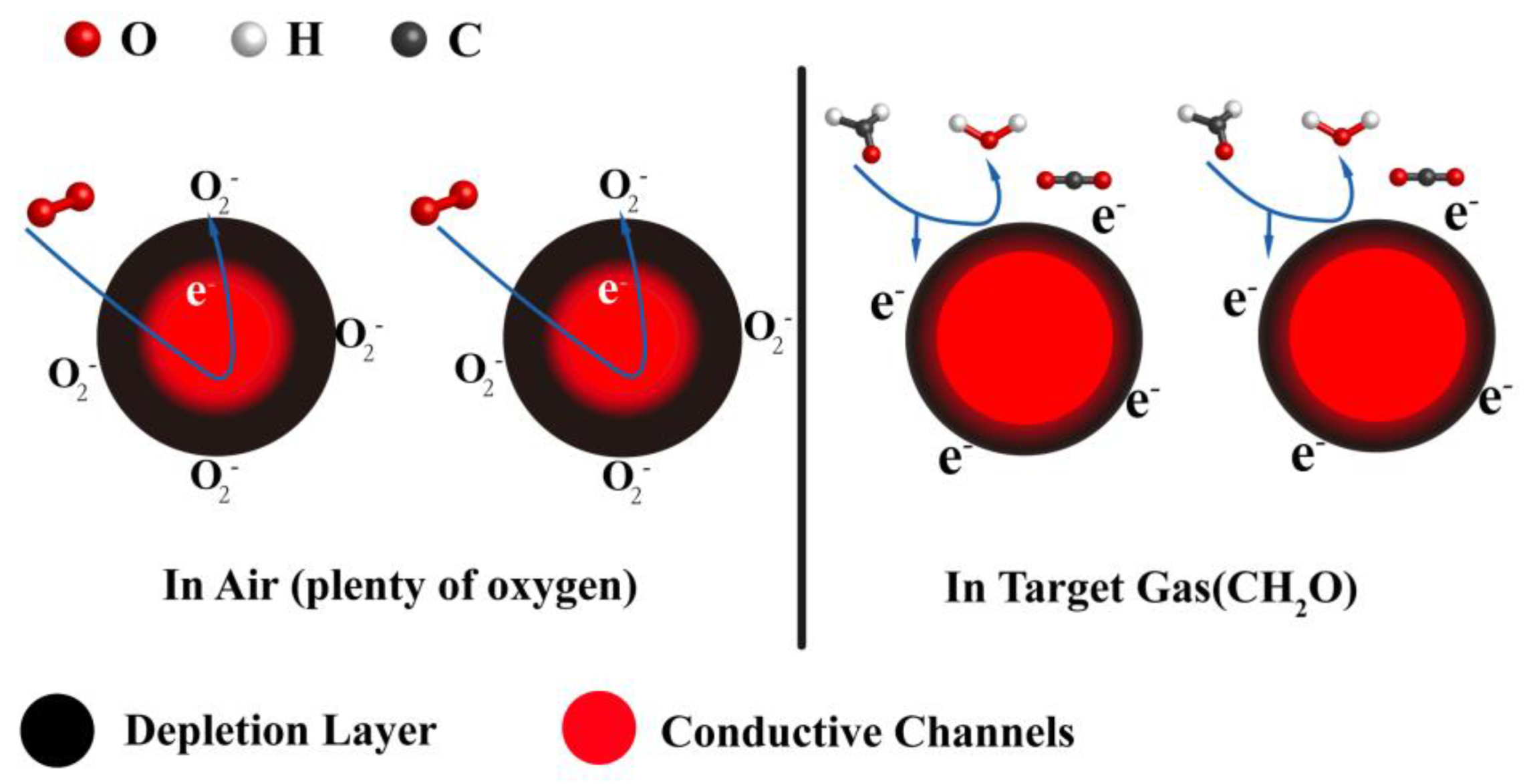
3. Results and Discussion

The XRD patterns of the two types of MoS2 prepared by different grinding solvents are shown in Figure 1. Compared with JCPDS Card No. 73-1508, the lattice constants were: a = 3.15938 Å, b = 3.15938 Å, and c = 12.28962 Å. The diffraction peaks 14.39°, 29.02°, 32.69°, 33.51°, 35.88°, 39.56°, 44.27°, 49.81°, and 56.01° in the figures correspond to (002), (004), (100), (101), (102), (103), (104), (105), and (106) crystal planes of MoS2, indicating that the materials were well-crystallized MoS2. The peak intensity of MoS2 nanosheets weakened, and the FWHM broadening (Figure S2) of the peaks appeared after liquid-phase exfoliation, indicating that the MoS2 nanosheets were able to be exfoliated, and thus, the size of MoS2 decreases [36,37,38,39].
Raman spectroscopy is effective in distinguishing bulk from exfoliated 2D materials. Figure 2 shows the Raman spectra of bulk MoS2: S1 and S2. The two Raman peaks correspond to the high-energy A 1 g mode and lower-energy E 2 g 1 mode. As shown in Figure 2a, all the samples displayed the E 2 g 1 and A 1 g peaks of MoS2. Comparing with peaks of bulk MoS2, a red shift of E 2 g 1 peak and a blue shift of the A 1 g peak were observed for both S1 and S2, respectively. These shifts are associated with nanosheets obtained with NMP and ACN [40,41]. Figure 2b presents two very broad and intense Raman peaks (1360 and 1580- cm−1) of S2, which may be assigned to NMP [31,36] that was not completely removed from the surface of MoS2 nanosheets although it was heated and reduced at 60 °C for several hours. In contrast, S1 showed no broad peaks, indicating that ACN was almost removed.
We next investigated the effect of grinding solvents on the morphology of MoS2 nanosheets. The SEM image shown in Figure 3a,b reveals the morphology of the starting MoS2 powder as a thick layer with dimensions ranging from about 1 to 6.4 μm. The SEM images presented in Figure 3c,d clearly indicate that the lateral sizes and thicknesses of layered MoS2 were reduced by combined grinding and sonication. The MoS2 nanosheets were obtained by grinding with ACN (S1), as shown in Figure 3c,d, and the nanosheets were uniform in size and well-dispersed, with the majority measuring between 0.1 and 0.5 μm. As shown in Figure 3e,f, exfoliation with NMP (S2) also produced nanosheets with good dispersion with lateral dimensions of 0.4–1.6 μm. The MoS2 nanosheets obtained by grinding with ACN were smaller than NMP-ground MoS2 nanosheets, which is consistent with the results reported in the literature [34] and the results of XRD patterns (Figure 1 and Figure S2).
The gas-sensitive properties of MoS2 nanosheets loaded on ceramic substrates were tested at room temperature. The results shown in Figure 4a,c indicate gas-sensitive properties and response time (Figure 4b,d) of S1 and S2 at 98% relative humidity (RH) and 1000 ppm of formaldehyde (CH2O), acetone (C3H6O), and ethanol (C2H6O). The MoS2 layers exfoliated with both the grinding solvents showed good stability in three continuous response–recovery cycles at room temperature. Both of them completed a response–recovery cycle in 40 s and returned completely each time with almost no drift.
Figure 5 shows the average response, response time, and recovery time of S1 and S2 for the target analyte. As can be seen from Figure 5a, the sensitivities of MoS2 nanosheets exfoliation with NMP (S2) were 2.4, 1.4, 1.9, and 2.7 times higher than exfoliation with ACN (S1) to CH2O, C3H6O, C2H6O, and 98%RH, respectively. These results prove that the MoS2 nanosheets obtained by grinding with NMP have higher gas-responsive properties than the MoS2 nanosheets with ACN although NMP was not removed completely. At the same time, it can be seen from Figure 5b,c that both samples have faster response time to the four analytes, which did not exceed 35 s, and the recovery time did not exceed 4 s.
In order to further evaluate the real-time monitoring capability of MoS2 nanosheets obtained by grinding with NMP (S1), the responses of the S2-based sensor under different concentrations (50–2000 ppm) of CH2O vapor were evaluated (Figure 6a). The response of S2 increased with the increase of CH2O concentration. Figure 5b shows a linear response to changing CH2O concentration, and the correlation coefficient R2 was 0.99, which facilitated gas-sensing application. Figure 6a,b show that the response time and recovery time of S2 were only 18 s and 0.5 s to 50 ppm CH2O, respectively, and only 11 s and 0.6 s to 100 ppm CH2O.
In order to comprehensively evaluate the gas-sensing performance of MoS2 nanosheets obtained by grinding with NMP, the performances of the MoS2 nanosheet-based sensors were compared (Table 1). As shown in Table 1, the response time and recovery time of MoS2 nanosheets obtained by grinding with NMP for 50 ppm CH2O were 18 s and 0.51 s, respectively, which were close to the shortest response time (11 s) and recovery time (8 s) shown by ZnS and In2O3/MoS2 [42,43]. Nevertheless, compared with the operating temperature (295 °C) of ZnS, the operating temperature of MoS2 nanosheets was at room temperature (25 °C). Therefore, the MoS2 nanosheets exhibited a robust sensing performance at a low working temperature, with rapid response and recovery. However, the sensitivity and limit of detection (LoD) of the sensor based on pure MoS2 nanosheets need to be improved.
Figure 7 shows the I–V curves of S1 and S2 measured with an applied bias voltage ranging from −2 to 2 V at 1000 ppm CH2O. The I–V curves demonstrated a good ohmic contact between the sensing layers and the electrodes for both samples, which indicates that the sensor response was attributed to the sensitive material and not the metal–semiconductor contact.
The conductivity of the sensing material depends on the adsorption and desorption of gas molecules on the surface. When the MoS2 sensor is exposed to air, the oxygen molecules are adsorbed on the surface of the MoS2 nanosheets. Because of the strong electronegativity of oxygen atom, the adsorbed oxygen molecule captures electrons from the conduction bands of MoS2 nanosheets and generates ionized oxygen radicals, such as O 2 ,   O , and O 2 [51]:
O 2 gas O 2 ads
O 2 ads + e O 2 ( 100   ° C )
O 2 ads + e 2 O ( 100 300   ° C )
O ads + e O 2 ( > 300   ° C )
The sensing mechanism of MoS2 nanosheet to CH2O, C3H6O, C2H6O, and 98%RH have been well-studied and described elsewhere [52,53,54,55]. According to these references, MoS2-nanosheets-based gas sensors exhibit n-type characteristics in our work. The possible sensing mechanism is as follows: The transfer of electrons from the conduction band to chemisorbed oxygen decreases the carrier density and increases the depletion layer, thereby increasing the resistance of the MoS2 nanosheets. At room temperature, when the MoS2-nanosheet-based sensor is exposed to the target gas, for example, CH2O, the gas is adsorbed on the surface of the MoS2 nanosheets. These chemisorbed molecules react with O 2 (ads) to form H2O and CO2. Therefore, the trapped electrons are released back into the MoS2 nanosheets, which increases the number of conductive channels, leading to a decrease in sensor resistance (Figure 8).

4. Conclusions

MoS2 nanosheets were prepared with two grinding solvents via grinding-assisted liquid-phase exfoliation. The effects of grinding solvents on the structure of MoS2 nanosheets as well as the gas-sensing performance were studied. The structural and gas-sensing properties of MoS2 were investigated using XRD, SEM, and Raman spectroscopy. The sensing performance of MoS2 toward four target gases, including CH2O, C3H6O, C2H6O, and 98% RH, was analyzed at room temperature. The experimental results proved that the MoS2 nanosheets exfoliated with NMP responded better than the MoS2 nanosheets exfoliated with ACN although NMP was not removed completely. The MoS2 nanosheet-based sensor also exhibited excellent response. However, the sensitivity and LoD of the sensor need to be improved. Accordingly, although NMP cannot be removed completely from the surface of MoS2, NMP exhibits good gas sensitivity compared with other materials. Therefore, NMP is preferred for the preparation of gas-sensitive materials in grinding-assisted liquid-phase exfoliation. The results provide an experimental basis for the preparation of two-dimensional materials and their application in gas sensors.

Supplementary Materials

The following are available online at https://www.mdpi.com/article/10.3390/nano12244485/s1, Figure S1: Schematic of sensing test system, Figure S2: The FWHM of XRD peak of the bulk MoS2, S1 and S2 from Figure 1. The FWHM of bulk MoS2, S1 and S2 are 0.1.31°, 0.1128°, 0.112°, respectively.

Author Contributions

H.W. designed the experiments, analyzed the data, and wrote the paper; X.X. performed the theoretical analysis; T.S. edited the manuscript and supervised the study. All authors have read and agreed to the published version of the manuscript.

Funding

This research was funded by (National Natural Science Foundation of China) grant number (62061046, 51403180) and (The Third “Tianshan Talents” Training Project of Xinjiang Uygur Autonomous Region).

Data Availability Statement

The data presented in this study are available on request from the corresponding author.

Conflicts of Interest

The authors have no conflict of interest to declare.

References

  1. Hur, J.H.; Park, J.; Kim, D.; Jeon, S. Model for the Operation of a Monolayer MoS2 Thin-Film Transistor with Charges Trapped near the Channel Interface. Phys. Rev. Appl. 2017, 7, 044030–044036. [Google Scholar] [CrossRef]
  2. Fabbri, F.; Rotunno, E.; Cinquanta, E.; Campi, D.; Bonnini, E.; Kaplan, D.; Lazzarini, L.; Bernasconi, M.; Ferrari, C.; Longo, M.; et al. Novel Near-infrared Emission from Crystal Defects in MoS2 Multilayer Flakes. Nat. Commun. 2016, 7, 13044–13051. [Google Scholar] [CrossRef] [PubMed] [Green Version]
  3. Mak, K.F.; Lee, C.; Hone, J.; Heinz, T.F. Atomically Thin MoS2: A New Direct-gap Semiconductor. Phys. Rev. Lett. 2010, 105, 136805. [Google Scholar] [CrossRef] [PubMed] [Green Version]
  4. Posudievsky, O.Y.; Khazieieva, O.A.; Cherepanov, V.V.; Dovbeshko, G.I.; Shkavro, A.G.; Koshechko, V.G.; Pokhodenko, V.D. Improved Dispersant-free Liquid Exfoliation down to the Graphene-like State of Solvent-free Mechanochemically DelamiNated Bulk MoS2. J. Mater. Chem. C 2013, 1, 6411–6415. [Google Scholar] [CrossRef]
  5. Chang, H.Y.; Yogeesh, M.N.; Ghosh, R.; Rai, A.; Sanne, A.; Yang, S.; Lu, N.; Banerjee, S.K.; Akinwande, D. Large-Area Monolayer MoS2 for Flexible Low-Power RF Nanoelectronics in the GHz Regime. Adv. Mater. 2016, 28, 1818–1823. [Google Scholar] [CrossRef]
  6. Wang, Q.H.; Kalantar-Zadeh, K.; Kis, A.; Coleman, J.N.; Strano, M.S. Electronics and Optoelectronics of Two-Dimensional Transition Metal Dichalcogenides. Nat. Nanotechnol. 2012, 7, 699–712. [Google Scholar] [CrossRef]
  7. Radisavljevic, B.; Radenovic, A.; Brivio, J.; Giacometti, V.; Kis, A. Single-Layer MoS2 Transistors. Nat. Nanotechnol. 2011, 6, 147–150. [Google Scholar] [CrossRef]
  8. Lee, G.H.; Yu, Y.J.; Cui, X.; Petrone, N.; Lee, C.H.; Choi, M.S.; Lee, D.Y.; Lee, C.; Yoo, W.J.; Watanabe, K.; et al. Flexible and Transparent MoS2 Field-Effect Transistors on Hexagonal Boron Nitride-Graphene Heterostructures. ACS Nano 2013, 7, 7931–7936. [Google Scholar] [CrossRef]
  9. Zhang, D.; Wu, J.; Li, P.; Cao, Y. Room-temperature SO2 Gas Sensing Properties Based on Metal-doped MoS2 Nanoflower:An Experimental and Density Functional Theory Investigation. J. Mater. Chem. A 2017, 5, 20666–20677. [Google Scholar] [CrossRef]
  10. Zhang, D.; Sun, Y.; Li, P.; Zhang, Y. Facile Fabrication of MoS2-modified SnO2 Hybrid Nanocomposite for Ultrasensitive Humidity Sensing. ACS App. Mater. Inter. 2016, 8, 14142–14149. [Google Scholar] [CrossRef]
  11. Zhang, D.; Wu, J.; Cao, Y. Ultrasensitive H2S Gas Sensing Properties of CuO Nanorods/MoS2 Nanosheets Nanoheterostructure with Synergistic Effect. Sens. Actuators B Chem. 2019, 287, 346–355. [Google Scholar] [CrossRef]
  12. Han, S.W.; Kwon, H.; Kim, S.K.; Ryu, S.; Yun, W.S.; Kim, D.H.; Hwang, J.H.; Kang, J.S.; Baik, J.; Shin, H.J.; et al. Band-Gap Transition Induced by Interlayer van Der Waals Interaction in MoS2. Phys. Rev. B 2011, 84, 045409. [Google Scholar] [CrossRef]
  13. Cheng, R.; Jiang, S.; Chen, Y.; Liu, Y.; Weiss, N.; Cheng, H.C.; Wu, H.; Huang, Y.; Duan, X. Few-Layer Molybdenum Disulfide Transistors and Circuits for High-Speed Flexible Electronics. Nat. Commun. 2014, 5, 5143. [Google Scholar] [CrossRef] [PubMed] [Green Version]
  14. Splendiani, A.; Sun, L.; Zhang, Y.; Li, T.; Kim, J.; Chim, C.-Y.; Galli, G.; Wang, F. Emerging Photoluminescence in Monolayer MoS2. Nano Lett. 2010, 10, 1271–1275. [Google Scholar] [CrossRef] [PubMed]
  15. Varghese, S.S.; Varghese, S.H.; Swaminathan, S.; Singh, K.K.; Mittal, V. Two Dimensional Materials for Sensing: Graphene and Beyond. Electronics 2015, 4, 651–687. [Google Scholar] [CrossRef] [Green Version]
  16. Kumar, M.; Agrawal, A.V.; Moradi, M.; Yousef, R. Chapter 6—Nanosensors for Gas Sensing Applications. In Nanomaterials for Air Remediation; Abdeltif, A., Assadi, A.A., Nguyen-Tri, P., Eds.; Elsevier: Amsterdam, The Netherlands, 2020; pp. 107–130. [Google Scholar]
  17. Wypych, F.; Schöllhorn, R. 1T-MoS2, A New Metallic Modifcation of Molybdenum Disulfde. J. Chem. Soc. Chem. Commun. 1992, 19, 1386–1388. [Google Scholar] [CrossRef]
  18. Eda, G.; Yamaguchi, H.; Voiry, D.; Fujita, T.; Chen, M.; Chhowalla, M. Photoluminescence from Chemically Exfoliated MoS2. Nano Lett. 2011, 11, 5111–5116. [Google Scholar] [CrossRef]
  19. Agrawal, A.V.; Kumar, N.; Kumar, M. Strategy and Future Prospects to Develop Room-Temperature-Recoverable NO2 Gas Sensor Based on Two-Dimensional Molybdenum Disulfide. Nano-Micro Lett. 2021, 13, 38. [Google Scholar] [CrossRef]
  20. Zhao, W.; Ribeiro, R.M.; Eda, G. Electronic Structure and Optical Signatures of Semiconducting Transition Metal Dichalcogenide Nanosheets. Acc. Chem. Res. 2015, 48, 91–99. [Google Scholar] [CrossRef]
  21. Barzegar, M.; Iraji-zad, A.; Tiwari, A. On the Performance of Vertical MoS2 Nanoflakes as a Gas Sensor. Vacuum 2019, 167, 90–97. [Google Scholar] [CrossRef]
  22. Ansari, S.A.; Fouad, H.; Ansari, S.G.; Pallashuddin Sk, M.; Cho, M.H. Mechanically Exfoliated MoS2 Sheet Coupled with Conductive Polyaniline as a Superior Supercapacitor Electrode Material. J. Colloid Interface Sci. 2017, 504, 276–282. [Google Scholar] [CrossRef] [PubMed]
  23. Joensen, P.; Frindt, R.F.; Morrison, S.R. Single-layer MoS2. Mater. Res. Bull. 1986, 21, 457–461. [Google Scholar] [CrossRef]
  24. Halim, U.; Chu, R.Z.; Yu, C. A Rational Design of Cosolvent Exfoliation of Layered Materials by Directly Probing Liquid-solid Interaction. Nat. Commun. 2013, 4, 2213. [Google Scholar] [CrossRef] [PubMed] [Green Version]
  25. Yao, Y.; Tolentino, L.; Yang, Z. High-Concentration Aqueous Dispersions of MoS2. Adv. Funct. Mater. 2013, 23, 3577–3583. [Google Scholar] [CrossRef]
  26. Kim, H.-S.; Kumar, M.D.; Kim, J.D. Lim Vertical Growth of MoS2 Layers by Sputtering Method for Efficient Photoelectric Application. Sens. Actuators A Phys. 2018, 269, 355–362. [Google Scholar] [CrossRef]
  27. Goni, F.; Chemelli, A.; Uhlig, F. High-Yield Production of Selected 2D Materials by Understanding Their Sonication-Assisted Liquid-Phase Exfoliation. Nanomaterials 2021, 11, 3253. [Google Scholar] [CrossRef]
  28. Lee, C.-S.; Shim, S.J.; Kim, T.H. Scalable Preparation of Low-Defect Graphene by Urea-Assisted Liquid-Phase Shear Exfoliation of Graphite and Its Application in Doxorubicin Analysis. Nanomaterials 2020, 10, 267. [Google Scholar] [CrossRef] [Green Version]
  29. Coleman, J.N.; Lotya, M.; O’Neill, A.; Bergin, S.D.; King, P.J.; Khan, U.; Young, K.; Gaucher, A.; De, S.; Smith, R.J.; et al. Two-Dimensional Nanosheets Produced by Liquid Exfoliation of Layered Materials. Science 2011, 331, 568–571. [Google Scholar] [CrossRef] [Green Version]
  30. Zhou, K.G.; Mao, N.N. A Mixed-Solvent Strategy for Efficient Exfoliation of Inorganic Graphene Analogues. Angew. Chem. Int. Ed. 2011, 50, 10839–10842. [Google Scholar] [CrossRef]
  31. Hernandez, Y.; Nicolosi, V.; Lotya, M.; Blighe, F.M.; Sun, Z.; De, S.; McGovern, I.T.; Holland, B.; Byrne, M.; Gun’ko, Y.K.; et al. High-Yield Production of Graphene by Liquid-Phase Exfoliation of Graphite. Nat. Nanotechnol. 2008, 3, 563–568. [Google Scholar] [CrossRef]
  32. Tang, Q.; Zhou, Z. Graphene-Analogous Low-Dimensional Materials. Prog. Mater. Sci. 2013, 58, 1244–1315. [Google Scholar] [CrossRef]
  33. Li, Y.; Yin, X.; Wu, W. Preparation of Few-Layer MoS2 Nanosheets via an Efficient Shearing Exfoliation Method. Ind. Eng. Chem. Res. 2018, 57, 2838–2846. [Google Scholar] [CrossRef]
  34. Emily, P.N.; Benjamin, J.C.; Torben, D.; Jian, Z.O.; Kay, L.; Serge, Z.; Kourosh, K. Investigation of Two-solvent Grinding-assisted Liquid Phase Exfoliation of Layered MoS2. Chem. Mater. 2015, 27, 53–59. [Google Scholar] [CrossRef]
  35. Xia, Y.; Wu, Z.; Qin, Z.; Chen, F.; Lv, C.; Zhang, M.; Shaymurat, T.; Duan, H. Wool-Based Carbon Fiber/MoS2 Composite Prepared by Low-Temperature Catalytic Hydrothermal Method and Its Application in the Field of Gas Sensors. Nanomaterials 2022, 12, 1105. [Google Scholar] [CrossRef] [PubMed]
  36. Lin, Y.; Liu, K.; Chen, Y.; Liu, L. Influence of Graphene Functionalized with Zine Dimethacrylate on the Mechanical and Thermal Properties of Natural Rubber Nanocomposites. Polym. Compos. 2014, 36, 1775–1785. [Google Scholar] [CrossRef]
  37. Shao, L.; Wu, Z.; Duan, H.; Talgar, S. Discriminative and rapid detection of ozone realized by sensor array of Zn2+ doping tailored MoS2 ultrathin nanosheets. Sens. Actuators B-Chem. 2018, 258, 937–946. [Google Scholar] [CrossRef]
  38. Cho, S.Y.; Kim, S.J.; Lee, Y.; Kim, J.S.; Jung, W.B.; Yoo, H.W.; Kim, J.; Jung, H.-T. Highly Enhanced Gas Adsorption Properties in Vertically Aligned MoS2 Layers. ACS Nano 2015, 9, 9314–9321. [Google Scholar] [CrossRef]
  39. Xie, J.; Zhang, J.; Li, S.; Grote, F.; Zhang, X.; Zhang, H.; Wang, R.; Lei, Y.; Pan, B.; Xie, Y. Controllable Disorder Engineering in Oxygen-Incorporated MoS2 Ultrathin Nanosheets for Efficient Hydrogen Evolution. J. Am. Chem. Soc. 2013, 135, 17881–17888. [Google Scholar] [CrossRef]
  40. Lin, H.; Wang, J.; Luo, Q.; Peng, H.; Luo, C.; Qi, R.; Huang, R.; Travas-Sejdic, J.; Duan, C. Rapid and Highly Efficient Chemical Exfoliation of Layered MoS2 and WS2. J. Alloys Compd. 2017, 699, 222–229. [Google Scholar] [CrossRef]
  41. Lee, C.; Yan, H.; Lee, C.; Yan, H.; Brus, L.E.; Heinz, T.F.; Hone, J.; Ryu, S. Anomalous Lattice Vibrations of Single-and Few-layer MoS2. ACS Nano 2010, 4, 2695–2700. [Google Scholar] [CrossRef]
  42. Hussain, S.; Liu, T.; Javed, M.S.; Aslam, N.; Zeng, W. Highly Reactive 0D ZnS Nanospheres and Nanoparticles for Formaldehyde Gas-sensing Properties. Sens. Actuators B Chem. 2017, 239, 1243–1250. [Google Scholar] [CrossRef]
  43. Cao, J.; Zhang, N.; Yang, S.; Xu, W.; Zhang, X.; Zhang, H.; Wang, S. Study on the Selectivity Difference of Formaldehyde and Ethanol Induced by Variation of Energy Gap in In2O3 Hierarchical Materials. Colloids Surf. A. 2022, 648, 129306. [Google Scholar] [CrossRef]
  44. Zhang, D.; Jiang, C.; Wu, J. Layer-by-layer Assembled In2O3 Nanocubes/flower-like MoS2 Nanofilm for Room Temperature Formaldehyde Sensing. Sens. Actuators B Chem. 2018, 273, 176–184. [Google Scholar] [CrossRef]
  45. Zhang, D.; Cao, Y.; Yang, Z.; Wu, J. Nanoheterostructure Construction and DFT Study of Ni-doped In2O3 Nanocubes/WS2 Hexagon Nanosheets for Formaldehyde Sensing at Room Temperature. ACS Appl. Mater. Interfaces 2020, 12, 11979–11989. [Google Scholar] [CrossRef]
  46. Zhang, S.; Zhao, L.; Huang, B.; Li, X. Enhanced Sensing Performance of Au-decorated TiO2 Nanospheres with Hollow Structure for Formaldehyde Detectionn at Room Temperature. Sens. Actuators B 2022, 358, 131465. [Google Scholar] [CrossRef]
  47. Li, X.; Wang, J.; Xie, D.; Xu, J.; Xia, Y.; Li, W.; Xiang, L.; Li, Z.; Xu, S.; Komarneni, S. Flexible Room-temperature Formaldehyde Sensors Based on rGO Film and rGo/MoS2 Hybrid film. Nanotechnology 2017, 28, 325501. [Google Scholar] [CrossRef] [PubMed]
  48. Shanmugasundaram, A.; Manorama, S.V.; Kim, D.S.; Jeong, Y.J.; Lee, D.W. Toward Point-of-care Chronic Disease Management: Biomarker Detection in Exhaled Breath Using an E-Nose Sensor Based on rGO/SnO2 Superstructures. Chem. Eng. J. 2022, 448, 137736. [Google Scholar] [CrossRef]
  49. Wang, D.; Zhang, D.; Chen, X.; Zhang, H.; Tang, M.; Wang, J. Multifunctional Respiration-driven Triboelectric Nanogenerator for Self-powered Detection of Formaldehyde in Exhaled Gas and Respiratory Behavior. Nano Energy 2022, 102, 107711. [Google Scholar] [CrossRef]
  50. Zhang, D.; Mi, Q.; Wang, D.; Li, T. MXene/Co3O4 Composite Based Formaldehyde Sensor Driven by ZnO/MXene Nanowire Arrays Piezoelectric Nanogenerator. Sens. Actuators B 2021, 339, 129923. [Google Scholar] [CrossRef]
  51. Xiao, C.; Liu, C.; Li, L.; Yu, L.; Wang, Z.; Bo, X. Tungsten Trioxide Nanotubes with High Sensitive and Selective Properties to Acetone. Sens. Actuators B Chem. 2014, 194, 33–37. [Google Scholar] [CrossRef]
  52. Cho, B.; Hahm, M.G.; Choi, M.; Yoon, J.; Kim, A.; Lee, Y.J.; Park, S.; Kwon, J.; Kim, C.; Song, M.; et al. Charge-transfer-based Gas Sensing Using Atomic-layer MoS2. Sci. Rep. 2015, 5, 8052–8058. [Google Scholar] [CrossRef] [PubMed] [Green Version]
  53. Liu, X.; Ma, T.; Pinna, N.; Zhang, J. Two-Dimensional Nanostructured Materials for Gas Sensing. Adv. Funct. Mater. 2017, 37, 1702168. [Google Scholar] [CrossRef]
  54. Zhou, Q.; Wu, H.; Wang, C.; Zhang, S.; Zhang, L.; Zhang, H.; Chen, J.; Pan, G. Ultrasonic-triggered Surface Morphological Reconstruction of MoS2 for Enhanced Ultrasensitive Humidity Sensing. ChemNanoMat 2020, 7, 122–127. [Google Scholar] [CrossRef]
  55. Tan, Y.; Yu, K.; Yang, T.; Zhang, Q.; Cong, W.; Yin, H.; Zhang, Z.; Chen, Y.; Zhu, Z. The Combinations of Hollow MoS2 Micro@nano-spheres: One-step Synthesis, Excellent Photocatalytic and Humidity Sensing Properties. J. Mater. Chem. C 2014, 2, 27, 5422–5430. [Google Scholar] [CrossRef]
Figure 1. XRD patterns of the bulk MoS2: S1 and S2.
Nanomaterials 12 04485 g001
Figure 2. Raman spectra of the bulk MoS2, S1 and S2. (a) Enlargement of the MoS2 peaks E 2 g 1 and A 1 g and (b) Enlargement of the MoS2 peaks from 1150 cm−1 to 1840 cm−1.
Nanomaterials 12 04485 g002
Figure 3. (a,b) SEM images of bulk MoS2. (c,d) MoS2 nanosheets obtained by grinding with ACN (S1). (e,f) MoS2 nanosheets obtained by grinding with NMP (S2).
Nanomaterials 12 04485 g003
Figure 4. Sensing curves in the presence of different target gases of S1 and S2. (a,c) Gas-sensitive properties of S1 and S2 at 98% RH and 1000 ppm of CH2O, C3H6O, and C2H6O, respectively. (b,d) Response time of S1 and S2 at 98% RH and 1000 ppm of CH2O, C3H6O, and C2H6O, respectively.
Nanomaterials 12 04485 g004
Figure 5. (a) Average responses; (b) response times; and (c) recovery times corresponding to the sensing curves of S1 and S2.
Nanomaterials 12 04485 g005
Figure 6. (a) Relationship between S2 response to CH2O at room temperature and the vapor concentration. (b) Fitted plot of response concentration of CH2O. (c,d) Response–recovery times of S2 at 50 ppm and 100 ppm, respectively.
Nanomaterials 12 04485 g006
Figure 7. I–V curve of sensor based on MoS2 at 1000 ppm CH2O.
Nanomaterials 12 04485 g007
Figure 8. Schematic illustration of the sensing mechanism of MoS2 before and after exposure to target gas.
Nanomaterials 12 04485 g008
Table 1. Sensing performances of recently reported CH2O sensors.
MaterialsStructureSensor TypesCon.
(ppm)
Response
(%)
LoD (ppb)Temperature (°C)Response Time (s)Recovery Time (s)Ref.
ZnS0D nanosphereResistance509440-295118[42]
In2O3/MoS2Nanocubes/nonfilmResistance5075200RT1422[43]
In2O3 NanospheresResistance13.510001801801000[44]
In2O3/WS2NanocompositesResistance57.5-RT98137[45]
Au/TiO2Hybrid filmsResistance58.5100RT36110[46]
rGo/MoS2Hybrid films Resistance102.8-RT--[47]
Ni-doped In2O3/WS2NanocompositesResistance2032150RT76123[45]
rGO/SnO2NanocompositesResistance0.53200101253162[48]
MXene/NH2-MWCNTsHybrid filmsSelf-powered voltage53510RT5157[49]
MXene/
Co3O4
Hybrid filmsSelf-powered voltage109.210RT835[50]
MoS2 NanosheetsResistance5066.4-RT180.5This work
Publisher’s Note: MDPI stays neutral with regard to jurisdictional claims in published maps and institutional affiliations.

