
Manganese Dioxide Nanoparticles Prepared by Laser Ablation as Materials with Interesting Electronic, Electrochemical, and Disinfecting Properties in Both Colloidal Suspensions and Deposited on Fluorine-Doped Tin Oxide
 , , , and
Abstract
:1. Introduction
2. Materials and Methods
2.1. Reagents
2.2. Synthesis of Colloidal MnO2 Nanoparticles by Laser Ablation
2.3. Characterization of Colloidal MnO2
2.4. This Involved the Deposition of Colloidal Nanoparticles on FTO As a Transparent Conductor
2.5. Characterization for MnO2 Deposited on FTO
2.6. Characterization of the Photocatalytic Properties of MnO2 Nanoparticles
3. Results and Discussion
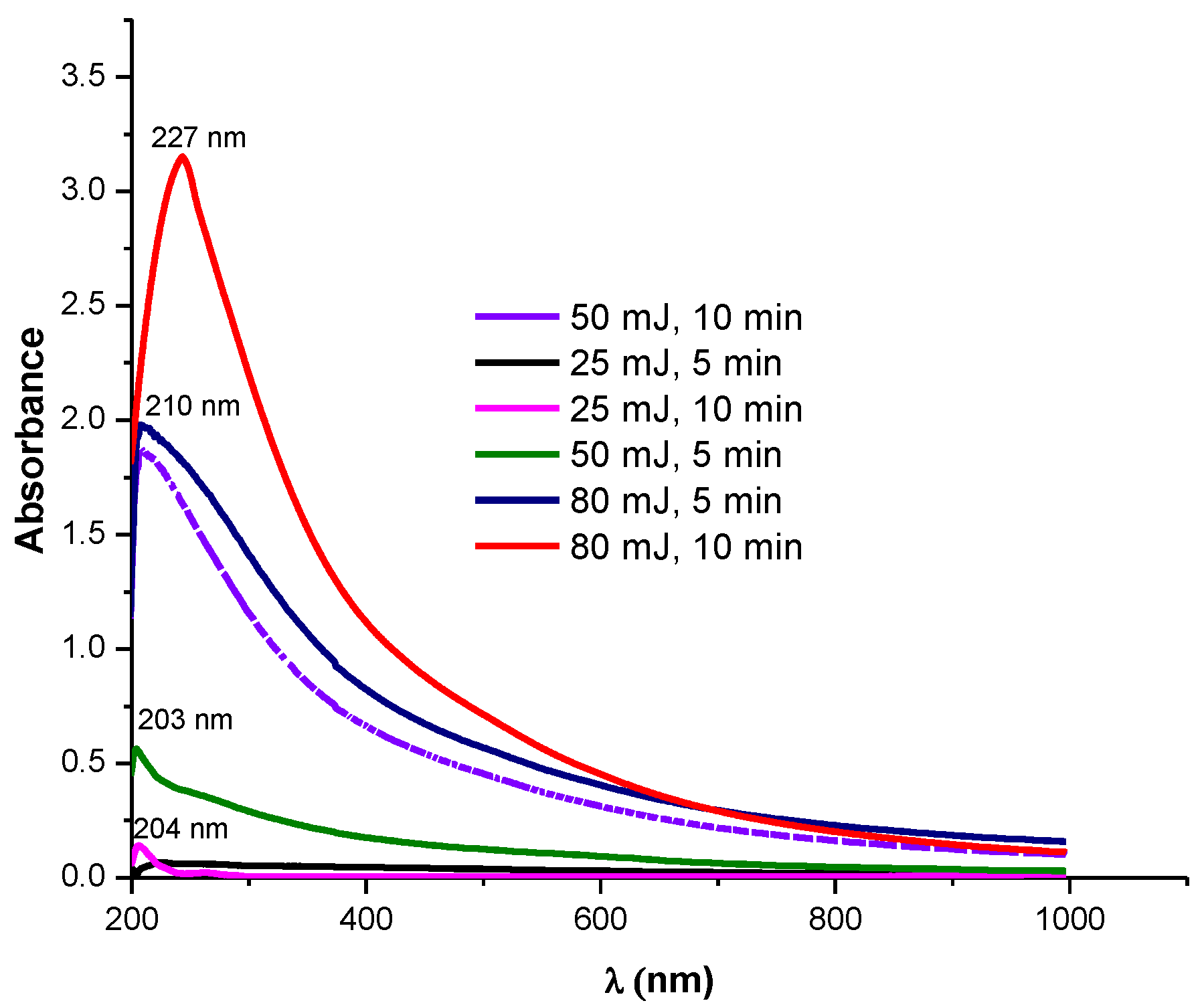
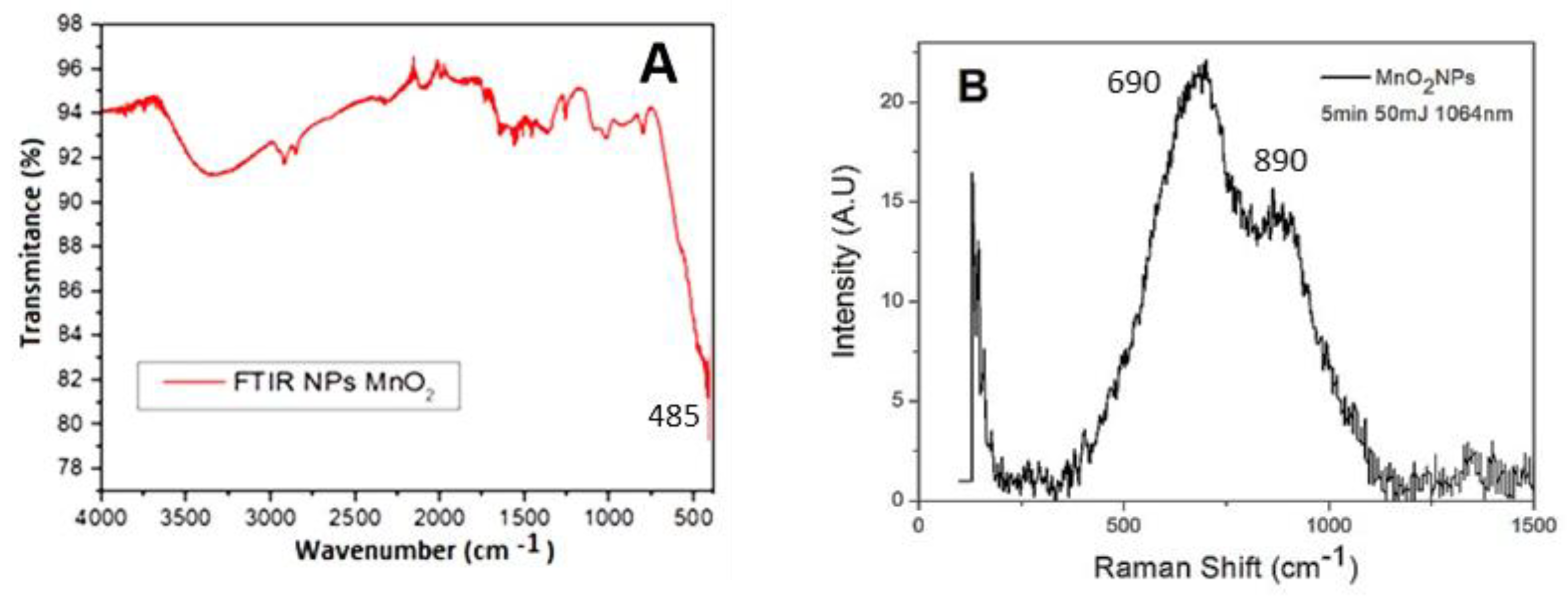
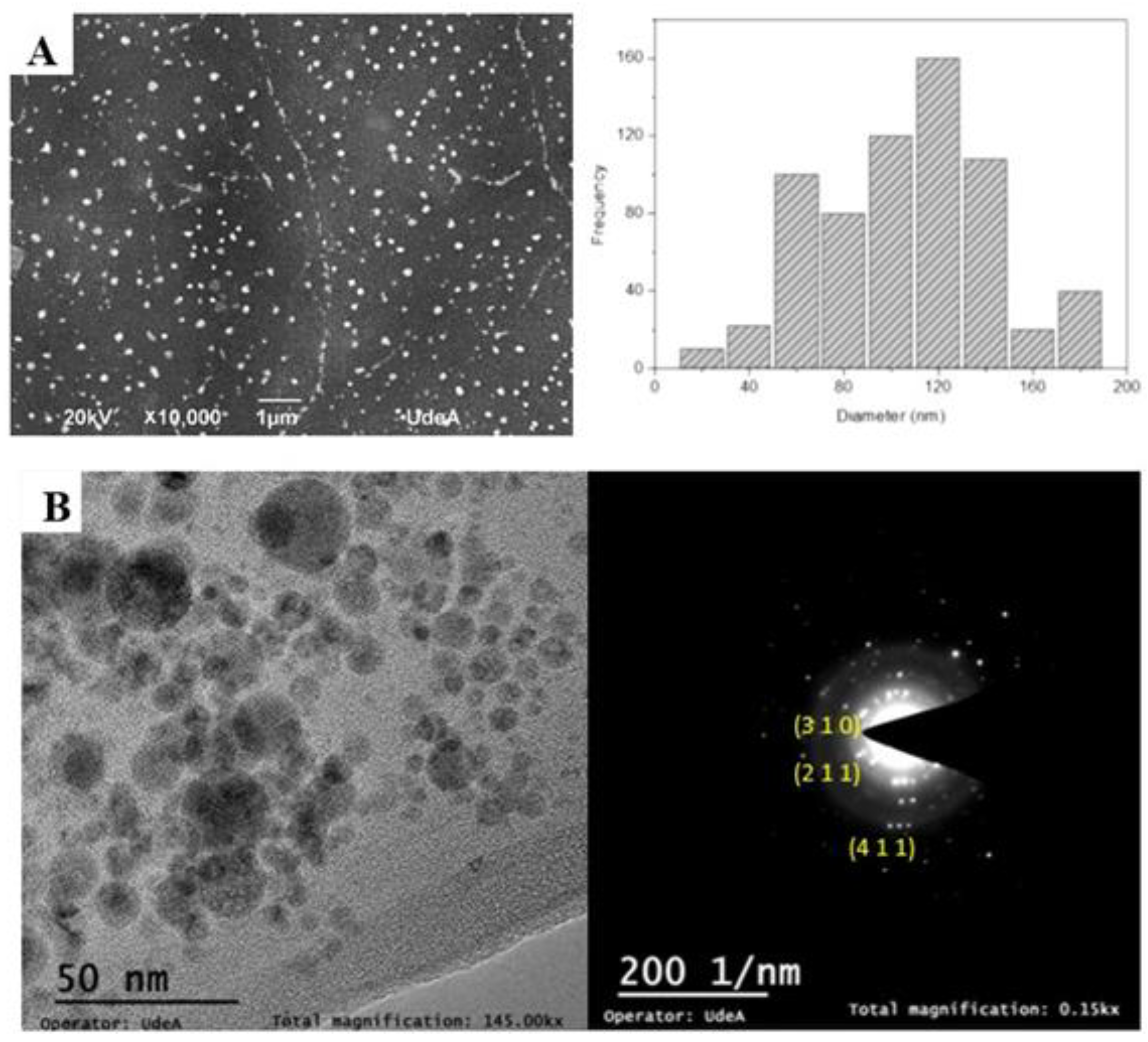
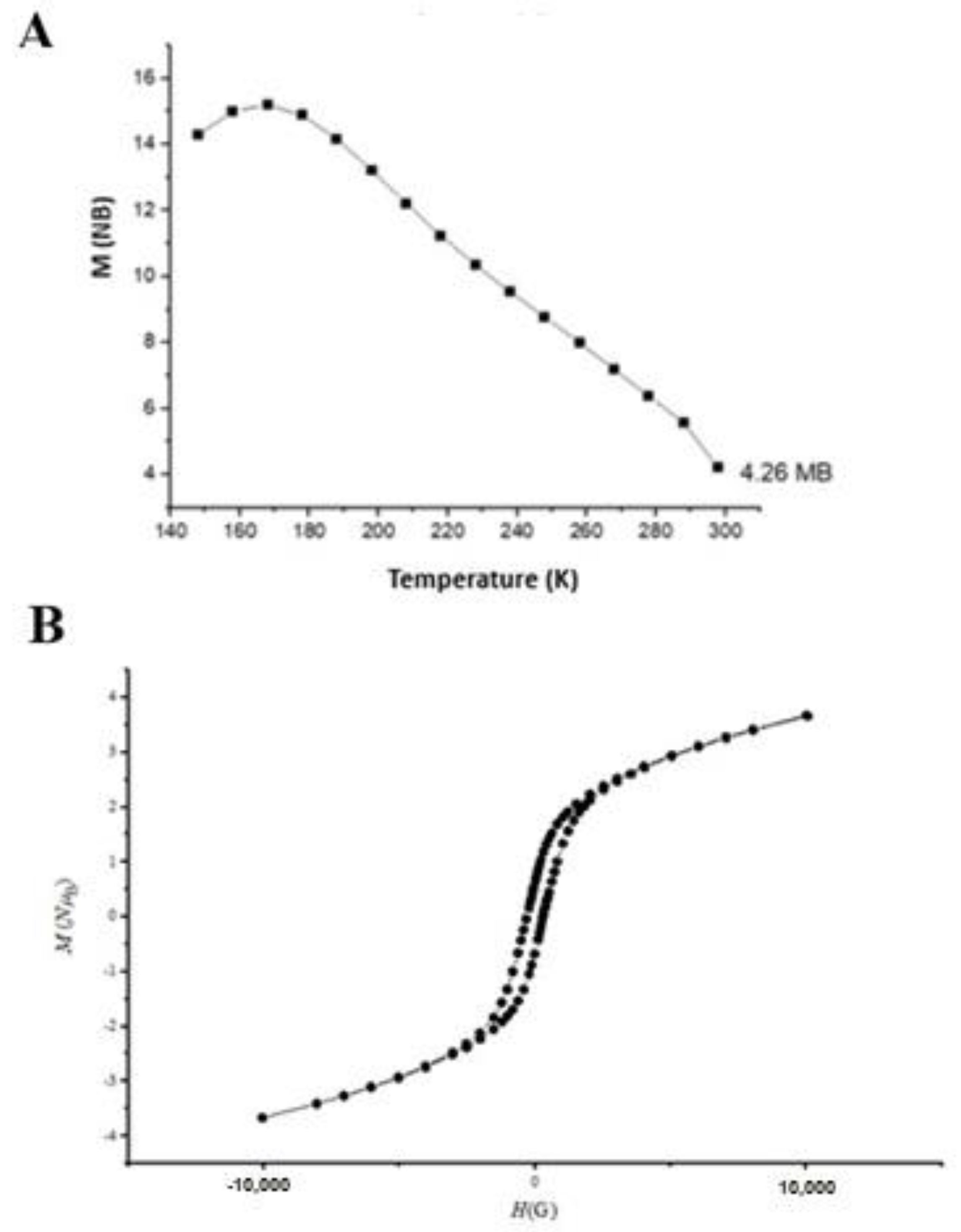
3.1. Characterization of Synthesized MnO2 Nanoparticles in Colloidal Suspension
3.2. Characterization of MnO2 NPs Deposited on FTO
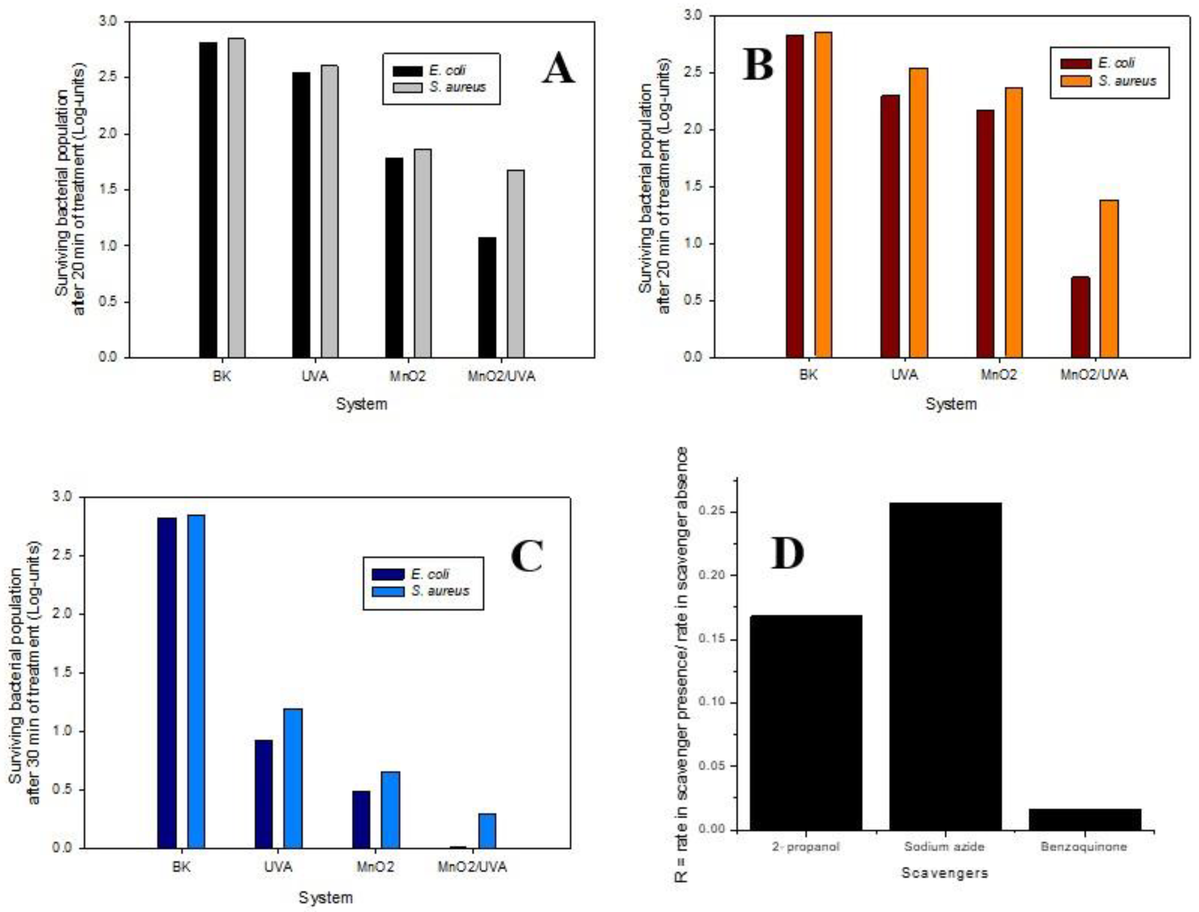
3.3. Photocatalytic Study on E. Coli and S. aureus
4. Conclusions
Supplementary Materials
Author Contributions
Funding
Data Availability Statement
Conflicts of Interest
References
- Vega, N.C.; Marin, O.; Tosi, E.; Grinblat, G.; Mosquera, E.; Moreno, M.S.; Tirado, M.; Comedi, D. The shell effect on the room temperature photoluminescence from ZnO/MgO core/shell nanowires: Exciton–phonon coupling and strain. Nanotechnology 2017, 28, 275702. [Google Scholar] [CrossRef] [Green Version]
- Marin, O.; Soliz, T.; Gutierrez, J.A.; Tirado, M.; Figueroa, C.; Comedi, D. Structural, optical and vibrational properties of ZnO:M (M=Al3+ and Sr2+) nano and micropowders grown by hydrothermal synthesis. J. Alloy. Compd. 2019, 789, 56–65. [Google Scholar] [CrossRef]
- Cruz, J.; Flórez, J.; Torres, R.; Urquiza, M.; Gutiérrez, J.A.; Guzmán, F.; Ortiz, C.C. Antimicrobial activity of a new synthetic peptide loaded in polylactic acid or poly(lactic-co-glycolic) acid nanoparticles againstPseudomonas aeruginosa, Escherichia coliO157:H7 and methicillin resistantStaphylococcus aureus(MRSA). Nanotechnology 2017, 28, 135102. [Google Scholar] [CrossRef]
- Voidazan, S.; Albu, S.; Toth, R.; Grigorescu, B.; Rachita, A.; Moldovan, I. Healthcare Associated Infections—A New Pathology in Medical Practice? Int. J. Environ. Res. Public Health 2020, 17, 760. [Google Scholar] [CrossRef] [PubMed] [Green Version]
- Davies, J.; Davies, D. Origins and Evolution of Antibiotic Resistance. Microbiol. Mol. Biol. Rev. 2010, 74, 417–433. [Google Scholar] [CrossRef] [PubMed] [Green Version]
- Khan, H.A.; Ahmad, A.; Mehboob, R. Nosocomial infections and their control strategies. Asian Pac. J. Trop. Biomed. 2015, 5, 509–514. [Google Scholar] [CrossRef] [Green Version]
- Carolina, C.; López, E.F.; John, V.; Jhon, G.; Yenny, Á.-T. Antimicrobial Surfaces of Metal Halides Immobilized on Polymeric Materials. J. Phys. Conf. Ser. 2020, 1541, 012004. [Google Scholar] [CrossRef]
- Chen, Y.; Fan, Z.; Zhang, Z.; Niu, W.; Li, C.; Yang, N.; Chen, B.; Zhang, H. Two-Dimensional Metal Nanomaterials: Synthesis, Properties, and Applications. Chem. Rev. 2018, 118, 6409–6455. [Google Scholar] [CrossRef] [PubMed]
- Petros, R.A.; DeSimone, J.M. Strategies in the design of nanoparticles for therapeutic applications. Nat. Rev. Drug Discov. 2010, 9, 615–627. [Google Scholar] [CrossRef]
- Kolahalam, L.A.; Viswanath, I.K.; Diwakar, B.S.; Govindh, B.; Reddy, V.; Murthy, Y. Review on nanomaterials: Synthesis and applications. Mater. Today Proc. 2019, 18, 2182–2190. [Google Scholar] [CrossRef]
- Valencia, I.; Ávila-Torres, Y.; Barba-Behrens, N.; Garzón, I.L. Circular dichroism and optical absorption spectra of mononuclear and trinuclear chiral Cu(II) amino-alcohol coordinated compounds: A combined theoretical and experimental study. J. Mol. Struct. 2015, 1085, 52–62. [Google Scholar] [CrossRef]
- Gutierrez, J.A.; Cruz, J.; Rondón, P.; Jones, N.; Ortiz, C. Small gold nanocomposites obtained in reverse micelles as nanoreactors. Effect of surfactant, optical properties and activity against Pseudomonas aeruginosa. New J. Chem. 2016, 40, 10432–10439. [Google Scholar] [CrossRef]
- Torres, R.; Serna, E.; Avila, Y. Nano-Materials as Photocatalysts for Degradation of Environmental Pollutants: Challenges and Possibilities. In Photochemical and Photocatalytical Degradation of Antibiotics in Water Promoted by Solar Irradiation; Elsevier: Amsterdand, The Netherlands, 2020; pp. 211–243. [Google Scholar] [CrossRef]
- Jelinkova, P.; Mazumdar, A.; Sur, V.P.; Kociova, S.; Dolezelikova, K.; Jimenez, A.M.J.; Koudelkova, Z.; Mishra, P.K.; Smerkova, K.; Heger, Z.; et al. Nanoparticle-drug conjugates treating bacterial infections. J. Control. Release 2019, 307, 166–185. [Google Scholar] [CrossRef] [PubMed]
- Bukhari, A.; Ijaz, I.; Gilani, E.; Nazir, A.; Zain, H.; Saeed, R.; Alarfaji, S.S.; Hussain, S.; Aftab, R.; Naseer, Y. Green Synthesis of Metal and Metal Oxide Nanoparticles Using Different Plants’ Parts for Antimicrobial Activity and Anticancer Activity: A Review Article. Coatings 2021, 11, 1374. [Google Scholar] [CrossRef]
- Qamar, H.; Saeed, A.; Owais, M.; Hussain, T.; Hussain, K.; Rahman, A.U.; Ahmed, S.; Kumar, S.; Khan, Z.A. CuO Bionanocomposite with Enhanced Stability and Antibacterial Activity against Extended-Spectrum Beta-Lactamase Strains. Materials 2021, 14, 6336. [Google Scholar] [CrossRef]
- Ameh, T.; Gibb, M.; Stevens, D.; Pradhan, S.H.; Braswell, E.; Sayes, C.M. Silver and Copper Nanoparticles Induce Oxidative Stress in Bacteria and Mammalian Cells. Nanomaterials 2022, 12, 2402. [Google Scholar] [CrossRef]
- Letchumanan, D.; Sok, S.; Ibrahim, S.; Nagoor, N.; Arshad, N. Plant-Based Biosynthesis of Copper/Copper Oxide Nanoparticles: An Update on Their Applications in Biomedicine, Mechanisms, and Toxicity. Biomolecules 2021, 11, 564. [Google Scholar] [CrossRef] [PubMed]
- Umoren, P.S.; Kavaz, D.; Nzila, A.; Sankaran, S.S.; Umoren, S.A. Biogenic Synthesis and Characterization of Chitosan-CuO Nanocomposite and Evaluation of Antibacterial Activity against Gram-Positive and -Negative Bacteria. Polymers 2022, 14, 1832. [Google Scholar] [CrossRef] [PubMed]
- Ul-Hamid, A.; Dafalla, H.; Hakeem, A.S.; Haider, A.; Ikram, M. In-Vitro Catalytic and Antibacterial Potential of Green Synthesized CuO Nanoparticles against Prevalent Multiple Drug Resistant Bovine Mastitogen Staphylococcus aureus. Int. J. Mol. Sci. 2022, 23, 2335. [Google Scholar] [CrossRef] [PubMed]
- Elkodous, M.A.; El-Sayyad, G.S.; Abdelrahman, I.Y.; El-Bastawisy, H.S.; Mohamed, A.E.; Mosallam, F.M.; Nasser, H.A.; Gobara, M.; Baraka, A.; Elsayed, M.A.; et al. Therapeutic and diagnostic potential of nanomaterials for enhanced biomedical applications. Colloids Surf. B Biointerfaces 2019, 180, 411–428. [Google Scholar] [CrossRef] [PubMed]
- Mitchell, M.J.; Billingsley, M.M.; Haley, R.M.; Wechsler, M.E.; Peppas, N.A.; Langer, R. Engineering precision nanoparticles for drug delivery. Nat. Rev. Drug Discov. 2020, 20, 101–124. [Google Scholar] [CrossRef]
- Feng, Y.; Liu, L.; Zhang, J.; Aslan, H.; Dong, M. Photoactive antimicrobial nanomaterials. J. Mater. Chem. B 2017, 5, 8631–8652. [Google Scholar] [CrossRef]
- Sivakumar, P.; Lee, M.; Kim, Y.-S.; Shim, M.S. Photo-triggered antibacterial and anticancer activities of zinc oxide nanoparticles. J. Mater. Chem. B 2018, 6, 4852–4871. [Google Scholar] [CrossRef] [PubMed]
- Fouda, A.; Eid, A.M.; Abdelkareem, A.; Said, H.A.; El-Belely, E.F.; Alkhalifah, D.H.M.; Alshallash, K.S.; Hassan, S.E.-D. Phyco-Synthesized Zinc Oxide Nanoparticles Using Marine Macroalgae, Ulva fasciata Delile, Characterization, Antibacterial Activity, Photocatalysis, and Tanning Wastewater Treatment. Catalysts 2022, 12, 756. [Google Scholar] [CrossRef]
- Bogdan, J.; Zarzyńska, J.; Pławińska-Czarnak, J. Comparison of Infectious Agents Susceptibility to Photocatalytic Effects of Nanosized Titanium and Zinc Oxides: A Practical Approach. Nanoscale Res. Lett. 2015, 10, 309. [Google Scholar] [CrossRef] [Green Version]
- Alzahrani, S.A.; Al-Thabaiti, S.A.; Al-Arjan, W.S.; Malik, M.A.; Khan, Z. Preparation of ultra long α-MnO 2 and Ag@MnO 2 nanoparticles by seedless approach and their photocatalytic performance. J. Mol. Struct. 2017, 1137, 495–505. [Google Scholar] [CrossRef]
- Ahn, M.-W.; Park, K.-S.; Heo, J.-H.; Park, J.-G.; Kim, D.-W.; Choi, K.J.; Lee, J.-H.; Hong, S.-H. Gas sensing properties of defect-controlled ZnO-nanowire gas sensor. Appl. Phys. Lett. 2008, 93, 263103. [Google Scholar] [CrossRef]
- Pottathara, Y.B.; Thomas, S.; Kalarikkal, N.; Grohens, Y.; Kokol, V. (Eds.) Nanomaterials Synthesis: Design, Fabrication and Applications; Elsevier: Amsterdam, The Netherlands, 2019. [Google Scholar] [CrossRef]
- Mergen, B.; Arda, E. Determination of Optical Band Gap Energies of CS/MWCNT Bio-nanocomposites by Tauc and ASF Methods. Synth. Met. 2020, 269, 116539. [Google Scholar] [CrossRef]
- Saunderson, A. A permanent magnet Gouy balance. Phys. Educ. 1968, 3, 272–273. [Google Scholar] [CrossRef]
- Wu, Y.; Liu, S.; Zhao, K.; He, Z.; Lv, K.; Ye, G. Synthesis of δ-MnO2 film on FTO glass with high electrochemical performance. Ionics 2015, 22, 637–647. [Google Scholar] [CrossRef]
- Banyamin, Z.Y.; Kelly, P.J.; West, G.; Boardman, J. Electrical and Optical Properties of Fluorine Doped Tin Oxide Thin Films Prepared by Magnetron Sputtering. Coatings 2014, 4, 732–746. [Google Scholar] [CrossRef] [Green Version]
- Moholkar, A.; Pawar, S.; Rajpure, K.; Almari, S.N.; Patil, P.; Bhosale, C. Solvent-dependent growth of sprayed FTO thin films with mat-like morphology. Sol. Energy Mater. Sol. Cells 2008, 92, 1439–1444. [Google Scholar] [CrossRef]
- Mahdieh, M.; Khosravi, A. Colloidal brass nanoparticles produced by pulsed laser ablation in deionized water and the effect of external electric field on particle size characteristics and ablation rate. Nano-Struct. Nano-Objects 2020, 24, 100580. [Google Scholar] [CrossRef]
- Jyothi, L.; Kuladeep, R.; Rao, D.N. Solvent effect on the synthesis of cobalt nanoparticles by pulsed laser ablation: Their linear and nonlinear optical properties. J. Nanophotonics 2015, 9, 93088. [Google Scholar] [CrossRef]
- Pandey, B.; Shahi, A.; Gopal, R. Synthesis, optical properties and growth mechanism of MnO nano structures. Appl. Surf. Sci. 2013, 283, 430–437. [Google Scholar] [CrossRef]
- Souri, M.; Hoseinpour, V.; Shakeri, A.; Ghaemi, N. Optimisation of green synthesis of MnO nanoparticles via utilising response surface methodology. IET Nanobiotechnol. 2018, 12, 822–827. [Google Scholar] [CrossRef]
- Gupta, S.K.; Kadam, R.; Gupta, R.; Sahu, M.; Natarajan, V. Evidence for the stabilization of manganese ion as Mn (II) and Mn (IV) in α-Zn2P2O7: Probed by EPR, luminescence and electrochemical studies. Mater. Chem. Phys. 2014, 145, 162–167. [Google Scholar] [CrossRef]
- Mozaffari, H.; Mahdieh, M.H. Enhancement of ablation rate and production of colloidal nanoparticles by irradiation of metals with nanosecond pulsed laser in presence of external electric field. Phys. Lett. A 2019, 383, 646–654. [Google Scholar] [CrossRef]
- Snaith, H.J.; Schmidt-Mende, L. Advances in Liquid-Electrolyte and Solid-State Dye-Sensitized Solar Cells. Adv. Mater. 2007, 19, 3187–3200. [Google Scholar] [CrossRef]
- Sannasi, V.; Subbian, K. Influence of Moringa oleifera gum on two polymorphs synthesis of MnO2 and evaluation of the pseudo-capacitance activity. J. Mater. Sci. Mater. Electron. 2020, 31, 17120–17132. [Google Scholar] [CrossRef]
- Selvam, M.; Srither, S.R.; Saminathan, R.K.; Rajendran, V. Chemically and electrochemically prepared graphene/MnO2 nanocomposite electrodes for zinc primary cells: A comparative study. Ionics 2014, 21, 791–799. [Google Scholar] [CrossRef]
- Zhou, C.; Wang, J.; Liu, X.; Chen, F.; Di, Y.; Gao, S.; Shi, Q. Magnetic and thermodynamic properties of α, β, γ and δ-MnO2. New J. Chem. 2018, 42, 8400–8407. [Google Scholar] [CrossRef]
- Shaaban, E.; Yahia, I.; El-Metwally, E. Validity of Swanepoel’s Method for Calculating the Optical Constants of Thick Films. Acta Phys. Pol. A 2012, 121, 628–635. [Google Scholar] [CrossRef]
- Zhang, L.; McMillon, L.; McNatt, J. Gas-dependent bandgap and electrical conductivity of Cu2O thin films. Sol. Energy Mater. Sol. Cells 2013, 108, 230–234. [Google Scholar] [CrossRef]
- Zheng, X.; Yu, L.; Lan, B.; Cheng, G.; Lin, T.; He, B.; Ye, W.; Sun, M.; Ye, F. Three-dimensional radial α-MnO 2 synthesized from different redox potential for bifunctional oxygen electrocatalytic activities. J. Power Sources 2017, 362, 332–341. [Google Scholar] [CrossRef]
- Toupin, M.; Brousse, T.; Bélanger, D. Charge Storage Mechanism of MnO2 Electrode Used in Aqueous Electrochemical Capacitor. Chem. Mater. 2004, 16, 3184–3190. [Google Scholar] [CrossRef]
- Ruiz, C.A.C.; Bélanger, D.; Rochefort, D. Electrochemical and Spectroelectrochemical Evidence of Redox Transitions Involving Protons in Thin MnO2 Electrodes in Protic Ionic Liquids. J. Phys. Chem. C 2013, 117, 20397–20405. [Google Scholar] [CrossRef]
- Julien, C.M.; Mauger, A. Nanostructured MnO2 as Electrode Materials for Energy Storage. Nanomaterials 2017, 7, 396. [Google Scholar] [CrossRef] [PubMed]
- Zhang, W.; Li, H.; Hopmann, E.; Elezzabi, A.Y. Nanostructured inorganic electrochromic materials for light applications. Nanophotonics 2020, 10, 825–850. [Google Scholar] [CrossRef]
- Ma, D.; Eh, A.L.-S.; Cao, S.; Lee, P.S.; Wang, J. Wide-Spectrum Modulated Electrochromic Smart Windows Based on MnO2/PB Films. ACS Appl. Mater. Interfaces 2021, 14, 1443–1451. [Google Scholar] [CrossRef] [PubMed]
- Sakai, N.; Ebina, Y.; Takada, K.; Sasaki, T. Electrochromic Films Composed of MnO. Nanosheets with Controlled Optical Density and High Coloration Efficiency. J. Electrochem. Soc. 2005, 152, E384–E389. [Google Scholar] [CrossRef]
- Xu, T.; Walter, E.C.; Agrawal, A.; Bohn, C.; Velmurugan, J.; Zhu, W.; Lezec, H.J.; Talin, A.A. High-contrast and fast electrochromic switching enabled by plasmonics. Nat. Commun. 2016, 7, 10479. [Google Scholar] [CrossRef] [Green Version]
- Giannakis, S.; López, M.I.P.; Spuhler, D.; Pérez, J.A.S.; Ibáñez, P.F.; Pulgarin, C. Solar disinfection is an augmentable, in situ -generated photo-Fenton reaction—Part 1: A review of the mechanisms and the fundamental aspects of the process. Appl. Catal. B Environ. 2016, 199, 199–223. [Google Scholar] [CrossRef]
- Du, T.; Chen, S.; Zhang, J.; Li, T.; Li, P.; Liu, J.; Du, X.; Wang, S. Antibacterial Activity of Manganese Dioxide Nanosheets by ROS-Mediated Pathways and Destroying Membrane Integrity. Nanomaterials 2020, 10, 1545. [Google Scholar] [CrossRef]
- Chiam, S.-L.; Pung, S.-Y.; Yeoh, F.-Y. Recent developments in MnO2-based photocatalysts for organic dye removal: A review. Environ. Sci. Pollut. Res. 2020, 27, 5759–5778. [Google Scholar] [CrossRef] [PubMed]
- Microbiology Info.com (Web site). Differences between Gram Positive and Gram Negative Bacteria. 15 August 2019. Available online: https://microbiologyinfo.com/differences-between-gram-positive-and-gram-negative-bacteria/ (accessed on 7 May 2022).
- Ortuño-Sahagún, D.; Pallàs, M.; Rojas-Mayorquín, A.E. Oxidative Stress in Aging: Advances in Proteomic Approaches. Oxidative Med. Cell. Longev. 2014, 2014, 573208. [Google Scholar] [CrossRef] [PubMed] [Green Version]
- Wang, F.; Feng, Y.; Chen, P.; Wang, Y.; Su, Y.; Zhang, Q.; Zeng, Y.; Xie, Z.; Liu, H.; Liu, Y.; et al. Photocatalytic degradation of fluoroquinolone antibiotics using ordered mesoporous g-C3N4 under simulated sunlight irradiation: Kinetics, mechanism, and antibacterial activity elimination. Appl. Catal. B Environ. 2018, 227, 114–122. [Google Scholar] [CrossRef]


| Laser Energy (mJ) | Ablation Time (min) | Bandgap (eV) |
|---|---|---|
| 25 | 5 | 5.75 |
| 10 | 5.82 | |
| 50 | 5 | 5.61 |
| 10 | 4.23 | |
| 80 | 5 | 4.00 |
| 10 | 3.84 |
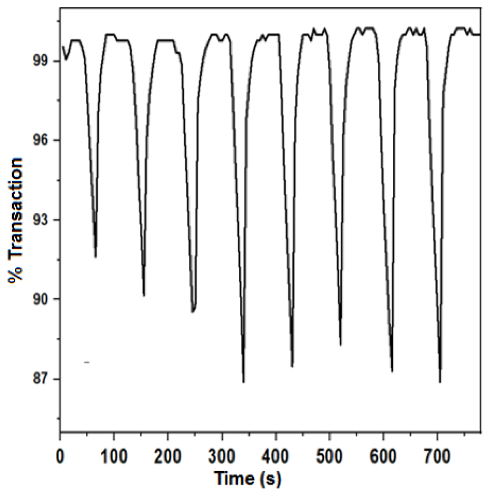
| Coloring Time (tc, in s) | Bleaching Time (tb, in s) | Contrast (ΔT, in%) | Optical Density (ΔOD) | Efficiency (η, in cm2 C−1) |
|---|---|---|---|---|
| 28.2 ± 1.9 | 22.9 ± 3.1 | 12.35 ± 0.61 | 0.059 ± 0.003 | 36.66 ± 0.40 |
Publisher’s Note: MDPI stays neutral with regard to jurisdictional claims in published maps and institutional affiliations. |
© 2022 by the authors. Licensee MDPI, Basel, Switzerland. This article is an open access article distributed under the terms and conditions of the Creative Commons Attribution (CC BY) license (https://creativecommons.org/licenses/by/4.0/).
Share and Cite
Corrales, J.; Acosta, J.; Castro, S.; Riascos, H.; Serna-Galvis, E.; Torres-Palma, R.A.; Ávila-Torres, Y. Manganese Dioxide Nanoparticles Prepared by Laser Ablation as Materials with Interesting Electronic, Electrochemical, and Disinfecting Properties in Both Colloidal Suspensions and Deposited on Fluorine-Doped Tin Oxide. Nanomaterials 2022, 12, 4061. https://doi.org/10.3390/nano12224061
Corrales J, Acosta J, Castro S, Riascos H, Serna-Galvis E, Torres-Palma RA, Ávila-Torres Y. Manganese Dioxide Nanoparticles Prepared by Laser Ablation as Materials with Interesting Electronic, Electrochemical, and Disinfecting Properties in Both Colloidal Suspensions and Deposited on Fluorine-Doped Tin Oxide. Nanomaterials. 2022; 12(22):4061. https://doi.org/10.3390/nano12224061
Chicago/Turabian StyleCorrales, Jhonatan, Jorge Acosta, Sandra Castro, Henry Riascos, Efraim Serna-Galvis, Ricardo A. Torres-Palma, and Yenny Ávila-Torres. 2022. "Manganese Dioxide Nanoparticles Prepared by Laser Ablation as Materials with Interesting Electronic, Electrochemical, and Disinfecting Properties in Both Colloidal Suspensions and Deposited on Fluorine-Doped Tin Oxide" Nanomaterials 12, no. 22: 4061. https://doi.org/10.3390/nano12224061