Article

Efficient Oxygen Vacancy Defect Engineering for Enhancing Visible-Light Photocatalytic Performance over SnO2−x Ultrafine Nanocrystals

1
Henan Province International Joint Laboratory of Materials for Solar Energy Conversion and Lithium Sodium Based Battery, Luoyang Institute of Science and Technology, Luoyang 471023, China
2
School of Materials Science and Engineering, Guilin University of Technology, Guilin 541004, China
3
Henan Province Engineering Research Center of Industrial Intelligent Vision, Luoyang Institute of Science and Technology, Luoyang 471023, China
4
Department of Nano Convergence Engineering, Jeonbuk National University, Jeonju 54896, Korea
*
Authors to whom correspondence should be addressed.
Nanomaterials 2022, 12(19), 3342; https://doi.org/10.3390/nano12193342
Submission received: 25 August 2022 / Revised: 16 September 2022 / Accepted: 22 September 2022 / Published: 25 September 2022
(This article belongs to the Special Issue Hydrothermal Synthesis and Application of Nanomaterials)

Abstract

:
Regardless of its good electron-transfer ability and chemical stability, pure Zn2SnO4 (ZSO) still has intrinsic deficiencies of a narrow spectral response region, poor absorption ability, and high photo-activated carrier recombination rate. Aiming to overcome the deficiencies above-mentioned, we designed a facile hydrothermal route for etching ZSO nanoparticles in a dilute acetic acid solution, through which efficient oxygen vacancy defect engineering was accomplished and SnO2−x nanocrystals were obtained with an ultrafine particle size. In comparison with the untreated ZSO nanoparticles, the specific surface area of SnO2−x nanocrystals was substantially enlarged, subsequently leading to the notable augmentation of active sites for the photo-degradation reaction. Aside from the above, it is worth noting that SnO2−x nanocrystals were endowed with a broad spectral response, enhancing light absorption capacity and the photo-activated carrier transfer rate with the aid of oxygen vacancy defect engineering. Accordingly, SnO2−x nanocrystals exhibited significantly enhanced photoactivity toward the degradation of the organic dye rhodamine B (RhB), which could be imputed to the synergistic effect of increasing active sites, intensified visible-light harvesting, and the separation rate of the photo-activated charge carrier caused by the oxygen vacancy defect engineering. In addition, these findings will inspire us to open up a novel pathway to design and prepare oxide compound photocatalysts modified by oxygen vacancy defects in pursuing excellent visible-light photoactivity.

1. Introduction

In the 21st century, human society has been entering into a new stage of rapid development, and the industrialization process has increasingly intensified, simultaneously accompanied by the two major serious problems of environment pollution and energy source shortage. Thus, how to reasonably address the two problems above-mentioned has become the focus of global concern. As is well-known, water pollution is one of the most important types of environment pollution, which originates from industrial discharge into the wastewater, containing pesticides, polychlorinated biphenyls, chlorophenols, dyes, and other organic pollutants [1,2,3,4,5,6]. Among these organic pollutants, dyes are considered as serious contributors to water pollution, because almost all textile industries excessively use different kinds of dyes, resulting in an excess of dyes being discharged into industrial waste water. Some of the dyes are hard to naturally decompose in water as their composite molecular structures can enable them to be more stable and resistant to biodegradation [7]. Therefore, it is extremely urgent to explore novel efficient methods to remove hazardous dyes.
In contaminated water without secondary pollution, among the physical, chemical, biological and acoustical methods of dye removal, advanced oxidation processes (AOPs) are reckoned as potentially effective strategies to remediate water pollution due to its main merits of easy control, ecological safety, and sufficient decomposition to different dyes. More interestingly, AOPs can also produce highly reactive peroxy and hydroxyl radicals to dominate the oxidation reaction of composite organic molecules [8,9,10,11,12,13,14,15]. Nowadays, the photodegradation process of dyes has become one of the most widely investigated AOPs, as it can covert solar energy into steady chemical energy and generate reactive species to decompose composite molecular structured substances [16,17,18]. Although the photodegradation of dyes has made great progress since the first discovery of photocatalytic hydrogen evolution over the TiO2 electrode [19], the photocatalytic efficiency toward the photodegradation of dyes is still limited to a narrow spectral response region, low solar-energy conversion efficiency, poor absorption ability, and high photo-activated carrier recombination rate of wide band-gap photo-catalysts. Therefore, developing novel catalysts with higher solar-energy conversion and photo-degradation efficiency to address the remediation of water pollution is the primary task for scientific researchers worldwide.
Oxygen defect engineering is a reliable and efficient approach to modulate the electronic structure of wide band-gap catalysts, resulting in narrowing the band-gap, enhancing the conductivity, and extending the light absorption range. Simultaneously, surface oxygen defects exert a critical influence on the efficient transfer and separation of photo-activated charge carriers [20,21,22,23]. Nowadays, oxygen defect engineering has been successfully utilized to modify TiO2, ZnO, SnO2, Bi2WO6, and other oxide compounds for the improvement of photodegradation efficiency [22,23,24,25,26,27,28,29,30,31,32,33,34,35,36,37,38]. For example, Feng et al. synthesized partially reduced TiO2 by using a facile one-step combustion method, and the reduced TiO2 exhibited improved hydrogen photocatalytic evolution under visible-light irradiation [24]. Li et al. previously designed and prepared a ternary structure of C@TiO2-TiO2−x via a solvothermal route with enhanced photocatalytic performance [25]. With respect to ZnO, Xu et al. introduced a surface oxygen defect to narrow the band gap to achieve excellent solar-driven photodegradation toward tetracycline with long-term stability [30]. Moreover, Wang and his coauthors prepared ZnO with oxygen vacancy defects by annealing ZnO2 under different atmospheres, and their results showed that oxygen vacancy defects in crystals facilitated the photo-decomposition of 2,4- dichlorophenol in the presence of visible light illumination [31]. As for SnO2−x, some recent research work has also focused on the modification of oxygen vacancy. Dong and his coauthors adopted a molten-aluminum reduction route to achieve SnO2−x nanoparticles with rich oxygen vacancy, and the SnO2−x nanoparticles exhibited significantly improved visible-light absorption capacity and electrical property [32]. Anuchai et al. obtained oxygen-deficient SnO2 nanocrystals at a relatively low temperature, and confirmed the obtained samples with enhanced photodegradation performance [33]. Xu et al. designed a synproportionation reaction to achieve Sn2+ self-doped SnO2−x with the improvement in the decomposition of methyl orange [34]. Chen et al. prepared SnO2−x/In2O3−y composites with different vacuum levels, and investigated the relationship between the vacuum levels and the formation of oxygen vacancy [35]. All of the above findings on the SnO2 system with oxygen vacancy defects are impressive and inspiring; however, the present synthetic processes of SnO2−x are still subject to multiple complicated steps, harsh conditions, and difficult control. Therefore, it still remains a great challenge to develop a facile approach for the large-scale synthesis of SnO2−x ultrafine nanocrystals with oxygen vacancy defects for the significant improvement of photodegradation toward dyes.
As is well-known, pure Zn2SnO4 (ZSO), with a relatively wide bandgap approximate to SnO2, has good electron-transfer ability and chemical stability [39]. However, pure ZSO is still restricted by its intrinsic deficiencies of narrow spectral response region, poor absorption ability, and high photo-activated carrier recombination rate. Up to now, few studies have previously investigated the evolution of SnO2−x ultrafine nanocrystals with oxygen vacancy defects from ZSO nanoparticles. Herein, we innovatively conceived a novel two-step hydrothermal route to achieve oxygen vacancy SnO2−x nanocrystals. First, ZSO nanoparticles were hydrothermally obtained using SnCl4·5H2O and Zn(CH3COO)2·2H2O as starting materials. Second, efficient oxygen vacancy defect engineering was launched to achieve SnO2−x nanocrystals by hydrothermally etching ZSO nanoparticles in a dilute acetic acid solution. In order to evaluate the effect of oxygen vacancies on the photoactivity, we carried out the photodegradation of rhodamine B (RhB) over different photocatalysts under visible-light irradiation. The results showed that SnO2−x ultrafine nanocrystals with oxygen vacancy defects exhibited a higher photodecomposition efficiency compared to the pure ZSO nanoparticles. To our knowledge, this investigation is, for the first time, concerned with the preparation and photodecomposition behavior of SnO2−x ultrafine nanocrystals originating from a hydrothermal etching process.

2. Materials and Methods

2.1. Materials

All of the chemicals used in this investigation including tin(IV) chloride penthydrate (SnCl4·5H2O), zinc acetate dehydrate (Zn(CH3COO)2·2H2O), sodium hydroxide (NaOH), and rhodamine B (RhB) were of analytical grade and purchased from Sinopharm Chemical Reagent Co. Ltd., Shanghai, China.

2.2. Synthesis of SnO2−x Ultrafine Nanocrystals

According to our previous studies [40,41,42], first, ZSO nanoparticles were prepared through a facile hydrothermal route. The obtained white product was collected through centrifuging, washed with deionized water and ethanol, and then dried at 60 °C for 12 h.
Second, 0.5 g of ZSO nanoparticles was dissolved into a mixed solvent containing 72 mL deionized water and 8 mL acetic acid (HAC) under constant stirring. After being stirred for 30 min, the resulting suspension was poured into a 100 mL Teflon-lined autoclave. Then, the autoclave was heated to different temperatures and kept for 15 h. After naturally being cooled down to room temperature, the resulting precipitates were treated by centrifuging, washing, and drying. By varying different temperature (65 °C, 85 °C, 105 °C, 125 °C), a series of SnO2−x ultrafine nanocrystals were obtained and respectively named as SnO2−x-65T, SnO2−x-85T, SnO2−x-105T, and SnO2−x-125T for convenience. For comparison, SnO2 nanoparticles were also synthesized via a hydrothermal route. The details for the synthetic process are described as follows. According to a previous study [43], a certain amount of SnCl4·5H2O and NaOH was dissolved into a mixed solvent containing 30 mL deionized water and 10 mL ethanol, respectively, then NaOH was added into the solution of SnCl4 until the pH reached 7. Then, the mixture was kept in a 100 mL Teflon-lined autoclave at 180 °C for 20 h. The final product was collected by centrifuging, washing, and drying.

2.3. Characterization

The X-ray diffraction patterns (XRD) of ZSO and SnO2−x samples were obtained on an X-ray diffractometer (D8 Advance, Bruker, Billerica, MA, USA) with Cu-Kα irradiation (1.5406Å). The morphological observation of the as-prepared catalysts was accomplished using a transmission electron microscope (JEM-2100F, JEOL Ltd., Tokyo, Japan). To explore the surface composition and chemical status, X-ray photoelectron spectroscopy (XPS) measurements were achieved on an X-ray photoelectron spectrometer (XPS, ESCALAB 250Xi, Thermo Fisher Scientific cooperation), taking C1s (284.6 eV) as the criterion to calibrate the binding energies of other elements. Through the virtue of the nitrogen adsorption and desorption method, the specific surface area measurement was realized on a Tristar II 3020 sorption analyzer (Micromeritics, Norcross, GA, USA). Taking BaSO4 as the reflectance standard reference, UV–Vis diffuse reflectance spectra (DRS) of ZSO and SnO2−x catalysts were recorded on a UV–Vis spectrophotometer (TU 1901, Puxi, Beijing, China). A LS55 fluorescence spectrometer (LS55, PE cooperation) was employed to measure the photoluminescence (PL) spectra of the ZSO and SnO2−x samples with an exciting wavelength of 320 nm.

2.4. Photoelectrochemical Measurements

The electrochemical property was measured on a CHI660E electrochemical workstation comprising a standard three-electrode system, in which the working electrode, counter electrode, and reference electrode were made of as-obtained catalysts, Pt wire, and saturated Ag/AgCl, respectively. The detailed procedure on the electrochemical measurements was demonstrated in our previously reported study [44,45].

2.5. Photocatalytic Experiments

As reported in our previous research work [46], RhB photodecomposition experiments were carried out in a home-made device. In each run, we took RhB aqueous solutions (60 mL, 1.0 × 10⁻⁵ mol L−1) combined with 100 mg of catalysts. After being irradiated for a certain time, the absorbance intensities at the characteristic peak (554 nm) were recorded on a UV–Vis spectrophotometer to reflect the concentration variation of the reacted solution.

3. Results

3.1. Formation Mechanism of SnO2−x Nanocrystals with Oxygen Vacancy

ZSO exhibits excellent chemical stability whether in an acid media or alkaline media under room temperature conditions. However, ZSO nanostructures are capable of being hydrothermally etched in alkaline media, which was verified in a previous study [46]. Similar to that of ZSO in alkaline media, the crystal structure of ZSO is likely to become unstable when undergoing a hydrothermal process in acid media. Thereby, a possible formation mechanism is proposed for the SnO2−x nanocrystals as follows. With an increase in the hydrothermal temperature and pressure, the solubility of ZSO is progressively enhanced according to the thermodynamics under the hydrothermal condition. Simultaneously, a large number of CH3COO groups and hydrogen protons (H+) could be generated in the solution through acetic acid ionization, and the transfer rate of the hydrogen protons and CH3COO groups was significantly accelerated with an increase in the temperature upon the effect of thermal convection. When subjected to a certain concentration solution of HAC, Zn atoms located at crystal lattice of ZSO were capable of being attacked by groups of CH3COO from the solution, as depicted in Figure 1a. In the hydrothermal condition, Zn2+ can coordinate with CH3COO to generate a soluble component of Zn(CH3COO)2, which would subsequently enter into the solution through the solid–liquid interface between ZSO and HAC. At the same time, O atoms located at the crystal lattice of ZSO are also capable of being attacked by hydrogen protons in a weak acid hydrothermal environment, resulting in the formation of H2O. Thereafter, the crystal structure of ZSO is about to gradually dissociate and form a novel crystal structure under the constant etching of CH3COO groups and hydrogen protons. However, hydrogen protons are more active and provided with more rapid transfer rate in the hydrothermal etching process compared with the CH3COO groups. The factors above-mentioned could lead to the fact that the ratio of O atoms to Zn atoms stripped from the crystal lattice of ZSO is larger than 1:1. Thus, Sn atoms with partial O atoms remain therein, although the integrity of the ZSO crystal structure is destroyed. After experiencing the dissolution–recrystallization and in-situ growth process, non-stoichiometry SnO2−x is about to form in a subsequent step, as depicted in Figure 1b.

3.2. Surface Composition and Defect Analysis

X-ray diffraction patterns were recorded to examine the variation of the crystal phase before and after undergoing an acid-assisted hydrothermal process. The XRD results are presented in Figure 2. For comparison, we also prepared pure SnO2 via a simple hydrothermal route, and its XRD pattern is presented in Figure 2. As clearly seen from Figure 2a, all diffraction peaks for the ZSO nanoparticles were relatively sharp and narrow, and they perfectly coincided with those of the hexagonal phase Zn2SnO4 (JCPDS No. 74-2184). After undergoing an acid-assisted hydrothermal process, obvious changes occurred in the diffraction patterns of the measured samples. The XRD diffraction patterns of SnO2−x-105T were almost in agreement with those of the tetragonal phase SnO2 (JCPDS No. 41-1445). Compared with those of ZSO and SnO2, the diffraction peaks became broader and weaker, suggesting that SnO2−x-105T might have a much smaller size, originating from the special acid-assisted hydrothermal process. Moreover, as is clearly seen from the (110) crystal plane in Figure 2b, the diffraction peak of SnO2−x-105T exhibited a slight shift in comparison with that of pure SnO2.
XPS measurements were executed to probe the surface composition and gain insights into the oxygen vacancy of the SnO2−x-105T sample. Figure 3 presents the high resolution XPS spectra of the Sn 3d and O1s of ZSO and SnO2−x-105T samples. It is very clear from Figure 3 that the SnO2−x-105T sample only consisted of Sn and O elements, coinciding with the results of the XRD measurements. Specifically, from Figure 3a, the Sn 3d orbital peaks for ZSO displayed two binding energies positioned at around 495.8 eV and 487.4 eV, respectively, corresponding to the Sn 3d5/2 and Sn 3d3/2 orbits. Obviously, Sn 3d5/2 and Sn 3d3/2 peaks of SnO2−x-105T (positioning at around 495.3 eV and 486.8 eV) exhibited a notable red-shift toward low energy direction compared with those of ZSO. According to the previously reported study [47], the binding energy (BE) of Sn 3d5/2 orbits for Sn2+ was in the region of 486.4 eV to 486.5 eV, while this value for Sn4+ ranged from 487.2 eV to 487.5eV. Thereby, it could reasonably be inferred that Sn4+, accompanied by Sn2+ ions, coexisted in the SnO2−x-105T sample because the BE of the Sn 3d peaks for SnO2−x-105T was slightly larger than those of Sn2+, but was a bit smaller than those of Sn4+ simultaneously. For this purpose, the Sn 3d3/2 spectrum could be deconvolved into two peaks centering at 495.8 eV and 495 eV, individually belonging to the Sn4+ and Sn2+ ions. As for the Sn 3d5/2 spectrum, it can be analogously split into two characteristic peaks positioned at around 487.4 eV and 486.6 eV. Furthermore, the O 1s spectra were deeply examined to disclose the oxygen vacancy defect. As can clearly be seen in Figure 3b, the O 1s spectrum of ZSO was obviously different to that of SnO2−x-105T. Referring to previous work, the O1s orbital peak for SnO2−x-105T could be well-fitted with three characteristic peaks of the crystal lattice oxygen (OCL), oxygen vacancy (OV), and absorbed hydroxyl oxygen (OAH), individually located at around 530.5 eV, 531.1 eV, and 532.3 eV. As for ZSO, the O1s spectrum could be perfectly divided into the OCL peak (530.5 eV) and OAH peak (532.3 eV). The aforementioned result could provide strong proof to verify the existence of oxygen vacancy in SnO2−x-105T. In our work, it is rational to speculate that oxygen vacancy might originate from excess oxygen atoms stripped by hydrogen protons to form H2O, which leads to the variation in the electron density around Sn–O bond. Hence, the bonds of Sn4+–O–Sn4+ are partially substituted by the bonds of Sn2+–O–Sn4+ in order to compensate the oxygen vacancy to make the whole maintain charge neutrality. Based on the above demonstration, the XPS results are in good agreement with the formation mechanism of non-stoichiometry SnO2−x nanocrystals.

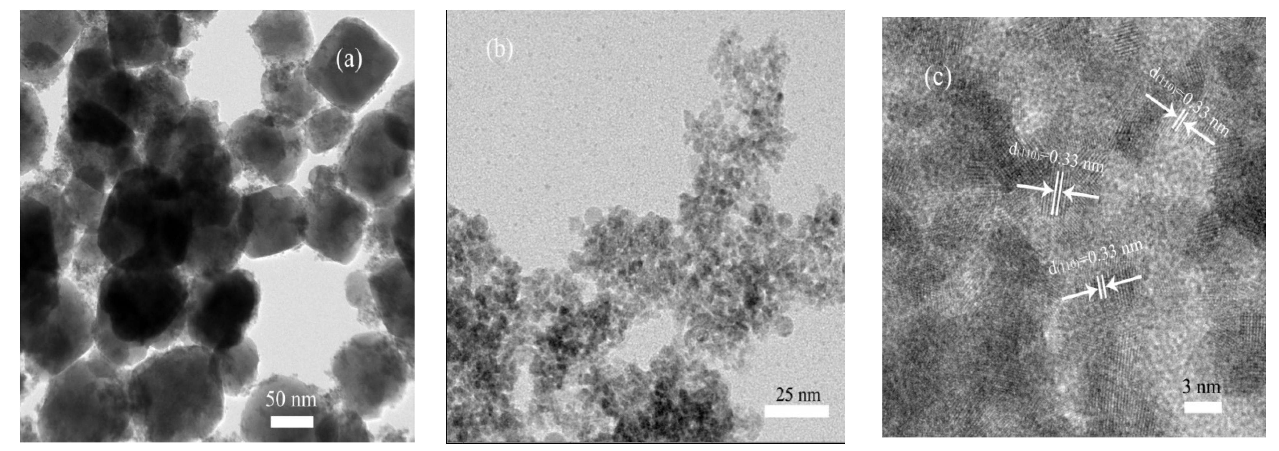
3.3. Morphology and Surface Characteristics

Figure 4 presents the TEM and HRTEM images of the ZSO and SnO2−x-105T samples. Through contrastive analysis from Figure 4a,b, an obvious difference was observed from the microstructure of the ZSO and SnO2−x-105T samples. Specifically, the ZSO sample was composed of numerous nanoparticles with relatively larger size, which was in good agreement with our previously reported study [40,41]. After undergoing the acid-assisted hydrothermal synthesis process, SnO2−x-105T exhibited a specially disordered mesostructure, and the crystal sizes significantly decreased, ranging from 5 nm to 7 nm. Such a special mesostructure of SnO2−x can be attributed to the dissolution–recrystallization and in situ growth process. From Figure 4c, it is quite evident that the lattice fringes with a d-spacing of 0.33 nm corresponded to the (110) crystallographic planes of tin oxide. This result also revealed the good crystalline characteristic of ultra-fine SnO2−x particles.
The decrease in the nanocrystal size could enlarge the surface area of the SnO2−x-105T sample, which was mostly likely to intensify the absorption capacity and provide more active sites for the photodegradation reaction. In order to accurately analyze the surface area, the nitrogen sorption isotherm, together with the corresponding Barret–Joyner–Helenda (BJH) pore size distribution plot of the SnO2−x-105T sample, are presented in Figure 5. This is very distinct from Figure 5a, where the isotherm belongs to a type IV isotherm accompanied by a typical H3 loop according to IUPAC classification, indicating the mesoporous structure feature of the SnO2−x-105T sample. Abiding by the Brunauer–Emmett–Teller (BET) method, the specific surface area for the SnO2−x-105T sample was determined to be about 174.3 m2/g. This value was two times higher than that of ZSO (70.5 m2/g), which was previously reported in our work [40]. Thus, the specific surface area of the SnO2−x nanocrystals was substantially enlarged. From Figure 5b, it is clear that two prominent peaks centered at about 1.6 nm and 5.2 nm were the main pore size distribution. Based on the analysis of microstructural observation and nitrogen sorption measurement, the formation of these tiny pores above-mentioned could probably be imputed to the stacking of ultra-fine SnO2−x nanocrystals.

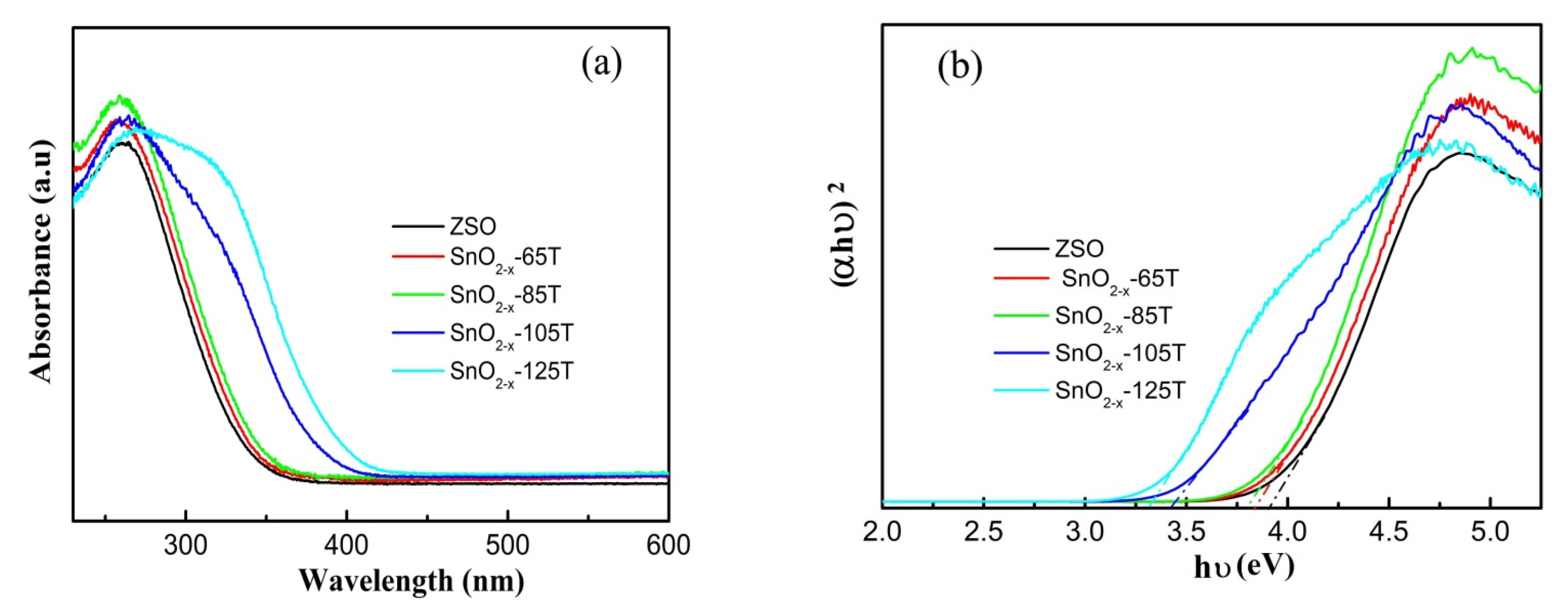
3.4. Optical Absorption and Band Energy Analysis

As is evident from previous studies [26,29,30,31,33,34,35], the construction of oxygen vacancy engineering could broaden the spectral response and enhance the visible-light absorption capacity. In order to evaluate the light absorption property of the SnO2−x samples, the UV–Vis diffraction reflectance spectra (DRS) of different catalysts are presented in Figure 6. A significant difference can be noticed between the ZSO and SnO2−x samples in the DRS curves from Figure 6a. Specifically, ZSO only exhibited a strong absorption capacity in a narrow UV light region due to its wide band-gap (3.91 eV from Figure 6b). As for the SnO2−x samples, the absorption edge displayed a stepwise redshift toward the visible-light region with the increase in the hydrothermal temperature compared with that of ZSO. Moreover, the extended tail located at the visible-light region in the absorbance curve was ascribed to the electron transfer associated with the oxygen vacancy-induced energy level near the valence band edge [48,49,50,51]. To better discuss the effect of the etching temperature on the band-gap energy (Eg) of the SnO2−x samples, the plots of photon energy (hν) with respect to (αhν)2 are presented in Figure 6b. As seen from Tauc plots, the Eg value was remarkably influenced with the increase in the etching temperature. Based on the results of the Tauc plots, the Eg values for SnO2−x samples were respectively determined to be about 3.3, 3.41, 3.79, and 3.84 eV, which were smaller than the reported value (3.95 eV) of the SnO2 nanoparticles [34]. However, it should be clearly noted that the Eg values for the SnO2−x samples were a little larger than those reported in previous work [33], mainly originating from the fine sizes of the nanoparticles. Based on the above analysis, the result of the UV–Vis DRS, combined with the XPS result, could favor the presence of oxygen vacancy defects and demonstrate the effect on the band-gap energy and energy level.

3.5. Evaluation of Photo-Degradation Performance

As one of the most important basic/cationic dyes, RhB is widely applied in the fields of textile, leather, printing paper, and pharmaceutics, which often result in the contamination of water sources due to its excess use. Due to its universal application and long-term stability, RhB was determined as the model pollutant in our work. Before visible-light illumination, RhB solution was placed in the dark for 1 h to make the reaction system reach the adsorption–desorption equilibrium. Figure 7a shows the RhB photo-decomposition plots over different catalysts. ZSO displayed inferior photodecomposition efficiency, and only 49% of the RhB molecules were decomposed upon 70 min of visible-light irradiation, probably originating from its wide band-gap and undesirable absorption property. Compared with ZSO, a larger amount of RhB molecules was absorbed on the surface of the SnO2−x catalysts, suggesting their excellent absorption property. Simultaneously, a continuous enhancement in the photodecomposition efficiency of the SnO2−x catalysts can clearly be seen in Figure 7a. Based on the BET results, combined with XPS and UV–Vis analysis, a larger BET area endowed SnO2−x catalysts with excellent absorption capacity, and provided more active sites for the removal reaction of RhB molecules. Thus, the mesostructure, together with the energy level near valence band induced by the oxygen vacancy, mainly contributed to the substantial improvement in the removal rate of RhB molecules. Among the SnO2−x catalysts, SnO2−x-105T displayed the highest removal efficiency, and about 98.75% of RhB molecules could be completely removed within 60 min. Given that the photodegradation reaction of RhB could adhere to the pseudo-first order reaction kinetics, the diagram for kinetic constant (k) values over different catalysts is depicted in Figure 7b. Correspondingly, the k value of the SnO2−x-105T sample was the largest among the tested catalysts, reaching 0.073 min−1, which was approximately seven times larger than that of ZSO. For this purpose, the SnO2−x-105T sample was determined as the representative catalyst in the subsequent measurements. Taking SnO2−x-105T as the model catalyst, cycling experiments were carried out to testify the usability and stability. After five successive cycling runs, no evident loss of the removal rate of RhB was detectable, revealing the high stability of the SnO2−x-105T sample. In comparison, the crystal phase of the used catalysts was in good accordance with that of the fresh catalyst, as seen in the XRD patterns from Figure 7d. This result further demonstrates the excellent usability and stability of the SnO2−x-105T sample.

3.6. Possible Enhancement Mechanism

Based on the afore-mentioned analysis, the intensification of adsorption capacity and the augment of BET area exerted a significantly positive effect on the enhancement of photodegradation toward the RhB solution. Aside from the above, oxygen vacancy defects can modulate the electronic structure, and induce the generation of novel energy levels near the valence band position for the n-type semiconductor catalyst, which would further influence the recombination rate as well as the separation rate of photo-activated electron-hole pairs. In order to ascertain this issue, PL measurement was carried out on the ZSO and SnO2−x samples. Among all of the tested catalysts, ZSO possessed the strongest emission peak, fitting well with the worst removal rate of RhB. After the oxygen vacancy defects were introduced, the PL intensity of the SnO2−x samples significantly decreased with an increase in the hydrothermal temperature. Specifically, the SnO2−x-105T sample had the weakest PL intensity, whereas the PL intensity, in contrast, increased when the etching temperature was beyond 105 °C. It is reasonably inferred that a further increase in the etching temperature could induce the generation of more oxygen vacancy defects, which could act as the recombination center of photoactivated charge carriers. Thus, SnO2−x-125T had a stronger PL intensity than SnO2−x-105T. This variation trend in the PL spectra for different catalysts was in good agreement with that of the photodegradation performance.
In order to further the investigation of the photo-activated electron transfer process, electrochemical measurements were executed, and the results are shown in Figure 8b,c. Generally, a larger photocurrent is tightly associated with a lower recombination rate of charge carriers, and more effective photoelectron transfer. In comparison, it is clear from Figure 8b that the photocurrent of the SnO2−x-105T sample was about three times higher than that of ZSO, demonstrating that the SnO2−x-105T sample had a much higher separation and transfer rate of photo-activated charge carriers. Additionally, the SnO2−x-105T sample displayed a substantial enhancement in photocurrents, equally confirming the influence of oxygen vacancy defects on band gap narrowing. As is well-known, a smaller arc radius in an EIS Nyquist plot signifies a lower electrochemical charge migration resistance at the interface. Obviously, the SnO2−x-105T sample had a lower electrochemical charge resistance, because the semicircle of its EIS Nyquist plot was smaller than that of ZSO, as observed in Figure 8c. Likewise, the result of the EIS measurement revealed the better capacity of the photoelectron migration of SnO2−x-105T, which agreed well with the photodecomposition test results.
To determine which active species were dominant in the photo-degradation of RhB over the SnO2−x catalyst, radical trapping experiments were considered and performed in our work. Benzoquinone (BZQ 2 mmol L−1), tert-butyl-alcohol (t-BuOH 2 mmol L−1), ammonium oxalate (AO 2 mmol L−1), and FeSO4-EDTA (FS-EDTA, 0.2 mmol L−1) were taken as scavengers to capture·O2, OH, h+, and H2O2, respectively, and the results are depicted in Figure 8d. Summarily, the restriction effect on the removal rate with the addition of four scavengers adhered to the order of FS-EDTA > AO > t-BuOH > BZQ. Clearly, the removal rate of RhB was remarkably suppressed upon exposure to FS-EDTA or AO, demonstrating that the contribution of h+ and H2O2 was primary radicals in the photo-decomposition process. Most notably, the RhB removal rate dwindled extraordinarily and significantly from approximately 98% to 20% due to the addition of FS-EDTA, demonstrating that the H2O2 radicals were the critical species to dominate the oxidation reaction system of the RhB and SnO2−x catalyst.
In consultation with previously reported work [52], the conduction band (CB) edge potential for SnO2 was approximate to 0.05 eV. Thus, the valence band (VB) edge potential of SnO2 could be determined by the virtue of the equation of EVB = ECB + Eg (ECB and EVB represent CB and VB edge potential, respectively, while Eg refers to the band-gap). According to the demonstration above, a possible enhancement mechanism for the photodecomposition behavior is schematically presented in Figure 9. Herein, the novel energy level induced by oxygen vacancy defects is defined as “Vos”. Upon visible-light illumination, the electrons from the Vos position are capable of being activated, and transfer to the CB position. Note that the accumulated photo-activated electrons cannot reduce O2 to generate active radicals of ·O2 because the CB potential of SnO2−x was more positive than the potential of E0 ((O2/O2) −0.33 eV vs. normal hydrogen electrode (NHE)) [53,54]. However, the CB potential of SnO2−x was negative enough to make photoelectrons react with oxygen and generate the active radicals of H2O2 (O2/H2O2, 0.695 eV), which is mainly responsible for the photo-oxidation reaction of RhB. Simultaneously, the remaining photo-activated holes in Vos can directly oxidize RhB molecules to form CO2 and H2O. Therefore, the oxygen vacancy-induced energy level can act as an electron trap, further facilitating the generation and efficient transfer of photoelectrons, and finally produce more active species to significantly enhance the photo-activity toward the removal of RhB.

4. Conclusions

By controlling the ratio of O atoms to Zn atoms stripped from the crystal lattice of ZSO, we designed a novel two-step hydrothermal route to accomplish oxygen vacancy defect engineering. SnO2−x ultrafine nanocrystals with oxygen vacancy defects were obtained after experiencing the dissolution–recrystallization and in situ growth process. Oxygen vacancy defects can adjust the electronic structure and induce the generation of the novel energy level near the valence band position. Aside from the augmentation of active sites and the improvement in the visible-light absorption capacity, the introduction of oxygen vacancy defects can also facilitate the generation and efficient transfer of photoelectrons under visible-light irradiation. Therefore, the synergistic effects of increasing active sites, intensified visible-light harvesting, and the photo-activated charge carrier’ separation rate could mainly contribute to the significant enhancement in the photodecomposition of RhB solution. This research will provide some enlightenment for the design and fabrication of a novel oxide compound semiconductor with oxygen vacancies toward the efficient and tunable organic pollutant degradation in waste water.

Author Contributions

T.J. conceived and designed the experiments; C.S., D.Y., F.F., and S.H. carried out the synthetic experiment and photo-catalytic performance of the as-prepared samples; N.S. and F.L. provided precise instruction and analyzed the data. J.H., J.W., J.L., and B.D. measured the as-prepared samples; T.J. wrote the paper; F.L. revised the paper. J.H.L. provided precise instruction. All authors have read and agreed to the published version of the manuscript.

Funding

This research was funded by the Education Department of Henan Province for the fund support (21A430026) and the Department of Science and Technology of Henan Province, China (Henan Science and Technology Research Program, 222102520005).

Institutional Review Board Statement

Not applicable.

Informed Consent Statement

Not applicable.

Data Availability Statement

The data presented in this study are available in this article.

Conflicts of Interest

The authors declare no conflict of interest.

References

  1. Lianos, P. Production of electricity and hydrogen by photocatalytic degradation of organic wastes in a photoelectrochemical cell: The concept of the photofuel cell: A review of a re-emerging research field. J. Hazard. Mater. 2011, 185, 575–590. [Google Scholar] [CrossRef] [PubMed]
  2. Tang, J.; Zou, Z.; Ye, J. Efficient photocatalytic decomposition of organic contaminants over CaBi2O4 under visible-light irradiation. Angew. Chem. Int. Ed. 2004, 43, 4463–4466. [Google Scholar] [CrossRef] [PubMed]
  3. Reddy, V.L.P.; Kim, K.A. Review of photochemical approaches for the treatment of a wide range of pesticides. J. Hazard. Mater. 2015, 285, 325–335. [Google Scholar] [CrossRef]
  4. Shinde, S.S.; Bhosale, C.H.; Rajpure, K.Y. Photodegradation of organic pollutants using N-titanium oxide catalyst. J. Photochem. Photobiol. B 2014, 141, 186–191. [Google Scholar] [CrossRef] [PubMed]
  5. Jo, W.; Tayade, R.J. New generation energy-efficient light source for photocatalysis: LEDs for environmental applications. Ind. Eng. Chem. Res. 2014, 53, 2073–2084. [Google Scholar] [CrossRef]
  6. Jo, W.; Tayade, R.J. Recent developments in photocatalytic dye degradation upon irradiation with energy-efficient light emitting diodes. Chin. J. Catal. 2014, 35, 1781–1792. [Google Scholar] [CrossRef]
  7. Mckay, G. Waste colour removal from textile effluents. Am. Dyest. Rep. 1979, 68, 29–36. [Google Scholar]
  8. Hage, R.; Lienke, A. Applications of transition-metal catalysts to textile and wood-pulp bleaching. Angew. Chem. Int. Ed. 2006, 45, 206–222. [Google Scholar] [CrossRef]
  9. Hong, Q.; Hardcastle, J.L.; Mckeown, R.A.J.; Marken, F.; Compton, R.G. The 20 kHz sonochemical degradation of trace cyanide and dye stuffs in aqueous media. New J. Chem. 1999, 23, 845–849. [Google Scholar] [CrossRef]
  10. Wang, S.A. Comparative study of fenton and fenton-like reaction kinetics in decolourisation of waste water. Dyes Pigments 2008, 76, 714–720. [Google Scholar] [CrossRef]
  11. Ma, T.Y.; Qiao, S.Z. Acid—Base bifunctional periodic mesoporous metal phosphonates for synergistically and heterogeneously catalyzing CO2 conversion. ACS Catal. 2014, 4, 3847–3855. [Google Scholar] [CrossRef]
  12. Shi, B.; Li, G.; Wang, D.; Feng, C.; Tang, H. Removal of direct dyes by coagulation: The performance of preformed polymeric aluminum species. J. Hazard. Mater. 2007, 143, 567–574. [Google Scholar] [CrossRef] [PubMed]
  13. Maezawa, A.; Nakadoi, H.; Suzuki, K.; Furusawa, T.; Suzuki, Y.; Uchida, S. Treatment of dye wastewater by using photo-catalytic oxidation with sonication. Ultrason. Sonochem. 2007, 14, 615–620. [Google Scholar] [CrossRef] [PubMed]
  14. Zhao, Y.Y.; Deng, N.; Fan, Z.H.; Hu, Z.T.; Fan, L.; Zhou, J.; Huang, X. On-site H2O2 electro-generation process combined with ultraviolet: A promising approach for odorous compounds purification in drinking water system. Chem. Eng. J. 2022, 430, 132829. [Google Scholar] [CrossRef]
  15. Rui, J.C.; Deng, N.; Zhao, Y.Y.; Tao, C.; Zhou, J.Z.; Zhao, Z.; Huang, X. Activation of persulfate via Mn doped Mg/Al layered double hydroxide for effective degradation of organics: Insights from chemical and structural variability of catalyst. Chemosphere 2022, 302, 134849. [Google Scholar] [CrossRef]
  16. Wang, H.; Zhang, L.; Chen, Z.; Hu, J.; Li, S.; Wang, Z.; Liu, J.; Wang, X. Semiconductor heterojunction photocatalysts: Design, construction, and photocatalytic performances. Chem. Soc. Rev. 2014, 43, 5234–5244. [Google Scholar] [CrossRef]
  17. Yu, H.; Jiang, L.; Wang, H.; Huang, B.; Yuan, X.; Huang, J.; Zhang, J.; Zeng, G. Modulation of Bi2MoO6-based materials for photocatalytic water splitting and environmental application: A critical review. Small 2019, 15, 1901008. [Google Scholar] [CrossRef]
  18. Li, X.; Xie, J.; Jiang, C.J.; Yu, J.G.; Zhang, P.Y. Review on design and evaluation of environmental photocatalysts. Front. Environ. Sci. Eng. 2018, 12, 14. [Google Scholar] [CrossRef]
  19. Fujishima, A.; Honda, K. Electrochemical photolysis of water at a semiconductor electrode. Nature 1972, 238, 37–38. [Google Scholar] [CrossRef]
  20. Hu, W.; Liu, Y.; Withers, R.L.; Frankcombe, T.J.; Norén, L.; Snashall, A.; Kitchin, M.; Smith, P.; Gong, B.; Chen, H.; et al. Electron-pinned defect-dipoles for high-performance colossal permittivity materials. Nat. Mater. 2013, 12, 821–826. [Google Scholar] [CrossRef]
  21. Liu, S.D.; Kang, L.; Zhang, J.; Jun, S.C.; Yamauchi, Y. Carbonaceous anode materials for nonaqueous sodium- and potassium-ion hybrid capacitors. ACS Energy Lett. 2021, 6, 4127–4154. [Google Scholar] [CrossRef]
  22. Ning, J.; Mu, C.; Guo, X.; Yang, R.; Jonathan, R.; Jiao, W.; Wu, X.; Jian, X. Efficient defect engineering and in-situ carbon doping in ultra-fine TiO2 with enhanced visible-light-response photocatalytic performance. J. Alloys Compd. 2022, 901, 163490. [Google Scholar] [CrossRef]
  23. Yang, Y.; Yin, L.C.; Gong, Y.; Niu, P.; Wang, J.Q.; Gu, L.; Chen, X.; Liu, G.; Wang, L.; Cheng, H.M. An unusual strong visible-light absorption band in red anatase TiO2 photocatalyst induced by atomic hydrogen-occupied oxygen vacancies. Adv. Mater. 2018, 30, 1704479. [Google Scholar] [CrossRef]
  24. Zuo, F.; Wang, L.; Wu, T.; Zhang, Z.; Borchardt, D.; Feng, P.Y. Self doped Ti3+ enhanced photocatalytic for hydrogen production under visible-light. J. Am. Chem. Soc. 2010, 132, 11856–11857. [Google Scholar] [CrossRef]
  25. Li, Y.; Xue, J.; Shen, Q.; Jia, S.; Li, Q.; Li, Y.; Liu, X.; Jia, H. Construction of a ternary spatial junction in yolk-shell nanoreactor for efficient photo-thermal catalytic hydrogen generation. Chem. Eng. J. 2021, 423, 130188. [Google Scholar] [CrossRef]
  26. Wu, J.; Qiao, P.; Li, H.; Xu, Y.; Yang, W.; Yang, F.; Lin, K.; Pan, K.; Zhou, W. Engineering surface defects on two-dimensional ultrathin mesoporous anatase TiO2 nanosheets for efficient charge separation and exceptional solar-driven photocatalytic hydrogen evolution. J. Mater. Chem. C 2020, 8, 3476–3482. [Google Scholar] [CrossRef]
  27. Li, L.; Chen, X.H.; Wang, L.; Tao, C.Y.; Wu, X.P.; Du, J.; Liu, Z.H. Synthesis of Ti3+ self-doped mesoporous TiO2 cube with enhanced visible-light photoactivity by a simple reduction method. J. Alloys Compd. 2020, 845, 156138. [Google Scholar] [CrossRef]
  28. Sun, D.; Chi, D.; Yang, Z.; Xing, Z.; Chen, P.; Li, Z.; Pan, K.; Zhou, W. CdS quantum dots modified surface oxygen vacancy defect ZnO1−x-TiO2−x solid solution sphere as Z-Scheme heterojunctions for efficient visible light-driven photothermal-photocatalytic performance. J. Alloys Compd. 2020, 826, 15428. [Google Scholar] [CrossRef]
  29. Gurylev, V.; Perng, T.P. Defect engineering of ZnO: Review on oxygen and zinc vacancies. J. Eur. Ceram. Soc. 2021, 41, 4977–4996. [Google Scholar] [CrossRef]
  30. Xu, Y.C.; Li, H.; Sun, B.; Qiao, P.; Ren, L.; Tian, G.; Jiang, B.; Pan, K.; Zhou, W. Surface oxygen vacancy defect-promoted electron-hole separation for porous defective ZnO hexagonal plates and enhanced solar-driven photocatalytic performance. Chem. Eng. J. 2020, 379, 122295. [Google Scholar] [CrossRef]
  31. Wang, J.; Wang, Z.; Huang, B.; Ma, Y.; Liu, Y.; Qin, X.; Zhang, X.; Dai, Y. Oxygen vacancy induced band-gap narrowing and enhanced visible light photocatalytic activity of ZnO. ACS Appl. Mater. Interfaces 2012, 4, 4024–4030. [Google Scholar] [CrossRef] [PubMed]
  32. Dong, W.; Xu, J.; Wang, C.; Lu, Y.; Liu, X.; Wang, X.; Yuan, X.; Wang, Z.; Lin, T.; Sui, M.; et al. A robust and conductive black tin oxide nanostructure makes efficient lithium-Ion batteries possible. Adv. Mater. 2017, 29, 1700136. [Google Scholar] [CrossRef] [PubMed]
  33. Anuchai, S.; Phanichpahant, S.; Tantraviwat, D.; Pluengphon, P.; Bovornratanaraks, T.; Incessungvorn, B. Low temperature preparation of oxygen-deficient tin dioxide nanocrystals and a role of oxygen vacancy in photocatalytic activity improvement. J. Colloid Interface Sci. 2018, 512, 105–114. [Google Scholar] [CrossRef] [PubMed]
  34. Fan, C.M.; Peng, Y.; Zhu, Q.; Lin, L.; Wang, R.; Xu, A.W. Synproportionation reaction for the fabrication of Sn2+ self-doped SnO2−x nanocrystals with tunable band structure and highly efficient visible light photocatalytic activity. J. Phy. Chem. C 2013, 117, 24157–24166. [Google Scholar] [CrossRef]
  35. Liu, W.T.; Wu, B.; Lai, Y.; Tai, N.; Perng, T.; Chen, L.J. Enhancement of water splitting by controlling the amount of vacancies with varying vacuum level in the synthesis system of SnO2−x/In2O3−y heterostructure as photocatalyst. Nano Energy 2018, 47, 18–25. [Google Scholar] [CrossRef]
  36. Zhou, Z.H.; Liu, J.; Long, R.; Li, L.; Guo, L.J.; Prezhdo, O.V. Control of charge carriers trapping and relaxation in hematite by oxygen vacancy charge: Ab initio non-adiabatic molecular dynamics. J. Am. Chem. Soc. 2017, 139, 6707–6717. [Google Scholar] [CrossRef]
  37. Wu, J.X.; Qiao, P.; Li, H.; Ren, L.; Xu, Y.; Tian, G.; Li, M.; Pan, K.; Zhou, W. Surface-oxygen vacancy defect-promoted electron-hole separation of defective tungsten trioxide ultrathin nanosheets and their enhanced solar-driven photocatalytic performance. J. Colloid Interface Sci. 2019, 557, 18–27. [Google Scholar] [CrossRef]
  38. Kong, X.Y.; Lee, W.Q.; Mohamed, A.R.; Chai, S.P. Effective steering of charge flow through synergistic inducing oxygen vacancy defects and p-n heterojunctions in 2D/2D surface-engineered Bi2WO6/BiOI cascade: Towards superior photocatalytic CO2 reduction activity. Chem. Eng. J. 2019, 372, 1183–1193. [Google Scholar] [CrossRef]
  39. Tan, B.; Toman, E.; Li, Y.G.; Wu, Y.Y. Zinc stannate (Zn2SnO4) dye-sensitized solar cells. J. Am. Chem. Soc. 2007, 129, 4162–4163. [Google Scholar] [CrossRef]
  40. Jia, T.K.; Fu, F.; Long, F.; Min, Z.Y.; Zhao, J.W.; Chen, J.; Li, J.L. Synthesis, characterization and enhanced visible-light photocatalytic activity of Zn2SnO4/C nanocomposites with truncated octahedron morphology. Ceram. Int. 2016, 42, 3808–3815. [Google Scholar] [CrossRef]
  41. Jia, T.K.; Liu, M.; Yu, D.Y.; Long, F.; Mo, S.; Deng, Z.; Wang, W. A facile approach for the synthesis of Zn2SnO4/BiOBr nanocomposites with improved visible light photocatalytic performance. Nanomaterials 2018, 8, 313. [Google Scholar] [CrossRef] [PubMed] [Green Version]
  42. Jia, T.K.; Fu, F.; Li, J.; Wang, W.; Hu, X. Constructing a novel Zn2SnO4/C/AgBr nanocomposite with extended spectral Response and improved photocatalytic performance. J. Alloys Compd. 2019, 783, 687–696. [Google Scholar] [CrossRef]
  43. Tuan, P.V.; Hieu, L.T.; Nga, L.Q.; Dung, N.D.; Ha, N.N.; Khiem, T.N. Hydrothermal synthesis and characteristic photoluminescence of Er-doped SnO2 nanoparticles. Physica B 2016, 501, 34–37. [Google Scholar] [CrossRef]
  44. Jia, T.K.; Fu, F.; Li, J.L.; Deng, D.; Long, F.; Yu, D.; Cui, Q.; Wang, W.M. Rational construction of direct Z-scheme SnS-g-C3N4 hybrid photocatalyst for significant enhancement of visible-light photocatalyticactivity. Appl. Surf. Sci. 2020, 499, 143941. [Google Scholar] [CrossRef]
  45. Yu, D.; Jia, T.K.; Deng, Z.; Wei, Q.; Wang, K.; Chen, L.; Wang, P.; Cui, J. One-dimensional P-doped graphitic carbon nitride tube: Facile synthesis, effect of doping concentration, and enhanced mechanism for photocatalytic hydrogen evolution. Nanomaterials 2022, 12, 1759. [Google Scholar] [CrossRef]
  46. Jia, T.K.; An, J.C.; Yu, D.; Li, J.; Fu, F.; Wang, K.; Wang, W. Continuously improved photocatalytic performanceof Zn2SnO4/SnO2/Cu2O composites by structural modulation and band alignment modification. Nanomaterials 2019, 9, 1390. [Google Scholar] [CrossRef]
  47. Chetri, P.; Choudhury, B.; Choudhury, A. Room temperature ferromagnetism in SnO2 nanoparticles: An experimental and density functional study. J. Mater. Chem. C 2014, 2, 9294–9302. [Google Scholar] [CrossRef]
  48. Xu, Y.; Zheng, L.; Yang, C.; Zheng, W.; Liu, X.; Zhang, J. Oxygen vacancies enabled porous SnO2 thin films for highly sensitive detection of triethylamine at room temperature. ACS Appl. Mater. Interfaces 2020, 12, 20704–20713. [Google Scholar] [CrossRef]
  49. Song, M.; Wu, Y.; Zhao, Y.; Du, C.; Su, Y. Structural insight on defect-rich tin oxide for smart band alignment engineering and tunable visible-light-driven hydrogen evolution. Inorg. Chem. 2020, 59, 181–3192. [Google Scholar] [CrossRef]
  50. Li, P.; Liu, Y.; Zhang, Q.; Zhao, Z.; Pullerits, T.; Zheng, K.; Zhou, Y. Iodinated SnO2 quantum dots: A facile and efficient approach to increase solar absorption for visible-light photocatalysis. J. Phys. Chem. C 2016, 120, 9253–9262. [Google Scholar] [CrossRef]
  51. Wang, H.; Dou, K.; Teoh, W.-Y.; Zhan, Y.; Hung, T.-F.; Zhang, F.; Xu, J.; Zhang, R.; Rogach, A.L. Engineering of facets, band structure, and gas-sensing properties of hierarchical Sn2+-doped SnO2 nanostructures. Adv. Funct. Mater. 2013, 23, 4847–4853. [Google Scholar]
  52. Li, Y.; Wu, X.; Ho, W.; Lv, K.; Li, Q.; Li, M.; Lee, S.C. Graphene-induced formation of visible- light-responsive SnO2-Zn2SnO4 Z scheme photocatalyst with surface vacancy for the enhanced photoreactivity towards NO and acetone oxidation. Chem. Eng. J. 2018, 336, 200–210. [Google Scholar] [CrossRef]
  53. Kim, J.; Lee, C.W.; Choi, W. Platinized WO3 as an environmental photocatalyst that generates OH radicals under visible light. Environ. Sci. Technol. 2010, 44, 6849–6854. [Google Scholar] [CrossRef] [PubMed]
  54. Wang, X.; Li, S.; Yu, H.; Yu, J. In situ anion-exchange synthesis and photocatalytic activity of Ag8W4O16/AgCl-nanoparticle core–shell nanorods. J. Mol. Catal. A Chem. 2011, 334, 52–59. [Google Scholar] [CrossRef]
Figure 1. The formation schematic diagram of SnO2−x nanocrystals in an acetic acid media under the hydrothermal condition. (a) ZSO structure; (b) SnO2−x structure.
Nanomaterials 12 03342 g001
Figure 2. (a) XRD patterns of the ZSO, SnO2, and SnO2−x-105T samples; (b) partially enlarged diffraction peak of (110) plane of the SnO2 and SnO2−x-105T samples.
Nanomaterials 12 03342 g002
Figure 3. High resolution XPS spectra of the ZSO and SnO2−x-105T sample, (a) Sn 3d; (b) O 1s.
Nanomaterials 12 03342 g003
Figure 4. (a) TEM image of ZSO; (b) TEM image of SnO2−x-105T; (c) HRTEM image of SnO2−x-105T.
Nanomaterials 12 03342 g004
Figure 5. (a) The nitrogen adsorption–desorption isotherm and (b) the corresponding pore size and distribution curve of SnO2−x-105T.
Nanomaterials 12 03342 g005
Figure 6. (a) UV–Vis absorbance spectra of the ZSO and SnO2−x samples, (b) the derived plots of (αhυ)2 versus from the absorption spectrum for ZSO and SnO2−x samples.
Nanomaterials 12 03342 g006
Figure 7. (a) The RhB photodegradation curves over different catalysts; (b) the corresponding apparent reaction rate constant; (c) cycling performance of the photodegradation of RhB solution over the SnO2−x-105T sample; (d) XRD pattern of SnO2−x-105T before and after use.
Nanomaterials 12 03342 g007
Figure 8. (a) PL spectra of the ZSO and SnO2−x samples; (b) transient photocurrents and (c) electrochemical impedance spectra ZSO and SnO2−x-105T of electrodes under visible light irradiation; (d) the effect of various scavengers on the visible light photocatalytic performance of the SnO2−x-105T sample.
Nanomaterials 12 03342 g008
Figure 9. A schematic diagram of the proposed photodegradation mechanism over the SnO2−x system under visible-light illumination.
Nanomaterials 12 03342 g009
Publisher’s Note: MDPI stays neutral with regard to jurisdictional claims in published maps and institutional affiliations.

