Article

Development of Novel Colorful Electrorheological Fluids

1
Department of Chemical and Biological Engineering, Hanbat National University, Yuseong-gu, Daejeon 34158, Korea
2
Department of Polymer Science and Engineering, Inha University, Michuhol-gu, Incheon 22212, Korea
3
Program of Environmental and Polymer Engineering, Inha University, Michuhol-gu, Incheon 22212, Korea
4
McKetta Department of Chemical Engineering and Texas Material Institute, The University of Texas at Austin, Austin, TX 78712, USA
5
Department of Advanced Materials Science and Engineering, Hanseo University, Seosan-si 31962, Korea
*
Author to whom correspondence should be addressed.
Nanomaterials 2022, 12(18), 3113; https://doi.org/10.3390/nano12183113
Submission received: 17 August 2022 / Revised: 31 August 2022 / Accepted: 6 September 2022 / Published: 8 September 2022

Abstract

:
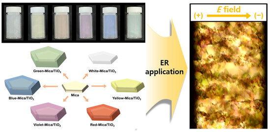
Herein, the electrorheological (ER) performances of ER fluids were correlated with their colors to allow for the visual selection of the appropriate fluid for a specific application using naked eyes. A series of TiO2-coated synthetic mica materials colored white, yellow, red, violet, blue, and green (referred to as color mica/TiO2 materials) were fabricated via a facile sol–gel method. The colors were controlled by varying the thickness of the TiO2 coating layer, as the coatings with different thicknesses exhibited different light interference effects. The synthesized color mica/TiO2 materials were mixed with silicone oil to prepare colored ER fluids. The ER performances of the fluids decreased with increasing thickness of the TiO2 layer in the order of white, yellow, red, violet, blue, and green materials. The ER performance of differently colored ER fluids was also affected by the electrical conductivity, dispersion stability, and concentrations of Na+ and Ca2+ ions. This pioneering study may provide a practical strategy for developing new ER fluid systems in future.

Graphical Abstract

1. Introduction

Electrorheological (ER) fluids, which are well-dispersed suspensions of polarizable materials in insulating media, such as silicone oils, have received widespread attention owing to their various advantages, such as rapid response, low power consumption, and reversible phase transition [1,2,3,4,5]. When an external electric field (E-field) is applied, ER fluids exhibit a sudden and rapid change from liquids to solid-like states within a few milliseconds [6,7,8,9,10]. ER fluids can therefore be used in various industrial applications which require fast response times, including dampers, clutch systems, engine mounts, and haptic devices [11,12,13]. ER activities can be improved by controlling the mechanical and chemical properties of dispersing materials [14,15,16]. Additionally, the polarizability of the materials plays a pivotal role in ER performance [17,18]: in general, materials with high polarizability exhibit high ER efficiencies [19]. Various polarizable materials, including metallic, polymeric, organic, and inorganic materials, have been used as ER materials [20,21,22].
Among various inorganic and metal oxides, TiO2 is widely used as a dispersing material for ER fluids owing to its advantages, such as availability for mass production and simple fabrication process via the sol–gel method [23,24,25]. TiO2 and its derived materials also exhibit numerous chemical advantages, such as stability, resistance to corrosion, and photocatalytic ability [26,27,28,29,30]. Previous ER studies have developed numerous TiO2-derived materials, such as polymer-coated TiO2, shape-controlled TiO2, and alkaline earth metal-doped TiO2 [31,32]. Furthermore, TiO2 materials can be incorporated or coated onto a SiO2 template to fabricate multilayered materials to achieve core/shell structures, porous characteristics, and precursors for hollow-type materials [33,34,35]. Silicon-containing materials, such as SiO2, SiO3, and other silicon-containing oxides, are widely utilized as ER fluids. Studies have demonstrated the successful ER performances of silicon-containing oxide materials: Tian et al. reported a zeolite-based ER fluid with a high dielectric constant [36]; Ma et al. synthesized an anisotropic silicon-rubber-based ER elastomer with 30 vol% TiO2-urea core-shell materials in silicone oil [37]; Méheust et. al. reported the ER activity of synthetic clay/mica-based ER fluids [38].
Mica consists of layered silicate materials. It naturally occurs in the earth as a mineral, and various methods have been developed for fabricating synthetic mica (mica) [39]. Natural and synthetic mica materials are utilized in various industrial fields owing to their benign characteristics, including nontoxicity, chemical stability, transparency, and glossiness [40,41]. Therefore, mica materials are widely employed in cosmetics, coating systems, and printing inks [42,43,44]. Furthermore, metal oxides, such as TiO2 and SiO2, can be coated onto the surface of mica to induce the light interference effect and consequently obtain various color-expressing pearlescent materials [45]. The light interference effects are facilitated on the surface of the metal oxide layer owing to the differences in refractive indices of the mica and the coated materials [46,47]. In addition, the thickness of the metal oxide coating layer can be manipulated to alter the color of pearlescent pigments.
While TiO2, silicon-containing oxides, and mica have been previously used as ER materials, research thus far has mainly focused on altering the shape, size, and porosity of materials [48]. In addition, numerous ER studies have demonstrated the incorporation or mixing of various materials, but the colors of ER fluids are confined to white, gray, and transparent. Therefore, negligible attention has been devoted to the synthesis of ER fluids of various colors. As the colors of conventional ER fluids are indistinguishable, confirming the performance of ER fluids with the naked eye and selecting an appropriate fluid for application are challenging. Therefore, developing ER fluids that can be selected for an application by correlating their color and performance is essential.
Herein, we developed six novel differently colored ER fluids by synthesizing various TiO2-coated colored mica (color mica/TiO2) materials. To the best of our knowledge, this is the first study to demonstrate the performances of ER fluids of various colors and their underlying mechanisms. The color mica/TiO2-based ER fluids were synthesized by coating TiO2 on synthetic base materials; the thickness of the TiO2 layer was manipulated to obtain a different color for each fluid—white, yellow, red, violet, blue, and green—owing to differences in the light interference effects. Significantly, the ER performance of each color mica/TiO2-based ER fluid changed according to the thickness of the TiO2 layer. Specifically, among the synthesized samples, the white mica/TiO2-based ER fluid demonstrated the highest ER activity (95.1 Pa), whereas green mica/TiO2-based ER fluids exhibited the lowest (51.1 Pa). Therefore, it was possible to select an appropriate ER fluid for a required application by simply evaluating the color. Additionally, the underlying ER mechanism for each color mica/TiO2-based ER fluid was revealed. The ER performance was directly affected by the amounts of Na+ and Ca2+ ions present in the ER fluids. In addition, the dispersion stability of each colored ER fluid was different, and the stability increased with decreasing TiO2 thickness. The synergistic contributions of ion polarizability and dispersion stability affect the ER performance of each ER fluid. In conclusion, we expect that these novel colored ER fluids will pave the way for the development of future smart stress control systems by allowing the facile selection of the appropriate ER fluid for an application by evaluating its color.

2. Materials and Methods

2.1. Materials

Mica (powder type) was purchased from CQV Co., Ltd. (Jincheon, Korea). Titanium tetrachloride (99.9%) and silicone oil (viscosity = 100 cSt) were purchased from Sigma-Aldrich (Burlington, MA, USA). Sodium hydroxide (40.0%) and ethanol (EtOH, 99.9%) were purchased from Samchun Chemical Company (Seoul, Korea). All chemicals were used as received without any additional purification.

2.2. Synthesis of Color-Mica/TiO2 Materials

Various color mica/TiO2 materials were synthesized using a typical sol–gel method. Mica (10.5 g) was dispersed in deionized water (100 mL) under vigorous magnetic stirring for 1 h. A constant heat of 90 °C was then applied to the solution, and TiCl4 (2.0 mL) was continuously added dropwise with constant stirring—the amount of TiCl4 added determined the color of the material. The sol–gel reaction proceeded for 4 h, during which NaOH (20.0%) was slowly added to the suspension to maintain the pH value at 1.8; pH preservation is vital for the successful coating of the TiO2 layer on mica. The resulting color mica/TiO2 materials were collected via vacuum filtration, washed several times with water to remove residues, and dried in an oven overnight at 90 °C. The final dry materials obtained were: white mica/TiO2, yellow mica/TiO2, red mica/TiO2, violet mica/TiO2, blue mica/TiO2, and green mica/TiO2. Figure 1 shows a schematic of the synthesis process.

2.3. Characterization

The morphological structures of the mica and color mica/TiO2 materials were investigated via field emission scanning electron microscopy (FE-SEM, S-4800, Hitachi, Tokyo, Japan); cross-sectional samples of the color mica/TiO2 materials were prepared for the analysis using a focused ion beam (FIB, LYRA3 GMH, TESCAN, Brno, The Czech Republic) system. The elemental compositions (Si and Ti) of the color mica/TiO2 materials were obtained using an energy-dispersive X-ray spectroscopy (EDS) system (EX-250, HORIBA, Ltd., Kyoto, Japan) integrated with the FE-SEM instrument. The molecular structures of the materials were analyzed using a Fourier-transform infrared (FT-IR) instrument (Nicolet iS10, Thermo Fisher Scientific, Waltham, MA, USA). The concentrations of Na+ and Ca2+ ions in the materials were determined using an ion chromatography system (930 Compact IC Flex, Metrohm, Herisau, Switzerland). The electrical conductivities of the materials in the pellet form were obtained using a two-point probe system (MCP-HT450, Mitsubishi, Tokyo, Japan).

2.4. Investigation of ER Properties

To prepare the color mica/TiO2-based ER fluids (3.0 wt%), the dried ER materials (0.3 g) were ultrasonically dispersed in silicone oil (11 mL, viscosity = 100 cST), followed by vigorous magnetic stirring overnight. No additional additives were added to the prepared ER fluids. The ER investigation of the samples was performed using a rheometer (MCR 302, Anton Parr, Graz, Austria) with a cup, concentric cylinder conical geometry, and high-voltage generator (HCN 7E-12500, Fug Elektronik, Schechen, Germany). The gap between the cup and the geometry was set to 1.00 mm on each side without any interference. The ER measurements were initiated by adding well-dispersed ER fluids into the cup along with a rotor. A mechanical shear rate of 10.0 s−1 was applied for 5 min to achieve stability and homogeneous placement of the fluid in the cup. Finally, the E-field was applied to analyze the ER performances of the different ER fluids under various conditions.

3. Results and Discussion

3.1. Structure and Morphology of Color-Mica/TiO2 Materials

To visualize the color appearance, the color mica/TiO2 materials were dissolved in EtOH and observed by the naked eye. Clearly, each color mica/TiO2 material expressed a distinct color (Figure 2a). The color change of a color mica/TiO2 material can be explained as follows: When incident light penetrates the color mica/TiO2 material, a certain amount of light is reflected from the nica–TiO2 interface. This reflected light encounters new incident light, facilitating the light interference effect. Varying the thickness of the TiO2 layer changes the traveling distances of the incident light, causing different interference effects involving various wavelengths, thereby producing different colors. This correlation between the ER material color and TiO2 layer thickness is shown in Figure 2b. The refractive indices of mica and TiO2 are 1.6 and 2.5, respectively [49,50]. According to Fresnel’s law of reflection, the larger the difference in the refractive index between two materials, the higher the light reflectance at the interface of the materials [51,52].
The morphologies of the color mica/TiO2 materials were investigated using FE-SEM (Figure 3). The particle size of the color mica/TiO2 materials was determined to be approximately 25 μm. Significantly, a clear separation between the particles was observed for all the color mica/TiO2 materials, which is in contrast with the aggregated mica particles; this particle separation occurred owing to the presence of the TiO2 layer. Notably, the surfaces of mica and color mica/TiO2 materials were completely different: compared to the smooth surface of mica, the color mica/TiO2 materials displayed rough surfaces owing to the TiO2 coating. In addition, the size of the TiO2 particles increased with increasing TiO2 thickness.
To further investigate the layer of each color mica/TiO2 material, FIB analysis was conducted on cross-sectional samples of mica and the color mica/TiO2 materials, and the thickness of the TiO2 layers was determined (Figure 4). For the mica sample, the measured thickness was approximately 728.6 nm. For the color mica/TiO2 materials, the thicknesses of the TiO2 layers of the white mica/TiO2, yellow mica/TiO2, red mica/TiO2, violet mica/TiO2, blue mica/TiO2, and green mica/TiO2 materials were approximately 52.3, 84.4, 115.7, 138.8, 164.9, and 187.5 nm, respectively. Clearly, TiO2 layers were successfully introduced onto the mica in the color mica/TiO2 materials, and the color of each material changed according to the different light interference effects of the respective TiO2 layers.
EDS analysis was conducted to examine the elemental compositions (Si and Ti) of mica and various color mica/TiO2 materials. For mica, only Si was detected, and no Ti was detected. In contrast, Ti was detected in all the color mica/TiO2 materials. The Ti proportions for white mica/TiO2, yellow mica/TiO2, red mica/TiO2, violet mica/TiO2, blue mica/TiO2, and green mica/TiO2 were 21.0%, 31.5%, 41.0%, 49.2%, 55.7%, and 61.9%, respectively. The total Ti composition of each color mica/TiO2 material increased with increasing TiO2 thickness, which is in accordance with the cross-sectional FE-SEM observation. The detailed elemental compositions of mica and the color mica/TiO2 materials are listed in Table 1.
FT-IR analysis was conducted to investigate the molecular structures of mica and the color mica/TiO2 materials, as shown in Figure 5. In the case of mica, characteristic peaks were observed at 970, 800, and 695 cm−1, which correspond to silicon-containing oxides with asymmetric Si–O–Si stretching vibrations, Si–O symmetric stretching vibrations, and Si–O symmetric bending, respectively [53,54,55]. Significantly, the peaks characteristic to mica diminished in the absorbance curves of all the color mica/TiO2 materials, indicating the successful coating of TiO2. Moreover, the absorbance increased after 830 cm−1 for the color mica/TiO2 materials owing to the presence of the TiO2 layers, which absorbs in the near-infrared (NIR) region.

3.2. Suitability Tests of Color-Mica/TiO2 Materials for ER Application

The suitability of various color mica/TiO2 materials for ER applications was investigated by determining their ion concentration, electrical conductivity, and dispersion stability. First, ion chromatography was used to identify the ion concentrations of the color mica/TiO2 materials. Ion chromatography revealed the presence of Na+ and Ca2+ in all the color mica/TiO2 materials. Notably, the concentrations of both Na+ and Ca2+ ions decreased as the thickness of the TiO2 layer increased, indicating that the ion species originated from the mica material, which contains various minerals. Detailed concentrations of Na+ and Ca2+ ions in various color mica/TiO2 materials are listed in Table 2. Previous studies have demonstrated that ion species enhance the ER activity via ion polarization effects [17]. Therefore, the ER performance of color mica/TiO2 may be affected by the ion concentration, and a high concentration may have a benign effect on the ER performance.
The electrical conductivities of various color mica/TiO2 materials were investigated to examine their suitability for ER applications, as electrical conductivity is a key factor that affects the ER performance of a material. Previous studies have demonstrated that the electrical conductivity should be near ~10−9 S m−1 for achieving the ideal ER performance of a material [56]. Moreover, the electrical conductivity mismatch between the materials and dispersing medium can facilitate the ER effect [25,57]. The electrical conductivities for white mica/TiO2, yellow mica/TiO2, red mica/TiO2, violet mica/TiO2, blue mica/TiO2, and green mica/TiO2 were determined to be 2.4 × 10⁻⁹, 2.1 × 10⁻⁹, 1.7 × 10⁻⁹, 1.4 × 10⁻⁹, 1.1 × 10⁻⁹, and 0.9 × 10⁻⁹ S m−1, respectively; the conductivity of silicone oil is ~1 × 10⁻¹³ S m−1. Therefore, the electrical conductivities of various color mica/TiO2 materials were in a suitable range for ER applications, and the electrical conductivity of each material was at least two orders of magnitude higher than that of silicone oil, thereby consequently promoting the ER effect of the materials [25].
Finally, the dispersion stabilities of the color mica/TiO2 materials were examined (Figure 6). For ER applications, dispersion stability is another key factor affecting ER performance, as the sedimentation of materials from the media may disrupt the formation of rigid fibril-like structures. To examine the dispersion stability, each color mica/TiO2 material was thoroughly mixed with silicone oil at a concentration of 3.0 wt%. Each fluid was well-dispersed in the original state, but the dispersed materials gradually sedimented over time, reaching the equilibrium state at 20 h. After 20 h, each color mica/TiO2-based ER fluid exhibited favorable dispersion stability for ER application. The dispersion stability of the materials decreased in the order of white mica/TiO2 > yellow mica/TiO2 > red mica/TiO2 > violet mica/TiO2 > blue mica/TiO2 > green mica/TiO2, indicating that the dispersion stability of the material decreased with increasing TiO2 content.

3.3. ER Performances of Color-Mica/TiO2-Based ER Fluids

The ER characteristics of the six differently colored ER fluids were examined by determining the shear stress under various conditions: as a function of shear rate, in E field on–off conditions, and at various E field strengths (Figure 7). First, ER activities were measured as a function of shear rate when an E-field strength of 3.0 kV mm−1 was applied (Figure 7a). All the ER fluids experienced immediate shear stress upon the application of the E field, owing to the electrostatic forces between the dispersed materials in the dispersing media [58]; the shear stress resulted in the formation of fibril-like structures. In the low shear rate region, all ER fluids exhibited Bingham plastic behavior, which was ascribed to the electrostatic forces between materials that were stronger than the hydrodynamic forces generated by mechanical shear [59]. Past the critical shear rate (τc), Newtonian behavior was exhibited as the shear stress increased proportionally with shear rate, indicating the dominance of hydrodynamic forces over electrostatic forces [60]. The determined ER activities for the white, yellow, red, violet, blue, and green mica/TiO2-based ER fluids were 95.1, 81.9, 71.2, 63.3, 57.5, and 51.1 Pa, respectively; the ER performance of the white mica/TiO2-based ER fluid was the highest among the various color mica/TiO2-based ER fluids. This trend mirrors that of the ER performance, which was in the order of white > yellow > red > violet > blue > green materials. These trends in relation to ER activity are in accordance with the experimental results of various suitability tests. As discussed earlier, the white mica/TiO2 material displayed the highest ion concentrations, the largest electrical conductivity difference with the dispersing media, and excellent dispersion stability compared to the other color mica/TiO2 materials. In contrast, the green mica/TiO2 material demonstrated low ion concentrations, electrical conductivity, and dispersion stability, thereby exhibiting the poorest ER performance. The tentative mechanism for the different ER performances of white and green mica/TiO2-based ER fluids is shown in Figure 7b.
E-field on–off tests were performed to examine the reversibility of color mica/TiO2-based ER fluids (Figure 7c). Without an applied E field, all ER fluids were in the rest position, experiencing a shear stress of less than 1.0 Pa. However, when an E field of 3.0 kV mm−1 was applied, shear stresses were observed immediately for all the ER fluids. Similarly to previous ER measurements, the magnitude of shear stresses experienced by each fluid followed the order of white > yellow > red > violet > blue > green. When the applied E field was turned off, the shear stresses immediately returned to their initial states. Therefore, all the color mica/TiO2-based ER fluids clearly exhibited reversible ER features. Finally, dynamic yield stresses of various color mica/TiO2-based ER fluids were measured as a function of E field strength at a fixed shear rate of 0.1 s−1 (Figure 7d). The applied E field was then elevated from 1.0 to 4.0 kV mm−1 in increments of 1.0 kV mm−1. The dynamic yield stress of all the ER fluids increased by approximately 1.5 times the power of the E field strength in the region. All the ER fluids exhibited stable dynamic yield stresses without any electrical shorts.
To examine the real-time response of each ER fluid, optical microscopy (OM) was performed (Figure 8). The well-dispersed color mica/TiO2-based ER fluids were placed between two electrodes. When an E field of 1.0 kV mm−1 was applied, the dispersed materials formed fibril-like structures within a few tens of milliseconds. Notably, each structure exhibited the complementary color of the corresponding color mica/TiO2 material; specifically, the structures in the white, yellow, red, violet, blue, and green mica/TiO2-based ER fluids displayed complementary colors of black, blue, green, yellowish-green, yellow, and red, respectively. Therefore, the OM analysis proved the rapid response time of the ER fluids, while also clearly evidencing the successful synthesis of the color mica/TiO2-based ER fluids with their distinct colors.

4. Conclusions

In conclusion, Mica/TiO2 materials with various colors—white, yellow, red, violet, blue, and green—were successfully prepared by the facile coating of a TiO2 layer on mica material using a sol–gel method, utilizing TiCl4 as a precursor. To control the color of each material, the amount of TiCl4 added was adjusted during the reaction to vary the thickness of the TiO2 layer, and consequently vary the light interference effects. ER suitability tests, including ion concentration, electrical conductivity, and dispersion stability measurements, were conducted for each color mica/TiO2 material. The results obtained for all the color mica/TiO2 materials were in suitable ranges for use in ER applications. The ER activities of the color mica/TiO2-based ER fluids were measured using various ER testing methods. Notably, with increasing thickness of the TiO2 layer, the ER performance decreased: the white mica/TiO2-based ER fluid exhibited the highest ER performance (95.1 Pa) owing to its high ion concentration and dispersion stability, and the green mica/TiO2-based ER fluid demonstrated the lowest performance (51.1 Pa). Hence, our novel colored ER fluids showed a correlation of color with ER performance, which may establish a new standard for selecting ER fluids by visually evaluating the colors.

Author Contributions

C.-M.Y. and S.J.: Conceptualization, Formal Analysis, Investigation, Methodology, Validation, Visualization and Writing—Original Draft, Review and Editing. J.K., D.-H.K., M.-J.K., H.-Y.K. and M.-S.K.: Formal Analysis, Investigation, Methodology, Validation and Visualization. H.-J.C. and Q.L.: Conceptualization, Formal Analysis and Investigation. J.N.: Validation, Visualization, Writing—Original Draft. W.-C.O.: Conceptualization, Supervision, Review and Editing. All authors have read and agreed to the published version of the manuscript.

Funding

This research was supported by the research fund of Hanbat National University in 2021.

Data Availability Statement

Data are contained within the article.

Conflicts of Interest

The authors declare no conflict of interest.

References

  1. Lee, S.; Yoon, C.-M.; Hong, J.-Y.; Jang, J. Enhanced electrorheological performance of a graphene oxide-wrapped silica rod with a high aspect ratio. J. Mater. Chem. C 2014, 2, 6010–6016. [Google Scholar] [CrossRef]
  2. Negita, K.; Misono, Y.; Yamaguchi, T.; Shinagawa, J. Dielectric and Electrical Properties of Electrorheological Carbon Suspensions. J. Colloid Interface Sci. 2008, 321, 452–458. [Google Scholar] [CrossRef] [PubMed]
  3. Hao, T. Electrorheological Fluids. Adv. Mater. 2001, 13, 1847–1857. [Google Scholar] [CrossRef]
  4. Wu, J.; Xu, G.; Cheng, Y.; Liu, F.; Guo, J.; Cui, P. The Influence of High Dielectric Constant Core on the Activity of Core-Shell Structure Electrorheological Fluid. J. Colloid Interface Sci. 2012, 378, 36–43. [Google Scholar] [CrossRef]
  5. Zhang, Y.; Lu, K.; Rao, G.; Tian, Y.; Zhang, S.; Liang, J. Electrorheological fluid with an extraordinarily high yield stress. Appl. Phys. Lett. 2002, 80, 880–890. [Google Scholar] [CrossRef]
  6. Yoon, C.-M.; Lee, S.; Hong, S.H.; Jang, J. Fabrication of Density-Controlled Graphene Oxide-Coated Mesoporous Silica Spheres and Their Electrorheological Activity. J. Colloid Interface Sci. 2015, 438, 14–21. [Google Scholar] [CrossRef] [PubMed]
  7. Mclntyre, C.; Yang, H.; Green, P.F. Electrorheology of Suspensions Containing Interfacially Active Constituents. ACS Appl. Mater. Interfaces 2013, 5, 8925–8931. [Google Scholar] [CrossRef]
  8. Liu, Y.D.; Quan, X.; Hwang, B.; Kwon, Y.K.; Choi, H.J. Core-Shell-Structured Monodisperse Copolymer/Silica Particle Suspension and Its Electrorheological Response. Langmuir 2014, 30, 1729–1734. [Google Scholar] [CrossRef]
  9. Linzhi, L.I.; Shujuan, G.A.O. Polyaniline (PANI) and BaTiO3 Composite Nanotube with High Suspension Performance in Electrorheological Fluid. Mater. Today Commun. 2020, 24, 100993. [Google Scholar] [CrossRef]
  10. Kutalkova, E.; Mrlik, M.; Ilcikova, M.; Osicka, J.; Sedlacik, M.; Mosnacek, J. Enhanced and Tunable Electrorheological Capability Using Surface Initiated Atom Transfer Radical Polymerization Modification with Simultaneous Reduction of the Graphene Oxide by Silyl-Based Polymer Grafting. Nanomaterials 2019, 9, 308. [Google Scholar] [CrossRef] [Green Version]
  11. Kamelreiter, M.; Kemmetmüller, W.; Kugi, A. Digitally Controlled Electrorheological Valves and Their Application in Vehicle Dampers. Mechatronics 2012, 22, 629–638. [Google Scholar] [CrossRef]
  12. Gong, X.; Wu, J.; Huang, X.; Wen, W.; Sheng, P. Influence of Liquid Phase on Nanoparticle-Based Giant Electrorheological Fluid. Nanotechnology 2008, 19, 165602. [Google Scholar] [CrossRef] [PubMed]
  13. Wu, J.; Song, Z.; Liu, F.; Guo, J.; Cheng, Y.; Ma, S.; Xu, G. Giant Electrorheological Fluids with Ultrahigh Electrorheological Efficiency Based on a Micro/Nano Hybrid Calcium Titanyl Oxalate Composite. NPG Asia Mater. 2016, 8, e322. [Google Scholar] [CrossRef]
  14. Noh, J.; Yoon, C.-M.; Jang, J. Enhanced Electrorheological Activity of Polyaniline Coated Mesoporous Silica with High Aspect Ratio. J. Colloid Interface Sci. 2016, 470, 237–244. [Google Scholar] [CrossRef]
  15. Noh, J.; Hong, S.; Yoon, C.-M.; Lee, S.; Jang, J. Dual External Field-Responsive Polyaniline-Coated Magnetite/Silica Nanoparticles for Smart Fluid Applications. Chem. Commun. 2017, 53, 6645–6648. [Google Scholar] [CrossRef]
  16. Yoon, C.-M.; Jang, Y.; Noh, J.; Kim, J.; Jang, J. Smart Fluid System Dually Responsive to Light and Electric Fields: An Electrophotorheological Fluid. ACS Nano 2017, 11, 9789–9801. [Google Scholar] [CrossRef] [PubMed]
  17. Wang, B.; Zhao, X. Core/Shell Nanocomposite Based on the Local Polarization and Its Electrorheological Behavior. Langmuir 2005, 21, 6553–6559. [Google Scholar] [CrossRef]
  18. Yoon, C.-M.; Jang, Y.; Noh, J.; Kim, J.; Lee, K.; Jang, J. Enhanced Electrorheological Performance of Mixed Silica Nanomaterial Geometry. ACS Appl. Mater. Interfaces 2017, 9, 36358–36367. [Google Scholar] [CrossRef]
  19. Plachy, T.; Sedlacik, M.; Pavlinek, V.; Trchová, M.; Morávková, Z.; Stejskal, J. Carbonization of aniline oligomers to electrically polarizable particles and their use in electrorheology. Chem. Eng. J. 2014, 256, 398–406. [Google Scholar] [CrossRef]
  20. Yoon, C.-M.; Lee, K.; Noh, J.; Lee, S.; Jang, J. Electrorheological Performance of Multigram-Scale Mesoporous Silica Particles with Different Aspect Ratios. J. Mater. Chem. C 2016, 4, 1713–1719. [Google Scholar] [CrossRef]
  21. Shin, K.-Y.; Lee, S.; Hong, S.; Jang, J. Graphene Size Control via a Mechanochemical Method and Electroresponsive Properties. ACS Appl. Mater. Interfaces 2014, 6, 5531–5537. [Google Scholar] [CrossRef] [PubMed]
  22. Yoon, C.-M.; Lee, G.; Noh, J.; Lee, C.; Cheong, O.J.; Jang, J. A Comparative Study of the Electrorheological Properties of Various N-Doped Nanomaterials Using Ammonia Plasma Treatment. Chem. Commun. 2016, 52, 4808–4811. [Google Scholar] [CrossRef] [PubMed]
  23. Yoon, C.-M.; Noh, J.; Jang, Y.; Jang, J. Fabrication of a Silica/Titania Hollow Nanorod and Its Electroresponsive Activity. RSC Adv. 2017, 7, 19754–19763. [Google Scholar] [CrossRef]
  24. Kim, S.; Kim, C.; Hong, J.-Y.; Hwang, S.H. Enhanced electroheological performance of barium-doped SiO2/TiO2 hollow mesoporous nanospheres. RSC Adv. 2014, 4, 6821–6824. [Google Scholar] [CrossRef]
  25. Yoon, C.-M.; Jang, Y.; Lee, S.; Jang, J. Dual Electric and Magnetic Responsivity of Multilayered Magnetite-Embedded Core/Shell Silica/Titania Nanoparticles with Outermost Silica Shell. J. Mater. Chem. C 2018, 6, 10241–10249. [Google Scholar] [CrossRef]
  26. Chen, J.-F.; Wang, J.-X.; Liu, R.-J.; Shao, L.; Wen, L.-X. Synthesis of Porous Silica Structures with Hollow Interiors by Templating Nanosized Calcium Carbonate. Inorg. Chem. Commun. 2004, 7, 447–449. [Google Scholar] [CrossRef]
  27. Yin, J.; Zhao, X. Titanate Nano-Whisker Electrorheological Fluid with High Suspended Stability and ER Activity. Nanotechnology 2006, 17, 192–196. [Google Scholar] [CrossRef]
  28. Hua, W.; Kar, P.; Roy, P.; Bu, L.; Shoute, L.C.T.; Kumar, P.; Shankar, K. Resistance of Superhydrophobic Surface-Functionalized TiO2 Nanotubes to Corrosion and Intense Cavitation. Nanomaterials 2018, 8, 783. [Google Scholar] [CrossRef]
  29. Wei, M.; Li, Z.; Chen, P.; Sun, L.; Kang, S.; Dou, T.; Qu, Y.; Jing, L. N-Rich Doped Anatase TiO2 with Smart Defect Engineering as Efficient Photocatalysts for Acetaldehyde Degradation. Nanomaterials 2022, 12, 1564. [Google Scholar] [CrossRef]
  30. Fan, J.; Wang, T.; Wu, B.; Wang, C. Highly Active Amino-Fullerene Derivative-Modified TiO2 for Enhancing Formaldehyde Degradation Efficiency under Solar-Light Irradiation. Nanomaterials 2022, 12, 2366. [Google Scholar] [CrossRef]
  31. Oh, S.Y.; Oh, M.K.; Kang, T.J. Characterization and Electrorheological Response of Silica/Titania-Coated MWNTs Synthesized by Sol-Gel Process. Colloids Surfaces A Physicochem. Eng. Asp. 2013, 436, 354–362. [Google Scholar] [CrossRef]
  32. Yin, J.B.; Zhao, X.P. Electrorheological Fluids Based on Glycerol-Activated Titania Gel Particles and Silicone Oil with High Yield Strength. J. Colloid Interface Sci. 2003, 257, 228–236. [Google Scholar] [CrossRef]
  33. Choi, M.; Kim, C.; Jeon, S.O.; Yook, K.S.; Lee, J.Y.; Jang, J. Synthesis of titania embedded silica hollow nanospheres via sonication mediated etching and re-deposition. Chem. Commun. 2011, 47, 7092–7094. [Google Scholar] [CrossRef] [PubMed]
  34. Yoon, C.-M.; Lee, S.; Cheong, O.J.; Jang, J. Enhanced Electroresponse of Alkaline Earth Metal-Doped Silica/Titania Spheres by Synergetic Effect of Dispersion Stability and Dielectric Property. ACS Appl. Mater. Interfaces 2015, 7, 18977–18984. [Google Scholar] [CrossRef]
  35. Jang, Y.; Yoon, C.-M.; Kim, S.; Lee, I.; Jang, J. Single/Dual Alkaline Earth Metal-Doped Hollow Nanoparticles as Nanocarrier for Accelerating Neurite Development by Activating pERK and pJNK. Part. Part. Syst. Char. 2018, 35, 1800132. [Google Scholar] [CrossRef]
  36. Tian, Y.; Meng, Y.; Wen, S. Electrorheology of a zeolite/silicone oil suspension under dc fields. J. Appl. Phys. 2001, 90, 493–496. [Google Scholar] [CrossRef]
  37. Ma, N.; Zhang, Z.; Dong, X.; Wang, Q.; Niu, C.; Han, B. Dynamic viscoelasticity and phenomenological model of electrorheological elastomers. J. Appl. Polym. Sci. 2017, 134, 45407. [Google Scholar] [CrossRef]
  38. Méheust, Y.; Parmar, K.P.S.; Schjelderupsen, B.; Fossum, J.O. The electrorheology of suspensions consisting of Na-Fluorohectorite synthetic clay particles in silicon oil. J. Rheol. 2011, 55, 809–833. [Google Scholar] [CrossRef]
  39. Ohta, S.-I. Synthetic Mica and Its Applications. J-Stage 2006, 12, 119–124. [Google Scholar]
  40. Ahmad, R.; Mirza, A. Application of Xanthan gum/ n-acetyl cysteine modified mica bionanocomposite as an adsorbent for the removal of toxic heavy metals. Groundw. Sustain. Dev. 2018, 7, 101–108. [Google Scholar] [CrossRef]
  41. Ke, S.; Chen, C.; Fu, N.; Zhou, H.; Ye, M.; Lin, P.; Yuan, W.; Zeng, X.; Chen, L.; Huang, H. Transparent Indium Tin Oxide Electrodes on Muscovite Mica for High-Temperature-Processed Flexible Optoelectronic Devices. ACS Appl. Mater. Interfaces 2016, 8, 28406–28411. [Google Scholar] [CrossRef] [PubMed]
  42. Junru, T.; Yunfang, H.; Wenxiang, H.; Xiuzeng, C.; Xiansong, F. The preparation and characteristics of cobalt blue mica coated titania pearlescent pigment. Dyes Pigm. 2002, 52, 215–222. [Google Scholar] [CrossRef]
  43. Junru, T.; Xiansong, F.; Wenxiang, H.; Xiuzeng, C.; Li, W. The preparation and characteristics of a multi-cover-layer type, blue mica titania, pearlescent pigment. Dyes Pigm. 2003, 56, 93–98. [Google Scholar] [CrossRef]
  44. Guillén, I.; Marco, J.; Gutierrez, D.; Jakob, W.; Jarabo, A. A General Framework for Pearlescent Materials. ACM Trans. Graph. 2020, 39, 253. [Google Scholar] [CrossRef]
  45. Nadal, M.E.; Early, E.A. Color Measurements for Pearlescent Coatings. Color Res. Appl. 2002, 29, 38–42. [Google Scholar] [CrossRef]
  46. Bayat, N.; Baghshahi, S.; Alizadeh, P. Synthesis of white pearlescent pigments using the surface response method of statistical analysis. Ceram. Int. 2008, 34, 2029–2035. [Google Scholar] [CrossRef]
  47. Du, H.; Liu, C.; Sun, J.; Chen, Q. An investigation of angle-dependent optical properties of multi-layer structure pigments formed by metal-oxide-coated mica. Powder. Technol. 2008, 185, 291–296. [Google Scholar] [CrossRef]
  48. Cho, K.-D.; Lee, I.; Han, J.-H. Dynamic Characteristics of ER Fluid-filled Composite Plate Using Multielectrode Configuration. J. Intel. Mat. Syst. Str. 2005, 16, 411–419. [Google Scholar] [CrossRef]
  49. Bailey, A.I.; Kay, S.M. Measurement of refractive index and dispersion of mica, employing multiple beam interfaces techniques. Brit. J. Appl. Phys. 1965, 16, 39–44. [Google Scholar] [CrossRef]
  50. Taherniya, A.; Raoufi, D. The annealing temperature dependence of anatase TiO2 thin films prepared by the electron-beam evaporation method. Semicond. Sci. Technol. 2016, 31, 125012. [Google Scholar] [CrossRef]
  51. Chen, J.; Lu, H. Generalized Laws of Reflection and Refraction from Real Valued Boundary Conditions. Opt. Commun. 2011, 284, 3802–3807. [Google Scholar] [CrossRef]
  52. Luhn, S.; Hentschel, M. Analytical Fresnel Laws for Curved Dielectric Interfaces. J. Opt. 2020, 22, 015605. [Google Scholar] [CrossRef]
  53. Guan, W.; Ji, F.; Chen, Q.; Yan, P.; Pei, L. Synthesis and Enhanced Phosphate Recovery Property of Porous Calcium Silicate Hydrate Using Polyethyleneglycol as Pore-Generation Agent. Materials 2013, 6, 2846–2861. [Google Scholar] [CrossRef] [PubMed]
  54. Busigny, V.; Cartigny, P.; Philippot, P.; Javoy, M. Ammonium quantification in muscovite by infrared spectroscopy. Chem. Geol. 2003, 198, 21–31. [Google Scholar] [CrossRef]
  55. Anbalagan, G.; Prabakaran, A.R.; Gunasekaran, S. Spectroscopic Characterization of Indian Standard Sand. J. Appl. Spectrosc. 2010, 77, 86–94. [Google Scholar] [CrossRef]
  56. Cho, M.S.; Cho, Y.H.; Choi, H.J.; Jhon, M.S. Synthesis and Electrorheological Characteristics of Polyaniline-Coated Poly(methyl methacrylate) Microsphere: Size Effect. Langmuir 2003, 19, 5875–5881. [Google Scholar] [CrossRef]
  57. Lee, H.-J.; Chin, D.B.; Yang, S.-M.; Park, O.O. Surfactant Effect on the Stability and Electrorheological Properties of Polyaniline Particle Suspension. J. Colloid Interface Sci. 1998, 206, 424–438. [Google Scholar] [CrossRef]
  58. Liu, Y.D.; Fang, F.F.; Choi, H.J. Core-Shell Structured Semiconducting PMMA/Polyaniline Snowman-like Anisotropic Microparticles and Their Electrorheology. Langmuir 2010, 26, 12849–12854. [Google Scholar] [CrossRef]
  59. Seo, Y.P.; Choi, H.J.; Seo, Y. Analysis of the flow behavior of electrorheological fluids with the aligned structure reformation. Polymer 2011, 52, 5695–5698. [Google Scholar] [CrossRef]
  60. Kim, M.H.; Choi, H.J. Core-shell structured semiconducting poly(diphenylamine)-coated polystyrene microspheres and their electrorheology. Polymer 2017, 131, 120–131. [Google Scholar] [CrossRef]
Figure 1. Schematic of the synthesis of color mica/TiO2 materials via the sol–gel method.
Nanomaterials 12 03113 g001
Figure 2. (a) Digital photographs of white mica/TiO2, yellow mica/TiO2, red mica/TiO2, violet mica/TiO2, blue mica/TiO2, and green mica/TiO2 materials (from left to right) dispersed in EtOH. (b) Schematic for the underlying mechanism of the color mica/TiO2 materials with different colors.
Nanomaterials 12 03113 g002
Figure 3. FE-SEM micrographs of (a) mica, (b) white mica/TiO2, (c) yellow mica/TiO2, (d) red mica/TiO2, (e) violet mica/TiO2, (f) blue mica/TiO2, and (g) green mica/TiO2 (inset: FE-SEM images of the surface of each material).
Nanomaterials 12 03113 g003
Figure 4. Cross-sectional FE-SEM images of (a) mica, (b) white mica/TiO2, (c) yellow mica/TiO2, (d) red mica/TiO2, (e) violet mica/TiO2, (f) blue mica/TiO2, and (g) green mica/TiO2 prepared using a focused ion beam (FIB) system. Regions of Mica are marked as dotted orange lines (inset: low magnification FE-SEM images of cross-sectional samples).
Nanomaterials 12 03113 g004
Figure 5. FT-IR spectra of mica and color mica/TiO2 materials.
Nanomaterials 12 03113 g005
Figure 6. (a) Dispersion stabilities of color mica/TiO2-based ER fluids dispersed in silicone oil (3.0 wt%) and (b) digital images of color mica/TiO2-based ER fluids after 20 h.
Nanomaterials 12 03113 g006
Figure 7. (a) Shear stresses of various color mica/TiO2-based ER fluids (3.0 wt%) as a function of shear rate with an applied E field strength of 3.0 kV mm−1. (b) Tentative mechanism for high and low ER efficiency difference between white and green mica/TiO2-based ER fluids according to the ion concentration, electrical conductivity, and dispersion stability. (c) E-field on–off test results with a fixed shear rate of 0.1 s−1 and (d) yield stresses of various color mica/TiO2-based ER fluids (3.0 wt%) as a function of the E field strength with a fixed shear rate of 0.1 s−1.
Nanomaterials 12 03113 g007
Figure 8. Visual and OM images of fibril-like structures of (a) white, (b) yellow, (c) red, (d) violet, (e) blue, and (f) green mica/TiO2-based ER fluids (3.0 wt% in silicone oil) when an E field of 3.0 kV mm−1 was applied. Complementary colors were observed using an optical microscope.
Nanomaterials 12 03113 g008
Table 1. Elemental compositions of mica and color mica/TiO2 materials a.
SamplesElements (Atomic %)
SiTi
Mica100.0-
white mica/TiO279.021.0
yellow mica/TiO268.531.5
red mica/TiO259.041.0
violet mica/TiO250.849.2
blue mica/TiO244.355.7
green mica/TiO238.161.9
a Elemental compositions of samples was obtained using the EDS mode installed in the FE-SEM system, excluding all other elements except Si and Ti (beam current: 10.0 μA, accelerating voltage: 10.0 kV).
Table 2. Concentrations of Na+ and Ca2+ ions in various color mica/TiO2 materials a.
SampleIon Concentration (ppm)
Na+Ca2+
white mica/TiO221.813.2
yellow mica/TiO216.58.2
red mica/TiO212.15.8
violet mica/TiO211.02.7
blue mica/TiO29.01.3
green mica/TiO27.60.9
a Ion concentrations of samples were measured using a Metrohm 930 compact IC Flex system.
Publisher’s Note: MDPI stays neutral with regard to jurisdictional claims in published maps and institutional affiliations.