1. Introduction
Nanosized mixed metal oxides with high surface area and small particle size display unique properties [
1]. MFe
2O
4 (M = Zn, Co, Mn, Ni, etc.) type magnetic spinel ferrites with the general formula have numerous applications due to their high reactivity, chemical stability, optical, electrical and catalytic/ photocatalytic behaviors. Additionally, this type of magnetic nanoparticle is easily separated and recycled without important loss of their chemical activity [
1,
2].
Nickel ferrite (NiFe
2O
4) has an inverse spinel structure with Ni
2+ ions occupying octahedral (B) sites and Fe
3+ ions occupying tetrahedral (A) as well as octahedral (B) sites. It presents high saturation magnetization (
MS), resistivity and low losses over a large frequency range, that resulted in applications in diverse fields [
3,
4]. Manganese ferrite (MnFe
2O
4) is of great interest due to its good biocompatibility, coloristic properties, tunable magnetic properties, guidability in a magnetic field and excellent chemical stability. MnFe
2O
4 nanoparticles are also recognized as efficient agents for magnetic hyperthermia and magnetic resonance imaging [
1,
2,
3,
4,
5]. MnFe
2O
4 has a spinel crystal structure with Fe
3+ ions occupying the octahedral sites and Mn
2+ ions occupying the tetrahedral sites [
4]. At calcination temperatures above 900 °C, a part of the Mn
2+ ions migrate from tetrahedral (A) to octahedral (B) sites leading to a mixed spinel structure [
4,
6]. Both pure and doped MnFe
2O
4 tend to form anti-ferromagnetic α-Fe
2O
3 phase when are thermally treated at 200 °C in open air, but at higher calcination temperatures, the anti-ferromagnetic α-Fe
2O
3 phase is no longer remarked [
7].
The substitution of NiFe
2O
4 with magnetic divalent transition metal ions like Mn
2+ received considerable interest due to appealing magnetic and electrical features. Mixed Ni-Mn ferrites are frequently used, as besides the good magnetic properties, they also have large resistivity, permeability and small losses in comparison with other dielectrics [
3,
6]. Ni-Mn ferrites show interesting magnetic properties which recommend them to be used as hard or soft magnets and for high-frequency applications. The ferrite structure and magnetic properties are sensitive to synthesis methods, additive substitutions and calcination process [
8]. By adjusting the Mn to Ni ratio in the ferrite, the magnetic properties of the ferrite can be controlled [
3]. By substitution of Mn
2+ ions with Ni
2+ ions, Ni
2+ ions occupy octahedral (B) sites, while Mn
2+ ions are distributed between tetrahedral (A) and octahedral (B) sites [
7].
The particle size and shape have a critical role in determining the ferrite magnetic characteristics [
7]. Nanosized magnetic materials have a so-called critical particle size below which the crossover from a single- to a multidomain structure is possible. In single-domain systems, below the so-named blocking temperature, the magnetic anisotropy governs the spin alignment along the magnetization axis [
7]. The presence of Mn
2+ ions in Ni ferrites changes their structural, magnetic, electrical and dielectric properties [
9]. Surface spins, spin canting and reduction of particle sizes also influence the magnetic properties [
8].
The wide-scale applications of nanosized ferrites boosted the development of numerous synthesis methods. Generally, the spinel ferrites are synthesized by the ceramic technique which involves high temperatures and produces particles with low specific surface area. Alternative synthesis methods are co-precipitation, sol-gel, hydrothermal, micro-emulsion, heterogeneous precipitation, sonochemistry, solid-state, combustion, etc. [
1,
2,
3,
4]. Generally, the chemical methods allow the production of fine-grained ferrites, but requests a long reaction time and post-synthesis thermal treatment, and produces ferrites with poor crystallinity and broad particle size distribution [
1]. Recently, the development of low-cost synthesis methods that allow the production of nano-sized, single-crystalline and single-phase powders has become of great interest [
4].
The sol-gel route is an easy way to produce ferrite-based NCs (nanocomposites) as it is a simple low-cost process and concedes the control of structure and properties [
5]. The sol-gel method allows the production of nanosized composite materials containing highly dispersed magnetic ferrite particles [
9]. For a better control of the particle size and particle agglomeration reduction, the coating of ferrite with silica (SiO
2) is often used. The SiO
2 coating also improves the magnetic properties and biocompatibility of the ferrites due to its bio-inert behavior in contact with living tissue [
5]. Most of the organic surfactants reduce the biocompatibility due to their inflammatory reactions. Oppositely, the SiO
2 is bioinert and a widely accepted material by the living body, the SiO
2 coating of ferrite nanoparticles preventing the direct contact with the living tissue and diminishing the possible inflammatory risk. Moreover, the organic surfactant layer can be removed from the nanoparticles in contact with the living tissue, revealing the ferrite surface. The SiO
2 layer cannot be solved or removed by the living tissue maintaining the optimal biocompatibility of the ferrite nanoparticles [
10]. Tetraethyl orthosilicate (TEOS) is a network forming agent commonly used in the sol-gel synthesis, because it forms strong networks with moderate reactivity, permits the incorporation of various organic and inorganic molecules and offers short gelation time [
5,
11].
This study investigates the influence of the mixed Ni-Mn ferrite embedding in various contents of amorphous SiO2 matrix, at different calcination temperatures on the structure, morphology and magnetic properties of (Ni0.6Mn04Fe2O4)α(SiO2)100−α NCs using X-ray diffraction (XRD), Fourier transform infrared spectroscopy (FT-IR), atomic force microscopy (AFM) and vibrating sample magnetometry (VSM).
3. Results and Discussion
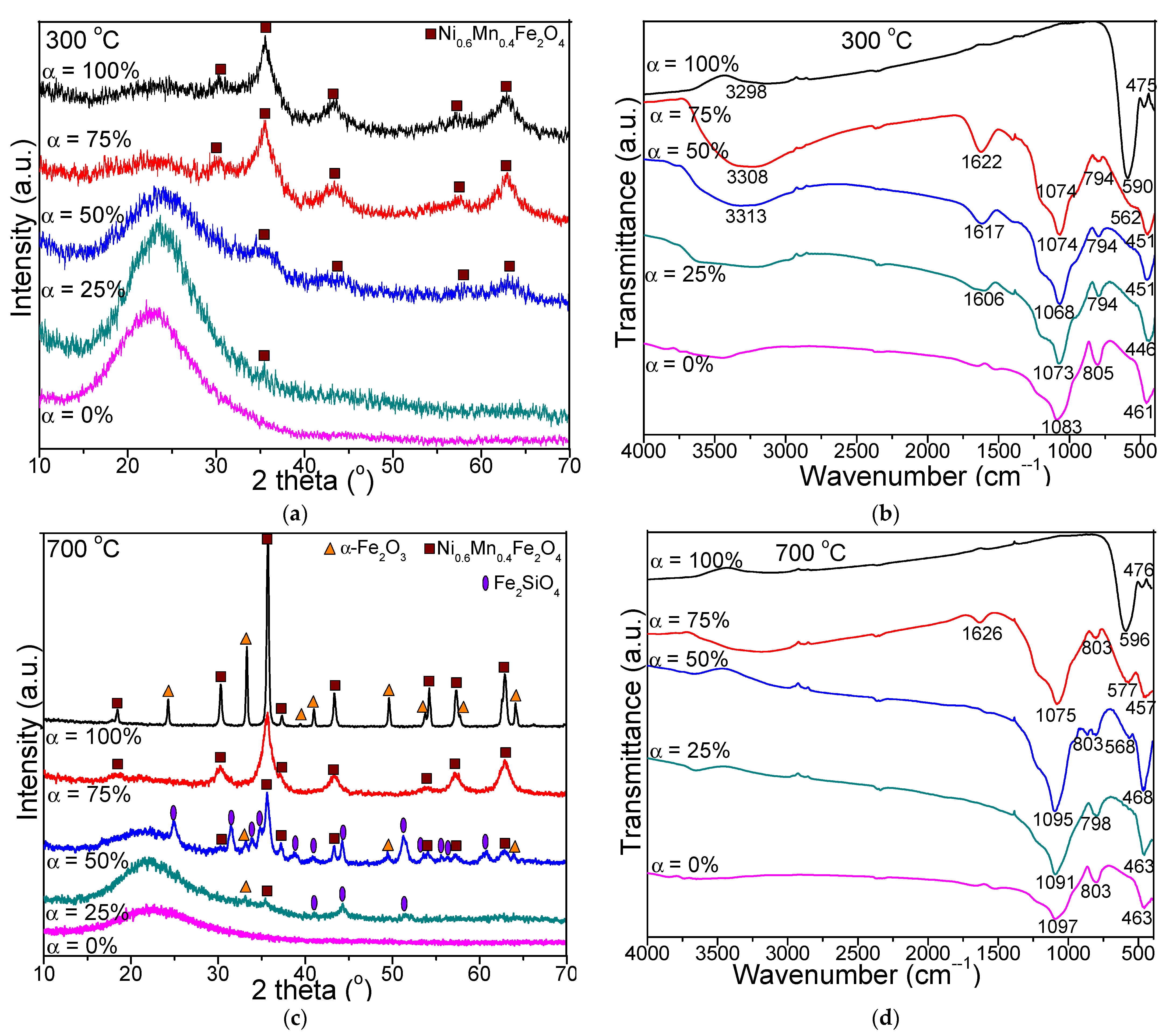
The XRD patterns and FT-IR spectra of the (Ni
0.6Mn
0.4Fe
2O
4)
α(SiO
2)
100−α (α = 0, 25, 50, 75, 100%) NCs calcined at 300, 700 and 1100 °C are presented in
Figure 1. At all calcination temperatures, in case of NCs with α = 0%, the formation of amorphous SiO
2 matrix is supported by the broad halo located at 2θ = 15–30° in the XRD pattern. At 300 °C, the NC with α = 25% is amorphous, the nano-crystalline state developing by increasing the value of α (
Figure 1a). In case of the NCs calcined at 700 and 1100 °C (
Figure 1c,e), the observed peaks indicate the presence of the cubic spinel structure of Mn
xNi
1−xFe
2O
4. The MnFe
2O
4 (JCPDS card no. 74-2403) has a lattice parameter of 8.511 Å, while the NiFe
2O
4 (JCPDS card no. 10-0325) [
12] has a lattice parameter of 8.339 Å. The Mn
xNi
1−xFe
2O
4 is isostructural with the two structures mentioned above, Ni and Mn being in the same position with an occupancy factor of x for Mn and 1−x for Ni. The reflection planes (220), (311), (222), (400), (422), (511) (440) and (533) belonging to the angular positions at 2θ = 29.99°, 35.33°, 36.83°, 42.87°, 53.11°, 56.65°, 62.16° and 73.38° are consistent with the spinel structure corresponding to the Fd3m space group and match with the literature data [
13]. From the positions of diffraction lines for Mn
xNi
1−xFe
2O
4 result a lattice parameter of 8.44 Å. From the lattice parameter which has a linear dependence with x, results x = 0.6, and Ni
0.6Mn
0.4Fe
2O
4.
At 700 °C, in case of NC with α = 75%, the single and well-crystallized Ni
0.6Mn
04Fe
2O
4 is observed, while in the case of NC with α = 100%, the α-Fe
2O
3 (JCPDS card no. 89-0599 [
12]) secondary phase is also present. The presence of α-Fe
2O
3 might be attributed to partially embedding of the ferrite in the SiO
2 matrix, due to the low content or lack of SiO
2 and the short time or calcination temperature required to produce pure crystalline Ni
0.6Mn
04Fe
2O
4 phase [
5].
As the ferrite content decreases, in NCs with α = 25 and 50%, besides Ni
0.6Mn
04Fe
2O
4, the presence of α-Fe
2O
3 and Fe
2SiO
4 (JCPDS card no. 87-0315 [
12]) secondary phases is also noticed. We assume that the formation of Fe
2SiO
4 could be related to difficulty of oxygen diffusion from the pores of SiO
2 matrix and partial reduction of Fe
3+ into Fe
2+, which reacts with the SiO
2 matrix and forms Fe
2SiO
4 under the reducing condition produced by the decomposition of carboxylate precursors.
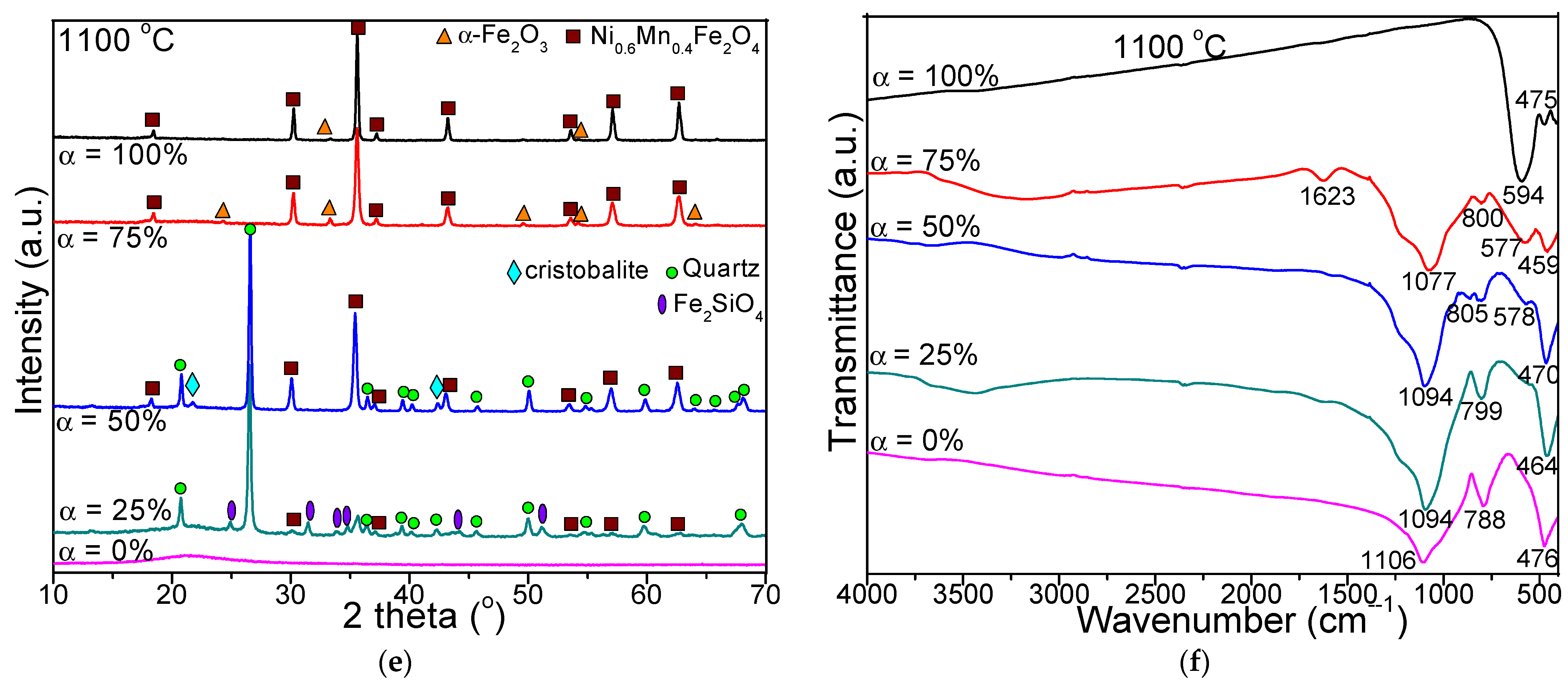
At 1100 °C, in case of NCs with α = 75–100% the well-crystallized Ni
0.6Mn
0.4Fe
2O
4 phase together with traces of α-Fe
2O
3 secondary phase are observed. In NCs with α = 50% containing ferrite and SiO
2 matrix in 1:1 molar ratio, besides the main phase of Ni
0.6Mn
04Fe
2O
4, the secondary phases of crystallized SiO
2 matrix are also noticed (cristobalite, JCPDS card no. 89-8936 and quartz, JCPDS card no. 89-8936 [
12]), while in NC with α = 25%, Fe
2SiO
4 is also obtained. Although it was reported that the thermal treatment may induce polymorphous transitions in Fe
2O
3, especially in the case of nanosized powders or nanoparticles embedded in amorphous and porous SiO
2 matrix, in our case only α-Fe
2O
3 was observed [
14]. The peaks corresponding to ferrite become more intense at 1100 °C, indicating high degree of crystallinity, crystallite size (due to the crystal coalescence process), nucleation rate and low effect of the inert surface layer [
5]. Also, the highest peak shifts to higher angles with increasing Ni
0.6Mn
04Fe
2O
4 content embedded in the SiO
2 matrix.
Among the available methods to estimate the crystallite size, those using the diffraction profile analysis, namely Williamson-Hall and Warren-Averbach procedures, require several diffraction profiles [
15,
16]. Considering that in our case, especially at low calcination temperatures, we have only few diffraction peaks, we estimated the average crystallite using the Scherrer method, which requires the full the width at half maximum (FWHM) for a single diffraction line [
9]. Though the X-ray profile analysis is an average method, it is still a reliable method for measuring the crystallite size, apart from transmission electron microscopy (TEM). The average crystallite size of NCs calculated using the Debye-Scherrer formula [
3,
17] are presented in
Table 1. The low ferrite content embedded in the amorphous SiO
2 matrix retards the expansion of the crystallite size, whereas high ferrite content favors both nucleation and growth of crystallite size at the nucleation centers, leading to higher crystallite size [
1]. By increasing the calcination temperature, the Ni
2+ and Fe
3+ ions tend to occupy specific positions in the crystal lattice of the ferrite [
18,
19]. The crystallites were more compact at low ferrite content embedding in SiO
2, since the smaller Ni
2+ ion can dissolve in the spinel lattice, while high ferrite content embedding in SiO
2 matrix causes the increase of the porosity leading to higher crystallite size [
18]. During the calcination process, coalescence occurs, the smaller crystallites being merged together to form the large crystallites [
7].
At all temperatures, the FT-IR spectra (
Figure 1b,d,f) of NCs with α = 25–100% show the absorption bands corresponding to the vibration of tetrahedral M–O (M=Ni, Mn) bonds at 568–596 cm
−1 and of octahedral M–O (M=Fe) bonds at 446–476 cm
−1 [
1,
3,
4,
17]. The different vibration frequencies of M–O groups are a consequence of the higher M–O bond length in octahedral (B) sites than that in tetrahedral (A) sites. The presence of these two absorption bands in FT-IR spectra confirms that the ferrites have cubic spinel structure. The intensity of the band at 568–596 cm
−1 is larger than that of 446-476 cm
−1, indicating that the vibration of tetrahedral M–O is higher than of octahedral M–O groups [
3]. Generally, the Ni
2+ ions occupy the octahedral (B) sites, whereas Mn
2+/Fe
3+ ions prefer both octahedral (B) and tetrahedral (A) sites [
17]. The absorption bands shifting to lower values is accredited to the movement of Fe
3+, Mn
2+ and Ni
2+ ions corresponding to the O
2− ions in the octahedral (B) and tetrahedral (A) sites, and consequently the change of the Fe
3+–O
2−(M
3+–O
2−) and M
2+–O
2− bond length, respectively [
4]. The intensity of the vibrational band at 568–596 cm
−1 increases with the increasing calcination temperature, due to the increasing ferrite crystallinity, since the ferrites consist of crystals bonded to all adjacent neighbors through ionic, covalent or van der Waals forces [
5,
11,
20,
21].
The small shift of the vibrational band originates from the movement of ions among the tetrahedral (A) and octahedral (A) sites as a result of the increasing calcination temperature [
5,
11,
21]. The characteristic bands of the SiO
2 matrix were detected in the FT-IR spectra of NCs with α = 0–75%, as follows: 1068–1106 cm
−1 with a shoulder at about 1200 cm
−1 related to vibration of Si–O–Si chains, 788–805 cm
−1 related to the vibrations of SiO
4 tetrahedron and 446–476 cm
−1 related to the Si–O bond vibration and overlapping the band of Fe–O vibration [
5,
11]. The high intensity of these bands indicates a low polycondensation degree of the SiO
2 network [
5]. The broad peaks observed at 3298-3313 cm
−1 and at 1606–1626 cm
−1 are ascribed to the vibrations of the –OH group and hydrogen bonds from adsorbed water molecules [
1].
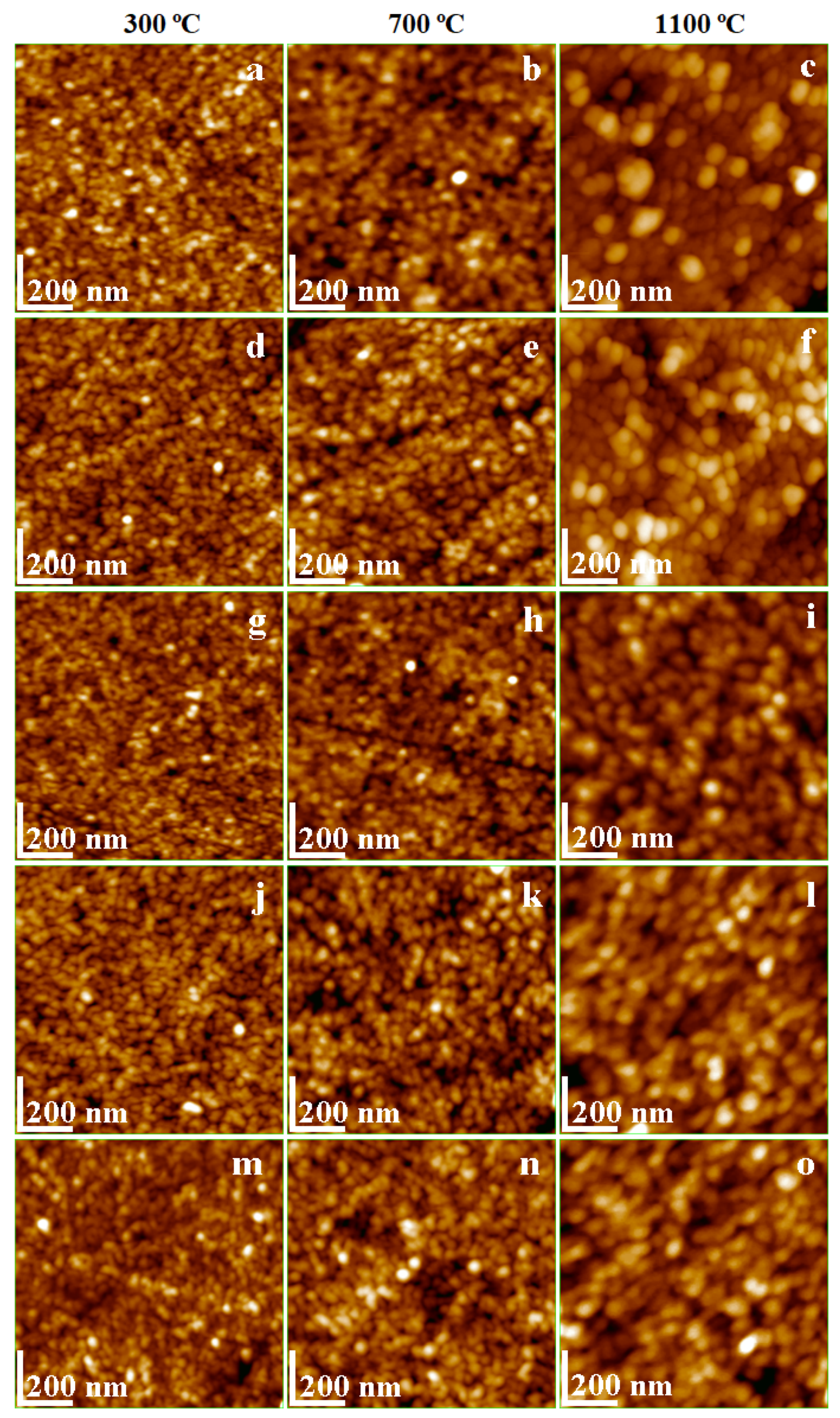
AFM was previously used to study the temperature effect on Ni and Mn ferrite nanoparticles transferred as thin film onto solid substrate. Ashiq et al. evidenced by AFM that Ni ferrite nanoparticles dispersion in liquid environment is proper to obtain well-structured thin films [
22]. Moreover, Tong et al. reported particle diameters of 25 nm at 400 °C; 44 nm at 500 °C and 65 nm at 700 °C, and surface roughness depending on the nanoparticle disposal in the topography [
23].
The use of Mn ferrite nanoparticles as dispersed matter into the liquid environment as magnetic ink was also reported [
24]. The printed thin film investigated with AFM revealed Mn ferrite nanoparticles of about 95 nm and the surface roughness depending on the particle diameter and on the observed agglomeration tendency [
24]. The AFM topographic images are presented in
Figure 2a–o. A dependence of nanoparticle diameter on the calcination temperature was observed for pure Ni
0.6Mn
04Fe
2O
4 (
Figure 2a–c). The diameter of the round shape particles increases from about 18 nm at 300 °C to 52 nm at 700 °C, and 75 nm at 1100 °C, respectively. The crystallite size increase with the temperature increase was also observed based on the XRD data. The particle size revealed by AFM correlation with XRD crystallite size of pure Ni-Mn ferrite indicates a polycrystalline state at low temperatures (crystallite size is considerably smaller than particle size) and monocrystalline state (crystallite size is very close to the particle diameter) at 1100 °C. Establishing a certain number of crystallites per particle requires a more enhanced investigation based on scanning electron microscopy (SEM) and Brunauer–Emmett–Teller (BET) analysis [
16].
XRD patterns show that the SiO
2 matrix is amorphous at all calcination temperatures. However, the particle size and shape evolution with increasing temperature may be observed using AFM.
Figure 2m reveals small round shape nanoparticles and a diameter increasing with the calcination temperature, i.e., about 12 nm at 300 °C, 28 nm at 700 °C and 35 nm at 1100 °C (
Figure 2n,o). Previous studies confirm the shape and sizes of the silica nanoparticles observed by AFM [
25,
26].
The NCs with α = 25–75% combine the morphological and structural features of both Ni-Mn ferrite and amorphous SiO
2 nanoparticles. The diameter of the round-shape nanoparticles is strongly influenced by the calcination temperature and composition (
Figure 2d–l). The lowest size particles were observed at 300 °C and the bigger ones at 1100 °C (
Table 1). The amorphous SiO
2 matrix increases the particle size compared to the ferrite crystallites due to the embedding effect. This effect is more visible at 300 °C than at 1100 °C. At higher calcination temperatures, the ferrite crystallite is well covered by an amorphous SiO
2 layer which forms the composite nanoparticle. The insulating behavior of the amorphous SiO
2 matrix prevents the overgrowth of magnetic domains and guarantees the nano-structural stability. A slight decrease of the nanoparticle size occurs by increasing the amorphous SiO
2 content. This decrease is most obvious at 1100 °C (
Figure 2c,f,i,l), where the amorphous SiO
2 matrix inhibits the development of bigger ferrite crystallites (
Table 1). A similar behavior was reported for other ferrite systems [
5,
11].
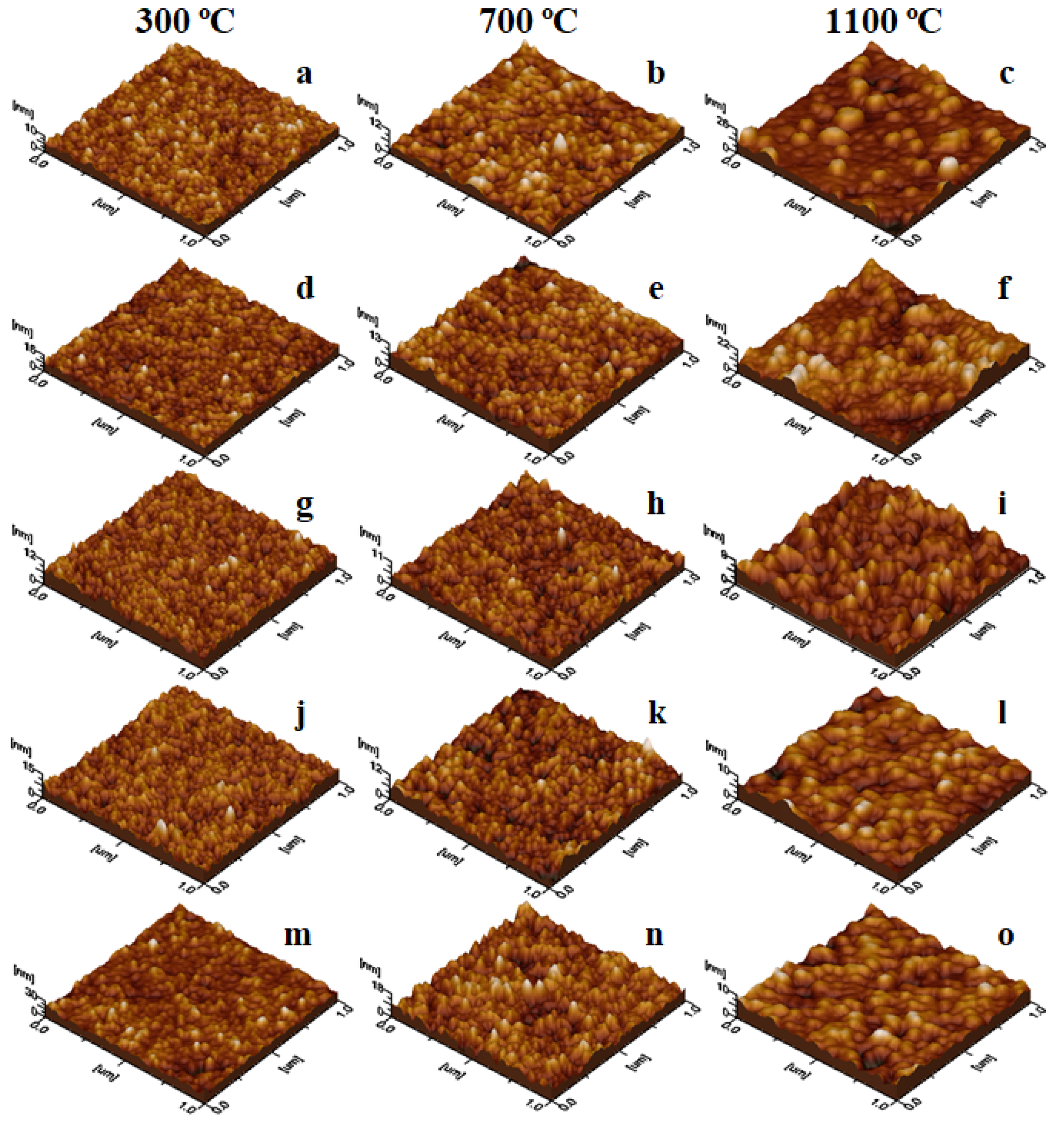
The powder dispersion in an aqueous environment facilitates the nanoparticle arrangement, assuring a uniform adsorption onto the solid substrate creating well-structured thin films [
27], as observed in
Figure 3a–o. The film roughness depends on the nanoparticle diameter and their disposal on the substrate surface. Thus, the lower roughness values are obtained at 200 °C (
Figure 3a,d,g,j,m) due to the uniform adsorption of fine nanoparticles. The particle diameter increases with the calcination temperature, while the adsorbed film uniformity depends on the local heights formed by bigger nanoparticles (
Figure 3c,f,i,l).
The morphological aspects of the nanoparticle thin films revealed by AFM correlated with the magnetic properties allow the design of functionalized surfaces for various applications where thermal deposition at high temperatures it is not possible, i.e., such as polymer coating.
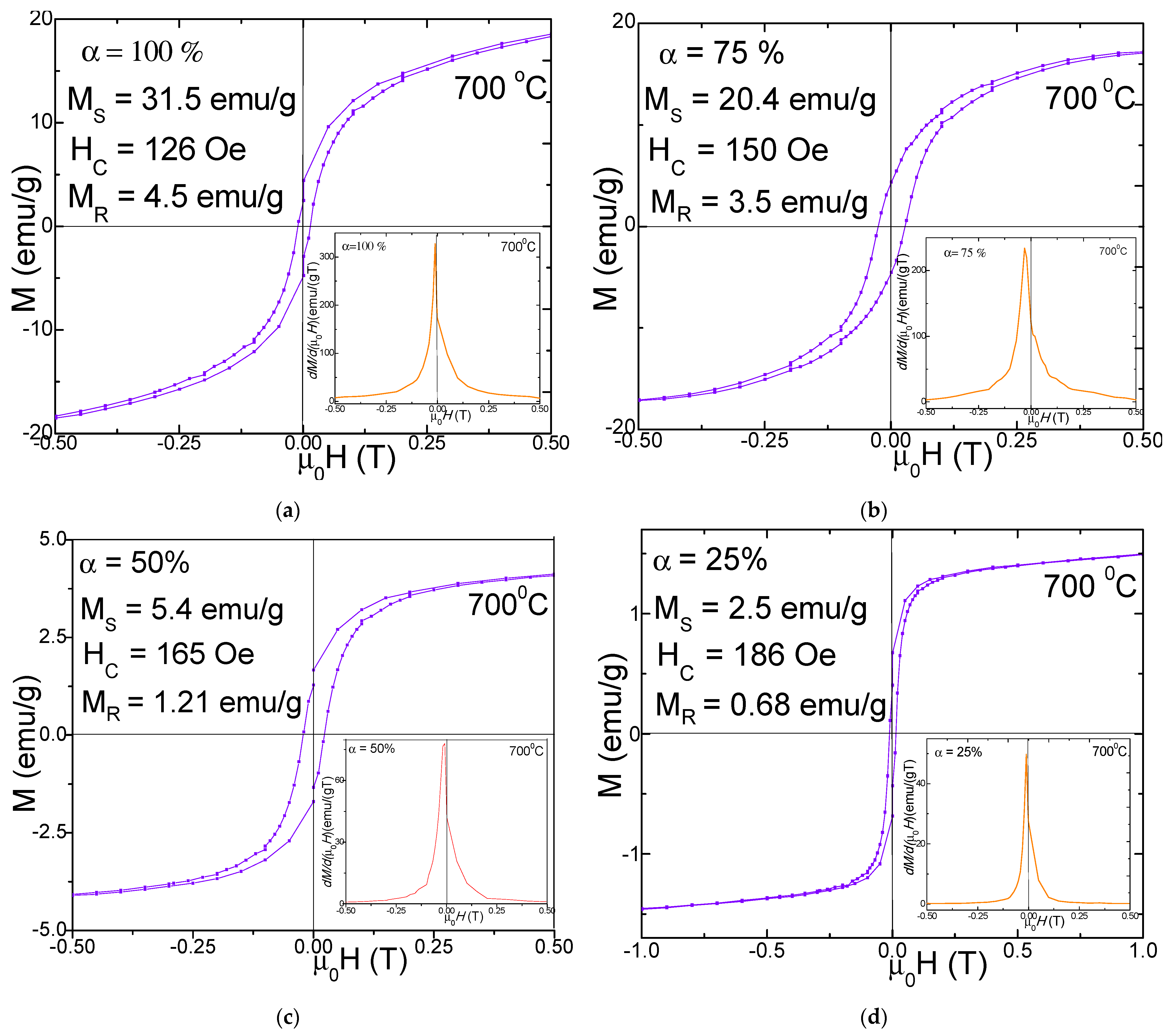
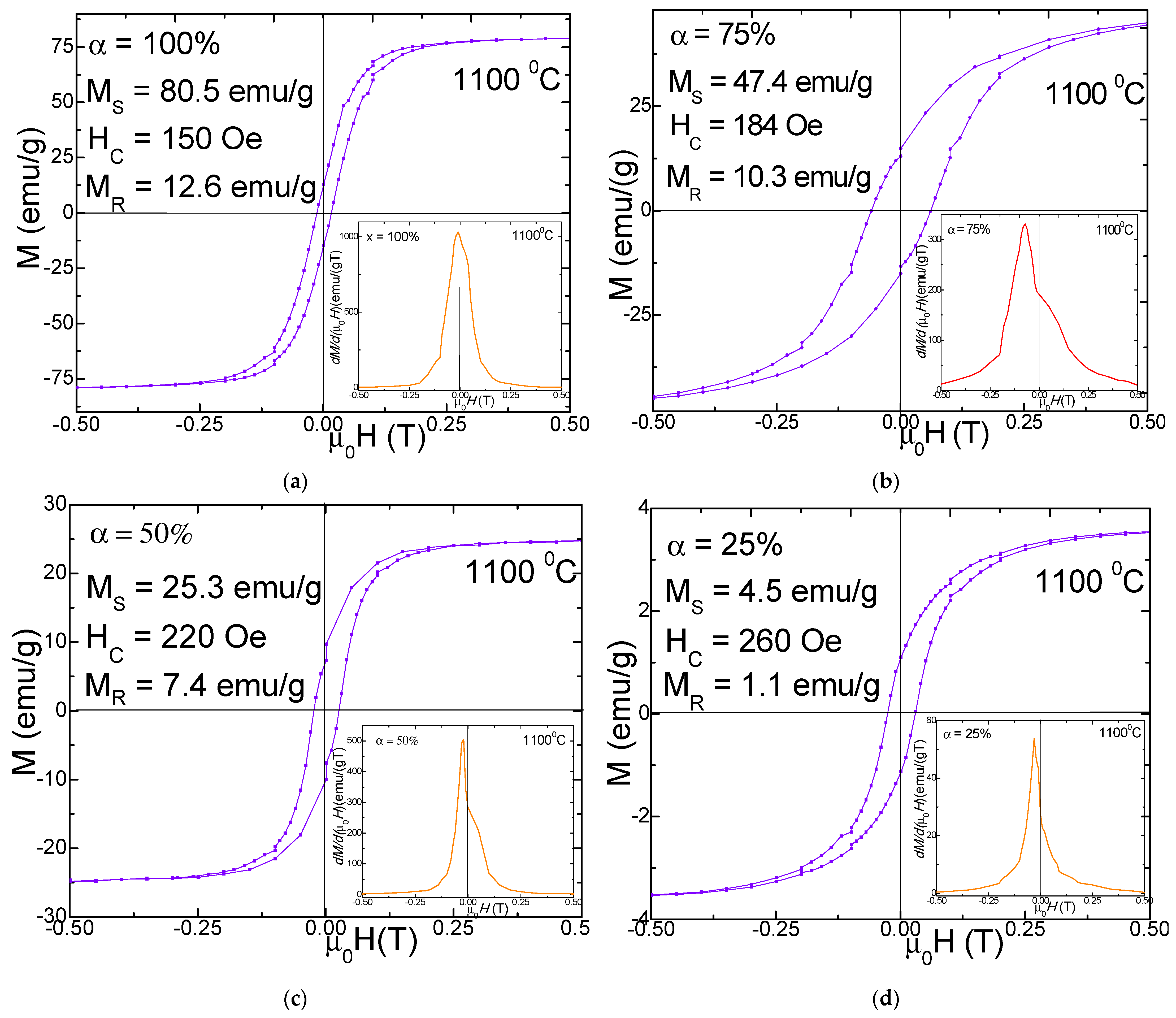
Figure 4 and
Figure 5 display the magnetic hysteresis loops and
dM/
d(
µ0H)) derivatives (in insets) as well as the saturation magnetization (
MS), remnant magnetization (
MR) and coercivity (
HC) values for (N
i0.
6Mn
0.4Fe
2O
4)
α(SiO
2)
100−α (α = 25–100%) NCs calcined at 700 and 1100 °C. The hysteresis loops are very narrow, indicating that the nanoparticles have soft magnetic behavior. The derivatives of the hysteresis loops (total susceptibility) represent the local slope of M-H curves. A single sharp maximum in the
dM/
d(
µ0H) vs. H curves suggests the presence of a single magnetic phase.
The peaks’ broadening indicates a larger distribution of the particle sizes. For the NCs with α = 25–100%,
dM/d(
µ0H) vs. H curves have a single and sharp peak. The morphology and the phase purity of NCs, as well as their magnetic properties, are strongly affected by the calcination temperature [
3,
5]. The SiO
2 matrix has diamagnetic behavior for both 700 and 1100 °C calcination temperatures. For the NCs with α = 100% (Ni
0.6Mn
0.4Fe
2O
4), typical hysteresis loops for ferromagnetic materials were obtained, for all the calcination temperatures, due to the presence of larger size crystallites and particles as found in XRD and AFM analyses. The unembedded Ni
0.6Mn
0.4Fe
2O
4 (α = 100%) has a much higher
MS, especially when it is calcined at 1100 °C, than the ferrites embedded in the SiO
2 matrix (α = 25–75%), with pretty narrow hysteresis loops, close to a superparamagnetic behavior.
The superparamagnetic-like behavior of the NCs is a consequence of the low sizes of the crystallites and of their low magnetic anisotropy which allow their easily thermal activation [
3,
4]. The increase of the calcination temperature can lead to the improvement of
MS and
MR, as a result of a better crystallinity of the ferrite, of proper interatomic lengths changing of the atomic coordination number, etc. The
MS values of NCs with high content of Ni
0.6Mn
0.4Fe
2O
4 embedded in the SiO
2 matrix are larger due to larger particles sizes which show reduced spin canting and other surface effects which are usually present in small size particles. The main mechanisms of the magnetization process are the domain wall motions and the magnetic moment rotations [
3]. The spin disorder on the nanoparticle surface can also strongly affect the
MS value. Moreover, the lattice defects can weaken the magnetic super-exchange interaction between the tetrahedral (A) and the octahedral (B) sites [
3]. The involved magnetic Fe
3+, Ni
2+ and Mn
2+ ions have magnetic moments with the following values: 5, 2 and 5 μ
B respectively [
20]. The distribution of cations between tetrahedral (A) and octahedral (B) sites of the spinel decides the magnetic moment per formula unit. The addition of Mn
2+ ions in the Ni ferrite can induce a migration of the Fe
3+ ions from the tetrahedral (A) to the octahedral (B) sites leading to a spin imbalance between the two sites, resulting in an increase of the magnetization at the octahedral (B) sites [
20]. The surface energy of nanosized particles is large and can modify the typical cation distribution between the A and B sites [
3,
5]. The SiO
2 matrix can partially dilute the magnetic matrix of the cations and it can create disorder at the surface of the particles and increase the number of defects, broken bonds, canted spins, and pinning of the magnetic field lines [
2,
5]. The nanoparticles calcined at 700 °C have rather low values of
MS since they show lower crystallinity, large defect concentration, reduced coordination number and increased interatomic spacing [
5]. The
MS values of NCs calcined at 700 °C increase with increasing N
i0.
6Mn
0.4Fe
2O
4 content, not far from a linear dependence, from 2.5 emu/g (α = 25%) to 31.5 emu/g (α = 100%). This behavior indicates that the main contribution to magnetization is given by the ferrite content in the samples. A possible explanation of the deviation from the linear dependence can be the disorder of magnetic moments on the surface of particles, mainly for the small size particles which have a higher surface-to-volume ratio [
2,
5]. The increase of
MS with increasing particle sizes is typical for nano-sized ferrites [
28]. Excepting the sample with α = 25%, there is a very good proportionality between particle and the crystallite sizes. The crystallite sizes also increase continuously with the ferrite content. This behavior suggests that the SiO
2 content has a negligible effect on the interaction between the magnetic moments of the cations from tetrahedral (A) and octahedral (B) sites, i.e., the magnetic order is not significantly changed by the SiO
2 matrix. The
HC decreases with increasing ferrite content, or with growing of the crystallite sizes as expected for multi-domain nanoparticles [
28,
29]. The
HC decreases from 185 Oe (α = 25%) to 126 Oe (α = 100%). The
MR decreases from 4.5 emu/g (α = 100%) to 0.68 emu/g (α = 25%) mainly due to the increasing disorder of the magnetic moments in the outer shell of the smaller sized particles [
2,
5]. The magnetic properties of these NCs are also affected by their bulk densities and by their grain sizes and grain size distributions. The strain released by the larger particles is higher than those of the smaller ones, resulting in lattice expansion. The pores can also contribute to the magnetic properties of the NCs, acting as pinning centers for the domain walls and for the magnetic moments of the cations [
5]. The observed
MS values are in good agreement with the cation distribution theory and Neel’s molecular field model [
1]. The lower values of
MS for some of the NCs can be explained by the effect of the spin canting in the frame of the non-colinear Yafet-Kittel model in the presence of Jahn-Teller cations [
8].
The coercive field,
HC, is given mainly by the magneto-crystalline anisotropy, but also by the exchange anisotropy due to the magnetic moment’s interaction from the particle surface [
15]. Generally, the M–H curves do not reach complete magnetic saturation, even in 10 T. For these cases, the
MS was estimated by using the law of approach to magnetic saturation [
30,
31]. The absence of complete saturation in ferromagnetic nanoparticles is generally related to the magnetic moments’ disorder in the surface layers of the particles which needs a larger magnetic field for saturation, in association with the lower anisotropy of the smaller sized particles [
10]. The
HC values are rather low, in the range from 126 to 260 Oe. As can be seen, the
MS increases for the NCs with lower SiO
2 matrix content. This behavior can be related to the decrease of the particle sizes with SiO
2 content increase and the associated micro-strains, and probably, the magnetic particles morphology and magnetic domain sizes [
3]. The
HC decreases nearly linearly with increasing SiO
2 content due to a continuous decrease of the crystallite sizes in the single-domain range under the influence of the SiO
2 matrix [
9]. The larger sized nanoparticles are composed of multi-domains, where the
HC decreases due to the formation of domain walls in the nanoparticles [
7]. The measured
MS values of our previously reported (Zn
0.6Mn
0.4Fe
2O
4)
α(SiO
2)
100−α (α = 100%) NCs [
5] are similar to those belonging to the (N
i0.
6Mn
0.4Fe
2O
4)
α(SiO
2)
100−α (α = 100%) NCs. The
MS of both systems keeps the same trend, decreasing with increasing SiO
2 matrix content, which results in decreasing particle sizes. The NCs calcined at 1100 °C from the both series behave similarly showing the enhancement of the
HC with increasing SiO
2 matrix content, in spite of the much larger values of
HC for the Zn
0.6Mn
0.4Fe
2O
4)
α(SiO
2)
100−α nanoparticles. These behaviors are typical for particle sizes belonging to the multi-domain range [
5,
28,
29]. The Ni-Mn ferrites calcined at 700 °C also belong to this category, while the previous Zn-Mn ferrites calcined at 700 °C (with smaller particle sizes) behave differently, with a
HC which depreciates with decreasing SiO
2 matrix content (or with increasing particle sizes), suggesting that most of the particles have sizes belonging to single-domain range.
The obtained Ni
0.6Mn
0.4Fe
2O
4)
α(SiO
2)
100−α NCs belong to an important group of materials with potential for technical application in many biomedical and industrial fields such as drug delivery [
30], hyperthermia and healthcare treatment [
31,
32], biocompatible magnetic fluids [
33], magnetic resonance imaging contrast enhancement [
34], magnetic data recording [
35], microwave applications [
36], supercapacitors [
37] since these nanoparticles (being passivated) have low toxicity and can be operated by magnetic and electric fields [
38,
39].


 ,
, 


