
Observation of Cu Spin Fluctuations in High-Tc Cuprate Superconductor Nanoparticles Investigated by Muon Spin Relaxation

 , , ,
, , ,
Abstract
:1. Introduction
2. Materials and Methods
3. Results and Discussion
4. Conclusions
Author Contributions
Funding
Institutional Review Board Statement
Informed Consent Statement
Data Availability Statement
Acknowledgments
Conflicts of Interest
References
- Chowdhuri, A.; Gupta, V.; Sreenivas, K.; Kumar, R.; Mozumdar, S.; Patanjali, P.K. Response speed of SnO2-based H2S gas sensors with CuO nanoparticles. Appl. Phys. Lett. 2004, 84, 1180–1182. [Google Scholar] [CrossRef]
- Switzer, J.A.; Kothari, H.M.; Polzot, P.; Nakanishi, S.; Bohannan, E.W. Enantiospecific electrodeposition of a chiral catalyst. Nature 2003, 425, 490–493. [Google Scholar] [CrossRef] [PubMed]
- Whitesides, G.M. The ‘right’ size in nanobiotechnology. Nat. Biotechnol. 2003, 21, 1161–1165. [Google Scholar] [CrossRef] [PubMed]
- Thurn-Albrecht, T.; Schotter, J.; Kastle, G.A.; Emley, N.; Shibauchi, T.; Krusin-Elbaum, L.; Guarini, K.; Black, C.T.; Tuominen, M.T.; Russell, T.P. Ultrahigh-density nanowire arrays grown in self-assembled diblock copolymer templates. Science 2000, 290, 2126–2129. [Google Scholar] [CrossRef] [PubMed] [Green Version]
- Chikazumi, S.; Taketomi, S.; Ukita, M.; Mizukami, M.; Miyajima, H.; Setogawa, M.; Kurihara, Y. Quelle magn. Flüssigkeit Invited paper physics of magnetic fluids. J. Magn. Magn. Mater. 1987, 65, 245–251. [Google Scholar] [CrossRef]
- Punnoose, A.; Magnone, H.; Seehra, M.S.; Bonevich, J. Bulk to nanoscale magnetism and exchange bias in CuO nanoparticles. Phys. Rev. B 2001, 64, 1–8. [Google Scholar] [CrossRef]
- Zheng, X.G.; Xu, C.N.; Nishikubo, K.; Nishiyama, K.; Higemoto, W.; Moon, W.J.; Tanaka, E.; Otabe, E.S. Finite-size effect on Néel temperature in antiferromagnetic nanoparticles. Phys. Rev. B 2005, 72, 014464. [Google Scholar] [CrossRef] [Green Version]
- Zheng, X.G.; Kubozono, H.; Yamada, H.; Kato, K.; Ishiwata, Y.; Xu, C.N. Giant negative thermal expansion in magnetic nanocrystals. Nat. Nanotechnol. 2008, 3, 724–726. [Google Scholar] [CrossRef]
- Nealon, G.L.; Donnio, B.; Greget, R.; Kappler, J.P.; Terazzi, E.; Gallani, J.L. Magnetism in gold nanoparticles. Nanoscale 2012, 4, 5244–5258. [Google Scholar] [CrossRef] [PubMed]
- Dehn, M.H.; Arseneau, D.J.; Buck, T.; Cortie, D.L.; Fleming, D.G.; King, S.R.; MacFarlane, W.A.; McDonagh, A.M.; McFadden, R.M.L.; Mitchell, D.R.G.; et al. Nature of magnetism in thiol-capped gold nanoparticles investigated with Muon spin rotation. Appl. Phys. Lett. 2018, 112, 053105. [Google Scholar] [CrossRef] [Green Version]
- Yamamoto, Y.; Miura, T.; Suzuki, M.; Kawamura, N.; Miyagawa, H.; Nakamura, T.; Kobayashi, K.; Teranishi, T.; Hori, H. Direct observation of ferromagnetic spin polarization in gold nanoparticles. Phys. Rev. Lett. 2004, 1–4. [Google Scholar] [CrossRef] [PubMed] [Green Version]
- Zheng, X.G.; Mori, T.; Nishiyama, K.; Higemoto, W.; Xu, C.N. Dramatic suppression of antiferromagnetic coupling in nanoparticle CuO. Solid State Commun. 2004, 132, 493–496. [Google Scholar] [CrossRef]
- Karthik, K.; Victor Jaya, N.; Kanagaraj, M.; Arumugam, S. Temperature-dependent magnetic anomalies of CuO nanoparticles. Solid State Commun. 2011, 151, 564–568. [Google Scholar] [CrossRef]
- Batsaikhan, E.; Lee, C.H.; Hsu, H.; Wu, C.M.; Peng, J.C.; Ma, M.H.; Deleg, S.; Li, W.H. Largely Enhanced Ferromagnetism in Bare CuO Nanoparticles by a Small Size Effect. ACS Omega 2020, 5, 3849–3856. [Google Scholar] [CrossRef] [PubMed] [Green Version]
- Yin, Y.; Liu, H.; Xie, L.; Su, T.; Teng, M.; Li, X. Coexistence of superconductivity and ferromagnetism in La2−xSrxCuO4 nanoparticles. J. Phys. Chem. C 2013, 117, 3028–3035. [Google Scholar] [CrossRef]
- Tanabe, Y.; Adachi, T.; Noji, T.; Koike, Y. Superconducting volume fraction in overdoped regime of La2−xSrxCuO4: Implication for phase separation from magnetic-susceptibility measurement. J. Phys. Soc. Jpn. 2005, 74, 2893–2896. [Google Scholar] [CrossRef] [Green Version]
- Adachi, T.; Oki, N.; Yairi, S.; Koike, Y.; Watanabe, I. Effects of Zn and Ni substitution on the Cu spin dynamics and superconductivity in La2−xSrxCu1−y(Zn,Ni)yO4 (x = 0.15–0.20): Muon spin relaxation and magnetic susceptibility study. Phys. Rev. B 2008, 78, 134515. [Google Scholar] [CrossRef] [Green Version]
- Croft, T.P.; Lester, C.; Senn, M.S.; Bombardi, A.; Hayden, S.M. Charge density wave fluctuations in La2−xSrxCuO4 and their competition with superconductivity. Phys. Rev. B 2014, 89, 1–8. [Google Scholar] [CrossRef] [Green Version]
- Keimer, B.; Kivelson, S.A.; Norman, M.R.; Uchida, S.; Zaanen, J. From quantum matter to high-temperature superconductivity in copper oxides. Nature 2015, 518, 179–186. [Google Scholar] [CrossRef]
- Koike, Y.; Adachi, T. μSR studies on magnetism in High-Tc cuprates. J. Phys. Soc. Jpn. 2016, 85, 1–12. [Google Scholar] [CrossRef]
- Watanabe, I.; Kawano, K.; Kumagai, K.; Nishiyama, K.; Nagamine, K. Magnetic order of Cu-moments in La2−xBaxCuO4 around x = 0.12 observed by positive muon spin rotation. J. Phys. Soc. Jpn. 1992, 61, 3058–3061. [Google Scholar] [CrossRef]
- Tranquada, J.M.; Sternlieb, B.J.; Axe, J.D.; Nakamura, Y.; Uchida, S. Evidence for stripe correlations of spins and holes in copper oxide superconductors. Nature 1995, 375, 561–563. [Google Scholar] [CrossRef]
- Mikuni, H.; Adachi, T.; Yairi, S.; Kato, M.; Koike, Y.; Watanabe, I.; Nagamine, K. 1/8 anomaly in the excess-oxygen-doped La1.8Nd0.2Cu1−yZnyO4+δ. Phys. Rev. B 2003, 68, 1–8. [Google Scholar] [CrossRef] [Green Version]
- Guguchia, Z.; Das, D.; Wang, C.N.; Adachi, T.; Kitajima, N.; Elender, M.; Brückner, F.; Ghosh, S.; Grinenko, V.; Shiroka, T.; et al. Using uniaxial stress to probe the relationship between competing superconducting states in a cuprate with spin-stripe order. Phys. Rev. Lett. 2020, 125, 97005. [Google Scholar] [CrossRef]
- Kopp, A.; Ghosal, A.; Chakravarty, S. Competing ferromagnetism in high-temperature copper oxide superconductors. Proc. Natl. Acad. Sci. USA 2007, 104, 6123–6127. [Google Scholar] [CrossRef] [Green Version]
- Sonier, J.E.; Kaiser, C.V.; Pacradouni, V.; Sabok-Sayr, S.A.; Cochrane, C.; MacLaughlin, D.E.; Komiya, S.; Hussey, N.E. Direct search for a ferromagnetic phase in a heavily overdoped nonsuperconducting copper oxide. Proc. Natl. Acad. Sci. USA 2010, 107, 17131–17134. [Google Scholar] [CrossRef] [Green Version]
- Gaur, A.; Varma, G.D. Sintering temperature effect on electrical transport and magnetoresistance of nanophasic La0.7Sr0.3MnO3. J. Phys. Condens. Matter 2006, 18, 8837–8846. [Google Scholar] [CrossRef]
- Li, Y.; Huang, J.; Cao, L.; Wu, J.; Fei, J. Optical properties of La2CuO4 and La2−xCaxCuO4 crystallites in UV—vis –NIR region synthesized by sol–gel process. Mater. Charact. 2012, 64, 36–42. [Google Scholar] [CrossRef]
- Kurashima, K.; Adachi, T.; Suzuki, K.M.; Fukunaga, Y.; Kawamata, T.; Noji, T.; Miyasaka, H.; Watanabe, I.; Miyazaki, M.; Koda, A.; et al. Development of ferromagnetic fluctuations in heavily overdoped (Bi,Pb)2Sr2CuO6+δ copper oxides. Phys. Rev. Lett. 2018, 121, 057002. [Google Scholar] [CrossRef] [Green Version]
- Toby, B.H.; Von Dreele, R.B. GSAS-II: The genesis of a modern open-source all purpose crystallography software package. J. Appl. Crystallogr. 2013, 46, 544–549. [Google Scholar] [CrossRef]
- Watanabe, I.; Adachi, T.; Yairi, S.; Koike, Y.; Nagamine, K. Change of the dynamics of internal fields in the normal state of La2-xSrxCuO4 observed by Muon-Spin-Relaxation. J. Phys. Soc. Jpn. 2008, 77, 1–6. [Google Scholar] [CrossRef]
- Tuilier, M.H.; Chevalier, B.; Tressaud, A.; Brisson, C.; Soubeyroux, J.L.; Etourneau, J. EXAFS study at the LaLIII X-ray absorption edge of superconducting materials obtained by fluorination of La2−xSrxCuO4 oxides (0 ≤ x ≤ 0.15). Phys. C 1992, 200, 113–121. [Google Scholar] [CrossRef]
- Winarsih, S.; Budiman, F.; Tanaka, H.; Adachi, T. Variable range hopping resistivity in La2−xSrxCuO4 nanoparticles evaluated by four point probe method. Key Eng. Mater. 2020, 860, 142–147. [Google Scholar] [CrossRef]
- Borchert, H.; Shevchenko, E.V.; Robert, A.; Mekis, I.; Kornowski, A.; Gru, G.; Weller, H. Determination of Nanocrystal Sizes: A Comparison of TEM, SAXS, and XRD studies of highly monodisperse CoPt3 particles. Langmuir 2005, 21, 1931–1936. [Google Scholar] [CrossRef] [PubMed]
- Winarsih, S.; Budiman, F.; Tanaka, H.; Adachi, T.; Goto, T.; Soegijono, B.; Kurniawan, B.; Watanabe, I. Growth of free-standing La2−xSrxCuO4 nanoparticles. Mater. Sci. Forum 2019, 966, 357–362. [Google Scholar] [CrossRef]
- Collomb, D.; Zhang, M.; Yuan, W.; Bending, S.J. Imaging of strong nanoscale vortex pinning in GdBaCuO high-temperature superconducting tapes. Nanomaterials 2021, 11, 1082. [Google Scholar] [CrossRef]
- Naik, S.P.K.; Muralidhar, M.; Jirsa, M.; Murakami, M. Enhanced flux pinning of single grain bulk (Gd, Dy)BCO superconductors processed by cold-top-seeded infiltration growth method. Mater. Sci. Eng. 2020, 253, 114494. [Google Scholar] [CrossRef]
- Adachi, T.; Yairi, S.; Koike, Y.; Watanabe, I.; Nagamine, K. Muon-spin-relaxation and magnetic-susceptibility studies of the effects of the magnetic impurity Ni on the Cu-spin dynamics and superconductivity in La2−xSrxCu1−yNiyO4 with x = 0.13. Phys. Rev. B 2004, 70, 13–16. [Google Scholar] [CrossRef] [Green Version]
- Adachi, T.; Yairi, S.; Takahashi, K.; Koike, Y.; Watanabe, I.; Nagamine, K. Muon spin relaxation and magnetic susceptibility studies of the effects of nonmagnetic impurities on the Cu spin dynamics and superconductivity in La2−xSrxCu1−yZnyO4 around x = 0.115. Phys. Rev. B 2004, 69, 184507. [Google Scholar] [CrossRef] [Green Version]


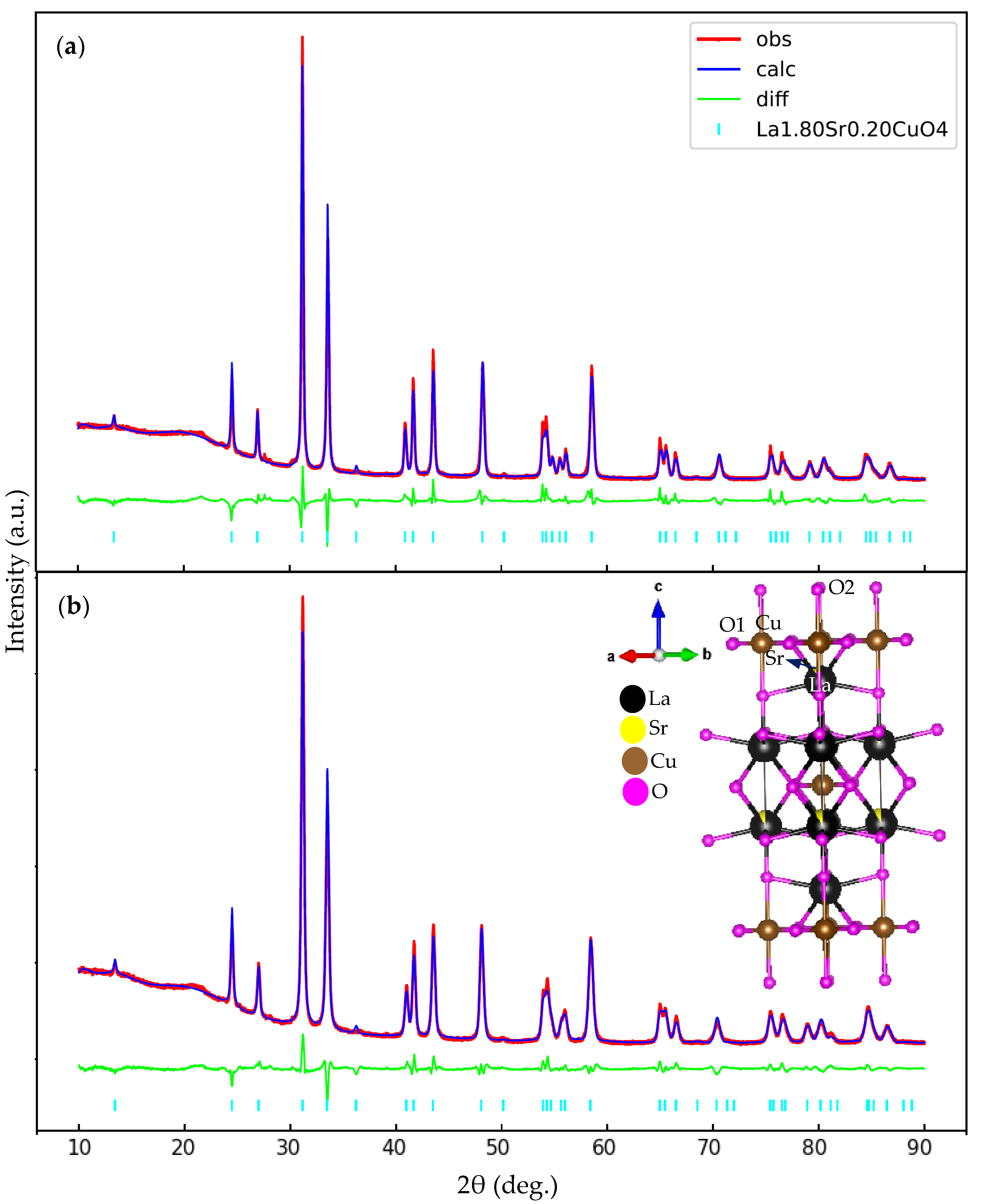
| Parameter | Sintering Condition | |
|---|---|---|
| 1073 K for 180 min | 973 K for 180 min | |
| Crystal structure | Tetragonal | Tetragonal |
| Space Group | I4/mmm | I4/mmm |
| Lattice constant a = b (Å) | 3.7722 | 3.7819 |
| Lattice constant c (Å) | 13.2295 | 13.2086 |
| Volume of unit cell (Å3) | 188.249 | 188.923 |
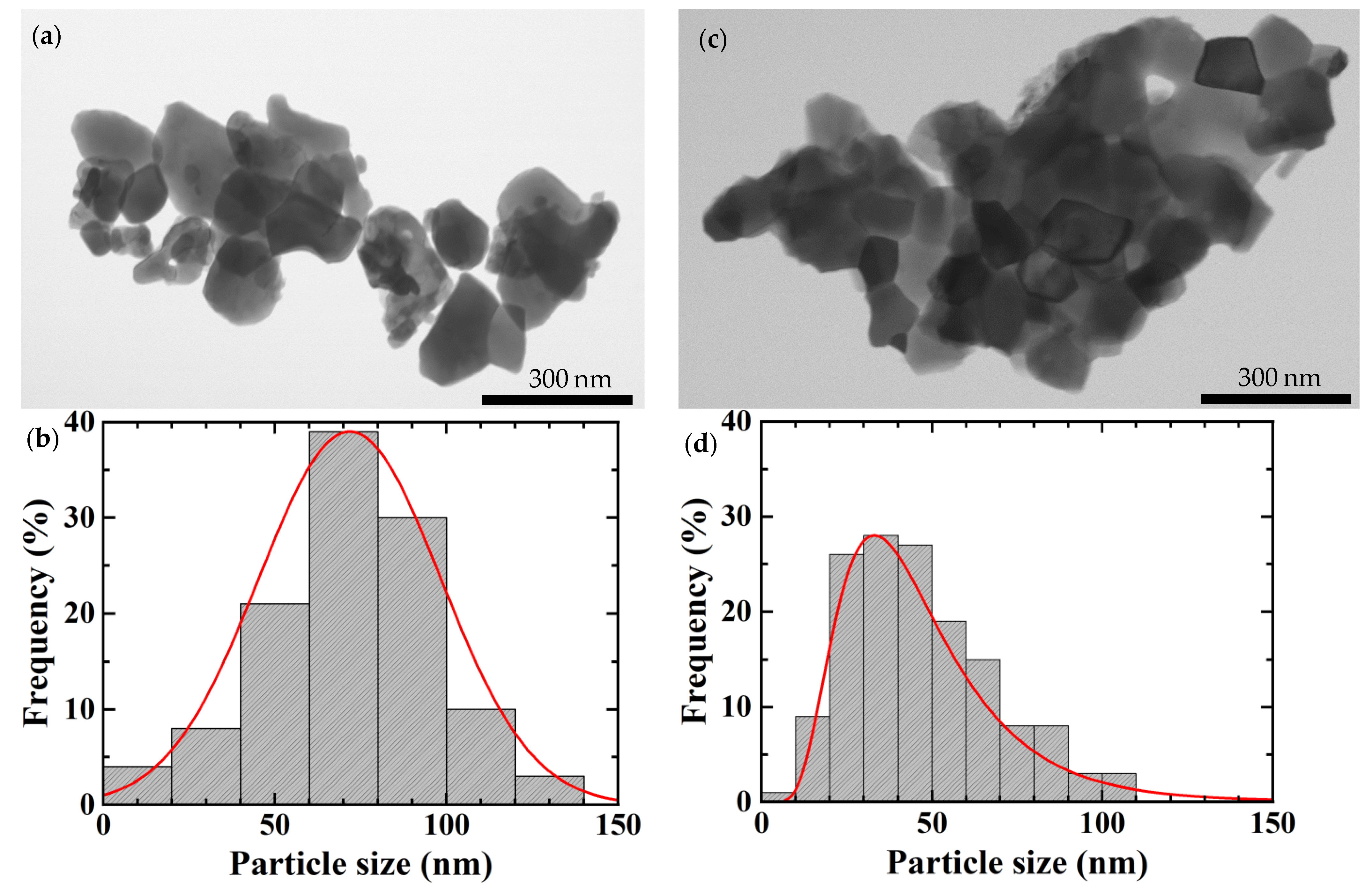
| Average crystallite size (nm) | 64 | 39 |
| Atom | Wyckoff Position | Site Symmetry | x | y | z | Site Occupation Factor |
|---|---|---|---|---|---|---|
| La1 | 4e | 4 mm (z) | 0 | 0 | 0.3609 (4) | 0.9 |
| Sr1 | 4e | 4 mm (z) | 0 | 0 | 0.3609 (4) | 0.1 |
| Cu1 | 2a | 4/mmm (z) | 0 | 0 | 0 | 1.0 |
| O1 | 4c | mmm | 0 | 1/2 | 0 | 1.0 |
| O2 | 4e | 4 mm (z) | 0 | 0 | 0.184 (4) | 1.0 |
| Name of Sample | Bond Distances (Å) | ||
|---|---|---|---|
| La1|Sr1–O2 | Cu1–O1 | Cu1–O2 | |
| Sample Reference [32] | 2.3351 | 1.8896 | 2.4288 |
| 64 nm | 2.3402 | 1.8861 | 2.4342 |
| 39 nm | 2.3366 | 1.8910 | 2.4304 |
Publisher’s Note: MDPI stays neutral with regard to jurisdictional claims in published maps and institutional affiliations. |
© 2021 by the authors. Licensee MDPI, Basel, Switzerland. This article is an open access article distributed under the terms and conditions of the Creative Commons Attribution (CC BY) license (https://creativecommons.org/licenses/by/4.0/).
Share and Cite
Winarsih, S.; Budiman, F.; Tanaka, H.; Adachi, T.; Koda, A.; Horibe, Y.; Kurniawan, B.; Watanabe, I.; Risdiana, R. Observation of Cu Spin Fluctuations in High-Tc Cuprate Superconductor Nanoparticles Investigated by Muon Spin Relaxation. Nanomaterials 2021, 11, 3450. https://doi.org/10.3390/nano11123450
Winarsih S, Budiman F, Tanaka H, Adachi T, Koda A, Horibe Y, Kurniawan B, Watanabe I, Risdiana R. Observation of Cu Spin Fluctuations in High-Tc Cuprate Superconductor Nanoparticles Investigated by Muon Spin Relaxation. Nanomaterials. 2021; 11(12):3450. https://doi.org/10.3390/nano11123450
Chicago/Turabian StyleWinarsih, Suci, Faisal Budiman, Hirofumi Tanaka, Tadashi Adachi, Akihiro Koda, Yoichi Horibe, Budhy Kurniawan, Isao Watanabe, and Risdiana Risdiana. 2021. "Observation of Cu Spin Fluctuations in High-Tc Cuprate Superconductor Nanoparticles Investigated by Muon Spin Relaxation" Nanomaterials 11, no. 12: 3450. https://doi.org/10.3390/nano11123450