Sensors play a critical role in monitoring human health and environmental safety. Unusually high or low levels of biological molecules, such as glucose and dopamine, can cause severe damage to the human body. High concentrations of pollutants, such as H2O2, nitrogen-based toxins, and metal ions, harm humans and the environment. Various LDH-based sensors have been developed as fast, affordable, and accurate sensors for many critical chemical markers. Herein, the design and modifications of various LDHs and LDH composites are examined, emphasizing the effects of LDH morphology, crystallinity, and composition on analytical performance.
4.1. Glucose
The ability to quickly and accurately monitor glucose levels in the blood is critical for diagnosing, managing, and treating diabetes [
113]. If left unchecked, people with diabetes may experience fatal kidney failures, blindness, heart attacks, and strokes [
114]. Electrochemical glucose sensors detect electron flow from glucose oxidation into gluconolactone, promoted by a catalyst, such as an enzyme, noble metal, transition metals, or metal oxides [
115]. Various LDHs have been fabricated as catalysts for electrochemical and optical glucose detection.
One of the largest disadvantages of fabricating LDH-based glucose sensors is the reduced conductivity due to the insulative property of LDHs and the binders used to immobilize the LDHs onto the conductive substrate. One method of improving the electrochemical glucose detection mechanism is to enhance conductivity by adding conductive nanoparticles, such as Ni nanoparticles, to the LDHs. LDHs can be partially reduced to yield free metal nanoparticles that reduce the impedance of the electrochemical probe. CoNi-LDHs have received much attention as efficient catalysts for glucose electrooxidation, owing to the similar potentials between Co and Ni that synergize to increase the composition of their electronic states. However, CoNi-LDHs exhibit poor conductivity that can be improved by LDHs comprised of conductive nanomaterials. Chen et al. extracted Ni nanoparticles by partially reducing CoNi-LDHs to yield a nanocomposite with enhanced conductivity for improved glucose detection [
116]. The CoNi-LDH was first synthesized via a hydrothermal method with Ni(NO
3)
2·6H
2O, Co(NO
3)
2·6H
2O, and hexamethylenetetramine (HMT). The CoNi-LDH underwent partial hydrothermal reduction with Na
2HPO
4 and NaOH at 160 °C for 4 h to yield free Ni nanoparticles from the CoNi-LDHs (
Figure 4A). The LDHs comprised large nanoflowers formed by interconnected nanosheets with 10–20 nm thickness, 200 nm lateral length, and tiny intercalated Ni nanospheres. A suspension of the CoNi-LDH/Ni nanocomposite was drop-cast onto a polished GCE with a chitosan binder, yielding an LDH-based glucose probe. The application of a chitosan binder and insulative CoNi-LDHs on the conductive GCE increased electrical impedance. However, the free Ni nanoparticles in the LDH-based film decreased the electron transfer resistance and facilitated glucose electrooxidation, owing to the high conductivity of the Ni nanoparticles and their ability to act as active sites for glucose electrooxidation. Thus, the CoNi-LDH/Ni composite exhibited a higher response current than the bare CoNi-LDH. Chronoamperometry found that the CoNi-LDH/Ni composite sensor exhibited excellent analytical ability, indicated by the low LOD of 1.6 μM and two wide linear detection ranges: 0.005–1.2 and 1.2–14.8 mM. Commonly interfering biomolecules such as DA, uric acid (UA), and ascorbic acid (AA) had no significant effect on glucose detection. A standard addition assay on blood samples exhibited a good recovery range between 97.3% and 101.5%. LDHs that are already excellent catalysts for glucose electrooxidation can be easily enhanced by extracting some of their metal centers via partial reduction to yield conductive nanoparticles adsorbed into the LDH nanosheets for improved conductivity.
Another way to reduce electrical impedance for more sensitive electrochemical glucose detection is to directly synthesize LDHs on conductive substrates to avoid using an insulative binder material. While it is challenging to directly synthesize LDHs due to poor adhesion between the lamellar LDHs and smooth metal substrates, CoNi-LDHs have been successfully grown on conductive electrodes using conductive nanotubes as nucleation points for LDH crystal growth. Shahrokhian et al. used vertical Cu(OH)
2 nanotubes stably grown on a GCE as a template on which to synthesize the CoNi-LDHs [
48]. The composition with Cu(OH)
2 nanotubes removed the need for a binder and improved the conductivity of the electrode as electrons moved freely along the length of the Cu(OH)
2 nanotubes. A GCE was first electrodeposited with a layer of Cu that was subsequently oxidized into Cu(OH)
2 nanotubes with (NH
4)
2S
2O
8 (
Figure 4B). CoNi-LDH sheets were directly grown on the Cu(OH)
2-coated GCE via electrodeposition, using the Cu(OH)
2 nanotubes as essential nucleation sites. The hydroxyl groups of Cu(OH)
2 attracted Co
2+ and Ni
2+ for homogeneous LDH nucleation. The resulting Cu(OH)
2/CoNi-LDH core/shell nanostructure is shown in
Figure 4C–E. The Cu(OH)
2 nanotubes, which had a 50–250 nm diameter and 2–3 μm length, provided a hollow nanostructure that promoted ion intercalation and access to additional active sites. Moreover, the direct adhesion between CoNi-LDHs and Cu(OH)
2 improved the electrical conductivity, owing to the absence of an insulative binder material. Electrodeposition durations of 60 s resulted in loosely formed LDH layers, whereas depositions longer than 75 s resulted in unproductive LDH agglomerations. Seventy-five seconds of LDH deposition resulted in a porous LDH shell with a 50–100 nm thickness.
Chronoamperometric analysis of the CoNi-LDH/Cu(OH)
2/GCE sensor determined a low glucose detection limit of 0.6 μM at a signal/noise ratio (S/N) of 3 and two linear ranges: 0.002–3.2 mM and 3.2–7.7 mM. The sensitivity decreased from 1895 to 1322 μA mM
−1 cm
−2 at higher glucose concentrations, owing to more gluconolactone aggregations on the LDH surface at higher glucose concentrations that inhibited additional glucose adsorption [
48]. The binder-less construction and highly porous architecture enabled fast electron and ion mobility, increasing the oxidation current response. The CoNi-LDH/Cu(OH)
2/GCE sensor exhibited excellent anti-interference from other biomolecules and saccharides, such as DA, UA, AA fructose, sucrose, and lactose. The binder-less LDH glucose sensor demonstrated good stability, indicated by the 6.37% RSD for five repeated glucose measurements. A standard addition test using human blood samples exhibited recoveries between 103.5% and 108.6%. Thus, the direct growth of catalytic CoNi-LDHs on conductive substrates is an effective means of improving electrochemical glucose detection.
While the direct synthesis of CoNi-LDHs on conductive substrates using conductive nanotubes is advantageous by virtue of avoiding the insulative binder material, the advantages of the conductive nanotube/LDH core/shell nanostructures must be emphasized. Zhao et al. improved the conductivity of a CoNi-LDH-based sensor via the in situ growth of the LDH on a conductive Cu foam (CuF) substrate modified with cobalt copper carbonate hydroxide (CCCH) nanorods as nucleation points [
47]. The resulting CCCH/CoNi-LDH core/shell nanostructure possessed high surface area and conductivity for enhanced electrochemical glucose detection. A hydrothermal method was first used to coat CuF with homogeneously protruding, needle-like cobalt copper carbonate hydroxide (CCCH) nanorods. CoNi-LDH nanosheets were grown on the CCCH nanorods via a one-pot hydrothermal process via a reaction between CoCl
2·6H
2O, Ni(NO
3)
2·6H
2O, hexadecyl trimethyl ammonium bromide, and the CCCH/CuF substrate. The conductive CCCH/CuF template provided a porous microstructure and many nucleation points for homogeneous CoNi-LDH growth. While the core/shell nanostructure already provided a high surface-area template, the nanocomposite structure was optimized for maximum surface area by tuning the Co-to-Ni ratio. An LDH with a low Co content resulted in non-uniformly sized bulk nanostructures with a ~100 nm layer thickness. Increasing the Co ratio to 4:6 (Ni to Co) improved the alignment of the LDH layer with the CCCH nanorods, yielding more uniform nanosheets and decreased LDH layer thickness for more abundant active sites. Further increasing the Co content shifted the crystal structure from an LDH phase to a Co
2(OH)
3Cl phase, reducing the response current. The growth of the optimized CoNi-LDH nanosheets on the CCCH/CuF electrode increased the surface area by 1.85 times. The hydrothermal reaction duration also significantly influenced the growth of CoNi-LDHs, with reactions under 10 h yielding only tiny nanosheets. A 10 h crystallization period formed sufficient CoNi-LDH layers without significant aggregations, whereas longer durations resulted in neighboring LDHs connecting and decreasing the overall microporosity. The highly porous structure enabled fast ion diffusion and more active sites for glucose adsorption and electrooxidation. The highly conductive CCCF/CuF also facilitated electron transfer for improved redox kinetics. As such, chronoamperometry found that the optimized CoNi-LDH with a Co-to-Ni ratio of 4:6 exhibited a linear detection range of 0.001–1.5 mM, a high sensitivity of 10780 μA mM
−1 cm
−2, a low LOD of 0.68 μM, and a short recovery time of 2.4 s. Weekly use of the sensor over 42 days resulted in no significant change to the response current. The CoNi-LDH did not experience significant interference from common biomolecules, such as UA, citric acid, and fructose. The sensor exhibited excellent recoveries between 98.5% and 102.6% when tested on human serum samples via the standard addition method. CoNi-LDHs can be directly synthesized onto conductive substrates using stable nanotubes as nucleation points, yielding binder-less glucose probes. However, the performance of CoNi-LDH sensors can be further improved by optimizing their morphology for maximum surface area and electron mobility by tuning the metal composition and LDH growth duration.
The optimization of the Co-to-Ni ratio is vital for yielding highly catalytic LDHs with a porous structure. Kong et al. used a metal–organic framework (MOF) template and varied the Co-to-Ni ratio to yield hollow shell Co
xNi
1-x-LDHs with different nanostructures [
117]. Ni(NO
3)
2·6H
2O was added to a dispersion of ZIF-67 in ethanol, yielding thin nanosheets on the surface of the hollow ZIF-67. The template-synthesized Co
0.52Ni
0.48-LDH retained the original dodecahedral shape of the ZIF-67 MOF, resulting in a ZIF-67/LDH yolk/shell structure (
Figure 5A,B). Increasing the Ni content to Co
0.33Ni
0.67 and Co
0.21Ni
0.79 resulted in no yolk due to the growth of more CoNi-LDH nanosheets (
Figure 5C–F). Higher Ni compositions resulted in a higher BET surface area, increasing from 269 m
2 g
−1 (Co
0.33Ni
0.67) to 358 m
2 g
−1 (Co
0.21Ni
0.79), owing to the formation of denser LDH nanosheets with increased Ni content. Compared to a pure Co-LDH-based probe, the CoNi-LDH-based probe exhibited an enhanced catalytic activity, owing to its more porous architecture for abundant active sites and efficient ion diffusion. Sufficient Co content was essential in yielding maximum electrocatalytic activity as demonstrated by the decreased analytical ability of the Co
0.21Ni
0.79-LDH probe rather than the Co
0.33Ni
0.67-LDH probe despite it having a larger surface area and Ni content. Thus, the Co centers were primarily responsible for the glucose electrooxidation mechanism, whereas the Ni content improved LDH formation while contributing some electrooxidation. Chronoamperometric analysis found a linear detection range from 0.01 to 2 mM with a sensitivity of 242.9 μA mM
−1 cm
−2 and an LOD of 3.1 μM. The MOF-based CoNi-LDH exhibited excellent anti-interference against common biomolecules such as AA, DA, and UA, as well as other saccharides such as sucrose, fructose, and lactose. The CoNi-LDH sensor was also highly stable, as indicated by the 91.7% response retention after 7-day exposure to air. When dealing with CoNi-LDHs, the metal ratios must be tuned to yield LDHs with a high surface area and electrocatalytic activity.
The ratio between more dissimilar metals, such as Ni and Fe, may be less impactful to the overall structure of the LDHs, resulting in less influence on electrochemical glucose sensing. Instead, improving the conductivity of Ni-based LDHs is more crucial for enhanced detection, owing to the innate electrocatalytic ability of Ni. Moolayadukkam et al. found no significant morphological nor crystallinity changes of a NiFe-LDH when altering the Ni-to-Fe ratio from 2:1 to 4:1 [
118]. The Ni
xFe
1−x-LDHs were synthesized via urea hydrolysis. Each LDH exhibited a rhombohedral LDH crystal phase and no significant changes to the lateral length of the LDH platelets. While the morphology of the NiFe-LDH did not change, the selectivity for glucose electrooxidation against the oxygen evolution reactions (OERs) was optimized with a Ni-to-Fe ratio of 4:1, indicated by the widest separation between the glucose oxidation peak and OER peak. DFT simulations revealed that increasing Ni content raised the required hydrogen desorption energy that inhibited the OER. The various NiFe-LDHs were composited with rGO for improved conductivity without changing the morphology. Ni
4Fe-LDH/rGO
5 (5 wt% rGO) produced the maximum peak response current, owing to the improved conductivity and increased surface area provided by the rGO. However, further increasing rGO content reduced the response current due to less NiFe-induced glucose electrooxidation. The chronoamperometric analysis of the optimized LDH/rGO sensor determined a sensitivity of 176.8 μA mM
−1 cm
−2 in a linear detection range of 0–3.1 mM. The sensor also operated without interference from UA and Cl
−. Improving LDH conductivity via doping with conductive rGO may be more valuable than optimizing the Ni-to-Fe ratios in NiFe-LDHs.
The conductivity of NiFe-LDHs can also be enhanced by directly synthesizing the LDHs onto conductive substrates. In this case, NiFe-LDH morphologies must be optimized for maximum glucose adsorption. Lu et al. synthesized a NiFe-LDH-based glucose sensor via the in situ growth of NiFe-LDHs on Ni foam using a hydrothermal process [
46]. The growth of the NiFe-LDHs changed the color of the Ni foam from silver to bronze (
Figure 5G). The NiFe/Ni foam probe comprised vertically aligned NiFe-LDH nanosheets (
Figure 5I) homogeneously grown on the Ni foam template with minimal aggregations (
Figure 5J). While the exact Ni-to-Fe ratio may not drastically influence the structure, the Fe centers were crucial in reducing Ni self-aggregation. Thus, adequate Fe
3+ content was necessary for high porosity with pore sizes greater than 50 nm (
Figure 5H), enabling rapid ion diffusion and abundant active sites. Unlike in the previously analyzed CoNi-LDHs [
117], the Ni centers facilitated glucose electrooxidation, while the Fe centers optimized the structure. Chronoamperometry determined a wide 2–800 μM linear detection range with a high sensitivity of 3680.2 μA mM
−1 cm
−2 and an LOD of 0.59 μM. The sensor exhibited high selectivity against interfering biomolecules such as DA and lactose, even at a 1:1 ratio with glucose. The binder-less electrode demonstrated excellent stability and reproducibility, exemplified by a 98% sensitivity retention after 45 days and a low RSD of 5.37% between five identical electrodes. Standard addition assays on human serum samples found high 95.6–98% recoveries. Binder-less Ni-based LDH glucose sensors can exhibit enhanced electroanalytical performance and stability.
NiAl-LDHs benefit from the catalytic Ni metal centers synergizing with the highly conductive and lightweight Al
3+ sites. However, NiAl-LDHs exhibit high electrical impedance when deposited on conductive probes, reducing glucose detection sensitivity. The same methods of improving the conductivity for CoNi-LDHs and NiFe-LDHs can be applied to NiAl-LDHs. A common method of increasing NiAl-LDH conductivity is to compose the LDHs of conductive metals and carbon nanoparticles. Fu et al. improved the conductivity of NiAl-LDHs by doping Au nanoparticles in a NiAl-LDH and having Au-doped/LDHs comprised of CNT/graphene oxide (GO) [
119]. The NiAl-LDH/carbon nanocomposite was first synthesized via in situ co-precipitation, wherein the appropriate metal nitrate solutions were mixed with GO and CNT followed by precipitation with NaOH. The NaOH used during the co-precipitation process also reduced the GO to rGO, yielding more graphene-like characteristics for higher conductivity. The resulting LDHs were mixed with HAuCl
4 and polyvinylpyrrolidone, yielding an Au/LDH/CNT/rGO composite. The LDH-based composite comprised a 20–30 nm-long NiAl-LDH sheets homogeneously distributed on exfoliated rGO sheets. Long CNTs were interwoven between the large rGO sheets via π–π stacking. The LDH/carbon immobilization matrix hosted 0.5 wt% Au nanoparticles around 8.4 nm in diameter, resulting in a 3D hybrid material. The LDH-modified GCE exhibited a low charge transfer resistance of 2.25 Ω cm
2, a higher conductivity than pristine CNTs and rGO, owing to improved carbon dispersion facilitated by the LDH. The lamellar NiAl-LDHs provided abundant active sites for glucose electrooxidation, while the Au nanoparticles and carbon matrix improved electron mobility for faster reaction kinetics. The Au nanoparticles also helped adsorb hydroxide anions, which were required for glucose oxidation. Chronoamperometric analysis of the LDH-based probe found a broad linear detection range of 0.010–6.1 mM, a high sensitivity of 1989.0 μA mM
−1 cm
−2, and an LOD of 1.0 μM. The 3D architecture of the LDH nanocomposite enabled the fast diffusion of glucose to the lamellar LDH. The Au/LDH/CNT/rGO sensor demonstrated high selectivity with less than 3% current response deviations against similar biomolecules and saccharides, including AA, DA, sucrose, and lactose. The electrode exhibited high stability with a 95% current retention after 30 days and a 4.1% RSD for five consecutive glucose measurements. The glucose sensor demonstrated excellent reproducibility, with a 1.9% RSD for five identically constructed electrodes. Standard addition assays on human blood samples found that the LDH-based glucose sensor exhibited excellent recovery values between 98.4% and 101.1%. Shishegari similarly improved the conductivity of NiAl-LDHs by compositing with Pd nanoparticles and nitrogen-doped rGOs (NrGOs) [
120]. The NiAl-LDHs/Pd/NrGO composite was synthesized via the one-pot electrodeposition of PdCl
2, Ni(NO
3)
2, Al(NO
3)
3, and KNO
3 on a graphite substrate. Electrochemical characterization using [Fe(CN)
6]
3−/4− redox probe ions found a higher response current for the LDH/NrGO electrode than the NrGO-less probe, owing to the increased surface area and conductivity afforded by the NrGO. The addition of Pd nanoparticles further enhanced the nanocomposite’s conductivity, indicated by the decrease in the charge transfer resistance from 3765 Ω to 2840 Ω without and with Pd nanoparticles, respectively. The intercalated Pd nanoparticles also enhanced the glucose electrooxidation peak current by facilitating OH
− adsorption. Chronoamperometry was used to determine the analytical performance of the hybrid LDH electrode. The Pd–NiAl–LDH/NrGO sensor exhibited a linear detection range of 0.5–10 000 μM, a sensitivity of 315.46 μA mM
−1 cm
−2, and a 234 nM detection limit. The probe demonstrated excellent anti-interference against AA, UA, DA, and other common biomolecules. The advantages of the doped metal nanoparticles and carbon nanomaterials go beyond simply enhancing LDH conductivity. Catalytic Ag and Pd nanoparticles showed the ability to increase OH
− adsorption, which was required for the glucose electrooxidation reaction mechanism. Similar metal nanoparticles with adsorptive properties may be implemented in LDH-based sensors for increased electrocatalysis. Carbon nanoparticles such as CNTs and rGO provide a porous template for LDHs to grow on, enabling increased glucose diffusion and more active sites for electrooxidation.
Apart from using the LDHs to directly catalyze glucose electrooxidation, LDHs are excellent host materials probe molecules. Various Co-based LDHs have been used as hosts for both electrochemical and optical detection. Wu et al. synthesized CoAl-LDHs hosting alizarin red S/aminophenylboronic acid complexes (ARS-PBA) for electrochemical glucose detection [
121]. The CoAl-LDH was first produced via a hydrothermal reaction between Co(NO
3)
2·6H
2O and Al(NO
3)
3·9H
2O in NaOH. ITO glass was subsequently dip-coated with alternating layers of ARS-PBA and CoAl-LDH. The resulting probe comprised hexagonal plate-like microstructures with highly crystalline CoAl-LDHs. Ten alternating layers of 7.4 nm-thick CoAl-LDH/ARS-PBA bilayers exhibited the lowest electron transfer resistance of 26.71 Ω and the highest peak oxidation current. The CoAl-LDHs prevented ARS-PBA aggregation and increased the surface area for improved catalytic activity. The DPV of the LDH/ARS-PBA glucose sensor found a linear detection range of 0–1 μM and a low LOD of 4 nM. The LDH-based sensor demonstrated excellent selectivity against DA, UA, and AA. The sensor was also highly stable with minimal response current loss after 10 days, owing to the LDH inhibiting ARS-PBA from peeling off the ITO substrate. The excellent hosting ability of LDHs enables LDH-based sensors to adsorb catalytic molecules for electrochemical glucose detection. The role of the LDHs as hosts is to provide chemical and physical stability as well as increased surface area for improved catalysis.
LDHs can perform simultaneous optical and electrochemical detection as the LDHs act as hosts for optical probe molecules while directly oxidizing glucose with its transition metal centers, such as Co
2+. Cui et al. fabricated a simultaneous electrochemical and colorimetric glucose sensor based on a CoFe-LDH with adsorbed chromogenic TMB [
122]. Electrodeposition of CoFe-LDHs onto a Ni wire yielded vertically aligned LDH nanosheets with ~8 nm thickness and 250–300 nm lateral length (
Figure 6A,B). The Co metal centers oxidized glucose, which was detected as a change in peak current. While the Co in the CoFe-LDH contributed to most of the glucose electrooxidation, the Fe facilitated electron transfer by altering the coordination electron structure of the LDH. The chronoamperometric analysis of the LDH-based glucose sensor determined a linear detection range of 10–1000 μM, a detection limit of 0.27 μM, and a high sensitivity of 1.063 μA μM
−1 cm
−2 (
Figure 6C). The sensor demonstrated high selectivity against UA, AA, and DA (
Figure 6D). It also exhibited high cyclability, indicated by the 2.2% RSD for 10 cycles. The CoFe-LDH also oxidized TMB when exposed to glucose, converting the TMB from colorless to sky blue with a peak absorbance at a wavelength of 652 nm (
Figure 6E).
The colorimetric aspect had a smaller linear detection range of 1–20 μM and a higher detection limit of 0.47 μM. The colorimetric sensor exhibited high selectivity against DA, AA, and UA and good stability with a 95% absorbance retention after three repeated tests (
Figure 6F). Both the electrochemical and colorimetric analytical methods were successfully conducted on human urine samples, with both methods exhibiting the same linear detection range. By using LDHs as hosts and catalysts, sensors can benefit from accurate electrochemical detection and naked eye optical detection.
Various LDHs have been modified to improve their catalytic ability for electrochemical glucose detection. The performance of LDH-based glucose sensors is summarized in
Table 1. Among the largest drawbacks of LDH-based sensors is their poor conductivity; conductive metal nanoparticles or carbon nanostructures may be added to enhance electron mobility for faster redox kinetics. Moreover, catalytic metal nanoparticles facilitate OH
− adsorption for faster glucose oxidation. Carbon nanostructures provide a porous template for increased diffusion rates and abundant active sites. Different LDH materials may be directly grown on conductive substrates for lower electron transfer resistance and enhanced stability. LDHs are also fantastic hosts for electrochemical or optical probe molecules. Combined with the natural glucose oxidation ability of Co and Ni-based LDHs, LDH sensors enable simultaneous electrochemical and optical detection for accurate and naked-eye detection.
4.2. Dopamine
DA is a vital neurotransmitter that affects critical human organ systems, including the cardiovascular, endocrine, and renal systems [
123]. However, abnormal DA levels have been linked to various mental illnesses, including depression, schizophrenia, Alzheimer’s, and Parkinson’s disease [
124]. Therefore, developing accurate and affordable DA sensors is imperative for preventing, diagnosing, and treating such diseases [
125]. LDHs are promising materials for electrochemical and optical DA detection, with their low cost, high sensitivity, selectivity, and biocompatibility. However, the poor conductivity of LDHs and self-aggregating thick films reduce the catalytic ability of LDHs. Various strategies were developed to increase LDH conductivity and expand LDH nanostructures for enhanced DA detection.
One of the most important factors to improve in LDHs is electron mobility. Similar methods that improved the conductivity of LDH-based glucose sensors (see
Section 4.1), such as metal doping and carbon compositing, can be applied to LDH-based DA sensors. As DA has very similar oxidation potentials to other biological molecules, including UA and AA, it is crucial to improve the electron mobility of LDHs for clearer distinctions between the similar biomolecules. On the other hand, if the LDH sensor conductivity is enhanced, the high catalytic ability of LDHs enables the simultaneous detection of DA and other biomolecules. Asif et al. synthesized a lamellar ZnNiAl-LDH/rGO composite for the simultaneous detection of DA, AA, and UA [
42]. ZnNiAl–CO
3-LDHs were synthesized via a hydrothermal method and converted into ZnNiAl–NO
3-LDHs via ion exchange. The ZnNiAl-LDH was exfoliated in formamide and dispersed with exfoliated graphene oxide (GO) sheets for self-assembly into an LDH/GO superlattice. The LDH/GO composite was thermally reduced into LDH/rGO. The LDH/rGO material comprised flaky platelets with a 0.9 nm layer spacing and an increased basal spacing of 0.85 nm from 0.75 nm without the rGO. The addition of the rGO interlayers improved the conductivity, decreasing the charge transfer resistance from 1318 Ω to 745 Ω. The rGO also increased the surface area and porosity of the nanomaterial for greater catalytic ability. The ZnNiAl-LDH/rGO sensor’s detection limits were 0.1 nM for DA, 0.9 nM for UA, and 13.5 nM for AA. The LDH-based sensor exhibited excellent anti-interference from glucose and other common biomolecules within the 1–1000 nM linear detection range. The rGOs were crucial in providing a boost in conductivity and porous nanostructure for rapid ion diffusion. The electrooxidation peaks for DA, UA, and AA increased, resulting in clear peak separation in the CV curve. Same group grown CuMn LDH on CNT to detect H
2S from live cell [
126]. They have also developed a DA sensor with simultaneous biomolecule detection abilities by improving the electrocatalytic ability of ZnAl-LDHs with MWCNTs [
127]. The sensor detected DA, bisphenol A, and acetaminophen while selecting against glucose and AA. A ZnAl-LDH with intercalated clopyralid anions was synthesized via co-precipitation under a nitrogen atmosphere. Multi-walled carbon nanotubes (MWCNTs) were mixed with the ZnAl-clopyralid-LDH. The resulting ZnAl-clopyralid-LDH/MWCNT composite comprised flaky ZnAl-LDHs, with MWCNTs integrated well with the LDH. The 5 wt% ZnAl-clopyralid-LDH/MWCNT sensor exhibited a higher peak electrooxidation current, owing to the excellent catalytic activity of the ZnAl-clopyralid-LDHs and high carbon content. Further increasing the LDH content increased the layer thickness, resulting in decreased current response due to decreased electron mobility. SWV found that the ZnAl–clopyralid-LDH/MWCNTs simultaneously detected DA, bisphenol A, and acetaminophen with LODs of 0.17, 0.18, and 0.14 μM, respectively. The linear detection ranges of the analytes were 7–500 μM for DA, 3–500 μM for bisphenol A, and 30–500 μM for acetaminophen. The composite LDH probe exhibited high selectivity against other organic compounds, including AA, sodium salicylate, glucose, sucrose, glutamic acid, and captopril, even with 25-fold more interfering species. The clopyralid intercalants likely shifted the electrooxidation potential window of the ZnAl-LDHs to encompass DA, bisphenol A, and acetaminophen but not the other biomolecules. Moreover, MWCNTs are endowed with excellent electron mobility, increasing the oxidation peaks of DA, bisphenol A, and acetaminophen such that the peaks were distinct under SWV analysis. The fact that a high MWCNT content had a greater impact on the peak oxidation potential than high ZnAl-LDH content emphasized the importance of the increasing conductivity for maximum DA detection sensitivity.
While compositing LDHs with carbon materials is a facile and effective method, other more unusual strategies have been used for improving LDH-based DA sensors. An interesting method homogeneously involves doping catalytic metal hydroxides. Zhang et al. doped NiCo-LDHs with Ni(OH)
2 nanoboxes for improved DA detection [
128]. Thin NiCo-LDHs were synthesized via a hydrothermal process and reacted with NaOH and CuCl
2 to yield CuO nanocubes. Ni(OH)
2 nanoboxes were formed from the CuO nanocubes via coordinate etching and precipitation, resulting in a Ni(OH)
2/NiCo-LDH nanocomposite. The hybrid NiCo-LDHs comprised small Ni(OH)
2 nanoboxes homogeneously dispersed on the thin NiCo-LDH flower petal-like nanosheets. The Ni(OH)
2 nanoboxes possessed thin 30–40 nm walls enclosing a hollow interior. The NiCo-LDHs provided a porous nanostructure for abundant Ni(OH)
2 adsorption. While the Ni(OH)
2 nanoboxes themselves are not highly conductive, the homogeneously dispersed Ni(OH)
2 nanoboxes possessed large high surface areas that were accessible for DA electrooxidation. The synergistic effects between the NiCo-LDH nanosheets and Ni(OH)
2 nanoboxes improved electron mobility compared to bare NiCo-LDHs and bare Ni(OH)
2 nanoboxes. The enhancements were indicated with chronoamperometry that determined a wide linear range between 0.05 and 1080 μM with a 17 nM LOD.
Another interesting method of improving the electron mobility in LDHs is phosphorization. Metal phosphides, such as Ni
2P and Ni
xCo
yP, have exhibited excellent catalytic ability, owing to the P atom altering the electronic structure for more metallic character [
129]. Thakur et al. phosphorized a NiFe-LDH for enhanced conductivity, improving the catalytic activity for electrochemical DA detection [
130]. A phosphorized NiFe-LDH (NiFeP) was synthesized via a microwave-induced phosphorization reaction of NiFe-LDHs with red phosphorus. The NiFeP comprised thin, vertically aligned, and cross-linked nanosheets that aggregated into a 3D microflower structure. The NiFeP exhibited a lower charge transfer resistance than NiFe-LDHs, resulting in faster electron mobility for improved DA electrooxidation, indicated by the 2-fold-higher current response for the phosphorized NiFe-LDH than the regular NiFe-LDH. The SWV of the NiFeP DA probe determined a low LOD of 0.57 nM, with three distinct linear response ranges with varying sensitivities: 0.01–1 μM with a sensitivity of 427 μA mM
−1 cm
−2, 1–50 μM with a sensitivity of 32 μA mM
−1 cm
−2, and 100–500 μM with a sensitivity of 3.4 μA mM
−1 cm
−2. The NiFeP-LDH demonstrated high selectivity against AA, glucose, or UA, even at high concentrations of 1000, 3000, and 200 μM, respectively. The NiFeP-based DA sensor exhibited excellent stability, with no significant change to its morphology and electrochemical performance after 100 cycles. Ni-based LDHs have been modified to have unique morphologies and compositions for enhanced electrochemical DA detection.
LDH-based sensors have also been used to host spectroscopically active compounds for optical DA detection. Shi et al. intercalated a MgAl-LDH with N,N-Bis (carboxymethyl) aminomethylfluorescein (calcein) for CL DA detection [
131]. The MgAl–calcein-LDH was synthesized via a solvothermal process and subsequently coated on ITO glass as a thin film. The LDH comprised horizontally or vertically grown nanosheets with a 30 nm thickness and 0.5 μm height, forming an interconnected, maze-like microstructure. The calcein was oxidized by the hydroxides in the LDHs and subsequently reduced by DA, emitting chemiluminescence. The MgAl–calcein-LDHs with a 1.25% calcein mole fraction exhibited a peak emission intensity at a wavelength of 510 nm. Further increasing calcein content reduced the CL intensity and red-shifted the emissions to 530 nm due to calcein aggregation. The MgAl–calcein-LDH exhibited a linear detection range of 0.5–101 μM with a detection limit of 0.352 μM. The calcein-based LDH also demonstrated high selectivity for DA, even when detecting DA in the presence of 500-times-more AA. While no CL response was measured, the vertically aligned MgAl–calcein-LDH nanowalls exhibited a two-times-higher response current than the horizontally stacked MgAl-calcein-LDHs according to CV analysis. The vertically aligned architecture improved electron and mass transport for enhanced DA electrooxidation. This result showed the advantage of more accurate electrochemical detection methods. More importantly, the result emphasized the necessity of optimizing LDH morphology for optimal electrochemical response but not necessarily for optical detection.
Many LDH-based materials were developed with excellent analytical performance for DA detection, owing to their high catalytic activity and versatile hosting abilities. The performance of many LDH-based DA sensors is summarized in
Table 2. Improving the conductivity of LDHs with carbonaceous materials or doping other catalytic molecules yielded greater sensitivity for electrochemical DA detection. The stability of spectroscopically active molecules in the LDH also enables optical DA sensing.
4.3. H2O2
Monitoring H
2O
2 concentrations is valuable as H
2O
2 plays a critical role in many biological and industrial processes [
132]. Current methods for H
2O
2 detection are prohibitive, requiring expensive precious metals and large analytical instruments [
133]. Various LDHs have shown promise as low-cost electrochemical and optical H
2O
2 sensors, owing to their excellent catalytic and hosting abilities. Electrochemical H
2O
2 detection often relies on catalytic Co and Ni transition metals with conductive metal dopants or redox molecules to improve electron mobility. However, it is possible to employ reliable H
2O
2 detection with LDHs alone if the structure and conductivity of the LDHs are optimized.
High crystallinity of LDHs is crucial as purer LDH crystal phases with high crystallinity facilitate electron transfer during electrochemical redox. The ability to improve crystallinity via heat treatment and aging has already been thoroughly discussed (see
Section 2.2 and
Section 2.3). Here, controlling the purity of the crystal phases via tuning the metal ratios is discussed with an exemplary study. Farhat et al. optimized the ratio between Co and Mn in CoMn-LDHs synthesized via co-precipitation [
40]. Co
3Mn-LDH (Co-to-Mn ratio of 3:1) exhibited the purest LDH crystal phase. Increasing Co content resulted in cobalt hydrate crystal phases, whereas Mn-rich compositions yielded manganese carbonate phases. Most Co
xMn-LDHs close to the Co-to-Mn ratio of 3:1 comprised regular hexagonal plates with a 40–60 nm thickness and 200–580 nm lateral length. However, Co
1Mn-LDHs (Co-to-Mn ratio of 1:1) and Co
5Mn-LDHs (Co-to-Mn ratio of 5:1) resulted in irregularly sized nanosheets with significant aggregation, owing to impurities in their crystal phases. Thus, the presence of alternate crystal phases reduces the electron mobility and surface area, resulting in poor electrochemical redox and analyte adsorption, respectively. Co
3Mn-LDHs exhibited the highest electrochemical response due to the purity of its LDH crystal phase. Based on chronoamperometric analysis, the optimized Co
3Mn-LDH-modified probe possessed a linear range of 0.11–1.2 mM, an LOD of 86 μM, a sensitivity of 20 μA mM
−1 cm
−2, and selectivity against glucose, phosphate, and ascorbate. Controlling the crystal phases and crystallinity of the LDHs enables electrochemical H
2O
2 detection without dopants.
Because electrochemical LDH-based probes require LDHs to be applied to conductive electrodes, such as GCE, the increased impedance from electrode fabrication must be minimized. The main strategies of reducing impedance involved thinner LDH films and binder-less LDH adhesion. Briefly, in terms of LDH film thickness, thinner films allow the faster diffusion of H
2O
2 and other involved redox species in and out of the LDHs. However, a thick enough film was required for sufficient LDH-catalyzed H
2O
2 oxidation. The thickness of the LDH films is easily tuned by controlling the LDH loading. In the previously discussed study by Farhat et al., increasing the Co
3Mn-LDHs loading from 10 to 20 μg increased film thickness and electrochemical response [
40]. Doubling the LDH loading to 40 μg decreased the electrochemical response current and decreased electrode stability.
The LDH-electrode impedance can also be decreased via binder-less adhesion of catalytic LDHs on conductive substrates for dopant-less H
2O
2 detection. You et al. grew NiFe-LDHs on Ni foam via in situ hydrothermal urea hydrolysis [
134]. The NiFe-LDH/Ni foam exhibited a bronze color due to the nucleation of spherical NiFe-LDH microspheres on the outer foam surface. SEM micrographs in
Figure 7 revealed the formation of different LDH nanostructures, depending on the location on the Ni foam. On the outer portion of the Ni foam, the NiFe-LDH/Ni foam electrode comprised cross-linked nanosheets that produced a porous, flower-like structure (
Figure 7A–D). However, the inner surfaces of the Ni foam comprised vertically aligned NiFe-LDH nanosheets instead of nanospheres (
Figure 7E,F), suggesting different growth mechanisms depending on the diffusion rate of the metal ions. Electron transfer was enhanced because the NiFe-LDHs were directly synthesized on the Ni foam, resulting in a high sensitivity of 1704 μA mM
−1 cm
−2. Chronoamperometry determined a linear detection range of 0.5–840 μM with a low detection limit of 0.5 μM. Moreover, the structure of the conductive substrate can be utilized to enhance catalytic ability. The porous architecture of the Ni foam enabled the fast diffusion of O
2, a byproduct of H
2O
2 oxidation. The sensor demonstrated excellent selectivity against DA, UA, and glucose. The in situ growth of LDH nanosheets on a conductive substrate improves electron transfer and ion diffusion for enhanced H
2O
2 detection.
While electrochemical methods are sensitive and accurate, optical methods may be more useful for naked-eye applications. Thus, colorimetric reactions, such as the TMB-H
2O
2 system, are common for fast and equipment-less H
2O
2 detection. TMB is a chromogenic compound that turns blue when oxidized by H
2O
2 when exposed to peroxidase-like LDHs. The colorimetric response may be enhanced by increasing the catalytic ability of LDHs. One method of improving the optical response is by exfoliating LDHs for many active sites and abundant TMB adsorption. Zhan et al. exfoliated thin NiFe-LDH nanosheets with TMB intercalants for the colorimetric detection of H
2O
2 [
50]. Bulk NiFe–CO
3-LDHs underwent ion exchange to yield NiFe–NO
3-LDHs that were more easily exfoliated in L-asparagine, owing to weaker intermolecular forces between the brucite-like layers. The exfoliated LDHs comprised 2 nm-thick and irregularly sized nanoflakes instead of the typical regular hexagon shape of bulk LDH. A higher H
2O
2 concentration yielded a darker blue solution. The exfoliated NiFe-LDH exhibited an absorbance almost 4-fold-higher than that of bulk NiFe-LDH, owing to increased peroxidase-like activity. The NiFe-LDH-based H
2O
2 sensor exhibited a linear determination range of 0.01–0.5 mM with an LOD of 4.4 μM. The exfoliation of bulk LDHs yield smaller nanoparticles with a higher surface area and more active sites for enhanced catalytic activity.
LDHs can also be doped with catalytic materials that may also improve the structure of LDHs for increased optical response to H
2O
2. Cao et al. synthesized Pt-doped CoNi-LDHs for colorimetric H
2O
2 detection [
52]. The LDHs were synthesized directly onto Ni foam via a hydrothermal process and immersed in a Na
2PtCl
6 solution for Pt doping. The pristine CoNi-LDHs comprised a flaky nanostructure (
Figure 7G). In contrast, the Pt-doped CoNi-LDHs comprised a more densely cross-layered porous structure (
Figure 7H) with Pt nanoparticles homogeneously adsorbed by the LDH nanosheets (
Figure 7I). The Pt facilitated TMB electrooxidation when exposed to H
2O
2, whereas the CoNi-LDH alone could not. Interestingly, the highly catalytic CoNi-LDHs exhibited poor peroxidase-like ability contrary to a previous study [
62]. Thus, catalytic dopants may be essential for consistent optical H
2O
2 detection. The colorimetric probe exhibited a linear detection range of 10–90 mM and a detection limit of 0.76 mM. An additional benefit of the TMB-H
2O
2 system is the secondary ability to detect glutathione, as glutathione reverts the oxidized blue TMB to colorless TMB. The Pt-doped CoNi-LDH detected glutathione concentrations in a linear detection range of 50–500 mM. Colorimetric H
2O
2 sensing using LDHs is fast and requires no additional equipment for simple detection purposes. The optical response may be enhanced by exfoliating LDHs for a higher surface area and adding catalytic dopants for more reliable TMB oxidation.
Ni and Co-based LDHs are excellent catalysts for sensitive electrochemical H
2O
2 detection. Their catalytic abilities may be optimized by improving crystallinity and directly synthesizing the LDHs onto conductive substrates. Furthermore, LDHs can host or directly catalyze spectroscopically active molecules for optical H
2O
2 detection. The performance of recent LDH-based H
2O
2 sensors is summarized in
Table 3.
4.4. Nitrogen-Based Toxins
Various nitrogen-based toxins, such as ammonia, nitrogen oxides, nitrites, and melamine, are prolific due to industrialization and urbanization. Thus, the detection of these nitrogen-based toxins is essential for protecting the environment and preventing severe illnesses. LDHs are versatile materials that can effectively capture nitrogen-based toxins in the air or water for electrochemical or optical detection.
Nitrogen-based gasses, such as ammonia and nitrogen oxides, are dangerous even at low concentrations. For gas sensors, high porosity is one of the most important characteristics as it provides many active sites for electrochemical detection. Ammonia is a nitrogen-based, toxic gas that is frequently used in industrial and agricultural sectors. However, exposure to over 25 ppm of ammonia can damage people’s eyes, skin, liver, and respiratory tract [
135]. LDHs are promising host materials, owing to their high adsorption capacity, high surface area, and ability to host ammonia-reacting molecules, such as PANI. Qin et al. prepared a porous ZnTi-LDH/PANI composite for ammonia detection via a hydrothermal process [
136]. ZnTi-LDH possessed a porous 3D framework due to the partial decomposition of the hierarchical ZnTi-LDH nanosheets during the acidic polymerization of the aniline monomers. Exposure to ammonia increased the sensor’s resistance because the ammonia deprotonated PANI, resulting in a conversion from a conductive emeraldine salt form to a less conductive emeraldine base form. The ZnTi-LDH provided a porous structure that hosted abundant PANI for sensitive detection and allowed fast ammonia diffusion, resulting in high sensitivity with a low LOD of 200 ppb. Increasing ammonia exposure resulted in higher probe resistance, which could be calibrated for two linear ranges: 0.2–2 ppm and 2–50 ppm. The sensor also demonstrated high selectivity against interfering gasses, such as methane, hydrogen, methanol, and acetone. Due to the stable 3D architecture of the ZnTi-LDH, the composite probe exhibited high stability, indicated by the 88.4% response retention after 15 days.
Nitrogen oxides, such as NO
2, are toxic gasses that threaten humans and the environment with health complications, acid rain, and photochemical smog. LDHs are promising gas sensors for NO
2, owing to their excellent stability and catalytic activity towards NO
2 [
137]. While LDHs alone provide a large surface area for gas detection, LDHs may be composed of highly porous carbon nanostructures for improved conductivity and increased surface area. Qin et al. synthesized a ZnTi-LDH/rGO composite to detect NO
2 gas [
138]. A ZnTi-LDH was first synthesized via a solvothermal reaction involving TiCl
4, Zn(NO
3)
2·6H
2O, and GO precursors in a urea/ethanol aqueous solution. The resulting composite comprised flower-like LDH nanosheet stacks grown on larger rGO nanosheets. LDH-rich compositions yielded thicker LDH nanosheet stacks, so the addition of sufficient rGO was critical in growing homogeneously spaced LDH crystal nanosheets. The highly porous structure and large surface area (275 m
2 g
−1) afforded by the rGO and LDH nanosheets enabled effective gas sensing, enabling unhindered diffusion and abundant surface active sites. The rGO also enhanced the probe’s sensitivity response by improving the conductivity of the LDH composite. Exposure to NO
2 gas decreased the resistance of the ZnTi-LDH/rGO probe, exhibiting a 0.2–10 ppm linear response range, a 50 ppb detection limit, a <2 s response time, and a 189 s recovery time. Conclusively, LDHs that are excellent host materials can be further enhanced for electrochemical gas sensing by combining with conductive and porous carbon nanostructures.
Melamine is a nitrogen-based compound commonly used in plastics, adhesives, and coatings, but overexposure to melamine can cause dangerous and even fatal damage to the renal system [
139]. While LDHs may not directly react with melamine, LDHs are excellent hosts for fluorescent compounds, such as dye-functionalized Ag nanoparticles. The primary issue that LDHs alleviate is the self-aggregating property of Ag nanoparticles that reduce fluorescence. Ren et al. synthesized a composite bilayer thin-films comprising MgAl-LDHs and Ag/chromotropic acid (CTA) nanoparticles for melamine detection [
51]. Exfoliated MgAl-LDHs and Ag–CTA nanoparticles were deposited onto a quartz glass substrate via alternating dip-coating. The resulting thin film comprised 2 nm-thick bilayers of MgAl-LDH/Ag–CTA nanoplatelets. The LDHs increased the fluorescence response by almost 2-fold because the LDHs immobilized the Ag–CTA to reduce agglomerations for more reactive sites and reduced non-radiative states. The fluorescent intensity linearly increased with increased melamine concentrations in a linear range of 30–100 nM, with a low LOD of 4 nM. Standard addition assays of melamine in milk found high recoveries between 97.5% and 102.3% with an RSD < 2.45%. LDHs are excellent hosts for spectroscopically active compounds that tend to aggregate.
Nitrites are nitrogen-based ions commonly used as food additives or corrosion inhibitors. However, overexposure to nitrites can cause hemoglobin damage and cancer [
140]. Thus, the affordable and accurate detection of nitrites in water is essential. Nitrites may be electrochemically detected via an electrochemical redox reaction with various LDHs. These LDHs, however, are often composited with carbon nanostructures for increased conductivity and surface active sites. Xiang et al. developed a MgAl-LDH-based sensor for electrochemical nitrite detection [
141]. MgAl-LDHs were grown in situ on carbon cloth via hydrothermal process, yielding 10 μm-diameter carbon nanofibers coated with flower-like MgAl-LDH nanosheets (
Figure 8A–D). The LDH nanoflower growths (
Figure 8C) afforded abundant active sites for ion adsorption, and the porous 3D architecture enabled fast ion diffusion. Based on CV analysis, the sensor exhibited a linear detection range of 3.7–117.4 μM and a low 30 nM detection limit. The LDH-based probe demonstrated high selectivity against common interfering anions such as SO
42−, CO
32−, and Cl
−, even at 100-fold the nitrite concentration. In a similar work, Ma et al. fabricated a NiFe-LDH/carbon cloth composite probe for electrochemical nitrite detection [
142]. The vertically aligned NiFe-LDH nanosheets were synthesized via an in situ hydrothermal method with NH
4F (
Figure 8E). The LDHs comprised 15–20 nm-long nanosheets perpendicular to the carbon nanofiber base (
Figure 8F,G).
Decreasing the Ni-to-Fe ratio resulted in thinner LDH nanosheets and decreased surface areas from 7.2 cm2 (Ni-to-Fe ratio of 5:1) to 5 cm2 (Ni-to-Fe ratio of 1:3). NiFe-LDH with a Ni-to-Fe ratio of 3:1 produced the maximum nitrite oxidation current owing to its high electroactive surface area and Ni content. Chronoamperometric analysis determined a 5–1000 μM linear detection range, a sensitivity of 803.6 μA mM−1 cm−2, 3 s response time, and a 20 nM detection limit. In both studies, the carbon nanofiber template reduced LDH aggregation by providing well-spaced nucleation sites. The porous 3D fiber structure enabled fast ion diffusion and abundant active sites for faster redox kinetics. Moreover, binder-less probe fabrication reduced electrical impedance, increasing electron mobility for a higher peak response current. While the carbon nanofiber structure was the template for LDH growth, the nanostructure of the LDHs was optimized for a high surface area by manipulating the metal ratios.
LDH materials are promising sensor materials for different nitrogen-based gasses because of their high adsorptive capability and catalytic activity. LDHs are excellent hosts that can adsorb various spectroscopically active compounds, enabling the optical detection of nitrogen-based toxins. Electrochemical LDH-based sensors have excellent catalytic reactivity with some nitrogen-based toxins. Their catalytic ability can be improved by directly synthesizing the LDHs onto porous and conductive carbon substrates and tuning the metal ratios in the LDHs for maximum surface area. The different strategies for improving LDH-based electrochemical and optical detection for nitrogen-based toxins are summarized in
Table 4.
4.5. Metal Ions
Hg
2+ and other metal ions are toxic pollutants that can cause severe environmental and biological harm—even at low concentrations. Thus, various LDH sensors have been designed to detect low concentrations of various metal ions using both electrochemical and optical sensing methods. Because the presence of these metal ions in drinking water is especially hazardous for humans, both detection and extraction are desired. LDHs enable the simultaneous extraction and detection of heavy metal ions with their highly lamellar structure and excellent hosting ability. Shamsayei et al. synthesized ZnCr-LDHs intercalated with NO
3− and Nafion for Hg
2+ extraction and detection [
143]. The resulting LDHs comprised irregularly sized nanoplates with a large basal spacing of 24.65 Å, owing to the Nafion intercalants, as well as an LDF surface from 49.92 to 61.50 m
2 g
−1. The ZnCr–Nafion-LDHs exhibited a high Hg
2+ adsorption capacity of 302.14 mg g
−1. The high surface area exposed many active hydroxyl sites for Hg
2+ adsorption. The sulfonate groups in the Nafion exhibited a high adsorption affinity for Hg
2+. Its extraction ability was calibrated for Hg
2+ detection. The ZnCr–Nafion-LDH sensor exhibited a linear detection range of 0.013–500 μg L
−1 and a low detection limit of 4 ng L
−1. The sensor demonstrated high reproducibility with a low RSD under 4.2% for five repeated measurements in one day. The ZnCr–Nafion-LDH also exemplified good stability, as indicated by the 5.8% RSD for five daily measurements. The extraction-based detection mechanism should be considered if the sensors are expected to be deployed for high-risk applications such as ensuring the safety of human drinking water.
LDH-based optical sensors are also advantageous for high-risk applications, owing to their visual indication of heavy metal ions. The excellent hosting abilities of LDHs are promising for improving fluorescent emissions. Chen et al. synthesized a Mg
2Al-LDH/primuline dye composite thin-film probe for fluorescent Hg
2+ detection [
144]. Mg
2Al-LDHs synthesized via a hydrothermal process were exfoliated in formamide. An alternating dip-coating method deposited bilayers of Mg
2Al-LDH and primuline dye on a clean quartz glass substrate. The resulting thin-film probe comprised a uniform and smooth surface with 2.48 nm-thick LDH-primuline bilayers. The LDH-primuline exhibited enhanced fluorescence intensity compared to bare primuline, owing to the LDHs suppressing the non-radiative rotation and vibration energy states of the primuline. The LDH layer also reduced primuline aggregations for increased fluorescence intensity. The Mg
2Al-LDHs also shielded the primuline dye from UV-induced bleaching, increasing the decay time from 50 min to 10 h. The LDH with 25 bilayers exhibited an excellent 0.13 pM LOD, a wide linear detection range of 2.5–100 nM, and a fast response rate. Increasing the film thickness reduced the fluorescent response due to the slower Hg
2+ diffusion. LDHs are highly adsorptive nanomaterials that enable effective Hg
2+ detection by hosting spectroscopically active guest dyes.
LDH-based optical sensors were also fabricated to detect multiple metal ions that pose serious health risks. Wang et al. fabricated an MgAl-LDH-based colorimetric sensor that detected Fe
3+, Cd
2+, Cu
2+, and Pb
2+ [
145]. The MgAl–NO
3-LDHs were directly synthesized onto the filter paper via a hydrothermal method and intercalated Fe(CN)
64− or S
2− via ion exchange. The resulting MgAl–anion-LDH comprised cellulose fibers from the filter paper coated with disc-like nanosheets approximately 20 nm thick and 100–200 nm in diameter. Depending on the intercalated anion, the concentrations of Fe
3+, Cd
2+, Cu
2+, and Pb
2+ could be determined via colorimetric analysis. MgAl–Fe(CN)
6-LDHs were initially yellow but turned blue when exposed to Fe
3+ (
Figure 9A) and brown when exposed to Cu
2+ (
Figure 9B). Similarly, the MgAl–S-LDHs were initially clear but turned grey when exposed to Pb
2+ and yellow when exposed to Cd
2+. Increased metal ion concentrations produced a darker color, and having multiple metal ions present resulted in one dominant color, suggesting an imbalance of attraction between the intercalated anion and each analyte. LDHs with either Fe(CN)
64− or S
2− demonstrated high stability, producing the same color-depth after eight months of exposure to air. Because the colorimetric MgAl-LDH sensor exhibited poor selectivity and no precise way to determine the metal ion concentrations, this detection method is not advised for high-accuracy applications. Rather, this type of multi-analyte sensor is useful for detecting poisonous metal ions in drinking water or other applications that require an immediate and obvious indication of many harmful substances.
Indeed, one of the most valuable properties of LDHs is their ability to host detection-enabling molecules. While their ability to host fluorescent or color-inducing anions for metal detection has been discussed, their ability to host multi-purpose molecules must be emphasized. Dyes are frequently doped into LDHs for optical detection, but the redox reactions of various dyes produce electrochemical signals. Lajevardi Esfahani et al. fabricated a simultaneous electrochemical and optical Al
3+ sensor via a layer-by-layer assembly of MgAl-LDHs and alizarin red S (ARS) on an ITO/PET substrate [
57]. A five-layer electrode exhibited the lowest sheet resistance, with successive layers decreasing the conductivity and ion diffusion. The Al
3+ reacted with the ARS to yield an ARS–Al
3+ complex, which was detected as a distinct oxidation peak. The MgAl-LDH-based electrochemical sensor exhibited a linear response range of 0.2–120 μM, a low detection limit of 10.1 nM, and excellent selectivity against other metal ions such as Co
2+, Ni
2+, and Cu
3+. The adsorbed ARS molecules simultaneously functioned as the fluorescent probe molecule. The immobilized ARS molecules exhibited increased fluorescence intensity when immobilized by the LDH compared to when in solution, owing to the LDH suppressing nonradiative states. Increasing Al
3+ exposure increased the fluorescence intensity, resulting in the same linear detection range of 0.2–120 μM with a 23 nM detection limit, significantly higher than the electrochemical LOD. Many optical detection strategies have been developed, but electrochemical methods may allow for more sensitive detection with lower LODs. Sensor studies involving LDHs–dye interactions for optical metal ion detection should also test for an electrochemical response.
Some LDHs also possess an innate fluorescence response that can be used for optical metal ion detection. LDHs often host fluorescent molecules or catalyze fluorescent reactions that emit light when exposed to the analyte. However, the natural fluorescence of some LDHs can be used in reverse, wherein the analyte absorbs some irradiation to reduce the fluorescent reaction from the LDHs. Wani et al. synthesized a trimetallic ZnAlNd-LDH supported by a PANI template for the fluorometric Cr
6+ detection via a hydrothermal urea hydrolysis method—as illustrated in
Figure 9C [
146]. The trimetallic ZnAlNd-LDH/PANI nanocomposite exhibited a more porous structure, owing to fewer agglomerations because of Nd
3+ integration. The acid polymerization of aniline into PANI reduced LDH aggregation while simultaneously forming long PANI nanorods integrated into the LDH via strong electrostatic interactions. The ZnAlNd-LDHs exhibited a low fluorescence intensity, owing to their smooth surface topology. In contrast, the ZnAlNd-LDH/PANI exhibited a seven-fold-higher fluorescence intensity than the ZnAlNd-LDH due to the PANI forming abundant surface defects for photogenerated electron–hole recombination. Because Cr
6+ absorbed light in the same frequency range as the ZnAlNd-LDH/PANI, an increase in Cr
6+ decreased the fluorescence intensity. The fluorometric probe exhibited a 200–1000 ppb linear detection range with an 8 ppb detection limit. This absorption-type of fluorimetric detection may help selectively detect analytes with similar fluorescent emissions but different absorption ranges. The initial fluorimetric response can also be increased to yield a broader detection range.
The performance of different LDH-based sensors for different metal ions is summarized in
Table 5. Various LDH nanomaterials were designed as effective catalysts and hosts for optical metal ion detection. In particular, LDH–dye guest–host interactions are often used for optical detection but may exhibit detectable electrochemical responses to metal ions. The high adsorptive property of LDH materials enables simultaneous metal ion extraction and detection for high-risk applications. Some LDHs exhibit natural fluorescence, which can be used for detecting compounds with similar absorption spectra.
4.6. Organic Compounds
The detection and monitoring of organic molecules, such as naphthol, vitamin B6, and ethanol, are critical as these compounds play significant roles in the environment and human health. LDHs are easily adaptable nanomaterials that can be modified to electrochemically or optically detect a wide range of organic molecules. While the metals in the LDHs used to detect the various organic compounds may differ depending on the analyte, the principles behind optimizing the electrochemical or optical sensing performance are generally applicable. Thus, this section focuses on methods of altering LDHs for the improved detection of organic molecules with exemplary organic molecules.
One of the largest drawbacks of implementing LDHs for detecting organic compounds is their poor conductivity and tendency to aggregate. These properties often prevent sensitive electrochemical detection at low concentrations. Exfoliating LDHs reduces agglomerations and improves their compatibility with other materials for improved performance. Wang et al. found that highly porous ZIF-67 MOFs could only be synthesized from exfoliated CoAl-LDHs used for the simultaneous detection of α- and β-naphthol [
49]. CoAl–NO
3-LDHs were synthesized via co-precipitation, exfoliated in formamide, and reacted with 2-methylimidazole, yielding exfoliated CoAl-LDH/ZIF-67 nanocomposites. Bulk CoAl-LDH/ZIF-67 comprised regular hexagon-shaped nanoparticles, typical of hydrotalcite-like compounds, with negligible ZIF-67 nucleation. In comparison, exfoliated CoAl-LDH/ZIF-67 exhibited a dodecahedral morphology, owing to abundant ZIF-67 growth. The ZIF-67 modification increased the effective area to 0.314 cm
2 and lowered the charge transfer resistance to 70 Ω. Consequently, the exfoliated CoAl-LDH/ZIF-67 exhibited a higher peak oxidation current than bare LDH, ZIF-67, or bulk CoAl-LDH/ZIF-67. The exfoliated CoAl-LDHs possessed more accessible Co sites for integration with the MOF structure. Because minor variations of organic molecules—such as α- and β-naphthol—possess similar oxidation potentials, the improved electrocatalytic properties are necessary to yield distinguishable response peaks. DPV found that the exfoliated LDH/ZIF-67 sensor exhibited a low 62 nM LOD for α-naphthol with two linear detection ranges: 0.3–50 and 50–150 μM. The LOD for β-naphthol was higher at 94 nM with two linear detection ranges: 0.3–40 and 40–150 μM. The exfoliated LDHs can be more easily composited with other nanoparticles, altering their morphology and conductivity for enhanced analytical performance.
The improved ability to compose exfoliated LDHs is further exemplified by Zhan et al., who composed exfoliated Co
2Al-LDH with rGO to immobilize hemoglobin for electrochemical trichloroacetic acid detection [
147]. The Co
2Al–CO
3-LDHs synthesized via urea hydrolysis underwent ion exchange with NaNO
3, followed by exfoliation in formamide. The ion exchange step was necessary because urea hydrolysis results in adhesive CO
32− ions which prevent exfoliation. GO nanosheets were added to the exfoliated LDHs and reduced into rGOs with hydrazine monohydrate, yielding loosely held LDHs adsorbed onto the larger rGO nanosheets. Because the LDHs assembled onto the rGOs via intermolecular forces between the LDH and rGO sheets, LDH exfoliation increased the surface area for homogeneous adhesion and reduced self-aggregation. The rGOs were particularly important in improving the chemical stability of the exfoliated LDHs. A mixture of hemoglobin and LDH/rGO was deposited onto a CILE, yielding a hemoglobin-based biosensor. Hemoglobin was effectively immobilized in the LDH/rGO matrix via strong electrostatic interactions with the positively charged LDH and π–π stacking with rGO. The hemoglobin retained its biomolecular structure, indicating excellent biocompatibility with the LDH/rGO composite. The rGO improved electron mobility, and the LDH provided a large surface area for enhanced ion adsorption, resulting in a significantly lower electron transfer resistance and probe sensitivity than a bare hemoglobin/CILE electrode. The LDH/rGO composite provided a stable and conductive matrix for hemoglobin immobilization while the intercalated hemoglobin catalyzed the electroreduction of trichloroacetic acid. SWV was determined with a 0.82 mM LOD and a 2.5–410 mM linear detection range. The LDH/rGO provided a stable matrix which reduced hemoglobin denaturing, as indicated by the 92% response current retention after 4 weeks. LDHs alone would not have been a sufficiently conductive host for hemoglobin, resulting in significant adsorptive denaturing. Moreover, bulk LDHs would have yielded a denser lamellar structure with lower conductivity, which decreases biocompatibility. Thus, LDH exfoliation is useful when composing LDHs of other materials to increase their surface area and conductivity for more sensitive electrochemical detection.
While exfoliation is beneficial in compositing with carbon nanostructures, bulk LDHs still synergize well with various carbon materials. If a simpler synthesis process without any complex intercalation is desirable, then bulk LDHs may be comprised of carbon materials for improved conductivity, resulting in enhanced electrochemical detection. Wang et al. doped NiAl-LDHs with carbon quantum dots (CQDs) as an effective sensor for acetylcholine [
148]. The NiAl-LDHs synthesized via a hydrothermal process were mixed with varying amounts of CQDs. The resulting nanocomposite comprised cross-linked NiAl-LDH nanosheets that formed flower-like microspheres. The porous LDH nanosheets adsorbed the CQDs, owing to the strong electrostatic interactions between the negative surface charge of the CQDs and the positive surface charge of the brucite-like LDH layers. Increasing the CQD content to a CQD:LDH ratio of 0.025 enhanced the peak oxidation current, owing to increased conductivity. Moreover, the oxygen-containing functional groups on the CQDs attracted the positively charged acetylcholine, increasing acetylcholine adsorption onto the LDH nanosheets. The higher CQD content decreased the response current due to increased water solubility and lower NiAl-LDHs. Chronoamperometric analysis of the LDH/CQD probe determined a linear response range of 5–6885 μM, a sensitivity of 133.2 μA mM
−1 cm
−2, and an LOD of 1.7 μM. The CQDs-doped NiAl-LDH demonstrated good selectivity against negatively charged neurotransmitters with less than 12% current interference for DA, AA, and norepinephrine, unlike the CQD-less NiAl-LDH sensor, which suffered from interference as high as 61%. Compositing LDHs with small, negatively charged particles such as CQDs is an easy and environmentally friendly modification that enhances the selectivity and conductivity of LDH-based sensors.
Another example of bulk LDHs compositing with conductive carbon structures involves integrating smaller LDHs into a porous graphene structure. Zhang et al. doped CuAl-LDH into a graphene template, resulting in increased conductivity and reactivity [
149]. CuAl-LDHs synthesized via co-precipitation were dispersed with graphene nanosheets and drop-cast onto a GCE, yielding a glyphosate-sensing probe. The LDH/graphene nanocomposite comprised small CuAl-LDHs homogeneously dispersed on the 3D graphene structure. The LDH/graphene-modified electrode exhibited a low 54.5 Ω charge transfer resistance, lower than bare GCE (89.27 Ω). Furthermore, a pure LDH crystal phase was synthesized by tuning the Cu:Al ratio of 72:28, improving the nanocomposite’s conductivity. The CuAl-LDH/graphene nanocomposite exhibited the highest redox response, owing to the graphene’s increased conductivity and surface area. Exposure to glyphosate inhibited Cu oxidation during CV, indicated by the decrease in the oxidation peak. The glyphosate nearly doubled the charge transfer and adsorption resistances, owing to glyphosate chelation with the CuAl-LDH (
Figure 10A). DPV found that the CuAl-LDH/graphene sensor exhibited a linear detection range of 2.96–1180 nM with a 1 nM detection limit. Standard addition assays with filtered water found high recoveries of 97.65–108.08%, highlighting the CuAl-LDH/graphene sensor’s promising detection ability. Bulk LDHs synergize well with large or small carbon nanoparticles and can be easily composited for enhanced analytical performance.
Porous carbon structures, such as graphene, used as templates for LDH crystallization, may be replaced with other highly porous structures. Yadav et al. synthesized a porous NiFe-LDH based on an Fe-MIL-88 MOF template for kojic acid detection [
151]. A mixture of Fe-MIL-88 was reacted with Ni(CH
3COO)
2 in dimethylformamide under solvothermal conditions to yield NiFe-LDHs. The resulting MOF-based synthesis process yielded spindle-shaped nanoparticles that comprised a layered structure grown from the spindle-like MOF nanostructure. Electrochemical characterization using [Fe(CN)
6]
3−/4− redox probe ions found a higher oxidation response current for the MOF-template-formed NiFe-LDHs than bare Fe-MIL-88. The high surface area made many active sites available and increased the adsorption capacity, resulting in a stronger electrochemical signal. Chronoamperometric analysis determined an LOD of 0.73 μM with a sensitivity of 32 μA mM
−1 cm
−2 and two linear ranges: 0.001–1.5 mM and 1.5–4.5 mM. The LDH-based probe demonstrated excellent stability, indicated by the low 5.6% RSD after three repeated measurements and 95% signal retention after 1 week at room temperature. The NiFe-LDH exhibited high selectivity, with most interferants causing less than 5% change to the response current. The standard addition of kojic acid to tomato sauces from three different companies found reliable recoveries between 86% and 106%. The morphology of the LDH plays a critical role in determining the analytical performance of an LDH-based electrochemical sensor. Thus, controlling the structure of LDH nanoparticles via template-based synthesis is an effective way of enhancing electrochemical detection.
For detecting hydrophobic analytes, LDHs can be exchanged with large organic anions for improved analyte adsorption and selectivity. Kameni et al. intercalated bis(ethylhexyl) hydrogen phosphate (BEHP) into NiAl-LDH layers for the selective detection of methyl parathion (MP) against the structurally similar 4-nitrophenol (4-NP) [
152]. The NiAl–NO
3-LDHs underwent ion exchange with BEHP, reducing the crystallinity of LDHs while increasing the interlayer spacing. Intercalation durations under 16 h resulted in the partial BEHP intercalation, whereas long intercalation times resulted in BEHP de-intercalation, owing to the poor BEHP solubility. For intercalating large and complex organic molecules into LDHs, the intercalation duration must be optimized for maximum anion adsorption. Optimizing the ion exchange duration is especially critical for intercalating hydrophobic molecules such as BEHP. The NiAl–BEHP-LDHs exhibited a two-times-greater peak response current than the NiAl–NO
3-LDHs when exposed to MP because of the organophilic interactions between BEHP and MP. As 4-NP is the product of natural MP decomposition, it is essential to detect MP in the presence of structurally similar 4-NP. SWV found a 0.5–3.5 μM linear detection range of MP with minimal 4-NP interference and a 22.8 nM LOD. The hydrophobic BEHP intercalants selectively adsorb hydrophobic MP against the hydrophilic 4-NP. Organic molecules can be intercalated into LDHs to yield sensors with high selectivity for hydrophobic organic compounds against structurally similar derivatives.
Optical detection methods may not depend on the catalytic ability of MgAl-LDHs, eliminating the need to increase conductivity. Rather, the hosting ability of LDHs may be improved. LDHs have been extensively studied as an effective host material for various spectroscopic detection methods. Tian et al. fabricated an ultra-thin-film MgAl-LDH composite doped with Au and hygroscopic sodium polyacrylate (PAAS) for the spectroscopic detection of various organic molecules, including rhodamine G6, methylene blue, and Congo red [
150]. The MgAl-LDH immobilized surface-enhanced Raman scattering (SERS)-active Au nanoparticles, preventing Au aggregation. Instead of conductivity, controlling the layer thickness is more crucial for LDHs hosting probe materials. Thick films prevent effective analyte diffusion, whereas thin films lack sufficient probe molecules. The layer-by-layer fabrication of the LDH/Au@PAAS bilayer film was optimized to 10 bilayers (
Figure 10B), with each bilayer 16.81 nm thick. Increasing the Au@PAAS content increased the density of the Au@PAAS nanoparticles between the LDH sheets and increased the roughness of the thin film surface. While Au nanoparticles are effective SERS-active materials, suspended Au nanoparticles suffer from aggregations, limiting their analytical ability for various organic analytes [
150]. Immobilizing the Au nanoparticles homogeneously in the MgAl-LDH lattice prevented Au aggregation, increasing SERS response. Rhodamine G6, methylene blue, Congo red, crystal violet, acid red, and Nile blue were all detected with low detection limits of 1 nM, 1 nM, 0.1 nM, 0.1 mM, and 0.1 nM, respectively. Distinct SERS peaks are distinguishable for thiram (
Figure 10C) and malachite green (
Figure 10D) for concentrations as low as 0.1 mM and 0.1 pM, respectively. In another example, Jin et al. optimized the number of Zn
2Al-LDH/8-amino-1,3,6-naphthalenetrisulfonate (ANTS) bilayers to yield an optimized dextran-40 sensor [
153]. Exfoliated Zn
2Al-LDHs and ANTS were deposited on a quartz glass substrate via alternating dip-coating. The number of Zn
2Al-LDH/ANTS bilayers was optimized to 20, exhibiting a maximum absorption peak at 222 nm. Each LDH/ANTS bilayer had a 2.05 nm thickness—in total a 41 nm-thick, thin-film coating. Interestingly, 40 ZnAl
2-LDH/ANTs bilayers nearly exhibited fluorescence intensity, while 5, 10, and 15 bilayers showed significantly lower intensities. This suggested that 20 bilayers exposed the maximum amount of ANTs for reaction with dextran-40, and the balance between dextran-40 diffusion and accessible ANTs was balanced even beyond 20 layers. The addition of dextran-40 reduced the fluorescence of ANTS, owing to the formation of dextran-40/ANTS complexes. A linear detection range based on the diminished fluorescence was 0.1–100 mM, with an LOD of 2.7 μM. The minimum number of bilayers for maximum optical response is important to determine how much the amount of materials used needs be reduced for more light-weight and affordable sensors.
Successfully intercalating large probe molecules for optical detection often involves an ion exchange step that requires improvement. Fujimura et al. fabricated a thin-film MgAl-LDH with pyrene-1-sulfonate anions for fluorometric toluene detection [
154]. The MgAl–CO
3-LDH underwent ion exchange for replacement with propionate anions, second ion exchange with acetate, and final ion exchange with pyrene-1-sulfonate (Pyr). The lamellar MgAl–Pyr-LDH exhibited incomplete CO
32− de-intercalation despite the multiple ion exchange steps, owing to CO
32− having a high affinity for the positively charged brucite-like layers. If possible, fewer ion exchange steps are recommended for higher yields. Furthermore, urea hydrolysis should be avoided, and the synthesis should occur under a nitrogen atmosphere to reduce the initial CO
32− content. Exposure to higher concentrations of toluene decreased the fluorescence intensity due to the toluene intercalation displacing Pyr. The change in fluorescent intensity could be used to determine toluene concentrations. Thus, a higher initial Pyr content without partial CO
32− intercalation would have increased the detection range. For similar optical detection mechanisms, CO
32− generating synthesis methods should be avoided.
LDHs as hosts for spectroscopically active probe molecules are also more sensitive to the pH of their environment. As sensor applications for organic molecules may require use in a wide range of pHs, it is essential to discuss the effects of pH on LDH stability. Zhou et al. used a MgAl-LDH to stabilize photoluminescent europium complexes (Eu) for the fluorometric determination of tetracycline [
53].
Figure 10E illustrates the synthesis process and structure of the composite. Briefly, the MgAl-LDHs synthesized via a hydrothermal process underwent silanization and was subsequently reacted with Eu(NO
3)·6H
2O in a bicarbonate buffer solution, yielding a MgAl-LDH/Eu composite. The MgAl-LDH/Eu exhibited a disc-like, lamellar structure with an average size of 100 nm. The LDHs immobilized Eu, enhancing the thermal stability of Eu. While LDHs retained their structure in a wide pH range between 5 and 11, acidic environments dissolved the hydroxide precipitates. Thus, LDHs are practical for alkaline environments. However, a high pH of 12 resulted in Eu leaching. For different intercalants, the pH threshold for leaching may be different and must be accounted for. Increasing the tetracycline concentration increased the fluorescence intensity in a linear range between 0.1 and 10 μM, owing to increased tetracycline-Eu chelation. The LDH/Eu fluorophore exhibited a low LOD of 7.6 nM and high selectivity against common amino acids, metal cations, and polyatomic anions. Within the stable pH range, the MgAl-LDH/Eu probe exhibited excellent stability with little change to crystallinity and peak fluorescence after two months. LDHs provide a stable structure within more alkaline environments, which must be accounted for in organic molecule detection.
Bimetallic LDHs with M
2+ and M
3+ metal centers have the benefit of tuning the net positive surface charge by adjusting the ratios of the metals. Increasing the ratio of trivalent metals, such as Al
3+, leads to a more positive surface charge, which is useful for adsorbing negatively charged analytes. Guan et al. modified the Mg
2+ and Al
3+ ratios in MgAl-LDHs that catalyzed the CL reaction between sodium dodecylbenzene sulfonate (SDBS) detection [
155]. MgAl-LDHs with different metal ratios were first synthesized via the co-precipitation of Mg and Al hydroxides at different loadings.
Figure 11 illustrates the structure and catalytic role of the MgAl-LDH with a CL IO
4-H
2O
2 system catalyzed by the adsorbed SDBS. The MgAl-LDH with an Mg:Al ratio of 2:1 exhibited a higher CL intensity than an Mg:Al ratio of 4:1, owing to the increase in positive surface charge density with a higher Al
3+ content. The more positive brucite-like layers exhibited enhanced anionic exchange for SDBS instead of CO
32−. The optimized MgAl-LDH probe exhibited a linear detection range of 0.1–10 μM with a low 0.08 μM detection limit for SDBS. The standard addition of SDBS to river water resulted in excellent recoveries between 97% and 103.3%. MgAl-LDHs can adsorb and catalyze CL systems for effective optical detection. Conclusively, the ratio of M
2+ and M
3+ metal centers can be easily adjusted to be more positive for adsorbing negatively charged analytes and reversed for neutral or more positively charged compounds.
Beyond surface charge, the initial metal salt loadings play a significant role in LDH structure. For organic optical sensors, large nanostructures with a high surface area are desirable. Zhang et al. found that a minimal Al(NO
3)
3·9H
2O loading was required to yield structurally stable ZnAl-LDHs as stable hosts for SERS-active 4-mercaptobenzoic acid [
156]. The ZnAl-LDHs were synthesized via a hydrothermal process with Zn(NO
3)
2·6H
2O and varying Al(NO
3)
3·9H
2O concentrations. Low concentrations of Al(NO
3)
3·9H
2O yielded a nanorod structure, owing to the formation of Al-doped ZnO until at least 25 vol% of Al(NO
3)
3·9H
2O was added, wherein the desired plate-like ZnAl-LDH was formed. The ZnAl-LDH comprised the typical hexagonal lamellar plate structure with a thickness of 6 nm and a high specific surface area of 52.1 m
2 g
−1. The large ZnAl-LDHs adsorbed 4-mercaptobenzoic acid via hydrogen bonds and diffusion. The SERS signal of 4-mercaptobenzoic acid was enhanced when combined with the ZnAl-LDH compared to the Al-doped ZnO, owing to the increased adsorption and electron transfer afforded by the porous ZnAl-LDH structure. Unlike metal oxide hosts that suffer from instability, the doped ZnAl-LDH exhibited long-term stability, with no significant SERS intensity loss after 60 days. It is crucial to react the proper ratio of metal salts to yield structurally stable LDHs.
As LDHs are deposited on various substrates for optical and electrochemical detection, the morphology of the substrate may significantly influence LDH performance. Murai et al. synthesized nano-sized ZnAl-LDH on a plasmonic lattice [
157]. The co-precipitation reaction of the appropriate metal chloride salts with acetylacetone and ethanol yielded nano-ZnAl-LDHs, which exhibited a smaller 70 nm diameter than the typical micrometer-length ZnAl-LDHs with NaOH precipitation. A thin film of the nano-ZnAl-LDH was deposited onto a periodic lattice of Al cylinders via spin-coating. Fluorescent fluorescein was adsorbed into the LDHs. The nano-ZnAl-LDH were homogeneously coated onto the Al lattice with dense packing to limit light-scattering losses. The plasmonic Al lattice enhanced the fluorescence intensity of fluorescein by 18 times compared to that without the Al cylinders. The plasmonic lattice resonated the excitation and emission lights for a visible increase in emissions intensity.
LDHs can directly catalyze the redox reactions of various organic sensor materials for electrochemical detection. Without compositing the LDHs with carbon nanostructures, the electrochemical response can be enhanced by synthesizing a high surface area structure and doping with catalytic compounds. Amini et al. synthesized ZnAl-LDHs doped with electrocatalytic NiCo
2O
4 for the selective, electrochemical determination of pyridoxine (Vitamin B6) in the presence of other vitamins [
45]. The ZnAl-LDH and NiCo
2O
4 were prepared separately via hydrothermal reactions, yielding a NiCo
2O
4/ZnAl-LDH composite that comprised spherical nanoparticles with cauliflower-like surface morphology. The ZnAl-LDH retained its CO
32− intercalants and incorporated the small, spherical NiCo
2O
4 onto its lamellar nanostructure. The addition of NiCo
2O
4 increased the surface area by creating more folds in the agglomerated ZnAl-LDH nanospheres. The peak oxidation current depended on proton accessibility, as indicated by the decrease in response current with an increasing pH, optimized to a pH of 7. The ZnAl-LDH/NiCo
2O
4 composite exhibited a large peak oxidation current when exposed to Vitamin B6 due to the electrocatalytic activity of the LDHs and NiCo
2O
4. DPV determined a wide linear response range of 0.2–200 μM and a low 86 nM detection limit for Vitamin B6. The LDH-based sensor demonstrated excellent selectivity against common biomolecules, such as caffeine, and other vitamins, such as Vitamin B2, owing to the selectivity of NiCo
2O
4. The probe also exhibited good stability and replicability, as indicated by 95.2% current retention after 15 days, 2.7% RSD for 5 consecutive cycles, and a 3.8% RSD for 5 identical probes. A standard addition assay of Vitamin B6 in human blood samples found high recoveries between 96.4% and 106.2%. ZnAl-LDHs are promising materials as electrochemical sensors for organic compounds owing to their high catalytic activity and ability to host other catalytic compounds.
While the importance of the surface area and porosity for effective LDH-based gas sensors has been discussed (see
Section 4.4), other factors such as the film thickness and operating temperature must be considered. Xiao et al. fabricated a Zn
2Al-LDH/1,3,6,8-pyrenetetrasulfonate (PyrTS)/ZnS thin-film electrochemical ethanol gas sensor [
158]. Quartz glass was alternatingly dip-coated with a Zn
2Al-LDH suspension and PyrTS solution to yield a Zn
2Al-LDH/PyrTS LDH. The modified electrode underwent a sulfurization reaction with H
2S to convert a portion of the Zn
2Al-LDH into ZnS, yielding a Zn
2Al-LDH/PyrTS/ZnS probe. Increasing the number of LDH/PyrTS bilayers increased the film thickness and increased the final ZnS composition. The resistance of the Zn
2Al-LDH/PyrTS/ZnS film with 30 layers in clean air (R
a) versus in ethanol-doped air (R
e) was used to calibrate the sensor’s detection abilities. Increasing ethanol concentrations decreased the probe’s resistance, increasing the R
a/R
e ratio. The LDH-based sensor exhibited a 50 s response time and a 16 s recovery time after ethanol gas exposure. High operating temperatures increased the adsorption of oxygen gas, resulting in faster reaction kinetics. However, temperatures above 70 °C resulted in oxygen desorption, reducing the sensor response. The sulfurization reaction increased the response current, owing to the ZnS and PyrTS having a high affinity for oxygen. ZnAl-LDHs are porous nanomaterials that can be easily modified with more adsorptive compounds for electrochemical gas sensing. Moreover, the operating temperature can be controlled for enhanced sensitivity.
Methods of improving LDH-based sensors for organic molecules were analyzed, with emphasis on enhancing electrochemical and optical detection methods. The performance of various LDHs in detecting various organic compounds is summarized in
Table 6. LDHs are promising materials for different detection methods as they may be flexibly used as excellent electrochemical catalysts and/or hosts for other probe molecules. The adaptability of LDHs is one of the key features that enable their application to a wide range of organic molecules.


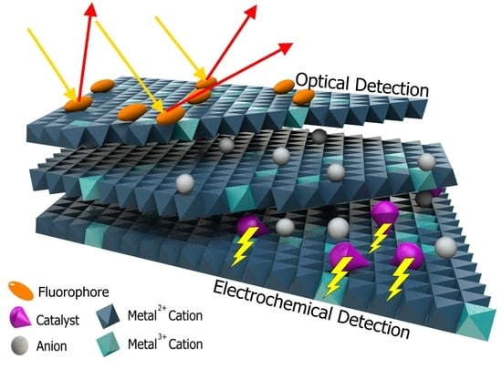


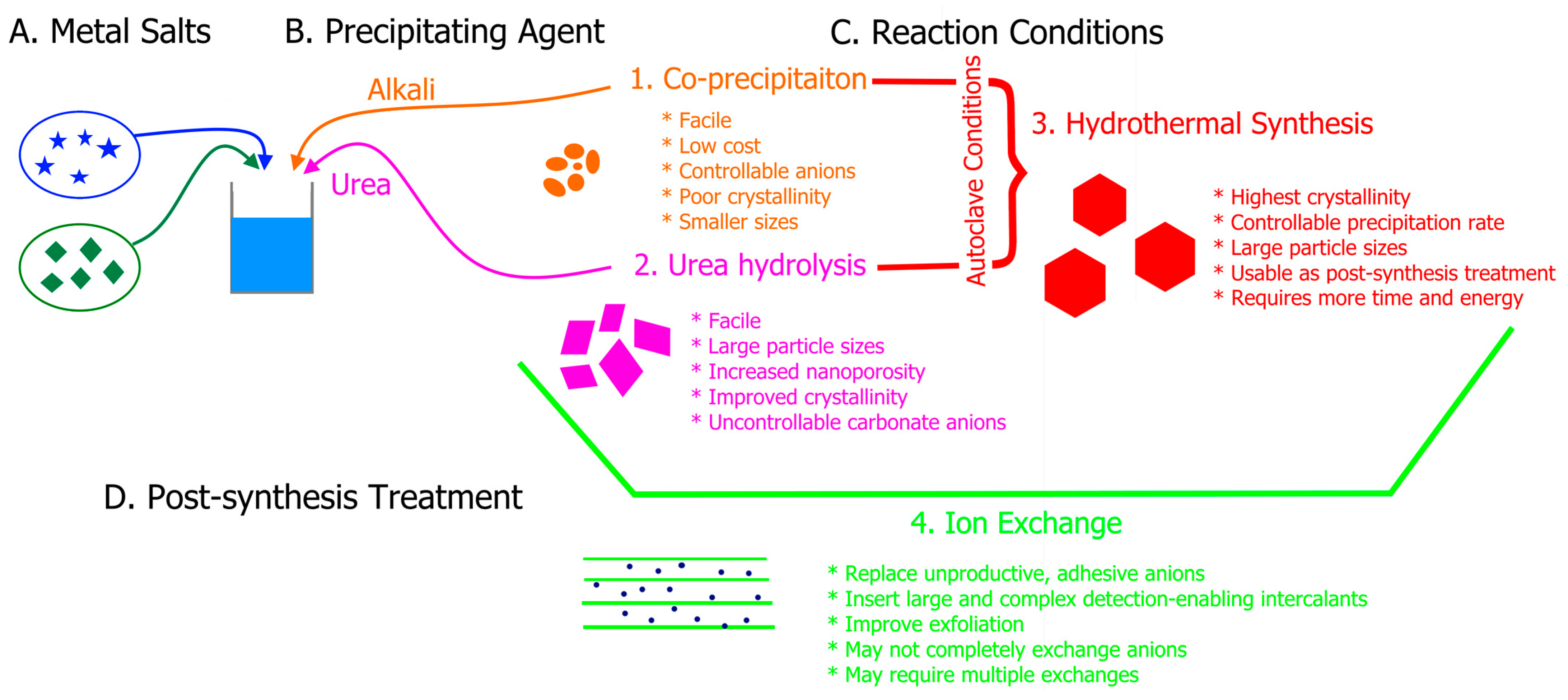
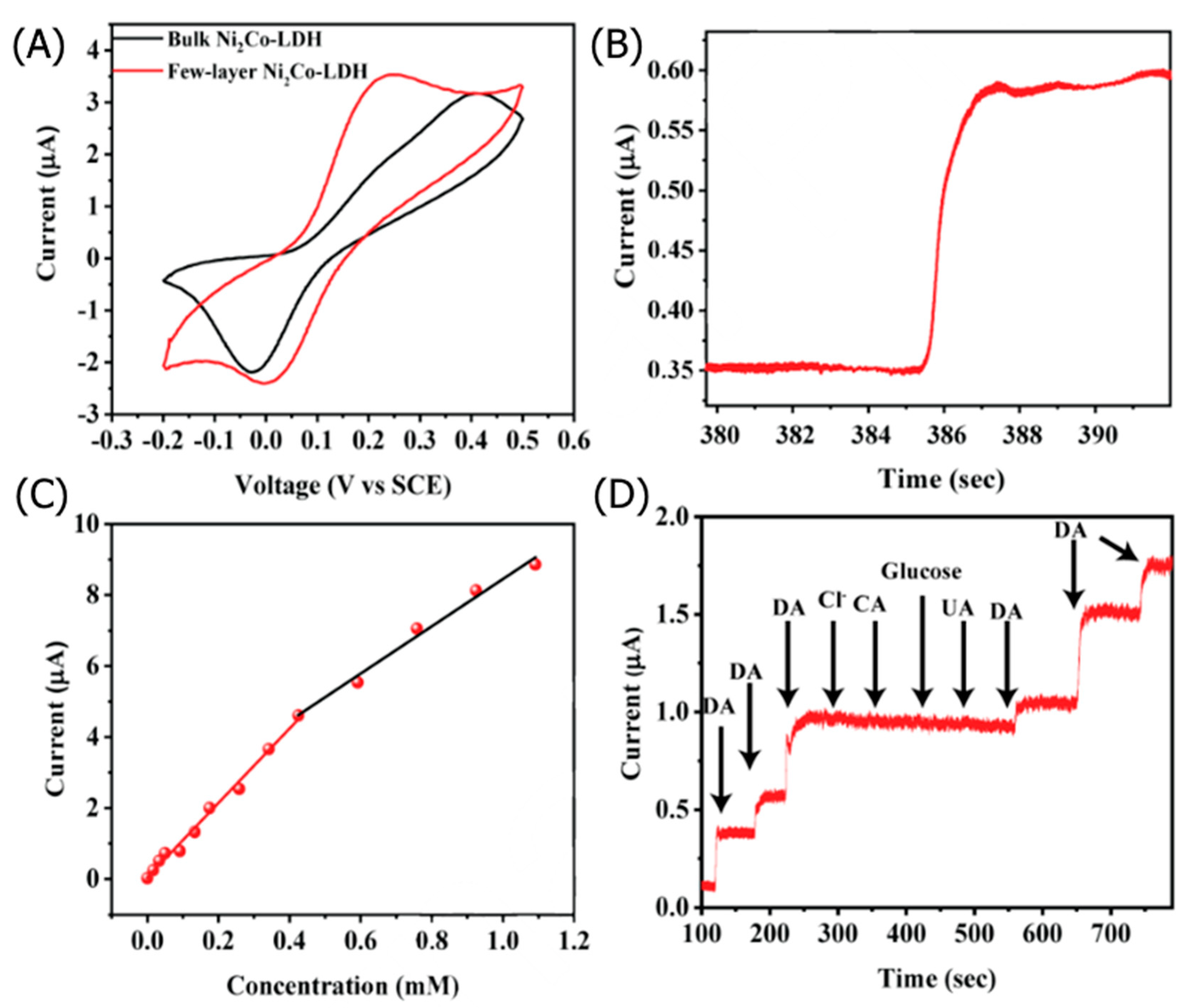
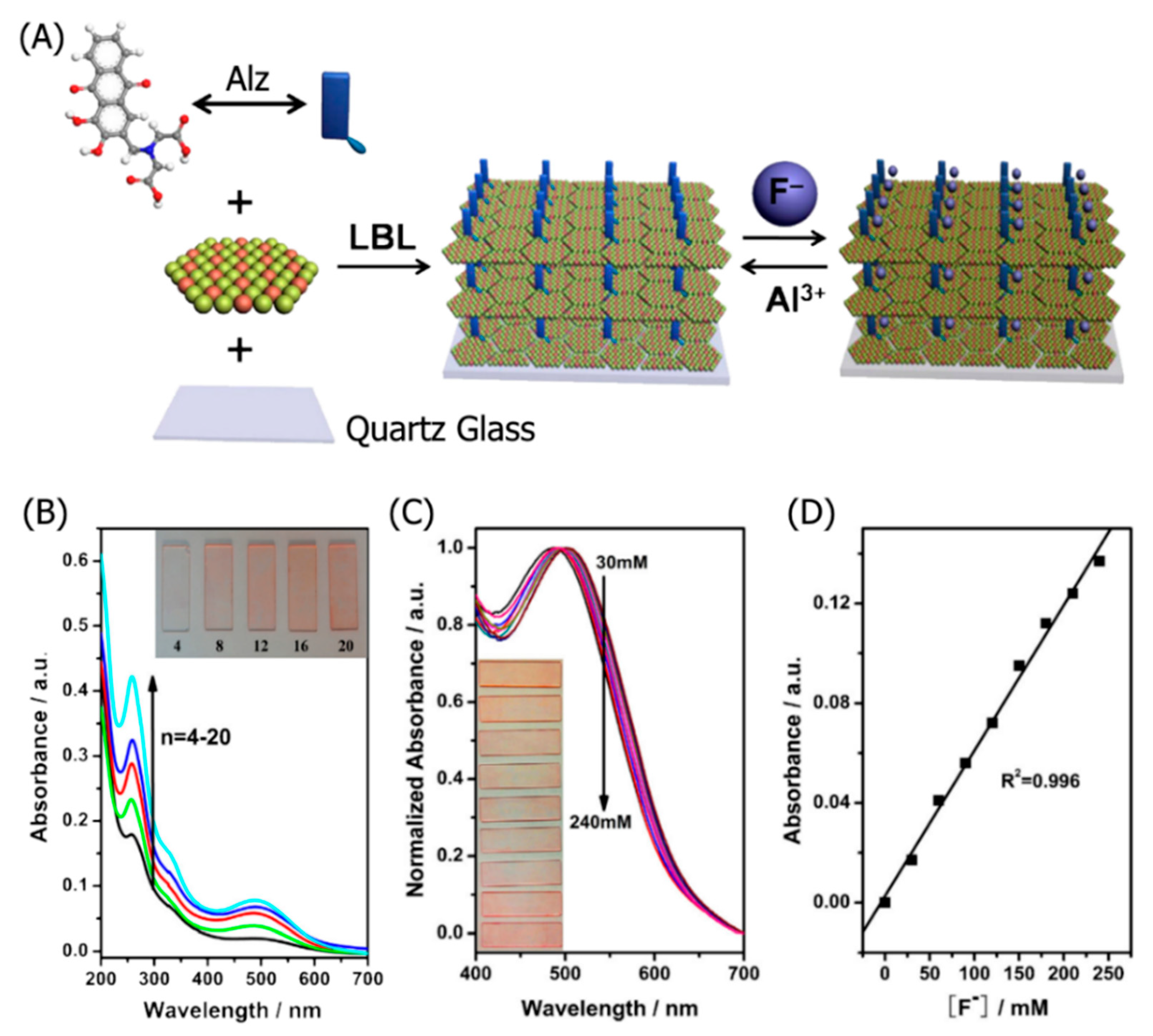
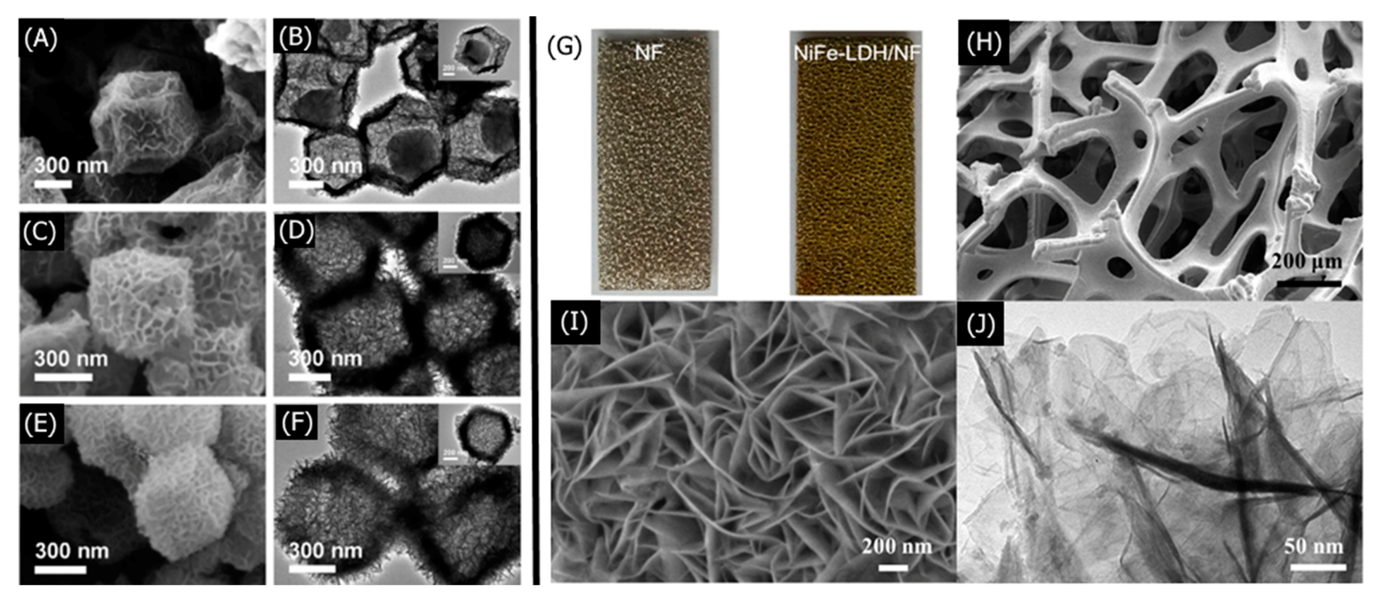
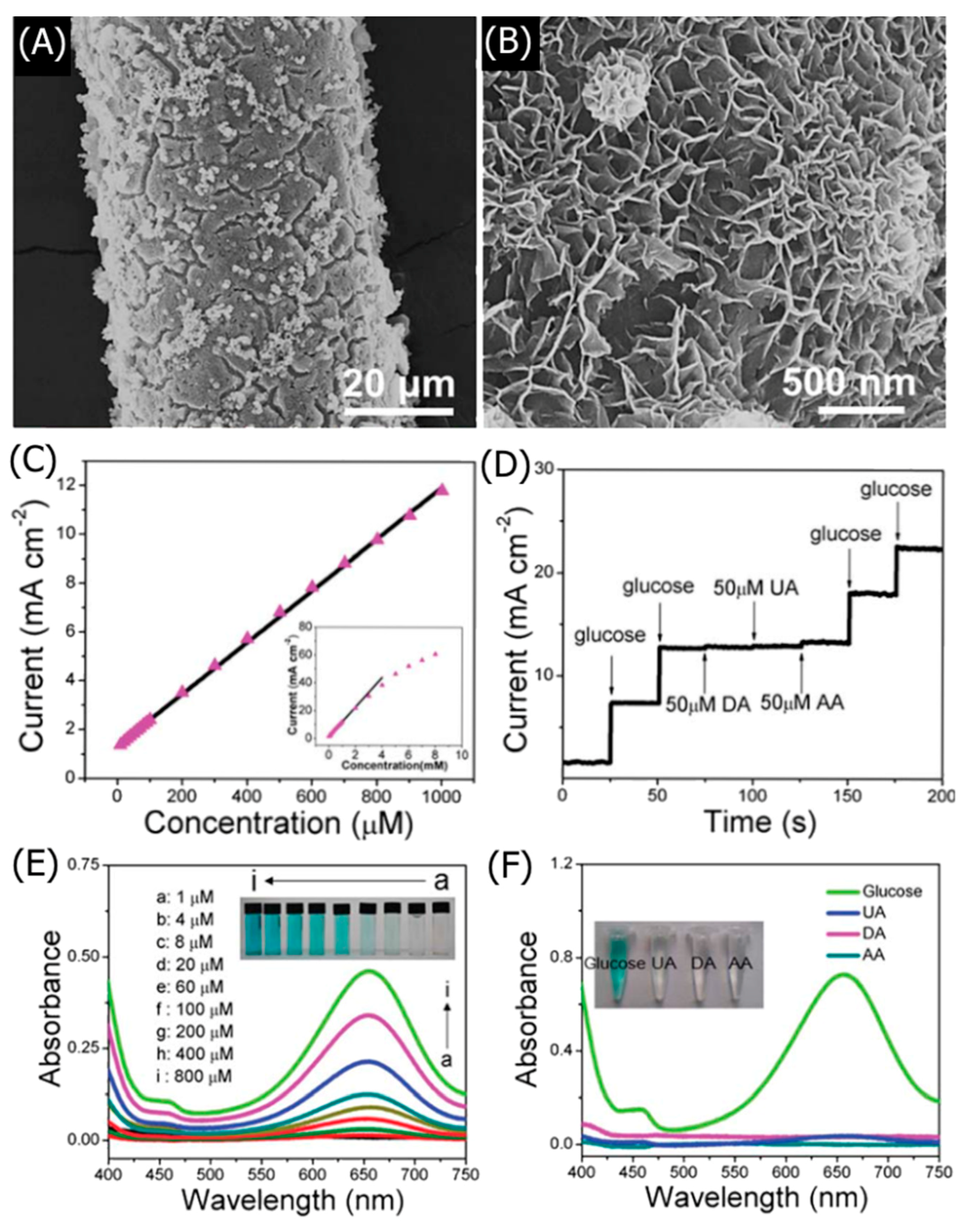
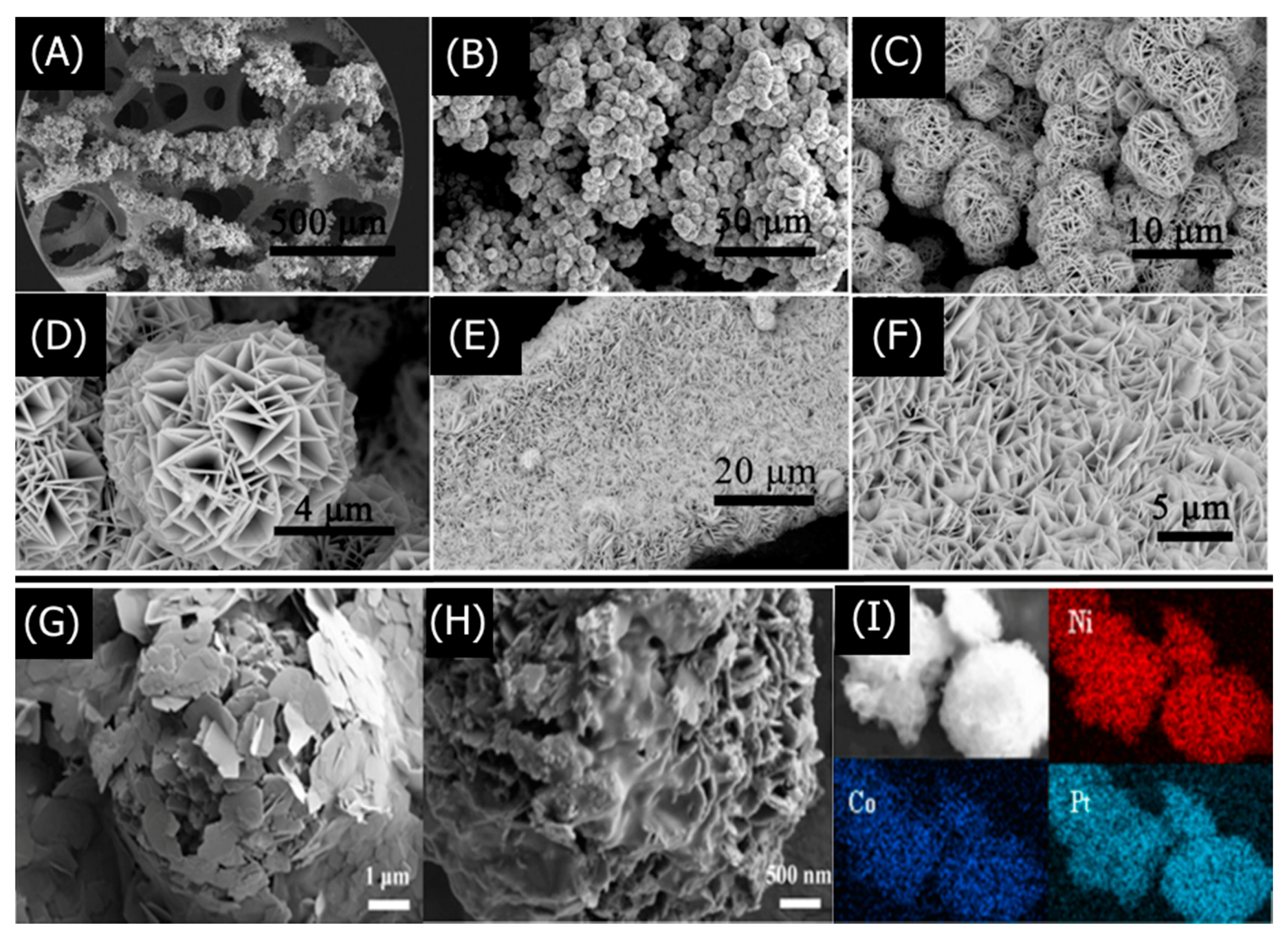
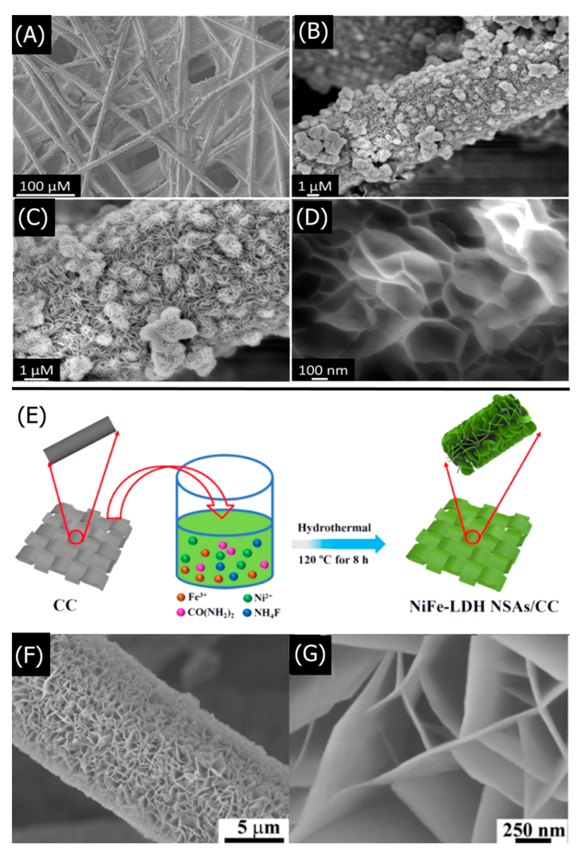
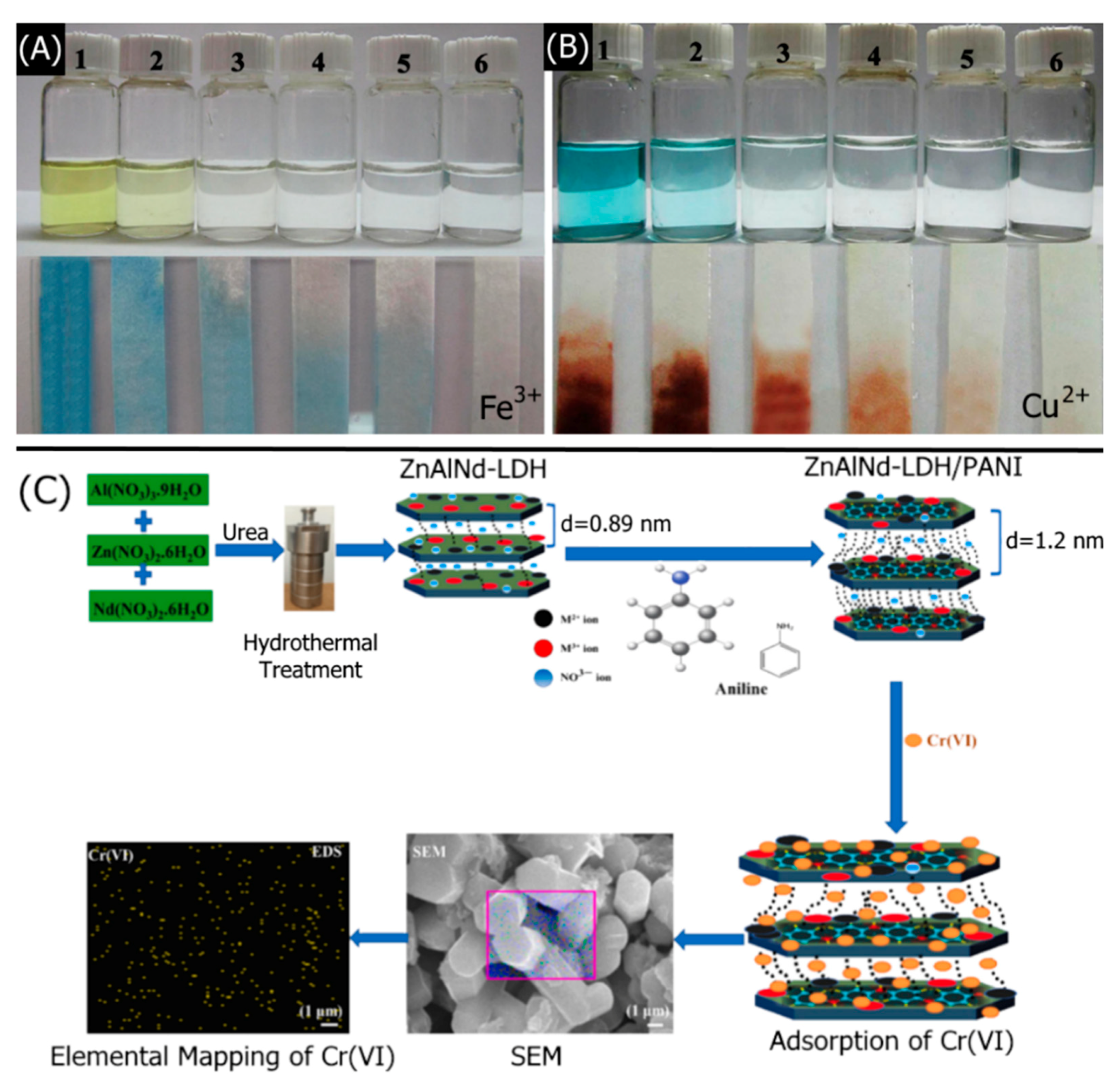
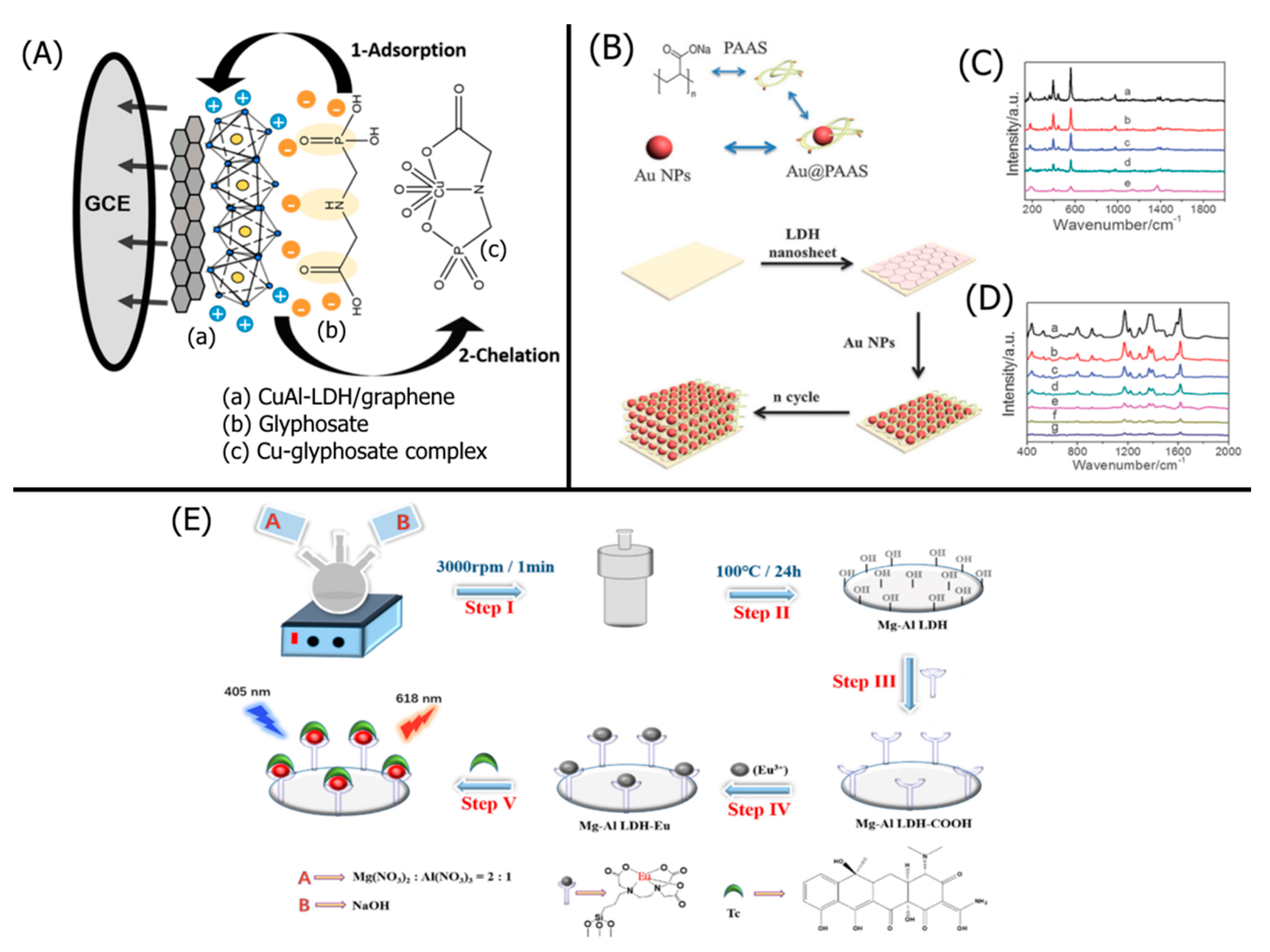
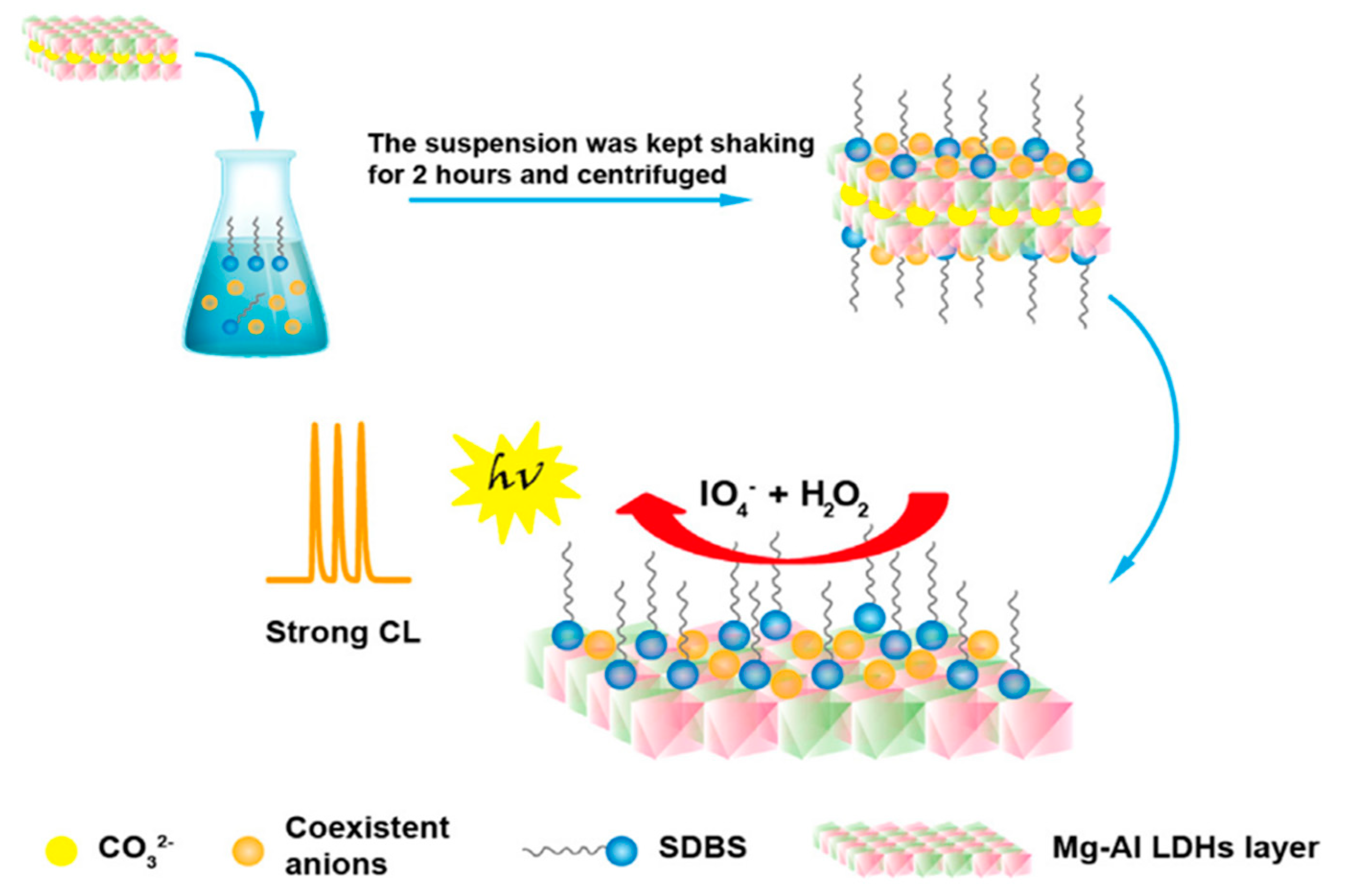