Article

Tuning Dielectric Loss of SiO2@CNTs for Electromagnetic Wave Absorption

1
Key Laboratory of In-Fiber Integrated Optics, College of Physics and Optoelectronic Engineering, Harbin Engineering University, Harbin 150001, China
2
School of Mechatronic Engineering, Daqing Normal University, Daqing 163712, China
3
Key Laboratory for Photonic and Electronic Bandgap Materials, Ministry of Education, School of Physics and Electronic Engineering, Harbin Normal University, Harbin 150025, China
4
School of Materials Science and Engineering, Zhengzhou University, Zhengzhou 450001, China
*
Authors to whom correspondence should be addressed.
Nanomaterials 2021, 11(10), 2636; https://doi.org/10.3390/nano11102636
Submission received: 20 September 2021 / Revised: 1 October 2021 / Accepted: 3 October 2021 / Published: 7 October 2021
(This article belongs to the Topic Advances and Applications of Carbon Nanotubes)

Abstract

:
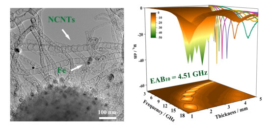
We developed a simple method to fabricate SiO2-sphere-supported N-doped CNTs (NCNTs) for electromagnetic wave (EMW) absorption. EMW absorption was tuned by adsorption of the organic agent on the precursor of the catalysts. The experimental results show that the conductivity loss and polarization loss of the sample are improved. Meanwhile, the impedance matching characteristics can also be adjusted. When the matching thickness was only 1.5 mm, the optimal 3D structure shows excellent EMW absorption performance, which is better than most magnetic carbon matrix composites. Our current approach opens up an effective way to develop low-cost, high-performance EMW absorbers.

Graphical Abstract

1. Introduction

With the development of science and technology, the rapid rise of artificial intelligence, the popularity of the smart home, and the extensive application of various electrical and electronic products, people’s work efficiency and quality of life has improved. However, at the same time, the widespread use of electronic products also hides huge harms: long-term exposure to electromagnetic radiation will damage human health, but also harms other electronic products’ electromagnetic interference, affecting their normal work. These hazards have attracted the attention of many countries in the world, and development of efficient electromagnetic absorption and shielding materials has become the main research direction. Therefore, it is necessary to develop a high-performance electromagnetic wave (EMW) absorber. To improve the efficiency of the unitizations, lightweight absorbers with a thin thickness are required. Carbonaceous materials such as graphene, carbon nanotubes (CNTs) and carbon nanofibers have attracted great attention because of their low mass density, good mechanical and chemical stability and high surface areas [1,2,3,4,5,6,7,8,9]. Carbon nanotubes have received extensive attention and in-depth studies in the field of EMW absorption due to their tubular structure suitable for electron transport, their light weight and good electrical conductivity [10,11,12,13,14,15,16,17]. For example, Lv et al. encapsulated Fe/Fe3C nanoparticles (NPs) into N-doped CNTs (NCNTs) and obtained the result that the sample had a reflection loss (RL) of −46.0 dB and a thickness of 4.97 mm at 3.6 GHz [10]. Chang et al. reported Fe3O4/PPy/CNT composites with an RL of −25.9 dB with a thickness of 3.0 mm [11]. The reflection loss of ZnFe2O4@CNT/PVDF composite film prepared by Li et al. was −54.5 dB, with a matching thickness of 2.4 mm [12]. Gong et al. reported SiCN(Fe) fibers with an RL of −47.64 dB and the effective absorption bandwidth of 4.28 GHz [13]. The minimum reflection loss of Fe3O4/CNTs prepared by Zeng et al. was −51.0 dB, with a matching thickness of 4.4 mm [14]. Recently, a series of magnetic metal alloys (Fe, Co, Ni, etc.) that encapsulated into NCNTs were designed for EMW absorption [15,16]. However, the impedance matching feature of the composites mentioned above needed to be precisely tuned due to highly conductive magnetic metals and CNTs in these composites [17]. Furthermore, there is still room to improve the EMW absorption property of CNT-based absorbers, such as stronger absorption at a lower filler ratio and thinner matching thickness.
Here, we propose a simple method for SiO2-sphere-supported NCNTs with embedded Fe3C/Fe nanoparticles (NPs) (SiO2@Fe3C/Fe@NCNT-GT) for EMW absorption. Fe(OH)X was first coated on the surface of SiO2 spheres [18], and then the organic solvent (terephthalic acid) was adsorbed on the Fe(OH)x surface to form relatively larger metal NPs for the growth of NCNTs with moderate diameters. Compared to the counterpart (SiO2@Fe3C/Fe@NCNT) without treatment in the organic solvent, the as-prepared SiO2@Fe3C/Fe@NCNT-GT showed significantly improved EMW absorption performance. At a filler ratio of 25%, the minimum RL (RL, min) and effective bandwidth of the SiO2@Fe3C/Fe@NCNT-GT reached −48.43 dB and 4.51 GHz, respectively, while the matching thickness was only 1.5 mm.

2. Materials and Methods

2.1. Materials

Tetraethoxysilane (TEOS, 99 wt%, analytical reagent, A.R.) was purchased from Tianjin Komiou Chemical Reagent Co., Ltd. (Tianjin, China). NH4OH (25 wt%, A.R.) was purchased from XiLong Scientific Co., Ltd. (Shantou, China). Absolute ethanol (99 wt%, analytical reagent, A.R.) was purchased from Tianjin Fuyu Fine Chemical Co., Ltd. (Tianjin, China). Terephthalic, N,N-Dimethylformamide (DMF) and dicyandiamide were purchased from Tianjin Guangfu Fine Chemical Research Institute (Tianjin, China). Ferric acetylacetonate was purchased from Sinopharm Chemical Reagent Co., Ltd. (Shanghai, China). Paraffin was purchased from Yuyang Wax Industry (Changge, China). It is worth noting that all chemicals were purchased without further treatment before use, and all aqueous solutions were prepared using ultrapure water.

2.2. Characterizations and Electromagnetic Parameter Measurement

The morphology and size of the samples were characterized using scanning electron microscopy (SEM; Hitachi SU70, Tokyo, Japan) and transmission electron microscopy (TEM; FEITecnai-F20, Hillsboro, USA). Energy dispersive X-ray spectroscopy (EDX) was performed to confirm the elemental contents of the samples. X-ray diffraction (XRD) data were measured using a Rigaku D/max 2550 V (Tokyo, Japan) with Cu Kα radiation (λ = 1.5418 Å). X-ray photoelectron spectroscopy (XPS) analyses were carried out by using a spectrometer with Mg Kα radiation (ESCALAB 250, Shanghai, China). Raman spectra were recorded on a Raman spectrometer (Xplora Plus, Paris, France) using a 488 nm He−Ne laser. The Brunauer–Emmett–Teller (BET) surface area and pore volume were tested with a Quantachrome Instruments Autosorb-iQ2-MP (Beijing, China) after the composites were vacuum dried at 200 ℃ for 10 h. Fourier-transform infrared (FTIR) spectra of samples were collected using a Nicolet FTIR510 spectrometer (KBr pellet method, 4 cm−1 resolution, Waltham, MA, USA). The electromagnetic wave absorption properties of the absorbing materials were measured using a vector network analyzer (Anritsu MS4644A Vectorstar, Kanagawa, Japan) in the 2–18 GHz range at room temperature.

2.3. Methods

2.3.1. Synthesis of the SiO2

Synthesis of the SiO2 was conducted following the Stöber method. Deionized water (18 mL), absolute ethanol (76 mL) and TEOS (14 mL) were dissolved into NH4OH (98 mL), and continuously stirred for 4 h. Then, the colloidal solution was centrifuged, and the resultant was placed in an oven at 100 °C for 12 h to obtain silica microspheres [19,20].

2.3.2. Synthesis of the SiO2@Fe(OH)x

SiO2 with diameters of about 400 nm (216 mg) were first dispersed in ethanol (72 mL), and ferric acetylacetonate (270.5 mg) was added to the mixture above, sequentially. Distilled water (3.6 mL) and ammonia (2 mL) were added to the mixture with sonication for 15 min. The as-prepared mixture was sealed in a conical flask and stirred at 80℃ for 10 h. The precipitate was washed with distilled water and ethanol several times, then centrifuged and dried in a vacuum oven at 40 °C to obtain the SiO2@Fe(OH)x [20].

2.3.3. Synthesis of the SiO2@Fe3C/Fe@NCNT

The SiO2@Fe(OH)x was annealed in Ar (the temperature was 800 °C, the time was 30 min and the ramp rate was 5 °C/min) to obtain SiO2@Fe3C/Fe@NCNT [20].

2.3.4. Synthesis of the SiO2@Fe(OH)x-GT

The SiO2@Fe(OH)x (200 mg), terephthalic acid (300 mg), pure water (3 mL) and ethanol (3 mL) were added into N,N-Dimethylformamide (DMF) solution (30 mL), and stirred for 30 min. Then, the mixed solution was placed into a 50 mL Teflon container and was treated at a high temperature of 150 °C for 12 h. The precipitate was washed and dried in a 40 °C vacuum oven to obtain the SiO2@Fe(OH)x-GT.

2.3.5. Synthesis of the SiO2@Fe3C/Fe@NCNT-GT

The SiO2@Fe(OH)x-GT was annealed in Ar (the temperature was 800 °C, the time was 30 min, and the ramp rate was 5 °C/min) to obtain the SiO2@Fe3C/Fe@NCNT-GT. The detailed experimental material, structural characterizations and the method are described in the Supplementary Materials.

3. Results and Discussion

The diameter of the prepared SiO2 microspheres were approximately 400 nm. After being coated with an Fe(OH)x layer on the surface of the SiO2 spheres, the diameter increased from 400 nm to about 500 nm (SiO2@Fe(OH)x). Scanning electron microscopy (SEM) imaging and transmission electron microscopy (TEM) imaging showed that the Fe(OH)x layer was uniformly coated on the surface of the SiO2 spheres (Figure 1a–c and Figure S1a, Supplementary Materials). In the X-ray diffraction (XRD) pattern, there were two obvious peaks at 2θ 35.02° and 62.72°, indicating that the outmost layer of the SiO2@Fe(OH)x was mainly composed of weakly crystalline Fe(OH)3, which corresponded to JCPDS card no. 22-0346 (Figure S2a). Energy dispersive X-ray spectroscopy (EDX) element mappings were also confirmed. As shown in Figure 1d, there were Si and O signals in the spherical core region, suggesting that the spherical core region materials were still SiO2 spheres. Fe and O single elements were obviously present in the area outside the sphere, confirming the composition of the SiO2@Fe(OH)3. The XRD pattern of the SiO2@Fe(OH)x-GT indicated that the weakly crystalline Fe(OH)3 (Figure S2a) remained after the treatment, but the rough surface became relatively smooth (Figure S1b). TEM images show that there was a lamellar structure on the surface of the SiO2@Fe(OH)3-GT, different from the SiO2@Fe(OH)3 (Figure 1e–g). The peak at 798 and 1431 cm−1 in the Fourier-transform infrared (FTIR) spectra of the SiO2@Fe(OH)3-GT corresponded to the C–H deformation vibration and the C=C stretching vibration (Figure S2b). The peak 1659 cm−1 in the spectrum of the SiO2@Fe(OH)3-GT corresponded to the C=O stretching vibration (Figure S2b). Thus, the FTIR results indicated the adsorption of terephthalic acid on the Fe(OH)3 layer. EDX element mappings also confirmed that Fe, O and C single elements were present in the area outside of the sphere, confirming the adsorption of terephthalic acid on the Fe(OH)3 layer (Figure 1h).
In order to analyze the composition and valence state of the SiO2@Fe3C/Fe@NCNT-GT, XRD and Raman spectra were performed. As shown in Figure 2a, there was a broad diffraction (2θ) from 10° to 30°, corresponding to amorphous SiO2 in the XRD pattern of the SiO2@Fe3C/Fe@NCNT-GT. In the XRD pattern of the SiO2@Fe3C/Fe@NCNT-GT, the peak at 2θ 44.7° and 42.9° can be indexed to (110) planes of the Fe NPs (JCPDS no. 06-0696) and (211) planes of the Fe3C (JCPDS no. 35-0772), in sequence, while the peak at 26.4° is attributed to the NCNTs (JCPDS no. 41-1487). The Raman spectra of the SiO2@Fe3C/Fe@NCNT-GT showed two distinguishable peaks: one at 1325 cm−1 (D band) and the other at 1585 cm−1 (G band) (Figure 2b). Their intensity ratios (ID/IG) for the SiO2@Fe3C/Fe@NCNT-GT was 1.002, which indicated rich defects in the sample. These defects in the SiO2@Fe3C/Fe@NCNT-GT can greatly improve the polarization relaxation and contribute to enhancing the absorption of electromagnetic waves [21]. As shown in Figure 2c, the N2-sorption isotherms of the SiO2@Fe3C/Fe@NCNT-GT displays type-IV loops, revealing that the mesopores existed in the prepared sample. Furthermore, the Brunauer–Emmett–Teller (BET) surface area of the SiO2@Fe3C/Fe@NCNT-GT was 243.54 m2 g−1. The illustration in Figure 2c displays pore size distribution diagrams. The pore sizes of the SiO2@Fe3C/Fe@NCNT-GT were centered at 15 nm, and pore volume was 0.568 cm³ g−1. X-ray photoelectron spectroscopy (XPS) spectra displayed that there were five elements (Fe, N, O, Si and C) in the SiO2@Fe3C/Fe@NCNT-GT (Figure S3). The peaks were at 398.5 (pyridine-N), 399.9 (pyrrolic-N), 401.1 (graphite-Nand) and 404.5 eV (oxide-N) in the XPS spectra of N 1s, respectively (Figure 2d) [22]. The peaks at 709.1 and 721.9 eV in the XPS spectra of Fe 2p can be indexed to metallic Fe. The peaks (717.3 and 733.2 eV) and satellite peaks (712.1 and 724.9 eV) reveal the oxidation state of Fe species in the sample [23,24,25] (Figure 2e). The binding energies of C–C (284.6 eV), C–N (285.7 eV) and C–O (288.7 eV) were observed on the surface of NCNT (Figure 2f) [26,27]. The carbon atom will tend to form unsaturated covalent bonds with the oxygen anion, increasing the charge state and increasing the band gap. This is due to the electron density shifts from the carbocation to the more electronegative oxygen anion, which in turn affects the electron structure.
SEM images indicated that the SiO2@Fe3C/Fe@NCNT-GT exhibited 3D morphology, where the NCNTs were grown on the surface of the SiO2 spheres (Figure S4). Bamboo-like NCNTs in the SiO2@Fe3C/Fe@NCNT-GT are also observed in Figure 3a,b with a length of approximately 1.5 μm. Magnified TEM images show that their average diameter and wall thickness were approximately 51 and 12 nm, respectively (Figure 3b,c and Figure S5). There were some NPs, with an average diameter of about 39 nm, embedded in the bamboo-like NCNTs. Figure 3c shows that the NPs were encapsulated in 25–30 layers of the graphene shell in the high-resolution TEM (HRTEM) images. The d-spacing of labeled lattice fringes of 0.20 nm corresponded to the (110) planes of Fe, while the d-spacing of 0.35 nm corresponded to the (002) planes of graphite–carbon (Figure 3d). Notably, a mass of defects were present in the NCNT walls and graphene shell of the SiO2@Fe3C/Fe@NCNT-GT. As is shown in Figure 3c, these defects are marked with a yellow frame. Defects including lattice distortion, lattice dislocation and fracture edges are considered to have a positive effect on the absorption property of the SiO2@Fe3C/Fe@NCNT-GT. The distribution of elements in the SiO2@Fe3C/Fe@NCNT-GT was analyzed by EDX element mapping. There were O and Si signals in the spherical core zone, indicating that the spherical core region mediums were still SiO2 spheres (Figure 3e). Fe, C and N single elements were present in the zone of the NCNTs, confirming the composition of NCNTs. Compared to the SiO2@Fe3C/Fe@NCNT [20], the average diameter of the NCNTs in the SiO2@Fe3C/Fe@NCNT-GT increased from 15 nm to 58 nm, the length of bamboo nodes increased from 15 nm to 50 nm, and the wall thickness increased from 3 to 12 nm. The adsorption of terephthalic acid limited the contact of the Fe(OH)3 with the reductive gases, leading to the formation of the larger metal NPs. Consequently, the diameter of NCNTs became larger compared to the counterpart without the adsorption of terephthalic acid.
The factors that may enhance the absorption performance of EMW were investigated through the comparison of electromagnetic parameters of the SiO2@Fe3C/Fe@NCNT-GT and the SiO2@Fe3C/Fe@NCNT, including complex permittivity and permeability. They can be expressed separately by the formula εr = ε′jε″ and μr = μ′jμ″. (ε′ is the real part of permittivity, ε″ is the imaginary part of permittivity, μ′ is the real part of permeability, and μ″ is the imaginary part of permeability) [28,29,30]. As shown in Figure 4a–c, the ε′ values of the SiO2@Fe3C/Fe@NCNT-GT varied in a range of 16.63−9.81, and the ε″ values of the SiO2@Fe3C/Fe@NCNT-GT varied in a range of 6.15−2.44. The permittivity for both samples gradually decreased with the increase in the frequency, which is due to the frequency dispersion effect. The ε′ and ε″ values of the SiO2@Fe3C/Fe@NCNT-GT were larger than those of the SiO2@Fe3C/Fe@NCNT. The dielectric loss tangent (tanδe = ε″/ε′) of the SiO2@Fe3C/Fe@NCNT-GT was also larger. Figure S6 shows that the two samples had very little difference in the real part of permeability, imaginary part of permeability and magnetic loss tangent (tan δm), with a value over 2–18 GHz. The saturation magnetization (Ms), remnant magnetization (Mr) and coercivity (Hc) of the SiO2@Fe3C/Fe@NCNT-GT were slightly larger than those of the SiO2@Fe3C/Fe@NCNT (Figure S7). Thus, the magnetic loss of the SiO2@Fe3C/Fe@NCNT-GT is not a determining factor for the EMW performance.
In general, the dielectric loss of the absorbing material includes the conduction loss and the polarization relaxation loss within the range of gigahertz. The former can be expressed by the formula (εc = σ/ε0ω); the characters σ, ε0 and ω represent the conductivity, the permittivity in a vacuum and the circular frequency, respectively. The latter is expressed by the formula (εp =ε″εc). The experimental results showed that the electrical conductivity of the SiO2@Fe3C/Fe@NCNT-GT was higher than that of the SiO2@Fe3C/Fe@NCNT (Table S1). Therefore, the SiO2@Fe3C/Fe@NCNT-GT had increased conductive loss compared to the SiO2@Fe3C/Fe@NCNT (Figure 4d). Meanwhile, the SiO2@Fe3C/Fe@NCNT-GT also had improved polarization losses compared to the SiO2@Fe3C/Fe@NCNT (Figure 4e) and had a higher attenuation coefficient (Figure 4f). As shown in Figure S8, multiple Cole–Cole semicircles could be found in the curve of the SiO2@Fe3C/Fe@NCNT-GT, confirming the existence of dipole polarization and interfacial polarization relaxation. Therefore, the increased dielectric relaxation loss of the SiO2@Fe3C/Fe@NCNT-GT is relevant to their enhanced dipole and interface polarizations [31,32,33,34,35]. Figure 5 shows the RLf curves of the two samples with d of 1.5–5.0 mm over 2–18 GHz. It can be found that the SiO2@Fe3C/Fe@NCNT-GT exhibited a better EMW absorption property than the SiO2@Fe3C/Fe@NCNT. It should be noted that all of the RL values of SiO2@Fe3C/Fe@NCNT-GT can exceed −20 dB (Figure 5a), where the minimum value was −48.43 dB with d of only 1.5 mm. However, the RL, min value for the SiO2@Fe3C/Fe@NCNT was only −16.63 dB with d of 5 mm (Figure 5b). Furthermore, the effective absorption bandwidth (EAB10, RL ≤ −10 dB) of the SiO2@Fe3C/Fe@NCNT-GT was 4.51 GHz, which is superior to that of the SiO2@Fe3C/Fe@NCNT (2.12 GHz) (Figure 5a,b). Thus, the SiO2@Fe3C/Fe@NCNT-GT showed a significantly enhanced EMW absorption property in the main parameters, including RL, min, EAB10 and d values, showing it has potential applications in practical EMW absorption. In addition, our prepared SiO2@Fe3C/Fe@NCNT-GT had comparable, or better, EMW absorption performance than reported carbon nanotube-based absorbent materials (Figure 5c, Table S2) [36,37,38,39,40,41,42,43,44,45,46,47,48,49,50,51,52,53]. The Mzf plot reveals that the SiO2@Fe3C/Fe@NCNT-GT had better impedance matching characteristics compared to the SiO2@Fe3C/Fe@NCNT (Figure S9). Therefore, the increase in diameter of NCNTs may also have a positive effect on the optimization of dielectric loss and impedance matching characteristics, thus enhancing the EMW absorption performance of the SiO2@Fe3C/Fe@NCNT-GT. Overall, compared to the counterpart (SiO2@Fe3C/Fe@NCNT) without treatment in the organic solvent, the as-prepared SiO2@Fe3C/Fe@NCNT-GT showed significantly improved EMW absorption performance. In addition, SiO2@Fe3C/Fe@NCNT-GT exhibited a decreased EMW absorption performance when the filling ratio was 20% or 30% (Figure S10). Therefore, the optimal filler ratio for EMW absorption is 25%.

4. Conclusions

In summary, we fabricated the SiO2@Fe3C/Fe@NCNT-GT with a moderate diameter for EMW absorption. Compared to the counterpart (SiO2@Fe3C/Fe@NCNT) without treatment in the organic solvent, the dielectric loss of the as-prepared SiO2@Fe3C/Fe@NCNT-GT was optimized, the impedance matching characteristics were adjusted, and the absorption performance of EMW was significantly improved. At a filler ratio of 25%, minimum, reflection loss can reach −48.43 dB. In the meantime, effective bandwidth of the SiO2@Fe3C/Fe@NCNT-GT can reach 4.51 GHz, while the matching thickness is only 1.5 mm, which is better than most magnetic carbon matrix composites. Our present approach opens up an effective way to develop low-cost, high-performance EMW absorbers.

Supplementary Materials

The following are available online at https://www.mdpi.com/article/10.3390/nano11102636/s1, Figure S1: SEM images of SiO2@Fe(OH)3 and SiO2@Fe(OH)3-GT, Figure S2: XRD patterns and FTIR spectras of SiO2@Fe(OH)3 and SiO2@Fe(OH)3-GT, Figure S3: XPS spectra of the SiO2@Fe3C/Fe@NCNT-GT, Figure S4: SEM image of SiO2@Fe3C/Fe@NCNT-GT, Figure S5: (a) TEM image, (b) Average diameter of NCNTs and NPs of SiO2@Fe3C/Fe@NCNT-GT, Figure S6: (a) μ′—f curves, (b) μ″—f curves, and (c) tanδmf of SiO2@Fe3C/Fe@NCNT-GT and SiO2@Fe3C/Fe@NCNT, Figure S7: Magnetization hysteresis loops of the SiO2@Fe3C/Fe@NCNT-GT and SiO2@Fe3C/Fe@NCNT, Figure S8: Cole-Cole semicircles of the (a) SiO2@Fe3C/Fe@NCNT-GT and (b) SiO2@Fe3C/Fe@NCNT, Figure S9: The Mzf curves of the (a) SiO2@Fe3C/Fe@NCNT-GT and (b) SiO2@Fe3C/Fe@NCNT, Figure S10: RLf curves of (a) the SiO2@Fe3C/Fe@NCNT-GT with a filler ratio of 20 wt.% and (b) 30 wt.%, Table S1: Electrical conductivity of absorbing materials, Table S2: EMW absorption properties of some representative materials.

Author Contributions

Y.C. and X.Z. (Xiao Zhang). conceived and supervised the project. F.C. carried out the experiments, analyzed the experimental data and wrote the first version of the manuscript. Q.O., X.Z. (Xinci Zhang) and X.Z. (Xitian Zhang) evaluated the data and made the intensive discussion. F.C., J.X. and B.L. contributed to electromagnetic parameter measurements. All authors have read and agreed to the published version of the manuscript.

Funding

This research was funded by the NNSF of China (grant number 51972077), the Fundamental Research Funds for the Central Universities (grant number 3072020CF2518, 3072020CFT2505, 3072021CFT2506, 3072021CF2523 and 3072021CF2524), Natural Science Foundation of Daqing Normal University (grant number 19ZR05), Daqing City Directive Science and Technology Plan Project (grant number zd-2020-04) and Heilongjiang Provincial Natural Resources Foundation Joint Guide Project (grant number LH2020E098).

Institutional Review Board Statement

Not applicable.

Informed Consent Statement

Not applicable.

Data Availability Statement

Data is contained within the article or Supplementary Materials.

Conflicts of Interest

The authors declare no conflict of interest.

References

  1. Cao, M.; Wang, X.; Zhang, M.; Cao, W.; Fang, X.; Yuan, J. Variable-Temperature Electron Transport and Dipole Polarization Turning Flexible Multifunctional Microsensor beyond Electrical and Optical Energy. Adv. Mater. 2020, 32, e1907156. [Google Scholar] [CrossRef] [PubMed]
  2. Yakovenko, O.; Matzui, L.; Vovchenko, L.; Lozitsky, O.; Prokopov, O.; Lazarenko, O.; Zhuravkov, A.; Oliynyk, V.; Launets, V.; Trukhanov, S. Electrophysical properties of epoxy-based composites with graphite nanoplatelets and magnetically aligned magnetite. Mol. Cryst. Liq. Cryst. 2018, 661, 68–80. [Google Scholar] [CrossRef]
  3. Yakovenko, O.S.; Matzui, L.Y.; Vovchenko, L.L.; Oliynyk, V.V.; Trukhanov, A.V.; Trukhanov, S.V.; Borovoy, M.O.; Tesel’ko, P.O.; Launets, V.L.; Syvolozhskyi, O.A.; et al. Effect of magnetic fillers and their orientation on the electrodynamic properties of BaFe12–xGaxO19 (x = 0.1–1.2)—epoxy composites with carbon nanotubes within GHz range. Appl. Nanosci. 2020, 10, 4747–4752. [Google Scholar] [CrossRef]
  4. Arief, I.; Biswas, S.; Bose, S. FeCo-anchored reduced graphene oxide framework-based soft composites containing carbon nanotubes as highly efficient microwave absorbers with excellent heat dissipation ability. ACS Appl. Mater. Interfaces 2017, 9, 19202–19214. [Google Scholar] [CrossRef] [PubMed]
  5. Vinnik, D.A.; Zhivulin, V.E.; Sherstyuk, D.P.; Starikov, A.Y.; Zezyulina, P.A.; Gudkova, S.A.; Zherebtsov, D.A.; Rozanov, K.N.; Trukhanov, S.V.; Astapovich, K.A.; et al. Electromagnetic properties of zinc–nickel ferrites in the frequency range of 0.05–10 GHz. Mater. Today Chem. 2021, 20, 100460. [Google Scholar] [CrossRef]
  6. Zdorovets, M.V.; Kozlovskiy, A.L.; Shlimas, D.I.; Borgekov, D.B. Phase transformations in FeCo–Fe2CoO4/Co3O4-spinel nanostructures as a result of thermal annealing and their practical application. J. Mater. Sci. Mater. Electron. 2021, 32, 16694–16705. [Google Scholar] [CrossRef]
  7. Thakur, A.; Sharma, N.; Bhatti, M.; Sharma, M.; Trukhanov, A.V.; Trukhanov, S.V.; Panina, L.V.; Astapovich, K.A.; Thakur, P. Synthesis of barium ferrite nano-particles using rhizome extract of Acorus Calamus: Characterization and its efficacy against different plant phytopathogenic fungi. Nano-Struct. Nano-Objects 2020, 24, 100599. [Google Scholar] [CrossRef]
  8. Kozlovskiy, A.; Zdorovets, M. Effect of doping of Ce4+/3+ on optical, strength and shielding properties of (0.5−x)TeO2-0.25MoO-0.25Bi2O3-xCeO2 glasses. Mater. Chem. Phys. 2021, 263, 124444. [Google Scholar] [CrossRef]
  9. Wu, Z.; Pei, K.; Xing, L.; Yu, X.; You, W.; Che, R. Enhanced Microwave Absorption Performance from Magnetic Coupling of Magnetic Nanoparticles Suspended within Hierarchically Tubular Composite. Adv. Funct. Mater. 2019, 29, 1901448. [Google Scholar] [CrossRef]
  10. Lü, Y.; Wang, Y.; Li, H.; Lin, Y.; Jiang, Z.; Xie, Z.; Kuang, Q.; Zheng, L. MOF-Derived Porous Co/C Nanocomposites with Excellent Electromagnetic Wave Absorption Properties. ACS Appl. Mater. Interfaces 2015, 7, 13604–13611. [Google Scholar] [CrossRef]
  11. Yang, R.B.; Reddy, P.M.; Chang, C.J.; Chen, P.A.; Chen, J.K.; Chang, C.C. Synthesis and characterization of Fe3O4/polypyrrole/carbon nanotube composites with tunable microwave absorption properties: Role of carbon nanotube and polypyrrole content. Chem. Eng. J. 2016, 285, 497–507. [Google Scholar] [CrossRef]
  12. Li, F.; Zhan, W.; Su, Y.; Siyal, S.H.; Bai, G.; Xiao, W.; Zhou, A.; Sui, G.; Yang, X. Achieving excellent electromagnetic wave absorption of ZnFe2O4@CNT/polyvinylidene fluoride flexible composite membranes by adjusting processing conditions. Compos. Part A: Appl. Sci. Manuf. 2020, 133, 105866. [Google Scholar] [CrossRef]
  13. Feng, Y.; Guo, X.; Lu, J.; Liu, J.; Wang, G.; Gong, H. Enhanced electromagnetic wave absorption performance of SiCN(Fe) fibers by in-situ generated Fe3Si and CNTs. Ceram. Int. 2021, 47, 19582–19594. [Google Scholar] [CrossRef]
  14. Zhu, L.; Zeng, X.; Chen, M.; Yu, R. Controllable permittivity in 3D Fe3O4/CNTs network for remarkable microwave absorption performances. RSC Adv. 2017, 7, 26801–26808. [Google Scholar] [CrossRef] [Green Version]
  15. Xu, J.; Zhang, X.; Yuan, H.; Zhang, S.; Zhu, C.; Zhang, X.; Chen, Y. N-doped reduced graphene oxide aerogels containing pod-like N-doped carbon nanotubes and FeNi nanoparticles for electromagnetic wave absorption. Carbon 2020, 159, 357–365. [Google Scholar] [CrossRef]
  16. Zhang, X.; Xu, J.; Yuan, H.; Zhang, S.; Ouyang, Q.; Zhu, C.; Zhang, X.; Chen, Y. Large-Scale Synthesis of Three-Dimensional Reduced Graphene Oxide/Nitrogen-Doped Carbon Nanotube Heteronanostructures as Highly Efficient Electromagnetic Wave Absorbing Materials. ACS Appl. Mater. Interfaces 2019, 11, 39100–39108. [Google Scholar] [CrossRef]
  17. Shu, R.; Li, W.; Zhou, X.; Tian, D.; Zhang, G.; Gan, Y.; Shi, J.-J.; He, J. Facile preparation and microwave absorption properties of RGO/MWCNTs/ZnFe2O4 hybrid nanocomposites. J. Alloy. Compd. 2018, 743, 163–174. [Google Scholar] [CrossRef]
  18. Yoshiyuki, N.; Kunihiro, S. Application of ferrite to electromagnetic wave absorber and its characteristics. IEEE Trans. Microw Theory Tech. 1971, 19, 65–72. [Google Scholar]
  19. Stöber, W.; Fink, A.; Bohn, E. Controlled growth of monodisperse silica spheres in the micron size range. J. Colloid Interface Sci. 1968, 26, 62–69. [Google Scholar] [CrossRef]
  20. Cao, F.; Yan, F.; Xu, J.; Zhu, C.; Qi, L.; Li, C.; Chen, Y. Tailing size and impedance matching characteristic of nitrogen-doped carbon nanotubes for electromagnetic wave absorption. Carbon 2020, 174, 79–89. [Google Scholar] [CrossRef]
  21. Wei, H.; Yin, X.; Li, X.; Li, M.; Dang, X.; Zhang, L.; Cheng, L. Controllable synthesis of defective carbon nanotubes/Sc2Si2O7 ceramic with adjustable dielectric properties for broadband high-performance microwave absorption. Carbon 2019, 147, 276–283. [Google Scholar] [CrossRef]
  22. Liu, L.; Yan, F.; Li, K.; Zhu, C.; Xie, Y.; Zhang, X.; Chen, Y. Ultrasmall FeNi3N particles with an exposed active (110) surface an-chored on nitrogen-doped graphene for multifunctional electrocatalysts. J. Mater. Chem. A. 2019, 7, 1083–1091. [Google Scholar] [CrossRef]
  23. Wu, H.; Yang, T.; Du, Y.; Shen, L.; Ho, G.W. Identification of Facet-Governing Reactivity in Hematite for Oxygen Evolution. Adv. Mater. 2018, 30, e1804341. [Google Scholar] [CrossRef]
  24. Almessiere, M.A.; Slimani, Y.; Trukhanov, A.V.; Baykal, A.; Gungunes, H.; Trukhanova, E.L.; Kostishin, V.G. Strong correlation between Dy3+ concentration, structure, magnetic and microwave properties of the [Ni0.5Co0.5](DyxFe2−x)O4 nanosized ferrites. J. Ind. Eng. Chem. 2020, 90, 251–259. [Google Scholar] [CrossRef]
  25. Kozlovskiy, A.; Egizbek, K.; Zdorovets, M.V.; Ibragimova, M.; Shumskaya, A.; Rogachev, A.A.; Ignatovich, Z.V.; Kadyrzhanov, K. Evaluation of the effi-ciency of detection and capture of manganese in aqueous solutions of FeCeOx nanocomposites doped with Nb2O5. Sensors. 2020, 20, 4851. [Google Scholar] [CrossRef] [PubMed]
  26. Quan, B.; Gu, W.; Sheng, J.; Lv, X.; Mao, Y.; Liu, L.; Huang, X.; Tian, Z.; Ji, G. From intrinsic dielectric loss to geometry patterns: Dual-principles strategy for ultrabroad band microwave absorption. Nano Res. 2020, 14, 1495–1501. [Google Scholar] [CrossRef]
  27. Zhang, Z.; Tan, J.; Gu, W.; Zhao, H.; Zheng, J.; Zhang, B.; Ji, G. Cellulose-chitosan framework/polyailine hybrid aerogel toward thermal insulation and microwave absorbing application. Chem. Eng. J. 2020, 395, 125190. [Google Scholar] [CrossRef]
  28. Dolmatov, A.; Maklakov, S.; Zezyulina, P.; Osipov, A.; Petrov, D.; Naboko, A.; Polozov, V.; Maklakov, S.; Starostenko, S.; Lagarkov, A. Deposition of a SiO2 Shell of Variable Thickness and Chemical Composition to Carbonyl Iron: Synthesis and Microwave Measurements. Sensors 2021, 21, 4624. [Google Scholar] [CrossRef] [PubMed]
  29. Sankaran, S.; Deshmukh, K.; Ahamed, M.; Pasha, S.K. Recent advances in electromagnetic interference shielding properties of metal and carbon filler reinforced flexible polymer composites: A review. Compos. Part A: Appl. Sci. Manuf. 2018, 114, 49–71. [Google Scholar] [CrossRef]
  30. Jacobo, S.E.; Aphesteguy, J.C.; Anton, R.L.; Schegoleva, N.; Kurlyandskaya, G. Influence of the preparation procedure on the properties of polyaniline based magnetic composites. Eur. Polym. J. 2007, 43, 1333–1346. [Google Scholar] [CrossRef]
  31. Liu, J.; Che, R.; Chen, H.; Zhang, F.; Xia, F.; Wu, Q.; Wang, M. Microwave Absorption Enhancement of Multifunctional Composite Microspheres with Spinel Fe3O4 Cores and Anatase TiO2 Shells. Small 2012, 8, 1214–1221. [Google Scholar] [CrossRef]
  32. Cao, M.; Song, W.; Hou, Z.; Wen, B.; Yuan, J. The effects of temperature and frequency on the dielectric properties, electro-magnetic interference shielding and microwave-absorption of short carbon fiber/silica composites. Carbon 2010, 48, 788–796. [Google Scholar] [CrossRef]
  33. Liu, P.; Gao, S.; Zhang, G.; Huang, Y.; You, W.; Che, R. Hollow Engineering to Co@N-Doped Carbon Nanocages via Synergistic Protecting-Etching Strategy for Ultrahigh Microwave Absorption. Adv. Funct. Mater. 2021, 31, 2102812. [Google Scholar] [CrossRef]
  34. Gu, W.; Cui, X.; Zheng, J.; Yu, J.; Zhao, Y.; Ji, G. Heterostructure design of Fe3N alloy/porous carbon nanosheet composites for efficient microwave attenuation. J. Mater. Sci. Technol. 2020, 67, 265–272. [Google Scholar] [CrossRef]
  35. Liu, Q.; Cao, Q.; Bi, H.; Liang, C.; Yuan, K.; She, W.; Yang, Y.; Che, R. CoNi@SiO2@TiO2 and CoNi@Air@TiO2 microspheres with strong wideband microwave absorption. Adv. Mate. 2016, 28, 486–490. [Google Scholar] [CrossRef]
  36. Feng, J.; Pu, F.; Li, Z.; Li, X.; Hu, X.; Bai, J. Interfacial interactions and synergistic effect of CoNi nanocrystals and nitrogen-doped graphene in a composite microwave absorber. Carbon 2016, 104, 214–225. [Google Scholar] [CrossRef]
  37. Zhao, T.; Ji, X.; Jin, W.; Wang, C.; Ma, W.; Gao, J.; Dang, A.; Li, T.; Shang, S.; Zhou, Z. Direct in situ synthesis of a 3D interlinked amorphous carbon nanotube/graphene/BaFe12O19 composite and its electromagnetic wave absorbing properties. RSC Adv. 2017, 7, 15903–15910. [Google Scholar] [CrossRef] [Green Version]
  38. Wang, H.; Xiang, L.; Wei, W.; An, J.; He, J.; Gong, C.; Hou, Y. Efficient and Lightweight Electromagnetic Wave Absorber Derived from Metal Organic Framework-Encapsulated Cobalt Nanoparticles. ACS Appl. Mater. Interfaces 2017, 9, 42102–42110. [Google Scholar] [CrossRef]
  39. Zhang, X.F.; Dong, X.L.; Huang, H.; Liu, Y.Y.; Wang, W.N.; Zhu, X.G.; Lv, B.; Lei, J.P. Microwave absorption properties of the car-bon-coated nickel nanocapsules. Appl Phys. Lett. 2006, 89, 053115. [Google Scholar] [CrossRef]
  40. Liu, X.; Cui, X.; Chen, Y.; Zhang, X.-J.; Yu, R.; Wang, G.-S.; Ma, H. Modulation of electromagnetic wave absorption by carbon shell thickness in carbon encapsulated magnetite nanospindles–poly(vinylidene fluoride) composites. Carbon 2015, 95, 870–878. [Google Scholar] [CrossRef]
  41. Mo, Z.C.; Yang, R.L.; Lu, D.W.; Yang, L.L.; Hu, Q.M.; Li, H.B.; Zhu, H.; Tang, Z.; Gui, X. Lightweight, three-dimensional carbon nanotube@TiO2 sponge with enhanced microwave absorption performance. Carbon 2019, 144, 433–439. [Google Scholar] [CrossRef]
  42. Wen, F.; Zhang, F.; Liu, Z. Investigation on Microwave Absorption Properties for Multiwalled Carbon Nanotubes/Fe/Co/Ni Nanopowders as Lightweight Absorbers. J. Phys. Chem. C 2011, 115, 14025–14030. [Google Scholar] [CrossRef]
  43. Zhou, Y.; Miao, J.; Shen, Y.; Xie, A. Novel porous FexCyNz/N-doped CNT nanocomposites with excellent bifunctions for cat-alyzing oxygen reduction reaction and absorbing electromagnetic wave. Appl. Surf. Sci. 2018, 453, 83–92. [Google Scholar] [CrossRef]
  44. Wang, L.; Jia, X.; Li, Y.; Yang, F.; Zhang, L.; Liu, L.; Ren, X.; Yang, H. Synthesis and microwave absorption property of flexible magnetic film based on graphene oxide/carbon nanotubes and Fe3O4 nanoparticles. J. Mater. Chem. A 2014, 2, 14940–14946. [Google Scholar] [CrossRef]
  45. Qi, X.; Xu, J.; Hu, Q.; Deng, Y.; Xie, R.; Jiang, Y.; Zhong, W.; Du, Y. Metal-free carbon nanotubes: Synthesis, and enhanced intrinsic mi-crowave absorption properties. Sci. Rep. 2016, 6, 28310. [Google Scholar] [CrossRef] [Green Version]
  46. Lin, H.; Zhu, H.; Guo, H.; Yu, L. Investigation of the microwave-absorbing properties of Fe-filled carbon nanotubes. Mater. Lett. 2007, 61, 3547–3550. [Google Scholar] [CrossRef]
  47. Zhang, T.; Zhong, B.; Yang, J.Q.; Huang, X.X.; Wen, G. Boron and nitrogen doped carbon nanotubes/Fe3O4 composite archi-tectures with microwave absorption property. Ceram. Int. 2015, 41, 8163–8170. [Google Scholar] [CrossRef]
  48. Zou, T.; Li, H.; Zhao, N.; Shi, C. Electromagnetic and microwave absorbing properties of multi-walled carbon nanotubes filled with Ni nanowire. J. Alloy. Compd. 2010, 496, L22–L24. [Google Scholar] [CrossRef]
  49. Xu, P.; Han, X.J.; Liu, X.R.; Zhang, B.; Wang, C.; Wang, X.H. A study of the magnetic and electromagnetic properties of γ-Fe2O3–multiwalled carbon nanotubes (MWCNT) and Fe/Fe3C–MWCNT composites. Mater. Chem. Phys. 2009, 114, 556–560. [Google Scholar] [CrossRef]
  50. Zhang, L.; Zhu, H.; Song, Y.; Zhang, Y.; Huang, Y. The electromagnetic characteristics and absorbing properties of multi-walled carbon nanotubes filled with Er2O3 nanoparticles as microwave absorbers. Mater. Sci. Eng. B 2008, 153, 78–82. [Google Scholar] [CrossRef]
  51. Zhang, L.; Zhu, H. Dielectric, magnetic, and microwave absorbing properties of multi-walled carbon nanotubes filled with Sm2O3 nanoparticles. Mater. Lett. 2009, 63, 272–274. [Google Scholar] [CrossRef]
  52. Green, M.; Van Tran, A.T.; Chen, X. Maximizing the microwave absorption performance of polypyrrole by data-driven discovery. Compos. Sci. Technol. 2020, 199, 108332. [Google Scholar] [CrossRef]
  53. Green, M.; Tran, A.T.; Chen, X. Obtaining strong, broadband microwave absorption of polyaniline through data-driven ma-terials discovery. Adv. Mater. Interfaces 2020, 7, 2000658. [Google Scholar] [CrossRef]
Figure 1. (ac) TEM images of structural characterizations and (d) EDX element mappings of the SiO2@Fe(OH)x. (eg) TEM image of structural characterizations and (h) EDX element mappings of SiO2@Fe(OH)x-GT.
Nanomaterials 11 02636 g001
Figure 2. (a) XRD pattern, (b) Raman spectra, (c) pore size distribution and N2-sorption isotherm, (df) N 1s, Fe 2p and C 1s XPS spectra of the SiO2@Fe3C/Fe@NCNT-GT.
Nanomaterials 11 02636 g002
Figure 3. (ac) TEM images, (d) HRTEM images, (e) TEM image and EDX elemental mappings of SiO2@Fe3C/Fe@NCNT-GT. The defects are marked by yellow dotted square (c).
Nanomaterials 11 02636 g003
Figure 4. (a) ε′—f curves, (b) ε″f curves, (c) tanδef curves, (d) εcf curves, (e) εpf curves and (f) α—f curves of SiO2@Fe3C/Fe@NCNT-GT and SiO2@Fe3C/Fe@NCNT.
Nanomaterials 11 02636 g004
Figure 5. RLf curves of (a) SiO2@Fe3C/Fe@NCNT-GT and (b) SiO2@Fe3C/Fe@NCNT, (c) the absorption performance of SiO2@Fe3C/Fe@NCNT-GT with previously reported absorbers.
Nanomaterials 11 02636 g005
Publisher’s Note: MDPI stays neutral with regard to jurisdictional claims in published maps and institutional affiliations.