Microalgae-Templated Spray Drying for Hierarchical and Porous Fe3O4/C Composite Microspheres as Li-ion Battery Anode Materials

 , , and
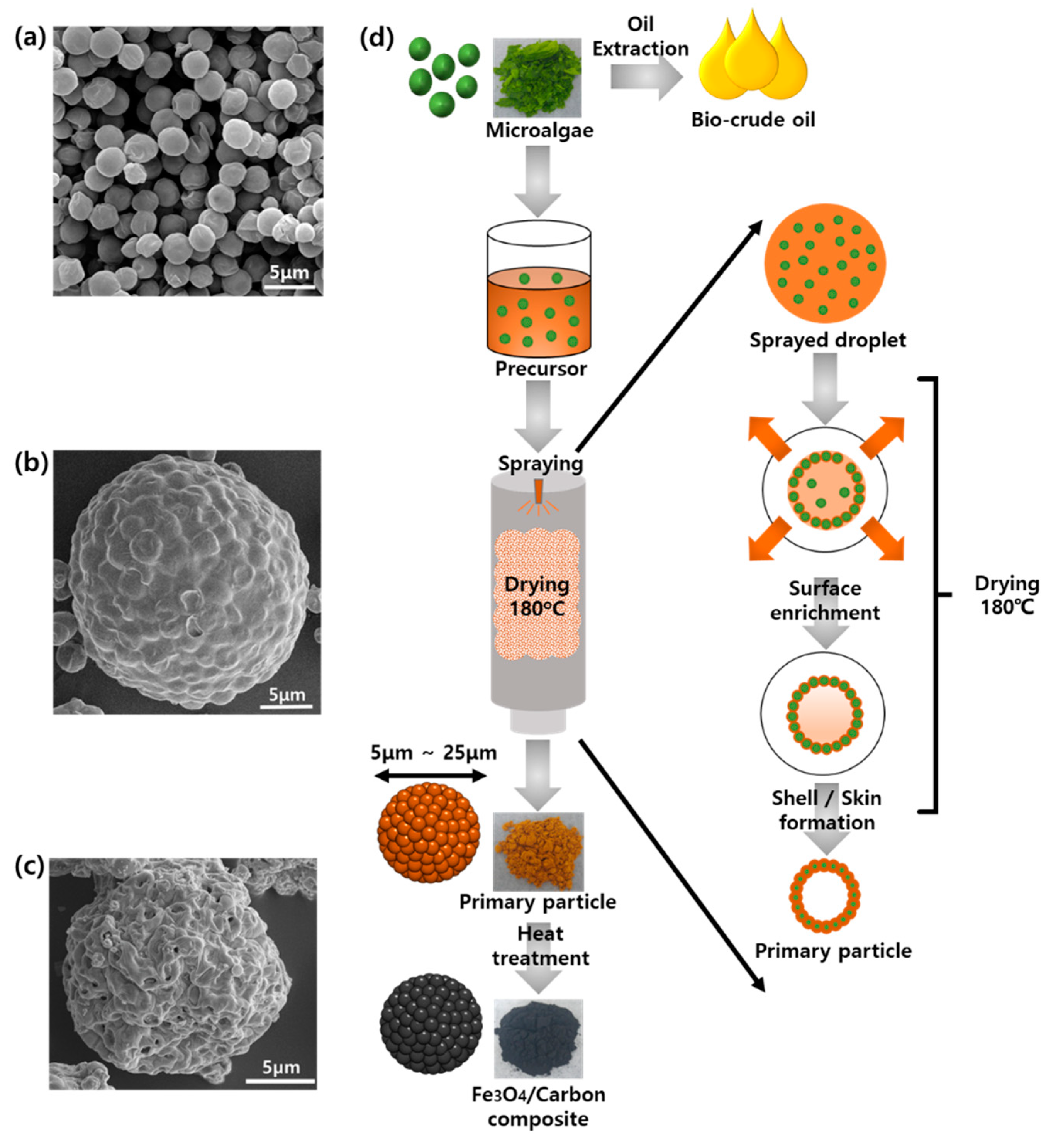
Abstract
:1. Introduction
2. Materials and Methods
2.1. Preparation of Lipid-Extracted Microalgae
2.2. Synthesis of Fe3O4/C Composite Microspheres
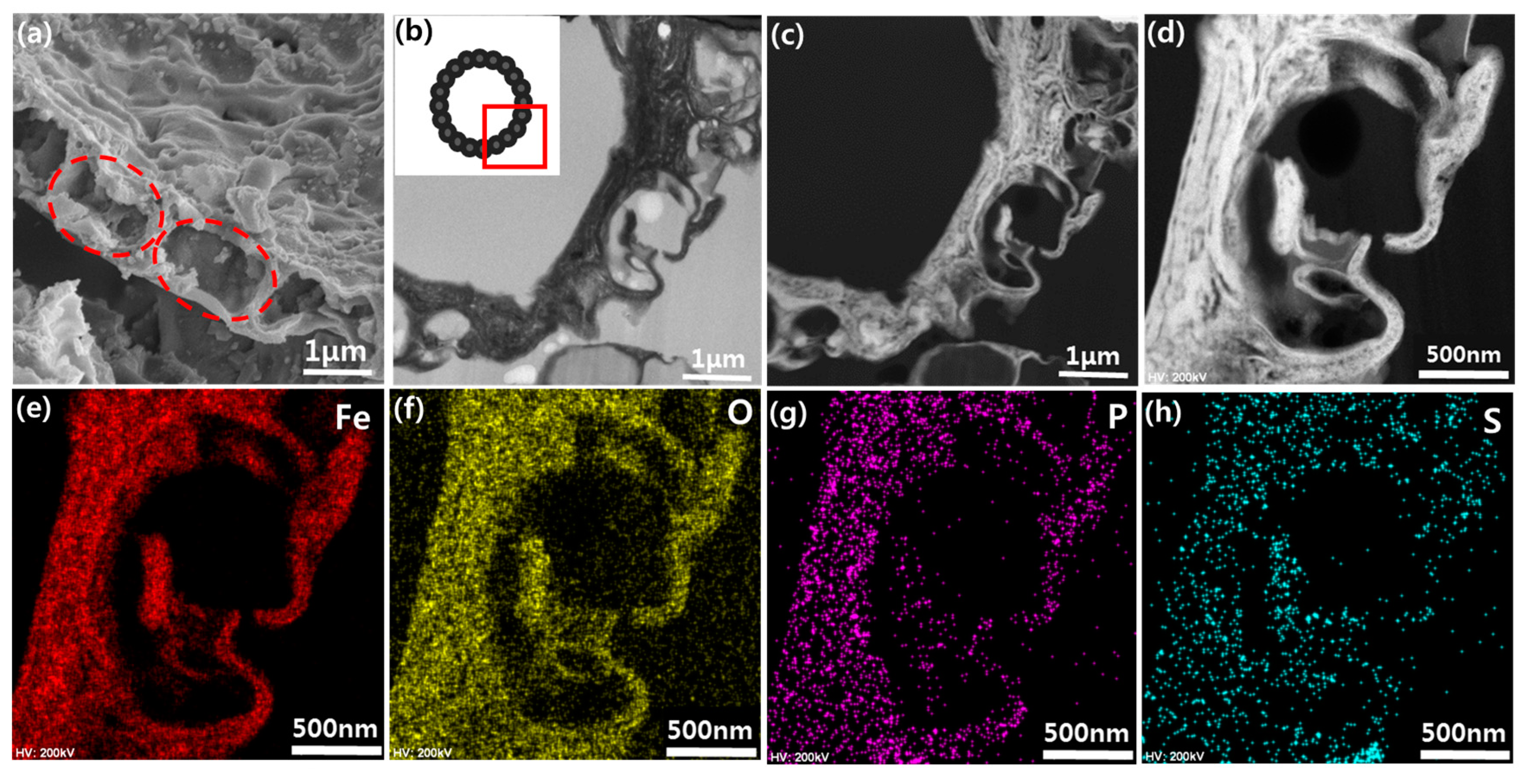
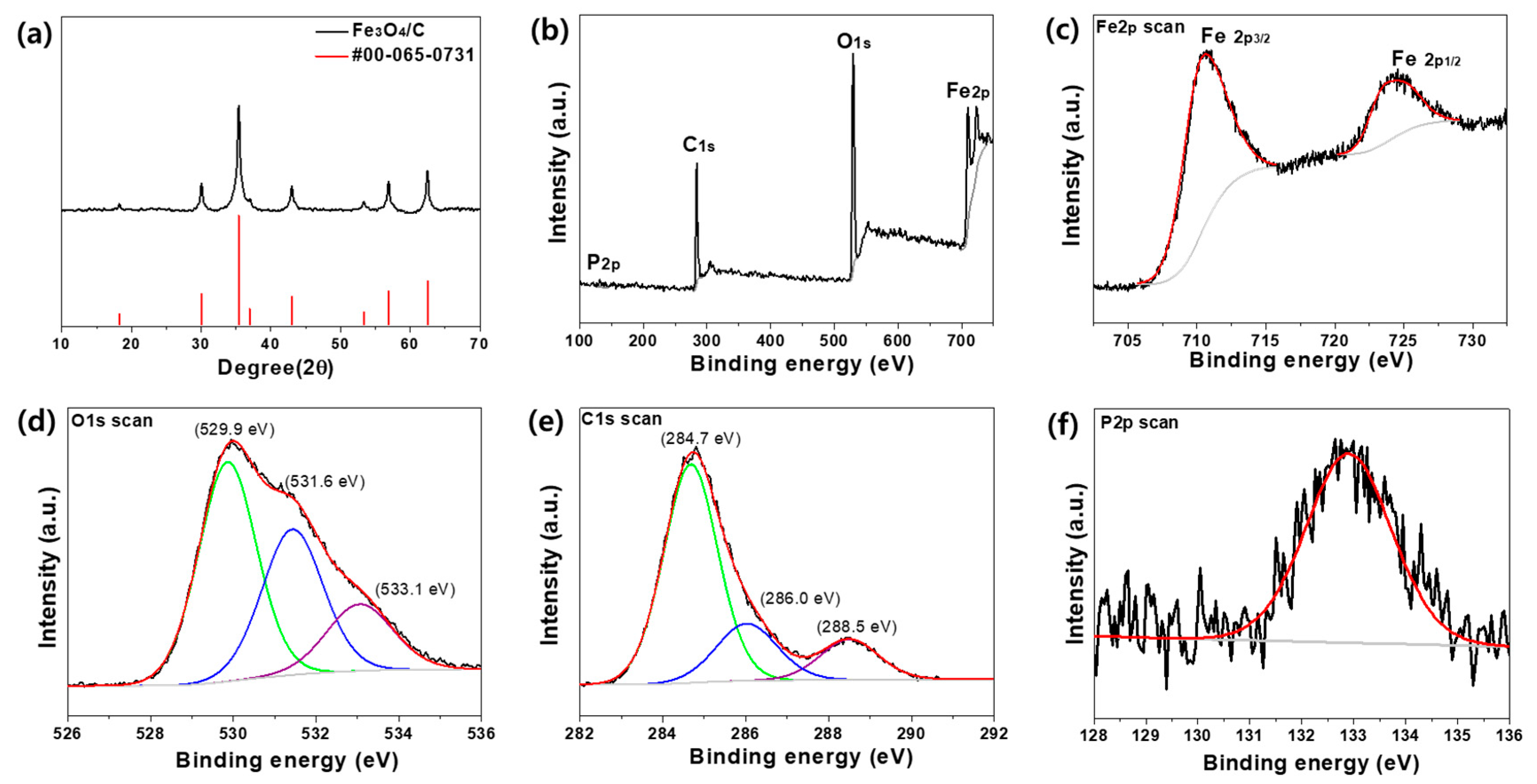
2.3. Characterization
2.4. Electrochemical Measurements
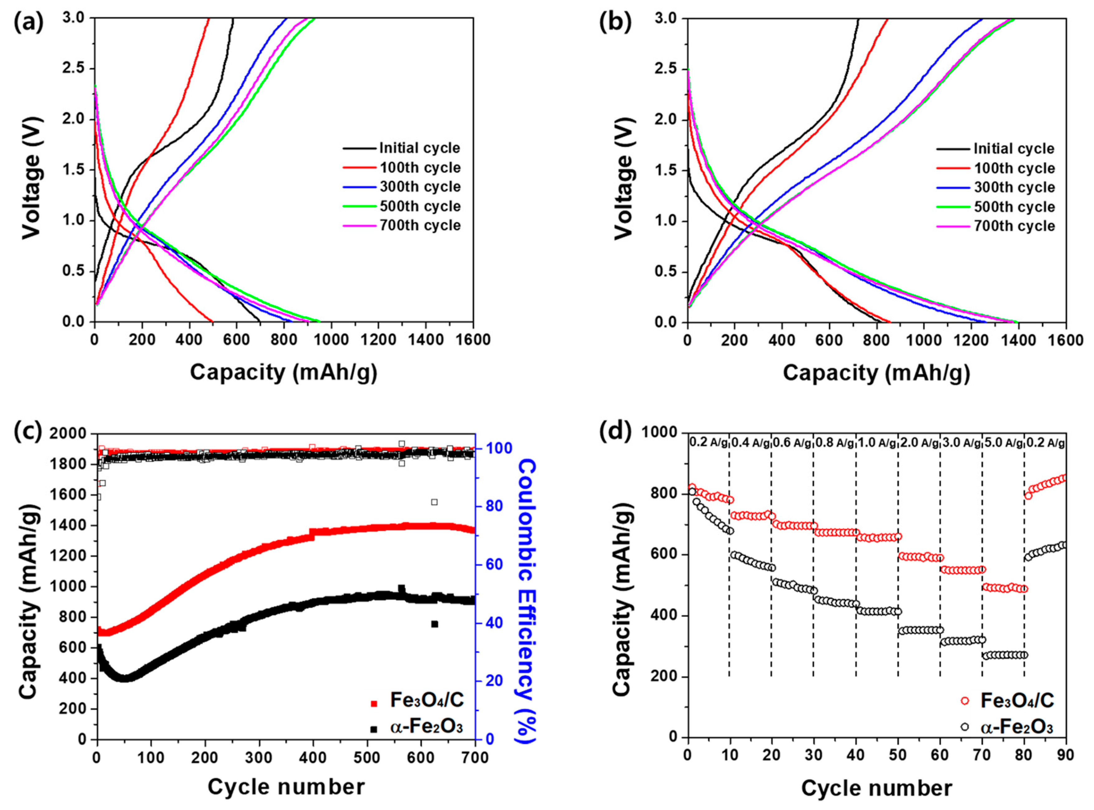
3. Results and Discussion
4. Conclusions
Supplementary Materials
Author Contributions
Funding
Conflicts of Interest
References
- Armand, M.; Tarascon, J.-M. Building better batteries. Nature 2008, 451, 652–657. [Google Scholar] [CrossRef] [PubMed]
- Manthiram, A. An outlook on lithium ion battery technology. ACS Cent. Sci. 2017, 3, 1063–1069. [Google Scholar] [CrossRef] [PubMed] [Green Version]
- Goodenough, J.B.; Park, K.-S. The Li-ion rechargeable battery: A perspective. J. Am. Chem. Soc. 2013, 135, 1167–1176. [Google Scholar] [CrossRef] [PubMed]
- Cabana, B.J.; Monconduit, L.; Larcher, D.; Palacín, M.R. Beyond Intercalation-Based Li-Ion Batteries: The State of the Art and Challenges of Electrode Materials Reacting Through Conversion Reactions. Adv. Mater. 2010, 22, 170–192. [Google Scholar] [CrossRef] [PubMed]
- Taberna, P.L.; Mitra, S.; Poizot, P.; Simon, P.; Tarascon, J.M. High Rate Capabilities Fe3O4-Based Cu Nano-Architectured Electrodes for Lithium-Ion Battery Applications. Nat. Mater. 2006, 5, 567–573. [Google Scholar] [CrossRef] [PubMed] [Green Version]
- Zhang, L.; Wu, H.B.; Wen, X.; Lou, D. Iron-Oxide-Based Advanced Anode Materials for Lithium- Ion Batteries. Adv. Energy Mater. 2014, 4, 1–11. [Google Scholar] [CrossRef]
- Chen, J.S.; Zhu, T.; Yang, X.H.; Yang, H.G.; Lou, X.W. Top-Down Fabrication of r-Fe2O3 Single-Crystal Nanodiscs and Microparticles with Tunable Porosity for Largely Improved Lithium Storage Properties. J. Am. Chem. Soc. 2010, 132, 13162–13164. [Google Scholar] [CrossRef]
- Liu, H.; Wang, G.; Wang, J.; Wexler, D. Magnetite / Carbon Core-Shell Nanorods as Anode Materials for Lithium-Ion Batteries. Electrochem. Commun. 2008, 10, 1879–1882. [Google Scholar] [CrossRef]
- Wang, L.; Yu, Y.; Chen, P.C.; Zhang, D.W.; Chen, C.H. Electrospinning Synthesis of C/Fe3O4 Composite Nanofibers and Their Application for High Performance Lithium-Ion Batteries. J. Power Sources 2008, 183, 717–723. [Google Scholar] [CrossRef]
- Piao, Y.; Hyeon, T. Direct Synthesis of Self-Assembled Ferrite/Carbon Hybrid Nanosheets for High Performance Lithium-Ion Battery Anodes. J. Am. Chem. Soc. 2012, 134, 15010–15015. [Google Scholar]
- Wu, H.B.; Chen, J.S.; Hng, H.H.; Lou, X.W. Nanostructured Metal Oxide-Based Materials as Advanced Anodes for Lithium-Ion Batteries. Nanoscale 2012, 4, 2526–2542. [Google Scholar] [CrossRef] [PubMed]
- Reddy, A.L.M.; Gowda, S.R.; Shaijumon, M.M.; Ajayan, P.M. Hybrid Nanostructures for Energy Storage Applications. Adv. Mater. 2012, 24, 5045–5064. [Google Scholar] [CrossRef] [PubMed]
- Lai, X.; Halpert, E.; Wang, D. Recent Advances in Micro-/Nano-Structured Hollow Spheres for Energy Applications: From Simple to Complex Systems. Energy Environ. Sci. 2012, 5, 5604–5618. [Google Scholar] [CrossRef]
- Koo, B.; Xiong, H.; Slater, M.D.; Prakapenka, V.B.; Balasubramanian, M.; Podsiadlo, P.; Johnson, C.S.; Rajh, T.; Shevchenko, E.V. Hollow Iron Oxide Nanoparticles for Application in Lithium Ion Batteries. Nano Lett. 2012, 12, 2429–2435. [Google Scholar] [CrossRef]
- Xiong, Q.Q.; Tu, J.P.; Lu, Y.; Chen, J.; Yu, Y.X.; Qiao, Y.Q.; Wang, X.L.; Gu, C.D. Synthesis of Hierarchical Hollow-Structured Single-Crystalline Magnetite (Fe3O4) Microspheres: The Highly Powerful Storage versus Lithium as an Anode for Lithium Ion Batteries. J. Phys. Chem. C 2012, 116, 6495–6502. [Google Scholar] [CrossRef]
- Wang, B.; Wu, H.B.; Zhang, L.; Lou, X.W. Self-Supported Construction of Uniform Fe3O4 Hollow Microspheres from Nanoplate Building Blocks. Angew. Chem.-Int. Ed. 2013, 52, 4165–4168. [Google Scholar] [CrossRef]
- Li, X.; Gu, M.; Hu, S.; Kennard, R.; Yan, P.; Chen, X.; Wang, C.; Sailor, M.J.; Zhang, J.; Liu, J. Mesoporous Silicon Sponge as an Anti-Pulverization Structure for High-Performance Lithium-Ion Battery Anodes. Nat. Commun. 2014, 5, 1–7. [Google Scholar] [CrossRef] [Green Version]
- Lv, Y.; Gan, L.; Liu, M.; Xiong, W.; Xu, Z.; Zhu, D.; Wright, D.S. A Self-Template Synthesis of Hierarchical Porous Carbon Foams Based on Banana Peel for Supercapacitor Electrodes. J. Power Sources 2012, 209, 152–157. [Google Scholar] [CrossRef]
- Zhou, H.; Fan, T.; Zhang, D. Hydrothermal Synthesis of ZnO Hollow Spheres Using Spherobacterium as Biotemplates. Microporous Mesoporous Mater. 2007, 100, 322–327. [Google Scholar] [CrossRef]
- Li, B.X.; Fan, T.; Zhou, H.; Chow, S.; Zhang, W.; Zhang, D.; Guo, Q.; Ogawa, H. Enhanced Light-Harvesting and Photocatalytic Properties in Morph -TiO2 from Green-Leaf Biotemplates. Adv. Funct. Mater. 2009, 19, 45–56. [Google Scholar] [CrossRef]
- Song, D.; Park, J.; Kim, K.; Seol, L.; Yoon, J.; Oh, Y.; Kim, Y.; Ryou, M.; Min, Y.; Lee, K. Recycling Oil-Extracted Microalgal Biomass Residues into Nano/Micro Hierarchical Sn/C Composite Anode Materials for Lithium-Ion Batteries. Electrochim. Acta 2017, 250, 59–67. [Google Scholar] [CrossRef]
- Seo, J.; Umirov, N.; Bin, S.; Lee, K.; Kim, S. Microalgae-Derived Hollow Carbon-MoS2 Composite as Anode for Lithium-Ion Batteries. J. Ind. Eng. Chem. 2019, 79, 106–114. [Google Scholar] [CrossRef]
- Chakravarty, R.; Banerjee, P.C. Mechanism of Cadmium Binding on the Cell Wall of an Acidophilic Bacterium. Bioresour. Technol. 2012, 108, 176–183. [Google Scholar] [CrossRef] [PubMed]
- Praveenkumar, R.; Kim, B.; Choi, E.; Lee, K.; Cho, S.; Park, J.H.J. Mixotrophic Cultivation of Oleaginous Chlorella Sp. KR-1 Mediated by Actual Coal-Fired Flue Gas for Biodiesel Production. Bioprocess Biosyst. Eng. 2014, 37, 2083–2094. [Google Scholar] [CrossRef] [PubMed]
- Nandiyanto, A.B.D.; Ogi, T.; Wang, W.N.; Gradon, L.; Okuyama, K. Template-Assisted Spray-Drying Method for the Fabrication of Porous Particles with Tunable Structures. Adv. Powder Technol. 2019, 30, 2908–2924. [Google Scholar] [CrossRef]
- Bayu, A.; Nandiyanto, D.; Okuyama, K. Progress in Developing Spray-Drying Methods for the Production of Controlled Morphology Particles: From the Nanometer to Submicrometer Size Ranges. Adv. Powder Technol. 2011, 22, 1–19. [Google Scholar]
- Bohström, Z.; Lillerud, K.P. Preparation of Chabazite with Mesopores Templated from a Cationic Polymer. Microporous Mesoporous Mater. 2018, 271, 295–300. [Google Scholar] [CrossRef]
- Yuan, S.M.; Li, J.X.; Yang, L.T.; Su, L.W.; Liu, L.; Zhou, Z. Preparation and Lithium Storage Performances of Mesoporous Fe3O4@C Microcapsules. ACS Appl. Mater. Interfaces 2011, 3, 705–709. [Google Scholar] [CrossRef]
- Chen, Y.; Song, B.; Li, M.; Lu, L.; Xue, J. Fe3O4 Nanoparticles Embedded in Uniform Mesoporous Carbon Spheres for Superior High-Rate Battery Applications. Adv. Funct. Mater. 2014, 24, 319–326. [Google Scholar] [CrossRef]
- Sun, G.; Dong, B.; Cao, M.; Wei, B.; Hu, C. Hierarchical Dendrite-Like Magnetic Materials of Fe3O4, γ-Fe2O3, and Fe with High Performance of Microwave Absorption. Chem. Mater. 2011, 23, 1587–1593. [Google Scholar] [CrossRef]
- Yamashita, T.; Hayes, P. Analysis of XPS Spectra of Fe2+ and Fe3+ Ions in Oxide Materials. Appl. Surf. Sci. 2008, 254, 2441–2449. [Google Scholar] [CrossRef]
- Wang, J. A Platinum Anticancer Theranostic Agent with Magnetic Targeting Potential Derived from Maghemite Nanoparticles. Chem. Sci. 2013, 4, 2605–2612. [Google Scholar] [CrossRef]
- Cheng, Y.; Guang, R.; Jun, M.; Cheng, H.; Kwan, C.; Dan, Q.; Yang, Y.; Antonio, J. Scalable Synthesis of Fe3O4 Nanoparticles Anchored on Graphene as a High-Performance Anode for Lithium Ion Batteries. J. Solid State Chem. 2013, 201, 330–337. [Google Scholar]
- Zhang, W.; Li, X.; Liang, J.; Tang, K.; Zhu, Y.; Qian, Y. One-Step Thermolysis Synthesis of Two-Dimensional Ultrafine Fe3O4 Particles/Carbon Nanonetworks for High-Performance Lithium-Ion Batteries. Nanoscale 2016, 8, 4733–4741. [Google Scholar] [CrossRef] [PubMed] [Green Version]
- Zheng, R.; Lin, L.; Xie, J.; Zhu, Y.; Xie, Y. State of Doped Phosphorus and Its Influence on the Physicochemical and Photocatalytic Properties of P-Doped Titania. J. Phys. Chem. C 2008, 112, 15502–15509. [Google Scholar] [CrossRef]
- Hao, S.; Zhang, B.; Wang, Y.; Li, C.; Feng, J.; Ball, S.; Srinivasan, M.; Wu, J.; Huang, Y. Hierarchical Three-Dimensional Fe3O4@porous Carbon Matrix/Graphene Anodes for High Performance Lithium Ion Batteries. Electrochim. Acta 2018, 260, 965–973. [Google Scholar] [CrossRef]
- Liu, L.; Zhang, H.; Liu, S.; Yao, H.; Hou, H.; Chen, S. Hollow Carbon Nanosphere Embedded with Ultra Fine Fe3O4 Nanoparticles as High Performance Li-Ion Battery Anode. Electrochim. Acta 2016, 219, 356–362. [Google Scholar] [CrossRef]
- Zhou, Z.; Xie, W.; Li, S.; Jiang, X.; He, D.; Peng, S.; Ma, F. Facile Synthesis of Porous Fe3O4@C Nanospheres as High-Performance Anode for Lithium-Ion Battery. J. Solid State Electrochem. 2015, 19, 1211–1215. [Google Scholar] [CrossRef]
- Paraknowitsch, J.P.; Thomas, A. Doping Carbons beyond Nitrogen: An Overview of Advanced Heteroatom Doped Carbons with Boron, Sulphur and Phosphorus for Energy Applications. Energy Environ. Sci. 2013, 6, 2839–2855. [Google Scholar] [CrossRef] [Green Version]
- Liu, Y.; Liu, P.; Wu, D.; Huang, Y.; Tang, Y.; Su, Y.; Zhang, F.; Feng, X. Boron-Doped, Carbon-Coated SnO2/Graphene Nanosheets for Enhanced Lithium Storage. Chem.-A Eur. J. 2015, 21, 5617–5622. [Google Scholar] [CrossRef]
- Wu, Z.-S.; Ren, W.; Xu, L.; Li, F.; Cheng, H.-M. Doped Graphene Sheets As Anode Materials with Superhigh Rate and Large Capacity for Lithium Ion. ACS Nano 2011, 5, 5463–5471. [Google Scholar] [CrossRef]


Publisher’s Note: MDPI stays neutral with regard to jurisdictional claims in published maps and institutional affiliations. |
© 2020 by the authors. Licensee MDPI, Basel, Switzerland. This article is an open access article distributed under the terms and conditions of the Creative Commons Attribution (CC BY) license (http://creativecommons.org/licenses/by/4.0/).
Share and Cite
Park, J.; Kim, J.; Jung, D.S.; Phiri, I.; Bae, H.-S.; Hong, J.; Kim, S.; Lee, Y.-G.; Ryou, M.-H.; Lee, K. Microalgae-Templated Spray Drying for Hierarchical and Porous Fe3O4/C Composite Microspheres as Li-ion Battery Anode Materials. Nanomaterials 2020, 10, 2074. https://doi.org/10.3390/nano10102074
Park J, Kim J, Jung DS, Phiri I, Bae H-S, Hong J, Kim S, Lee Y-G, Ryou M-H, Lee K. Microalgae-Templated Spray Drying for Hierarchical and Porous Fe3O4/C Composite Microspheres as Li-ion Battery Anode Materials. Nanomaterials. 2020; 10(10):2074. https://doi.org/10.3390/nano10102074
Chicago/Turabian StylePark, Jinseok, Jungmin Kim, Dae Soo Jung, Isheunesu Phiri, Hyeon-Su Bae, Jinseok Hong, Sojin Kim, Young-Gi Lee, Myung-Hyun Ryou, and Kyubock Lee. 2020. "Microalgae-Templated Spray Drying for Hierarchical and Porous Fe3O4/C Composite Microspheres as Li-ion Battery Anode Materials" Nanomaterials 10, no. 10: 2074. https://doi.org/10.3390/nano10102074