
pH-Activated Dissolvable Polymeric Coatings to Reduce Biofouling on Electrochemical Sensors
 ,
,
Abstract
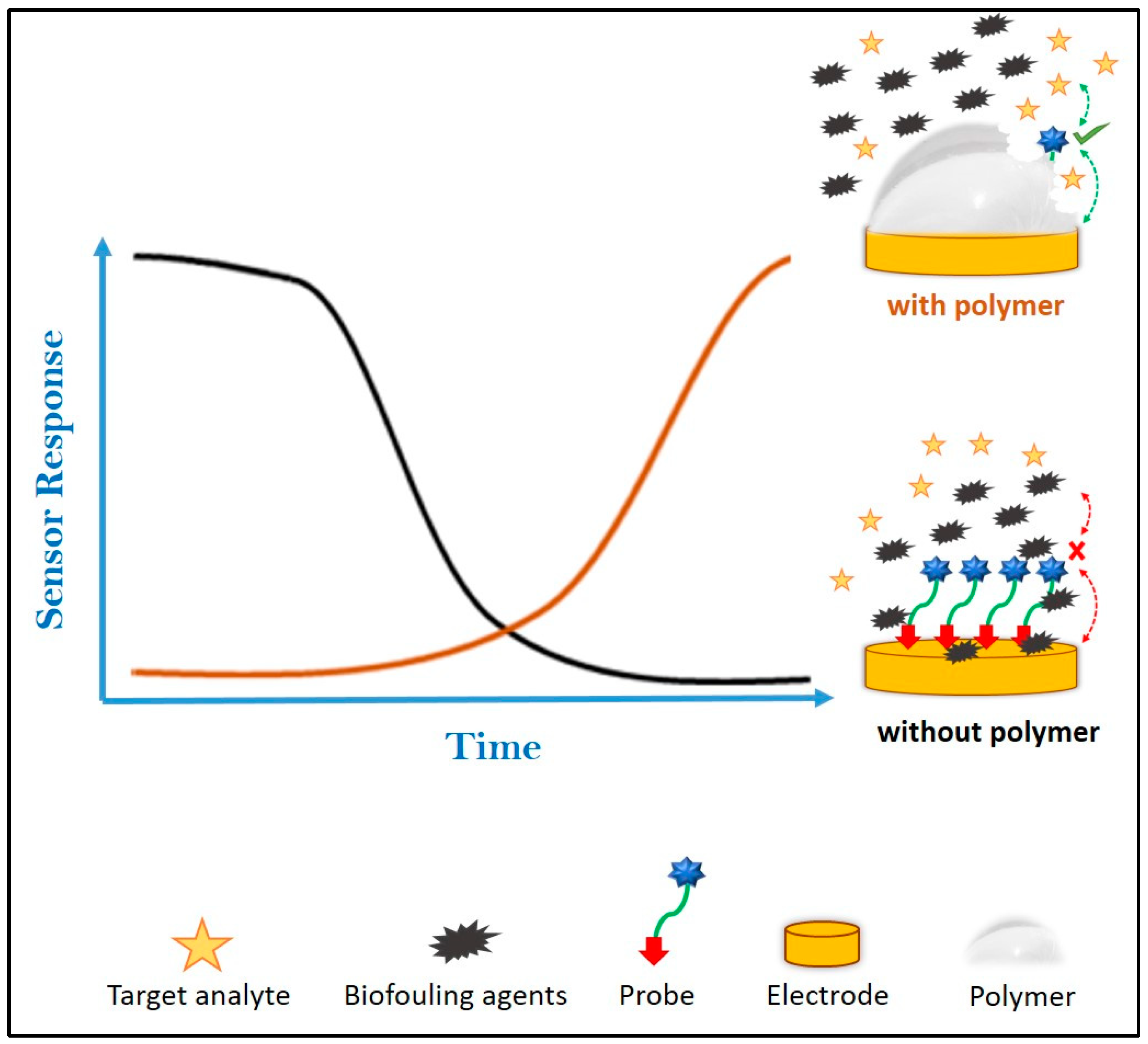
:1. Introduction
2. Materials and Methods
2.1. Instrumentation
2.2. Reagents and Materials
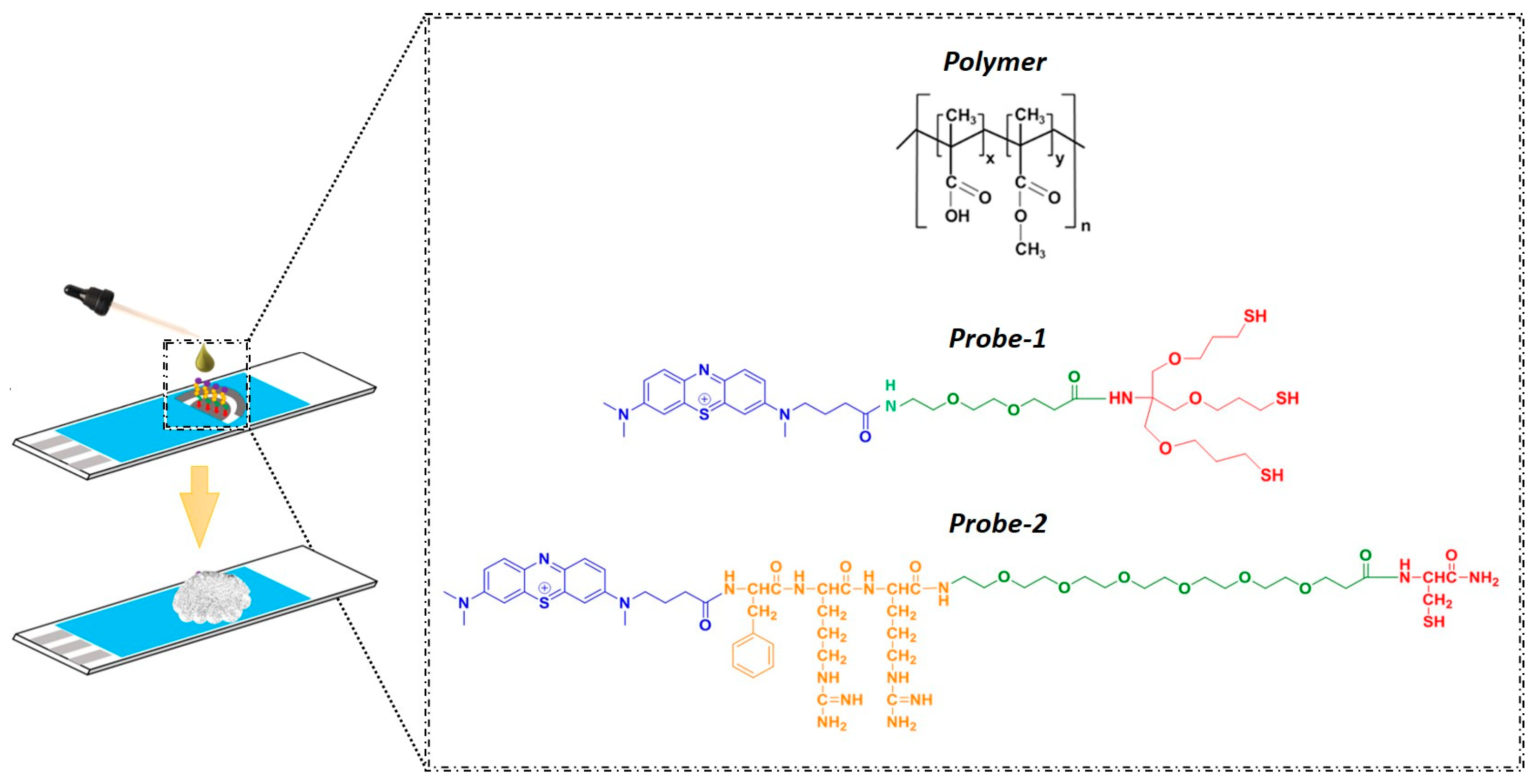
2.3. Synthetic Methods
2.4. Cleaning and Preparation of Electrodes
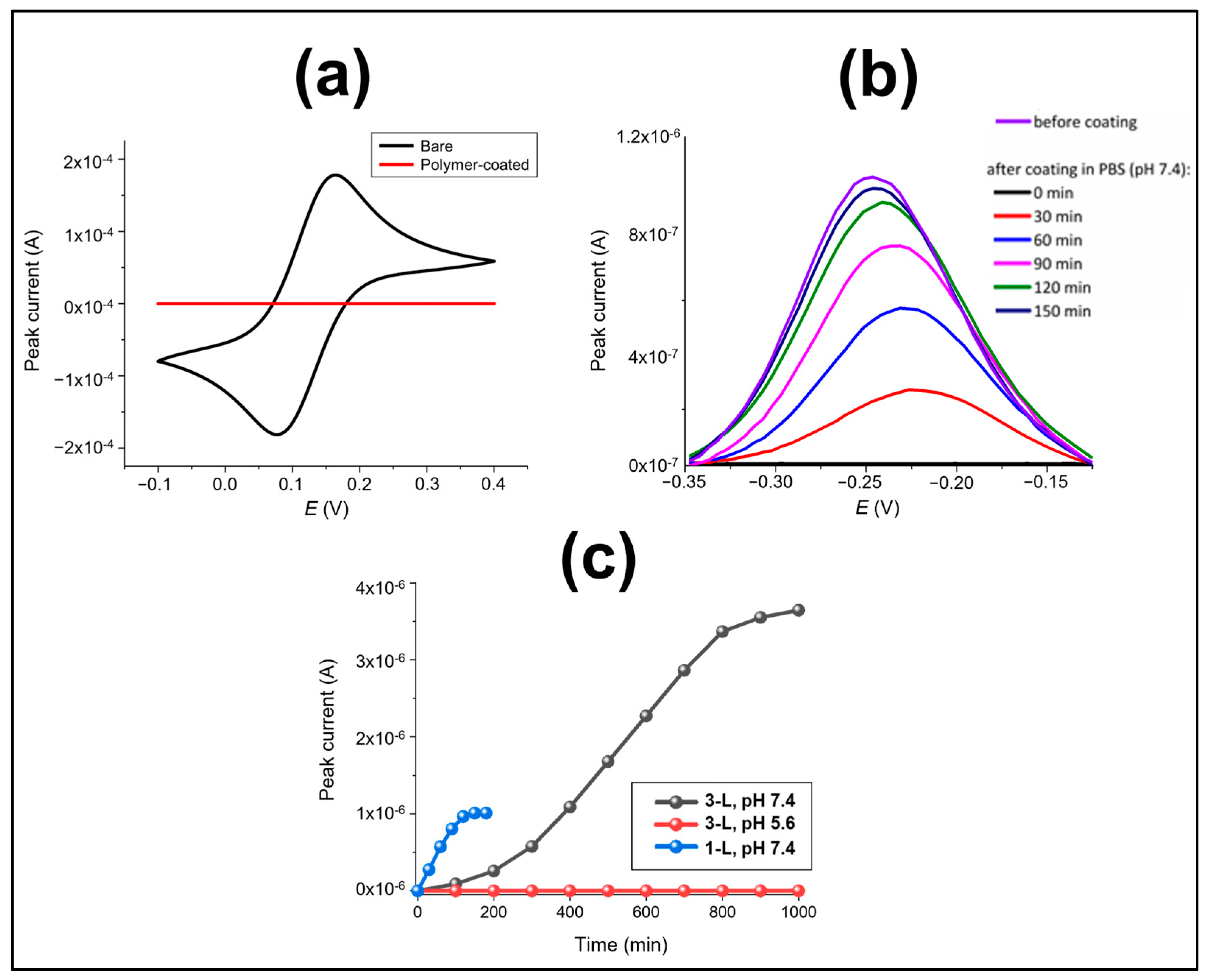
2.5. Electrochemical Characterisation of Polymer Dissolution
3. Results and Discussion
3.1. Characterisation of Polymer Dissolution
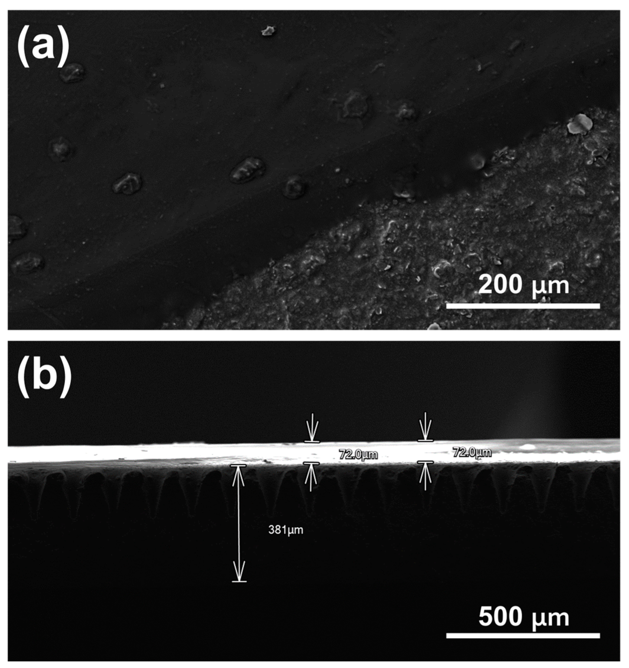
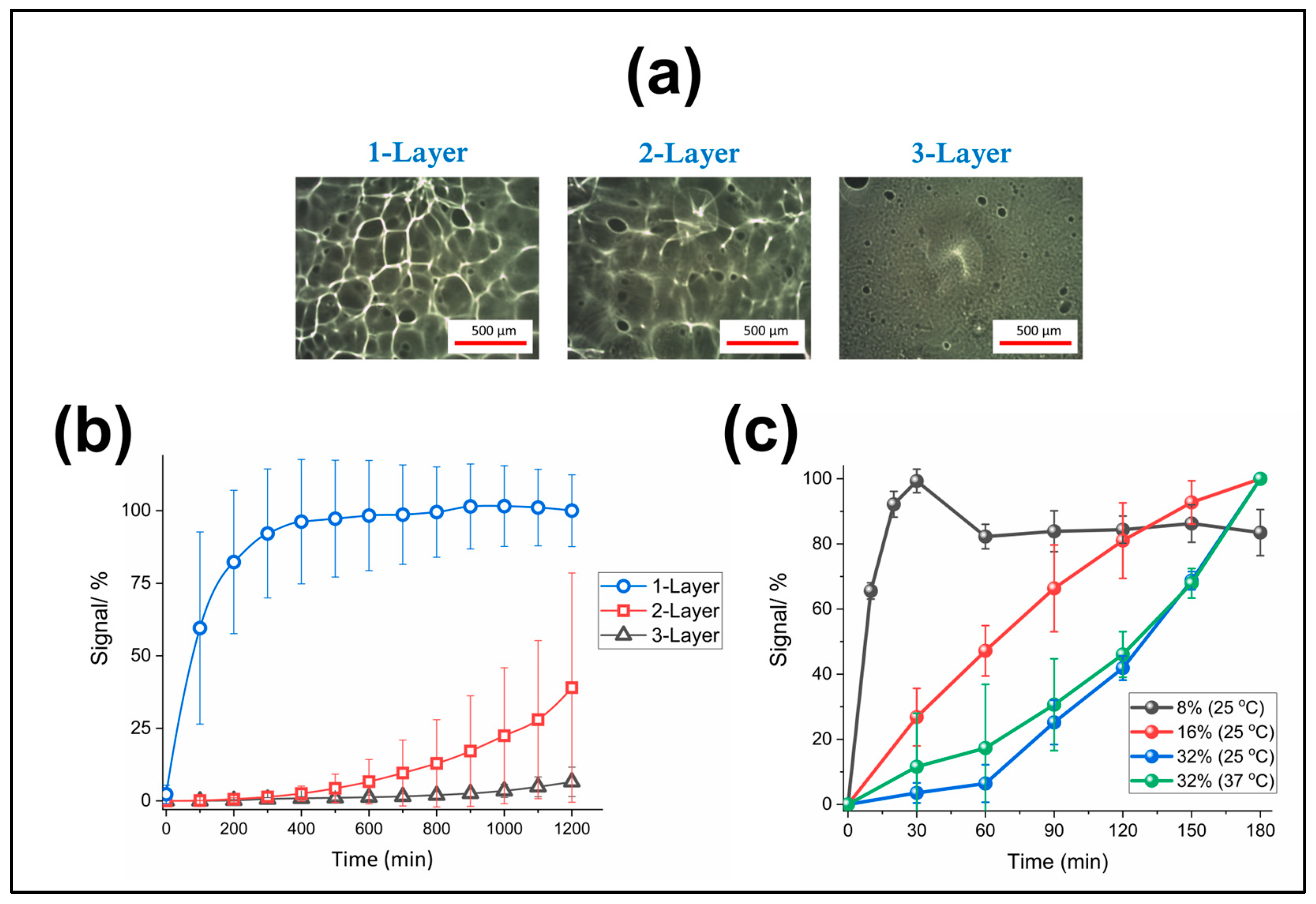
3.2. Optimisation of Polymer Coating Formation (Thickness and Concentration)
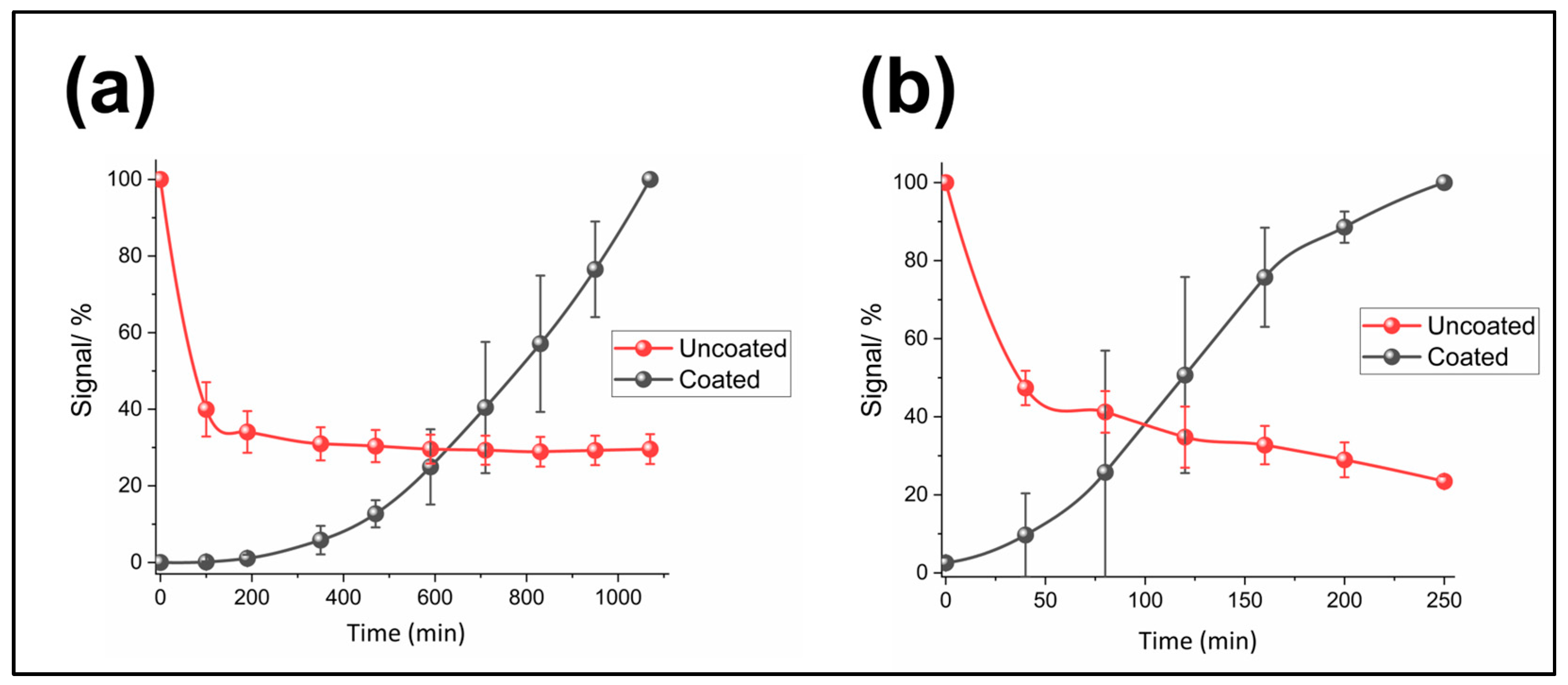
3.3. Biofouling Protection
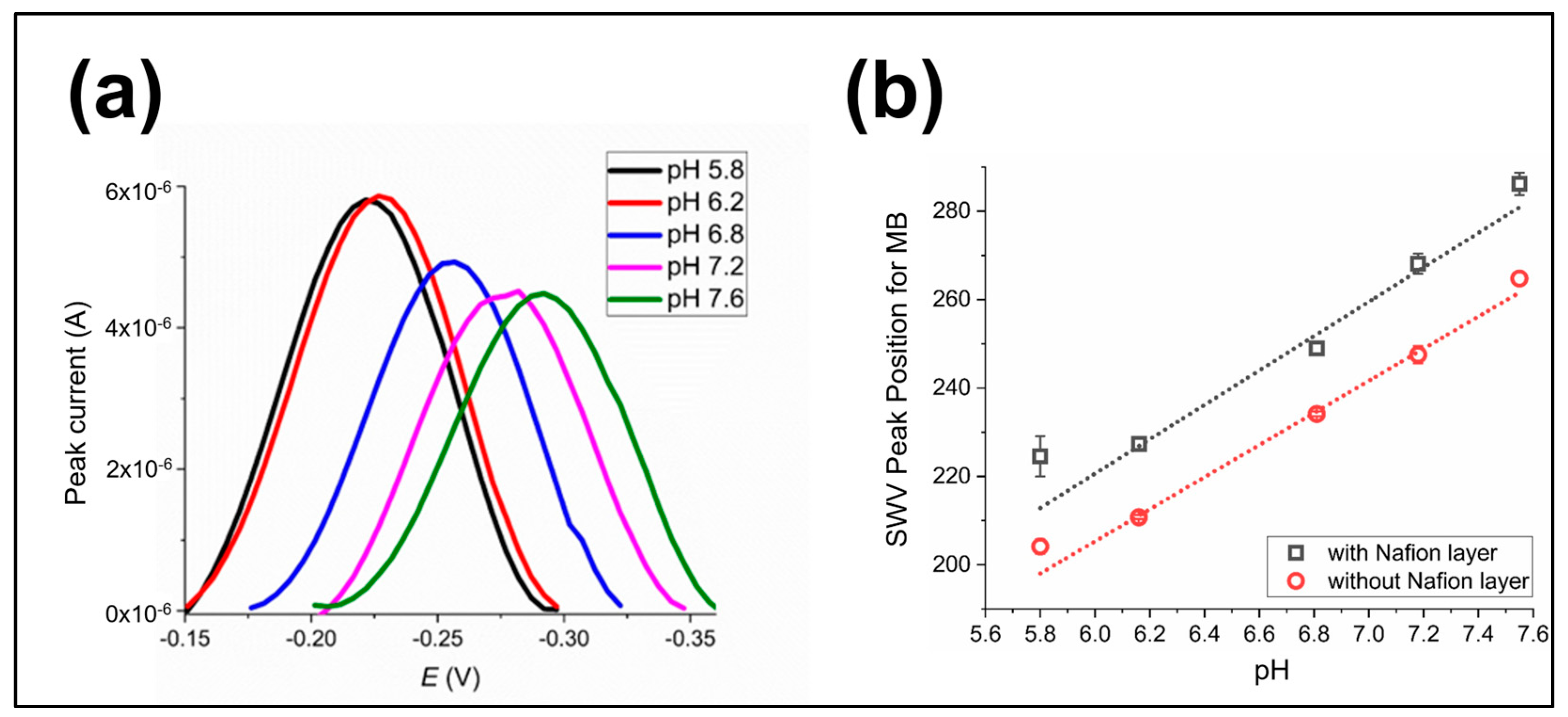
3.4. Analysis of Post-Dissolution Sensor Performance
4. Conclusions
Author Contributions
Funding
Data Availability Statement
Acknowledgments
Conflicts of Interest
References
- Windmiller, J.R.; Wang, J. Wearable Electrochemical Sensors and Biosensors: A Review. Electroanalysis 2013, 25, 29–46. [Google Scholar] [CrossRef]
- Kim, J.; Campbell, A.S.; Wang, J. Wearable Non-Invasive Epidermal Glucose Sensors: A Review. Talanta 2018, 177, 163–170. [Google Scholar] [CrossRef] [PubMed]
- Heller, A. Implanted Electrochemical Glucose Sensors for the Management of Diabetes. Annu. Rev. Biomed. Eng. 1999, 1, 153–175. [Google Scholar] [CrossRef] [PubMed]
- Ming Li, C.; Dong, H.; Cao, X.; Luong, T.; John, H.; Zhang, X. Implantable Electrochemical Sensors for Biomedical and Clinical Applications: Progress, Problems, and Future Possibilities. Curr. Med. Chem. 2007, 14, 937–951. [Google Scholar] [CrossRef]
- Voskerician, G.; Anderson, J. Sensor Biocompatibility and Biofouling in Real-Time Monitoring. In Wiley Encyclopedia of Biomedical Engineering; John Wiley & Sons, Inc.: Hoboken, NJ, USA, 2006. [Google Scholar]
- Wisniewski, N.; Moussy, F.; Reichert, W.M. Characterization of Implantable Biosensor Membrane Biofouling. Fresenius. J. Anal. Chem. 2000, 366, 611–621. [Google Scholar] [CrossRef]
- Zhang, D.; Chen, Q.; Shi, C.; Chen, M.; Ma, K.; Wan, J.; Liu, R. Dealing with the Foreign-body Response to Implanted Biomaterials: Strategies and Applications of New Materials. Adv. Funct. Mater. 2021, 31, 2007226. [Google Scholar] [CrossRef]
- Barfidokht, A.; Gooding, J.J. Approaches toward Allowing Electroanalytical Devices to Be Used in Biological Fluids. Electroanalysis 2014, 26, 1182–1196. [Google Scholar] [CrossRef]
- Jiang, C.; Wang, G.; Hein, R.; Liu, N.; Luo, X.; Davis, J.J. Antifouling Strategies for Selective in Vitro and in Vivo Sensing. Chem. Rev. 2020, 120, 3852–3889. [Google Scholar] [CrossRef]
- Lin, P.-H.; Li, B.-R. Antifouling Strategies in Advanced Electrochemical Sensors and Biosensors. Analyst 2020, 145, 1110–1120. [Google Scholar] [CrossRef]
- Teo, A.J.T.; Mishra, A.; Park, I.; Kim, Y.-J.; Park, W.-T.; Yoon, Y.-J. Polymeric Biomaterials for Medical Implants and Devices. ACS Biomater. Sci. Eng. 2016, 2, 454–472. [Google Scholar] [CrossRef]
- Ward, I.M.; Sweeney, J. Mechanical Properties of Solid Polymers; John Wiley & Sons: Hoboken, NJ, USA, 2012; ISBN 1119967112. [Google Scholar]
- Campuzano, S.; Pedrero, M.; Yáñez-Sedeño, P.; Pingarrón, J.M. Antifouling (Bio) Materials for Electrochemical (Bio) Sensing. Int. J. Mol. Sci. 2019, 20, 423. [Google Scholar] [CrossRef] [Green Version]
- Francolini, I.; Vuotto, C.; Piozzi, A.; Donelli, G. Antifouling and Antimicrobial Biomaterials: An Overview. Apmis 2017, 125, 392–417. [Google Scholar] [CrossRef] [Green Version]
- Walker, J.A.; Robinson, K.J.; Munro, C.; Gengenbach, T.; Muller, D.A.; Young, P.R.; Lua, L.H.L.; Corrie, S.R. Antibody-Binding, Antifouling Surface Coatings Based on Recombinant Expression of Zwitterionic EK Peptides. Langmuir 2018, 35, 1266–1272. [Google Scholar] [CrossRef]
- Zhang, B.; Nagle, A.R.; Wallace, G.G.; Hanks, T.W.; Molino, P.J. Functionalised Inherently Conducting Polymers as Low Biofouling Materials. Biofouling 2015, 31, 493–502. [Google Scholar] [CrossRef]
- Song, Z.; Sheng, G.; Cui, Y.; Li, M.; Song, Z.; Ding, C.; Luo, X. Low Fouling Electrochemical Sensing in Complex Biological Media by Using the Ionic Liquid-Doped Conducting Polymer PEDOT: Application to Voltammetric Determination of Dopamine. Microchim. Acta 2019, 186, 220. [Google Scholar] [CrossRef]
- Xue, C.-H.; Guo, X.-J.; Ma, J.-Z.; Jia, S.-T. Fabrication of Robust and Antifouling Superhydrophobic Surfaces via Surface-Initiated Atom Transfer Radical Polymerization. ACS Appl. Mater. Interfaces 2015, 7, 8251–8259. [Google Scholar] [CrossRef] [PubMed]
- Lee, J.-W.; Jung, J.; Cho, Y.H.; Yadav, S.K.; Baek, K.Y.; Park, H.B.; Hong, S.M.; Koo, C.M. Fouling-Tolerant Nanofibrous Polymer Membranes for Water Treatment. ACS Appl. Mater. Interfaces 2014, 6, 14600–14607. [Google Scholar] [CrossRef] [PubMed]
- Kyröläinen, M.; Håkanson, H.; Mattiasson, B.; Vadgama, P. Minimal-Fouling Enzyme Electrode for Continuous Flow Measurement of Whole Blood Lactate. Biosens. Bioelectron. 1997, 12, 1073–1081. [Google Scholar] [CrossRef] [PubMed]
- Du, M.-Q.; Peng, Y.-Z.; Ma, Y.-C.; Yang, L.; Zhou, Y.-L.; Zeng, F.-K.; Wang, X.-K.; Song, M.-L.; Chang, G.-J. Selective Carbon Dioxide Capture in Antifouling Indole-Based Microporous Organic Polymers. Chin. J. Polym. Sci. 2020, 38, 187–194. [Google Scholar] [CrossRef]
- Akthakul, A.; Salinaro, R.F.; Mayes, A.M. Antifouling Polymer Membranes with Subnanometer Size Selectivity. Macromolecules 2004, 37, 7663–7668. [Google Scholar] [CrossRef]
- Ruiz-Valdepeñas Montiel, V.; Sempionatto, J.R.; Esteban-Fernández de Ávila, B.; Whitworth, A.; Campuzano, S.; Pingarrón, J.M.; Wang, J. Delayed Sensor Activation Based on Transient Coatings: Biofouling Protection in Complex Biofluids. J. Am. Chem. Soc. 2018, 140, 14050–14053. [Google Scholar] [CrossRef]
- Montiel, V.R.-V.; Sempionatto, J.R.; Campuzano, S.; Pingarrón, J.M.; de Ávila, B.E.F.; Wang, J. Direct Electrochemical Biosensing in Gastrointestinal Fluids. Anal. Bioanal. Chem. 2019, 411, 4597–4604. [Google Scholar] [CrossRef] [PubMed]
- Evonik Industries EUDRAGIT® Functional Polymers for Oral Solid Dosage Forms. Available online: https://healthcare.evonik.com/product/health-care/en/products/pharmaceutical-excipients/EUDRAGIT/ (accessed on 23 January 2020).
- Duan, H.; Lü, S.; Gao, C.; Bai, X.; Qin, H.; Wei, Y.; Wu, X.; Liu, M. Mucoadhesive Microparticulates Based on Polysaccharide for Target Dual Drug Delivery of 5-Aminosalicylic Acid and Curcumin to Inflamed Colon. Colloids Surf. B Biointerfaces 2016, 145, 510–519. [Google Scholar] [CrossRef]
- Sawant, P.D.; Luu, D.; Ye, R.; Buchta, R. Drug Release from Hydroethanolic Gels. Effect of Drug’s Lipophilicity (Log P), Polymer–Drug Interactions and Solvent Lipophilicity. Int. J. Pharm. 2010, 396, 45–52. [Google Scholar] [CrossRef]
- Liu, S.; Tang, J.; Ji, F.; Lin, W.; Chen, S. Recent Advances in Zwitterionic Hydrogels: Preparation, Property, and Biomedical Application. Gels 2022, 8, 46. [Google Scholar] [CrossRef]
- González-Fernández, E.; Staderini, M.; Marland, J.R.K.; Gray, M.E.; Uçar, A.; Dunare, C.; Blair, E.O.; Sullivan, P.; Tsiamis, A.; Greenhalgh, S.N.; et al. In Vivo Application of an Implantable Tri-Anchored Methylene Blue-Based Electrochemical pH Sensor. Biosens. Bioelectron. 2022, 197, 113728. [Google Scholar] [CrossRef]
- González-Fernández, E.; Avlonitis, N.; Murray, A.F.; Mount, A.R.; Bradley, M. Methylene Blue Not Ferrocene: Optimal Reporters for Electrochemical Detection of Protease Activity. Biosens. Bioelectron. 2016, 84, 82–88. [Google Scholar] [CrossRef] [Green Version]
- Ucar, A.; González-Fernández, E.; Staderini, M.; Avlonitis, N.; Murray, A.F.; Bradley, M.; Mount, A.R. Miniaturisation of a Peptide-Based Electrochemical Protease Activity Sensor Using Platinum Microelectrodes. Analyst 2020, 145, 975–982. [Google Scholar] [CrossRef] [PubMed] [Green Version]
- Eisele, J.; Glasbrenner, B. EUDRAGIT® S100 Product Regulatory Datasheet; Evonik Operations GmbH: Essen, Germany, 2022. [Google Scholar]
- Evonik Industries Technical Information of Eudragit® L100 and Eudragit® S100. Available online: www.pharosproject.net/uploads/files/cml/1389279051.pdf (accessed on 23 January 2019).
- Nguyen, D.A.; Fogler, H.S. Facilitated Diffusion in the Dissolution of Carboxylic Polymers. AIChE J. 2005, 51, 415–425. [Google Scholar] [CrossRef]
- Vinner, G.K.; Vladisavljević, G.T.; Clokie, M.R.J.; Malik, D.J. Microencapsulation of Clostridium Difficile Specific Bacteriophages Using Microfluidic Glass Capillary Devices for Colon Delivery Using PH Triggered Release. PLoS ONE 2017, 12, e0186239. [Google Scholar] [CrossRef] [PubMed] [Green Version]
- Khan, M.Z.I.; Štedul, H.P.; Kurjaković, N. A pH-Dependent Colon-Targeted Oral Drug Delivery System Using Methacrylic Acid Copolymers. II. Manipulation of Drug Release Using Eudragit® L100 and Eudragit S100 Combinations. Drug Dev. Ind. Pharm. 2000, 26, 549–554. [Google Scholar] [CrossRef]
- Nikam, V.K.; Kotade, K.B.; Gaware, V.M.; Dolas, R.T.; Dhamak, K.B.; Somwanshi, S.B.; Khadse, A.N.; Kashid, V.A. Eudragit a Versatile Polymer: A Review. Pharmacologyonline 2011, 1, 152–164. [Google Scholar]
- Blaszykowski, C.; Sheikh, S.; Thompson, M. Surface Chemistry to Minimize Fouling from Blood-Based Fluids. Chem. Soc. Rev. 2012, 41, 5599–5612. [Google Scholar] [CrossRef] [PubMed]
- Koutsoumpeli, E.; Murray, J.; Langford, D.; Bon, R.S.; Johnson, S. Probing Molecular Interactions with Methylene Blue Derivatized Self-Assembled Monolayers. Sens. Bio-Sens. Res. 2015, 6, 1–6. [Google Scholar] [CrossRef] [Green Version]
- Gray, M.E.; Sullivan, P.; Marland, J.R.K.; Greenhalgh, S.N.; Meehan, J.; Gregson, R.; Clutton, R.E.; Cousens, C.; Griffiths, D.J.; Murray, A. A Novel Translational Ovine Pulmonary Adenocarcinoma Model for Human Lung Cancer. Front. Oncol. 2019, 9, 534. [Google Scholar] [CrossRef]
- Huang, Y.; Qian, X.; Wang, X.; Wang, T.; Lounder, S.J.; Ravindran, T.; Demitrack, Z.; McCutcheon, J.; Asatekin, A.; Li, B. Electrospraying Zwitterionic Copolymers as an Effective Biofouling Control for Accurate and Continuous Monitoring of Wastewater Dynamics in a Real-Time and Long-Term Manner. Environ. Sci. Technol. 2022, 56, 8176–8186. [Google Scholar] [CrossRef]
- Yang, C.; Yang, C.; Li, X.; Zhang, A.; He, G.; Wu, Q.; Liu, X.; Huang, S.; Huang, X.; Cui, G. Liquid-like Polymer Coating as a Promising Candidate for Reducing Electrode Contamination and Noise in Complex Biofluids. ACS Appl. Mater. Interfaces 2021, 13, 4450–4462. [Google Scholar] [CrossRef]
- Qi, L.; Jiang, T.; Liang, R.; Qin, W. Polymeric Membrane Ion-Selective Electrodes with Anti-Biofouling Properties by Surface Modification of Silver Nanoparticles. Sens. Actuators B Chem. 2021, 328, 129014. [Google Scholar] [CrossRef]


| Material | Electrode Type | Analyte | Significant Remarks | Ref. |
|---|---|---|---|---|
| Poly(trifluoroethyl methacrylate-random-sulfobetaine methacrylate) (PTFEMA-r-SBMA) | Solid-state ion-selective electrodes | NH4+ | Highly sensitive and long-term stable sensing performance was shown in real wastewater for 55 days. | [41] |
| Liquid-like polydimethylsiloxane (PDMS) | Integrated Au electrodes | Reactive oxygen species | Stable sensing performance was observed after 3 days of incubation with bacteria and sensitive ROS detection in bacteria-rich media over 24 h was achieved. | [42] |
| Ag NPs/ hydrophilic polydopamine | Glassy carbon electrodes with ion selective polymer membrane | Na+, Ca2+, Mg2+, Li+, Ag+ | Due to the anti-bacterial properties of Ag NPs, the sensor showed good sensing ability even after contact with bacterial suspension for 7 days. | [43] |
| Methacrylic acid and methyl methacrylate copolymer (Eudragit® L100) | Bare carbon or GOx-PB-graphite SPEs | Glucose | Controlled sequential sensor activation was found to delay biofouling for enzymatic glucose sensing in blood and undiluted saliva samples over a 2 h period. | [23] |
| Methacrylic acid and methyl methacrylate copolymer (Eudragit® S100) | Au SPEs | pH and protease | Up to 20 h delay against biofouling effects was achieved for electrodes with SAM-probes. | This study |
Disclaimer/Publisher’s Note: The statements, opinions and data contained in all publications are solely those of the individual author(s) and contributor(s) and not of MDPI and/or the editor(s). MDPI and/or the editor(s) disclaim responsibility for any injury to people or property resulting from any ideas, methods, instructions or products referred to in the content. |
© 2023 by the authors. Licensee MDPI, Basel, Switzerland. This article is an open access article distributed under the terms and conditions of the Creative Commons Attribution (CC BY) license (https://creativecommons.org/licenses/by/4.0/).
Share and Cite
Uçar, A.; González-Fernández, E.; Staderini, M.; Murray, A.F.; Mount, A.R.; Bradley, M. pH-Activated Dissolvable Polymeric Coatings to Reduce Biofouling on Electrochemical Sensors. J. Funct. Biomater. 2023, 14, 329. https://doi.org/10.3390/jfb14060329
Uçar A, González-Fernández E, Staderini M, Murray AF, Mount AR, Bradley M. pH-Activated Dissolvable Polymeric Coatings to Reduce Biofouling on Electrochemical Sensors. Journal of Functional Biomaterials. 2023; 14(6):329. https://doi.org/10.3390/jfb14060329
Chicago/Turabian StyleUçar, Ahmet, Eva González-Fernández, Matteo Staderini, Alan F. Murray, Andrew R. Mount, and Mark Bradley. 2023. "pH-Activated Dissolvable Polymeric Coatings to Reduce Biofouling on Electrochemical Sensors" Journal of Functional Biomaterials 14, no. 6: 329. https://doi.org/10.3390/jfb14060329