
Mesenchymal Stem Cells Combined with a P(VDF-TrFE)/BaTiO3 Scaffold and Photobiomodulation Therapy Enhance Bone Repair in Rat Calvarial Defects
 ,
,  ,
,  , , , , and
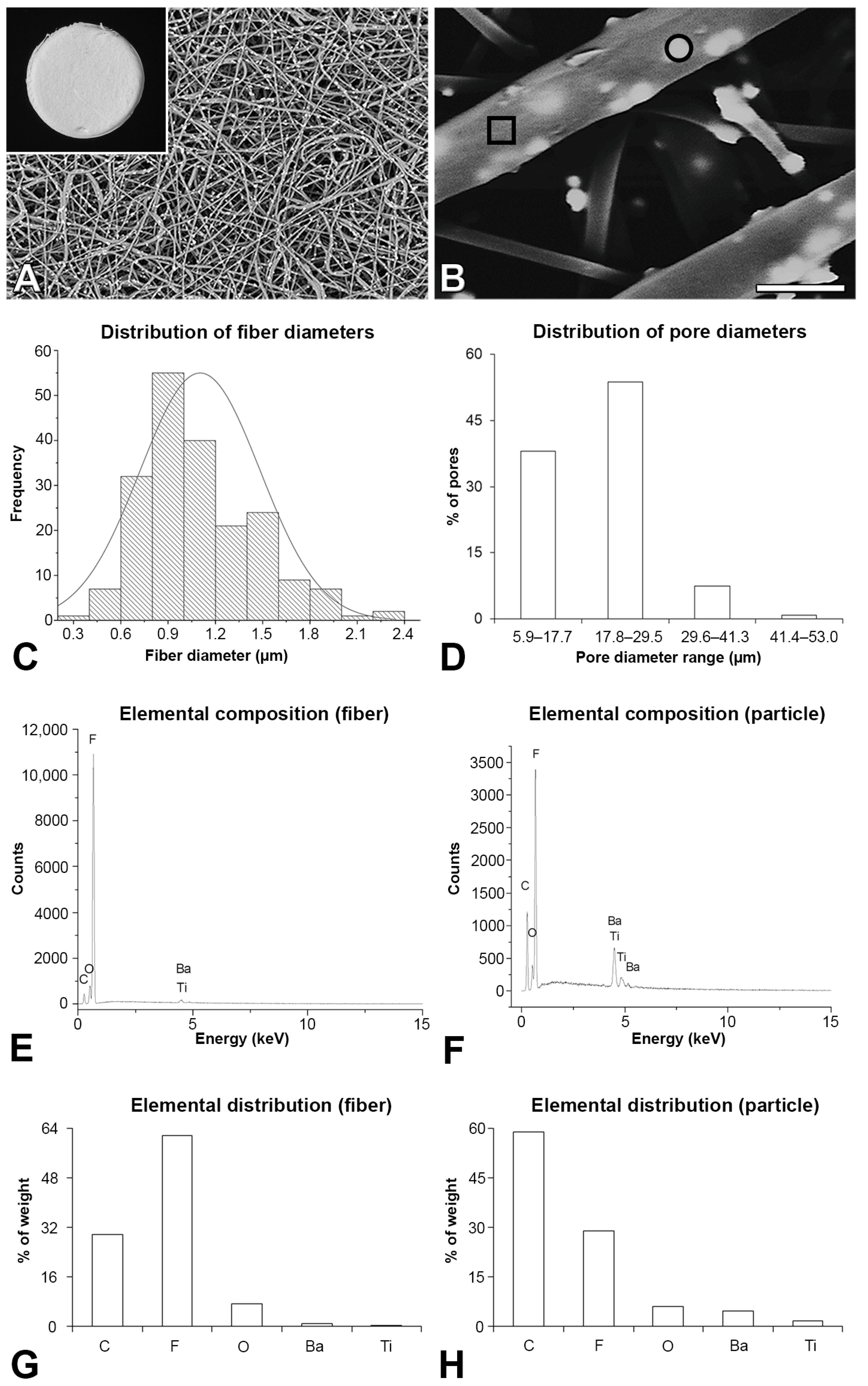
Abstract
:1. Introduction
2. Materials and Methods
2.1. Synthesis of the P(VDF-TrFE)/BaTiO3 Scaffold
2.2. P(VDF-TrFE)/BaTiO3 Scaffold Characterization
2.3. Evaluation of Bone Repair
2.3.1. Animals
2.3.2. Isolation and Culture of MSCs
2.3.3. Creation and Treatment of Calvarial Defects
2.3.4. µCT Analysis
2.3.5. Histological Analysis
2.4. Statistical Analysis
3. Results
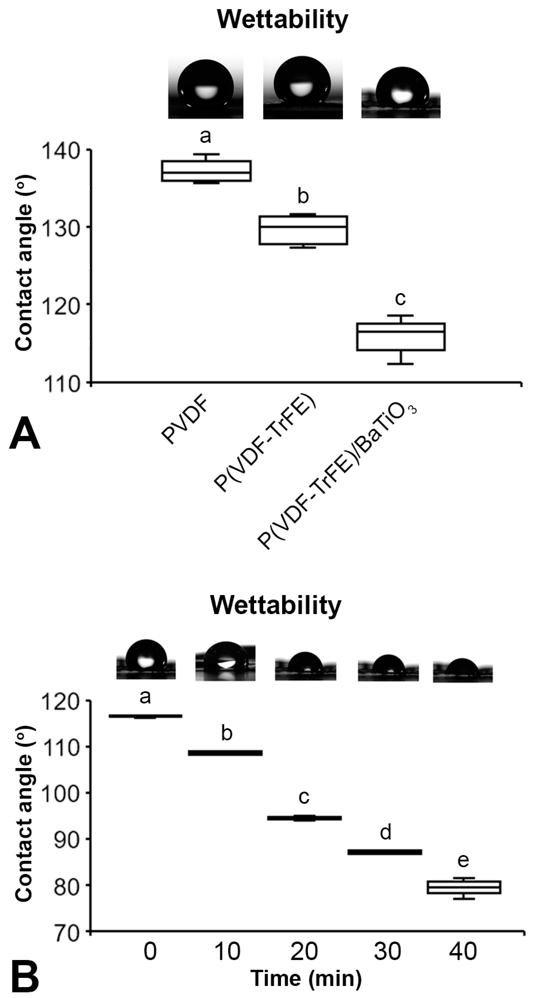
3.1. P(VDF-TrFE)/BaTiO3 Scaffold Characterization
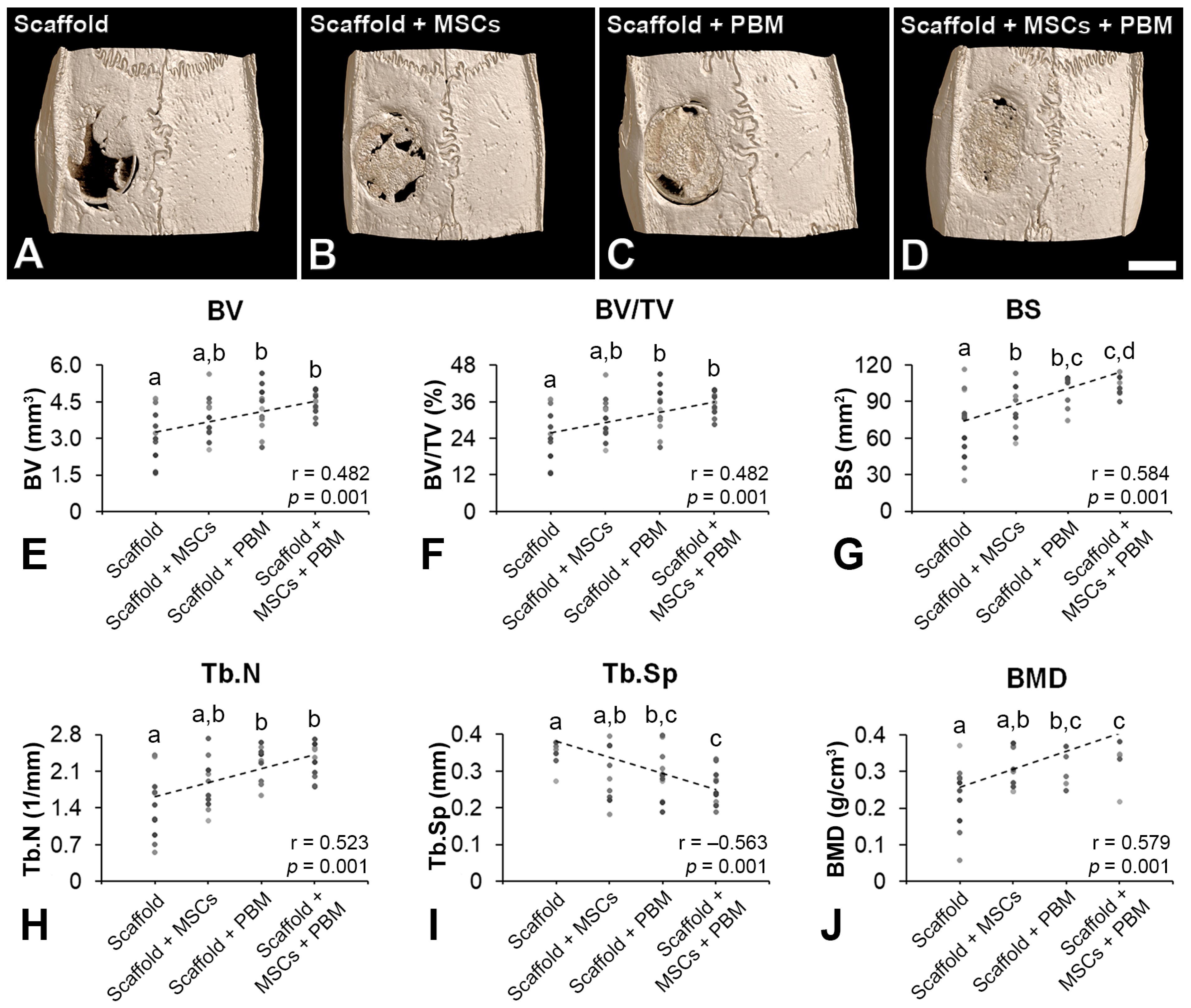
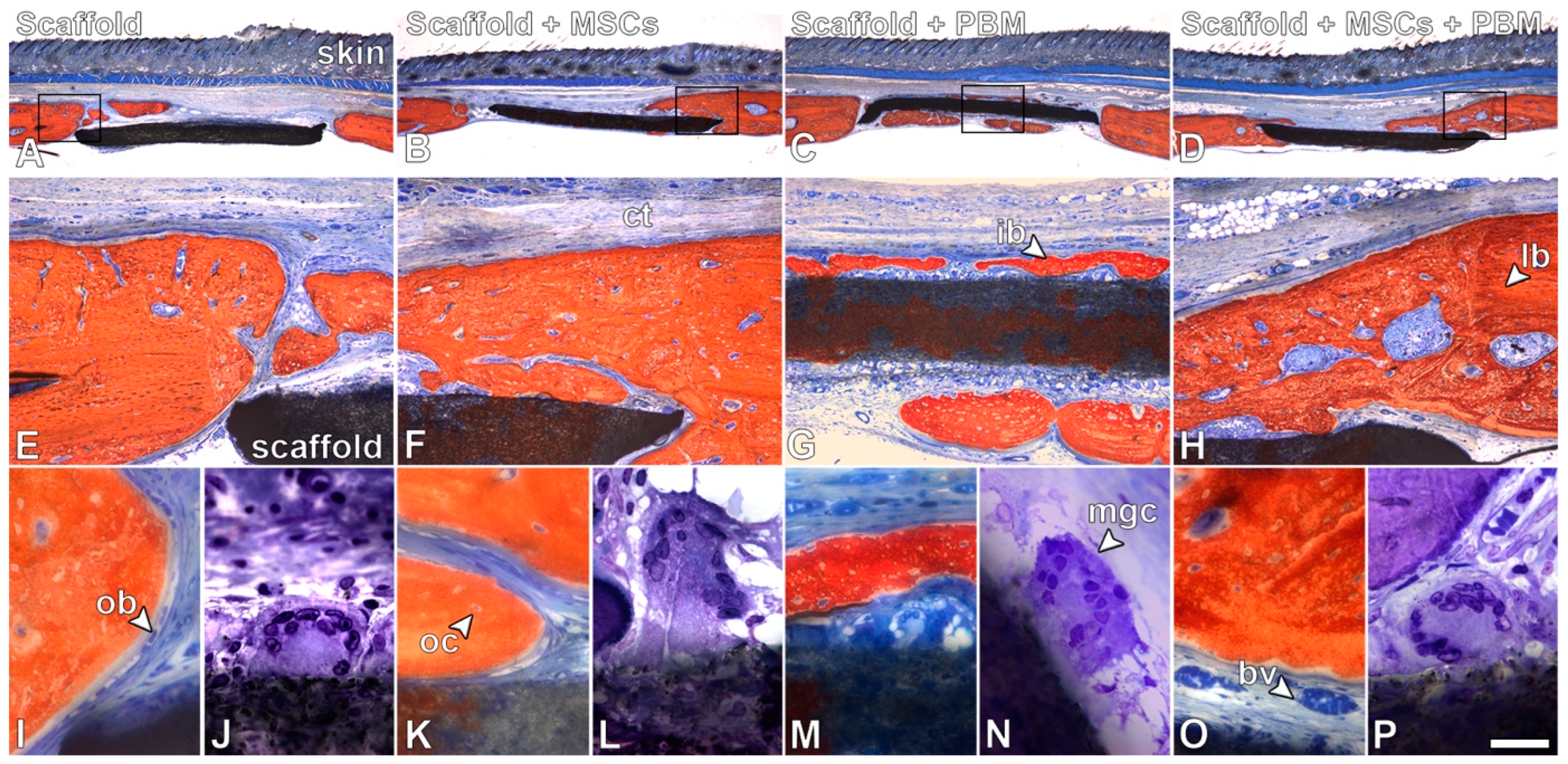
3.2. Evaluation of Bone Repair
4. Discussion
Author Contributions
Funding
Institutional Review Board Statement
Informed Consent Statement
Data Availability Statement
Acknowledgments
Conflicts of Interest
References
- Schuind, F.; Burny, F.; Quintin, J.; Potaznik, A.; Pasteels, J.L. Single stage reconstruction of a large tibial defect using a free vascularised osteomyocutaneous ulnar transfer. Int. Orthop. 1989, 13, 239–245. [Google Scholar] [CrossRef] [PubMed]
- Marcacci, M.; Kon, E.; Zaffagnini, S.; Giardino, R.; Rocca, M.; Corsi, A.; Benvenuti, A.; Bianco, P.; Quarto, R.; Martin, I.; et al. Reconstruction of extensive long-bone defects in sheep using porous hydroxyapatite sponges. Calcif. Tissue Int. 1999, 64, 83–90. [Google Scholar] [CrossRef]
- Tarr, J.T.; Hagan, M.; Zhang, B.; Tanna, N.; Andrews, B.T.; Lee, J.C.; Bradley, J.P. Syndrome of the trephined: Quantitative functional improvement after large cranial vault reconstruction. Plast. Reconstr. Surg. 2020, 145, 1486–1494. [Google Scholar] [CrossRef] [PubMed]
- Langer, R.; Vacanti, J.P. Tissue engineering. Science 1993, 260, 920–926. [Google Scholar] [CrossRef]
- Nevo, Z.; Robinson, D.; Horowitz, S.; Hasharoni, A.; Yayon, A. The manipulated mesenchymal stem cells in regenerated skeletal tissues. Cell Transplant. 1998, 7, 63–70. [Google Scholar] [CrossRef]
- Radice, M.; Brun, P.; Cortivo, R.; Scapinelli, R.; Battaliard, C.; Abatangelo, G. Hyaluronan-based biopolymers as delivery vehicles for bone-marrow-derived mesenchymal progenitors. J. Biomed. Mater. Res. 2000, 50, 101–109. [Google Scholar] [CrossRef]
- Langer, R.; Vacanti, J. Advances in tissue engineering. J. Pediatr. Surg. 2016, 51, 8–12. [Google Scholar] [CrossRef] [PubMed]
- Zhang, X.; Wang, G.; Wang, W.; Ran, C.; Piao, F.; Ma, Z.; Zhang, Z.; Zheng, G.; Cao, F.; Xie, H.; et al. Bone marrow mesenchymal stem cells paracrine TGF-β1 to mediate the biological activity of osteoblasts in bone repair. Cytokine 2023, 164, 156139. [Google Scholar] [CrossRef]
- Yamada, Y.; Ueda, M.; Naiki, T.; Takahashi, M.; Hata, K.; Nagasaka, T. Autogenous injectable bone for regeneration with mesenchymal stem cells and platelet-rich plasma: Tissue-engineered bone regeneration. Tissue Eng. 2004, 10, 955–964. [Google Scholar] [CrossRef]
- Suzuki, S.; Venkataiah, V.S.; Yahata, Y.; Kitagawa, A.; Inagaki, M.; Njuguna, M.M.; Nozawa, R.; Kakiuchi, Y.; Nakano, M.; Handa, K.; et al. Correction of large jawbone defect in the mouse using immature osteoblast-like cells and a 3D polylactic acid scaffold. PNAS Nexus 2022, 1, pgac151. [Google Scholar] [CrossRef]
- Liu, W.; Li, X.; Jiao, Y.; Wu, C.; Guo, S.; Xiao, X.; Wei, X.; Wu, J.; Gao, P.; Wang, N.; et al. Biological effects of a three-dimensionally printed Ti6Al4V scaffold coated with piezoelectric BaTiO3 nanoparticles on bone formation. ACS Appl. Mater. Interfaces 2020, 12, 51885–51903. [Google Scholar] [CrossRef] [PubMed]
- Lim, J.; Liu, Y.C.; Chu, Y.C.; Lin, Y.X.; Hwang, W.H.; Wang, J.L. Piezoelectric effect stimulates the rearrangement of chondrogenic cells and alters ciliary orientation via atypical PKCζ. Biochem. Biophys. Rep. 2022, 30, 101265. [Google Scholar] [CrossRef] [PubMed]
- Bhaskar, N.; Kachappilly, M.C.; Bhushan, V.; Pandya, H.J.; Basu, B. Electrical field stimulated modulation of cell fate of pre-osteoblasts on PVDF/BT/MWCNT based electroactive biomaterials. J. Biomed. Mater. Res. A 2023, 111, 340–353. [Google Scholar] [CrossRef]
- Beloti, M.M.; de Oliveira, P.T.; Gimenes, R.; Zaghete, M.A.; Bertolini, M.J.; Rosa, A.L. In vitro biocompatibility of a novel membrane of the composite poly(vinylidene-trifluoroethylene)/barium titanate. J. Biomed. Mater. Res. A 2006, 79, 282–288. [Google Scholar] [CrossRef] [PubMed]
- Teixeira, L.N.; Crippa, G.E.; Gimenes, R.; Zaghete, M.A.; de Oliveira, P.T.; Rosa, A.L.; Beloti, M.M. Response of human alveolar bone-derived cells to a novel poly(vinylidene fluoride-trifluoroethylene)/barium titanate membrane. J. Mater. Sci. Mater. Med. 2011, 22, 151–158. [Google Scholar] [CrossRef] [PubMed]
- Lopes, H.B.; Santos Tde, S.; de Oliveira, F.S.; Freitas, G.P.; de Almeida, A.L.; Gimenes, R.; Rosa, A.L.; Beloti, M.M. Poly(vinylidene-trifluoroethylene)/barium titanate composite for in vivo support of bone formation. J. Biomater. Appl. 2014, 29, 104–112. [Google Scholar] [CrossRef]
- Scalize, P.H.; Bombonato-Prado, K.F.; de Sousa, L.G.; Rosa, A.L.; Beloti, M.M.; Semprini, M.; Gimenes, R.; de Almeida, A.L.; de Oliveira, F.S.; Hallak Regalo, S.C.; et al. Poly(vinylidene fluoride-trifluorethylene)/barium titanate membrane promotes de novo bone formation and may modulate gene expression in osteoporotic rat model. J. Mater. Sci. Mater. Med. 2016, 27, 180. [Google Scholar] [CrossRef]
- Lopes, H.B.; Ferraz, E.P.; Almeida, A.L.; Florio, P.; Gimenes, R.; Rosa, A.L.; Beloti, M.M. Participation of MicroRNA-34a and RANKL on bone repair induced by poly(vinylidene-trifluoroethylene)/barium titanate membrane. J. Biomater. Sci. Polym. Ed. 2016, 27, 1369–1379. [Google Scholar] [CrossRef]
- Freitas, G.P.; Lopes, H.B.; Almeida, A.L.G.; Abuna, R.P.F.; Gimenes, R.; Souza, L.E.B.; Covas, D.T.; Beloti, M.M.; Rosa, A.L. Potential of osteoblastic cells derived from bone marrow and adipose tissue associated with a polymer/ceramic composite to repair bone tissue. Calcif. Tissue Int. 2017, 101, 312–320. [Google Scholar] [CrossRef]
- Almeida, A.L.G.; Freitas, G.P.; Lopes, H.B.; Gimenes, R.; Siessere, S.; Sousa, L.G.; Beloti, M.M.; Rosa, A.L. Effect of stem cells combined with a polymer/ceramic membrane on osteoporotic bone repair. Braz. Oral. Res. 2019, 33, e079. [Google Scholar] [CrossRef]
- Hu, Y.; Zhang, C.; Zhang, S.; Xiong, Z.; Xu, J. Development of a porous poly(L-lactic acid)/hydroxyapatite/collagen scaffold as a BMP delivery system and its use in healing canine segmental bone defect. J. Biomed. Mater. Res. A 2003, 67, 591–598. [Google Scholar] [CrossRef] [PubMed]
- Ekambaram, R.; Dharmalingam, S. Design and development of biomimetic electrospun sulphonated polyether ether ketone nanofibrous scaffold for bone tissue regeneration applications: In vitro and in vivo study. J. Biomater. Sci. Polym. Ed. 2022, 33, 947–975. [Google Scholar] [CrossRef] [PubMed]
- Valerini, D.; Tammaro, L.; Vitali, R.; Guillot, G.; Rinaldi, A. Sputter-deposited Ag nanoparticles on electrospun PLC scaffolds: Morphology, wettability and antibacterial activity. Coatings 2021, 11, 345. [Google Scholar] [CrossRef]
- Wilk, S.; Benko, A. Advances in fabricating the electrospun biopolymer-based biomaterials. J. Funct. Biomater. 2021, 12, 26. [Google Scholar] [CrossRef]
- Baldari, S.; Di Rocco, G.; Piccoli, M.; Pozzobon, M.; Muraca, M.; Toietta, G. Challenges and strategies for improving the regenerative effects of mesenchymal stromal cell-based therapies. Int. J. Mol. Sci. 2017, 18, 2087. [Google Scholar] [CrossRef]
- Lee, B.C.; Kang, K.S. Functional enhancement strategies for immunomodulation of mesenchymal stem cells and their therapeutic application. Stem Cell Res. Ther. 2020, 11, 397. [Google Scholar] [CrossRef]
- Hosseinpour, S.; Fekrazad, R.; Arany, P.R.; Ye, Q. Molecular impacts of photobiomodulation on bone regeneration: A systematic review. Prog. Biophys. Mol. Biol. 2019, 149, 147–159. [Google Scholar] [CrossRef]
- Fallahnezhad, S.; Jajarmi, V.; Shahnavaz, S.; Amini, A.; Ghoreishi, S.K.; Kazemi, M.; Chien, S.; Bayat, M. Improvement in viability and mineralization of osteoporotic bone marrow mesenchymal stem cell through combined application of photobiomodulation therapy and oxytocin. Lasers Med. Sci. 2020, 35, 557–566. [Google Scholar] [CrossRef]
- Bueno, N.P.; Copete, I.N.; Lopes, H.B.; Arany, P.R.; Marques, M.M.; Ferraz, E.P. Recovering the osteoblastic differentiation potential of mesenchymal stem cells derived from diabetic rats by photobiomodulation therapy. J. Biophotonics 2021, 14, e202000393. [Google Scholar] [CrossRef]
- Dalapria, V.; Marcos, R.L.; Bussadori, S.K.; Anselmo, G.; Benetti, C.; da Silva Santana, A.C.A.; Marinho, N.S.R.; Pinto, R.S.; de Sales, R.S.; de França, L.S.; et al. LED photobiomodulation therapy combined with biomaterial as a scaffold promotes better bone quality in the dental alveolus in an experimental extraction model. Lasers Med. Sci. 2022, 37, 1583–1592. [Google Scholar] [CrossRef]
- Khosravipour, A.; Amini, A.; Farahani, R.M.; Mostafavinia, A.; Asgari, M.; Rezaei, F.; Abrahamse, H.; Chien, S.; Bayat, M. Evaluation of the effects of preconditioned human stem cells plus a scaffold and photobiomodulation administration on stereological parameters and gene expression levels in a critical size bone defect in rats. Lasers Med. Sci. 2022, 37, 2457–2470. [Google Scholar] [CrossRef]
- Diniz, I.M.A.; Carreira, A.C.O.; Sipert, C.R.; Uehara, C.M.; Moreira, M.S.N.; Freire, L.; Pelissari, C.; Kossugue, P.M.; de Araújo, D.R.; Sogayar, M.C.; et al. Photobiomodulation of mesenchymal stem cells encapsulated in an injectable rhBMP4-loaded hydrogel directs hard tissue bioengineering. J. Cell. Physiol. 2018, 233, 4907–4918. [Google Scholar] [CrossRef] [PubMed]
- Martins, M.A.; Martins, M.D.; Lascala, C.A.; Curi, M.M.; Migliorati, C.A.; Tenis, C.A.; Marques, M.M. Association of laser phototherapy with PRP improves healing of bisphosphonate-related osteonecrosis of the jaws in cancer patients: A preliminary study. Oral. Oncol. 2012, 48, 79–84. [Google Scholar] [CrossRef] [PubMed]
- Alves, F.A.M.; Marques, M.M.; Cavalcanti, S.C.S.X.B.; Pedroni, A.C.F.; Ferraz, E.P.; Miniello, T.G.; Moreira, M.S.; Jerônimo, T.; Deboni, M.C.Z.; Lascala, C.A. Photobiomodulation as adjunctive therapy for guided bone regeneration. A microCT study in osteoporotic rat model. J. Photochem. Photobiol. B. 2020, 213, 112053. [Google Scholar] [CrossRef] [PubMed]
- Bouxsein, M.L.; Boyd, S.K.; Christiansen, B.A.; Guldberg, R.E.; Jepsen, K.J.; Müller, R. Guidelines for assessment of bone microstructure in rodents using micro-computed tomography. J. Bone Miner. Res. 2010, 25, 1468–1486. [Google Scholar] [CrossRef]
- Sheng, R.; Mu, J.; Chernozem, R.V.; Mukhortova, Y.R.; Surmeneva, M.A.; Pariy, I.O.; Ludwig, T.; Mathur, S.; Xu, C.; Surmenev, R.A.; et al. Fabrication and characterization of piezoelectric polymer composites and cytocompatibility with mesenchymal stem cells. ACS Appl. Mater. Interfaces 2023, 15, 3731–3743. [Google Scholar] [CrossRef] [PubMed]
- Ge, J.; Guo, L.; Wang, S.; Zhang, Y.; Cai, T.; Zhao, R.C.; Wu, Y. The size of mesenchymal stem cells is a significant cause of vascular obstructions and stroke. Stem Cell Rev. Rep. 2014, 10, 295–303. [Google Scholar] [CrossRef]
- Crick, C.R.; Parkin, I.P. Preparation and characterisation of super-hydrophobic surfaces. Chemistry 2010, 16, 3568–3588. [Google Scholar] [CrossRef]
- Li, R.; Wei, W.; Hai, J.; Gao, L.; Gao, Z.; Fan, Y. Preparation and electric-field response of novel tetragonal barium titanate. J. Alloys Compd. 2013, 574, 212–216. [Google Scholar] [CrossRef]
- Arima, Y.; Iwata, H. Effect of wettability and surface functional groups on protein adsorption and cell adhesion using well-defined mixed self-assembled monolayers. Biomaterials 2007, 28, 3074–3082. [Google Scholar] [CrossRef]
- Shojaei, S.; Shahgholi, M.; Karimipour, A. The effects of atomic percentage and size of Zinc nanoparticles, and atomic porosity on thermal and mechanical properties of reinforced calcium phosphate cement by molecular dynamics simulation. J. Mech. Behav. Biomed. Mater. 2023, 141, 105785. [Google Scholar] [CrossRef] [PubMed]
- Shahshahani, S.; Shahgholi, M.; Karimipour, A. The thermal performance and mechanical stability of methacrylic acid porous hydrogels in an aqueous medium at different initial temperatures and hydrogel volume fraction using the molecular dynamics simulation. J. Mol. Liq. 2023, 382, 122001. [Google Scholar] [CrossRef]
- Genchi, G.G.; Sinibaldi, E.; Ceseracciu, L.; Labardi, M.; Marino, A.; Marras, S.; De Simoni, G.; Mattoli, V.; Ciofani, G. Ultrasound-activated piezoelectric P(VDF-TrFE)/boron nitride nanotube composite films promote differentiation of human SaOS-2 osteoblast-like cells. Nanomedicine 2018, 14, 2421–2432. [Google Scholar] [CrossRef] [PubMed]
- Bai, Y.; Dai, X.; Yin, Y.; Wang, J.; Sun, X.; Liang, W.; Li, Y.; Deng, X.; Zhang, X. Biomimetic piezoelectric nanocomposite membranes synergistically enhance osteogenesis of deproteinized bovine bone grafts. Int. J. Nanomed. 2019, 14, 3015–3026. [Google Scholar] [CrossRef] [PubMed]
- Souza, A.T.P.; Freitas, G.P.; Lopes, H.B.; Ferraz, E.P.; Oliveira, F.S.; Beloti, M.M.; Rosa, A.L. Effect of cell therapy with allogeneic osteoblasts on bone repair of rat calvaria defects. Cytotherapy 2018, 20, 1267–1277. [Google Scholar] [CrossRef]
- Freitas, G.P.; Lopes, H.B.; Souza, A.T.P.; Gomes, M.P.O.; Quiles, G.K.; Gordon, J.; Tye, C.; Stein, J.L.; Stein, G.S.; Lian, J.B.; et al. Mesenchymal stem cells overexpressing BMP-9 by CRISPR-Cas9 present high in vitro osteogenic potential and enhance in vivo bone formation. Gene Ther. 2021, 28, 748–759. [Google Scholar] [CrossRef]
- Andalib, N.; Kehtari, M.; Seyedjafari, E.; Motamed, N.; Matin, M.M. In vivo bone regeneration using a bioactive nanocomposite scaffold and human mesenchymal stem cells. Cell Tissue Bank. 2021, 22, 467–477. [Google Scholar] [CrossRef]
- Naudot, M.; Garcia Garcia, A.; Jankovsky, N.; Barre, A.; Zabijak, L.; Azdad, S.Z.; Collet, L.; Bedoui, F.; Hébraud, A.; Schlatter, G.; et al. The combination of a poly-caprolactone/nano-hydroxyapatite honeycomb scaffold and mesenchymal stem cells promotes bone regeneration in rat calvarial defects. J. Tissue Eng. Regen. Med. 2020, 14, 1570–1580. [Google Scholar] [CrossRef]
- Yagi, H.; Soto-Gutierrez, A.; Parekkadan, B.; Kitagawa, Y.; Tompkins, R.G.; Kobayashi, N.; Yarmush, M.L. Mesenchymal stem cells: Mechanisms of immunomodulation and homing. Cell Transplant. 2010, 19, 667–679. [Google Scholar] [CrossRef]
- Cassiede, P.; Dennis, J.E.; Ma, F.; Caplan, A.I. Osteochondrogenic potential of marrow mesenchymal progenitor cells exposed to TGF-beta 1 or PDGF-BB as assayed in vivo and in vitro. J. Bone Miner. Res. 1996, 11, 1264–1273. [Google Scholar] [CrossRef]
- Tuby, H.; Maltz, L.; Oron, U. Induction of autologous mesenchymal stem cells in the bone marrow by low-level laser therapy has profound beneficial effects on the infarcted rat heart. Lasers Surg. Med. 2011, 43, 401–409. [Google Scholar] [CrossRef] [PubMed]
- Amid, R.; Kadkhodazadeh, M.; Ahsaie, M.G.; Hakakzadeh, A. Effect of low-level laser therapy on proliferation and differentiation of the cells contributing in bone regeneration. J. Lasers Med. Sci. 2014, 5, 163–170. [Google Scholar] [PubMed]
- De Marco, A.C.; Torquato, L.C.; Gonçalves, P.R.; Ribeiro, T.C.; Nunes, C.M.; Bernardo, D.V.; Gomes, M.F.; Jardini, M.A.N.; Santamaria, M.P. The Effect of photobiomodulation therapy in different doses on bone repair of critical size defects in rats: A histomorphometric study. J. Lasers Med. Sci. 2021, 12, e53. [Google Scholar] [CrossRef] [PubMed]
- Rufato, F.C.T.; de Sousa, L.G.; Scalize, P.H.; Gimenes, R.; Regalo, I.H.; Rosa, A.L.; Beloti, M.M.; de Oliveira, F.S.; Bombonato-Prado, K.F.; Regalo, S.C.H.; et al. Texturized P(VDF-TrFE)/BT membrane enhances bone neoformation in calvaria defects regardless of the association with photobiomodulation therapy in ovariectomized rats. Clin. Oral. Investig. 2022, 26, 1053–1065. [Google Scholar] [CrossRef]
- Calis, M.; Irmak, G.; Demirtaş, T.T.; Kara, M.; Üstün, G.G.; Gümüşderelioğlu, M.; Türkkanı, A.; Çakar, A.N.; Özgür, F. Photobiomodulation combined with adipose-derived stem cells encapsulated in methacrylated gelatin hydrogels enhances in vivo bone regeneration. Lasers Med. Sci. 2022, 37, 595–606. [Google Scholar] [CrossRef]
- Stancker, T.G.; Vieira, S.S.; Serra, A.J.; do Nascimento Lima, R.; Dos Santos Feliciano, R.; Silva, J.A., Jr.; Dos Santos, S.A.; Dos Santos Vieira, M.A.; Simões, M.C.B.; Leal-Junior, E.C.; et al. Can photobiomodulation associated with implantation of mesenchymal adipose-derived stem cells attenuate the expression of MMPs and decrease degradation of type II collagen in an experimental model of osteoarthritis? Lasers Med. Sci. 2018, 33, 1073–1084. [Google Scholar] [CrossRef] [PubMed]
- Park, I.S.; Kim, D.K.; Kim, J.H.; Bae, J.S.; Kim, E.H.; Yoo, S.H.; Chung, Y.J.; Lyu, L.; Mo, J.H. Increased anti-allergic effects of secretome of low-level light treated tonsil-derived mesenchymal stem cells in allergic rhinitis mouse model. Am. J. Rhinol. Allergy 2022, 36, 261–268. [Google Scholar] [CrossRef]
- Tanneberger, A.M.; Al-Maawi, S.; Herrera-Vizcaíno, C.; Orlowska, A.; Kubesch, A.; Sader, R.; Kirkpatrick, C.J.; Ghanaati, S. Multinucleated giant cells within the in vivo implantation bed of a collagen-based biomaterial determine its degradation pattern. Clin. Oral. Investig. 2021, 25, 859–873. [Google Scholar] [CrossRef]


Disclaimer/Publisher’s Note: The statements, opinions and data contained in all publications are solely those of the individual author(s) and contributor(s) and not of MDPI and/or the editor(s). MDPI and/or the editor(s) disclaim responsibility for any injury to people or property resulting from any ideas, methods, instructions or products referred to in the content. |
© 2023 by the authors. Licensee MDPI, Basel, Switzerland. This article is an open access article distributed under the terms and conditions of the Creative Commons Attribution (CC BY) license (https://creativecommons.org/licenses/by/4.0/).
Share and Cite
Adolpho, L.F.; Ribeiro, L.M.S.; Freitas, G.P.; Lopes, H.B.; Gomes, M.P.O.; Ferraz, E.P.; Gimenes, R.; Beloti, M.M.; Rosa, A.L. Mesenchymal Stem Cells Combined with a P(VDF-TrFE)/BaTiO3 Scaffold and Photobiomodulation Therapy Enhance Bone Repair in Rat Calvarial Defects. J. Funct. Biomater. 2023, 14, 306. https://doi.org/10.3390/jfb14060306
Adolpho LF, Ribeiro LMS, Freitas GP, Lopes HB, Gomes MPO, Ferraz EP, Gimenes R, Beloti MM, Rosa AL. Mesenchymal Stem Cells Combined with a P(VDF-TrFE)/BaTiO3 Scaffold and Photobiomodulation Therapy Enhance Bone Repair in Rat Calvarial Defects. Journal of Functional Biomaterials. 2023; 14(6):306. https://doi.org/10.3390/jfb14060306
Chicago/Turabian StyleAdolpho, Leticia Faustino, Larissa Mayra Silva Ribeiro, Gileade Pereira Freitas, Helena Bacha Lopes, Maria Paula Oliveira Gomes, Emanuela Prado Ferraz, Rossano Gimenes, Marcio Mateus Beloti, and Adalberto Luiz Rosa. 2023. "Mesenchymal Stem Cells Combined with a P(VDF-TrFE)/BaTiO3 Scaffold and Photobiomodulation Therapy Enhance Bone Repair in Rat Calvarial Defects" Journal of Functional Biomaterials 14, no. 6: 306. https://doi.org/10.3390/jfb14060306