
Poly(Aspartic Acid) Promotes Odontoblast-like Cell Differentiation in Rat Molars with Exposed Pulp
 , and
, and 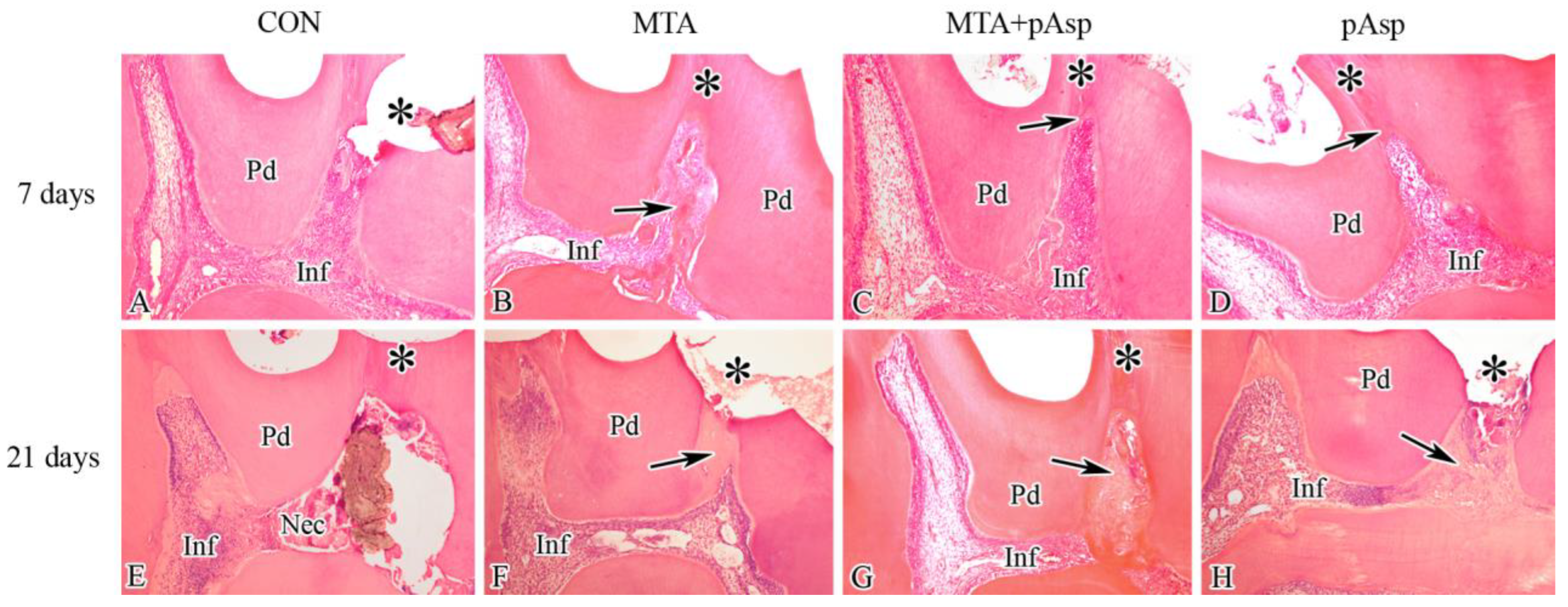
Abstract
:1. Introduction
2. Materials and Methods
2.1. Experimental Design
2.2. Anesthesia
2.3. Mechanical Pulp Exposure
2.4. Experimental Groups
- (1)
- Group C (negative control): after the mechanical pulpal exposure procedure, no capping material was applied and a piece of polytetrafluoroethylene (PTFE; Teflon tape—PS65, 3M, Sumaré, SP, Brazil) previously sterilized in autoclave at 121 °C for 40 min was placed at the exposure site to avoid direct contact between the pulp tissue and the coronal restoration made with a flowable resin composite (NT Premium, Vigodent-Coltene, Rio de Janeiro, RJ, Brazil) associated with a two-step adhesive system (Single Bond 2, 3M ESPE, St. Paul, MN, USA);
- (2)
- MTA group: pulp capping with mineral trioxide aggregate (MTA-Angelus; Angelus, Londrina, PR, Brazil), mixed with distilled water according to the manufacturer’s instructions. The cement was brought into position using an endodontic probe and accommodated without pressure with the narrow end of a sterile paper cone. The cavity was filled immediately with flowable resin composite, as described for the control group;
- (3)
- pAsp group: 20 μL of a solution containing 25 mg/mL of pAsp (27,000 Da, Alamanda Polymers Inc, Huntsville, AL, USA) was applied to the exposure site and left undisturbed for 20 s [34]. The excess solution was removed using sterile cotton balls, and a sterile piece of Teflon tape was placed at the exposure site before sealing the cavity with the resin composite;
- (4)
- MTA/pAsp group: MTA particles were mixed with pAsp in a 5:1 ratio (by mass) before the manipulation of the material with distilled water [39]. The material was applied to the pulpal exposure site, and the cavity was sealed as described for the MTA group.
2.5. Euthanasia and Sample Collection
2.6. Histological Processing
2.7. Histopathological Analysis
2.8. Immunohistochemical Analysis
2.9. Data Analysis
3. Results
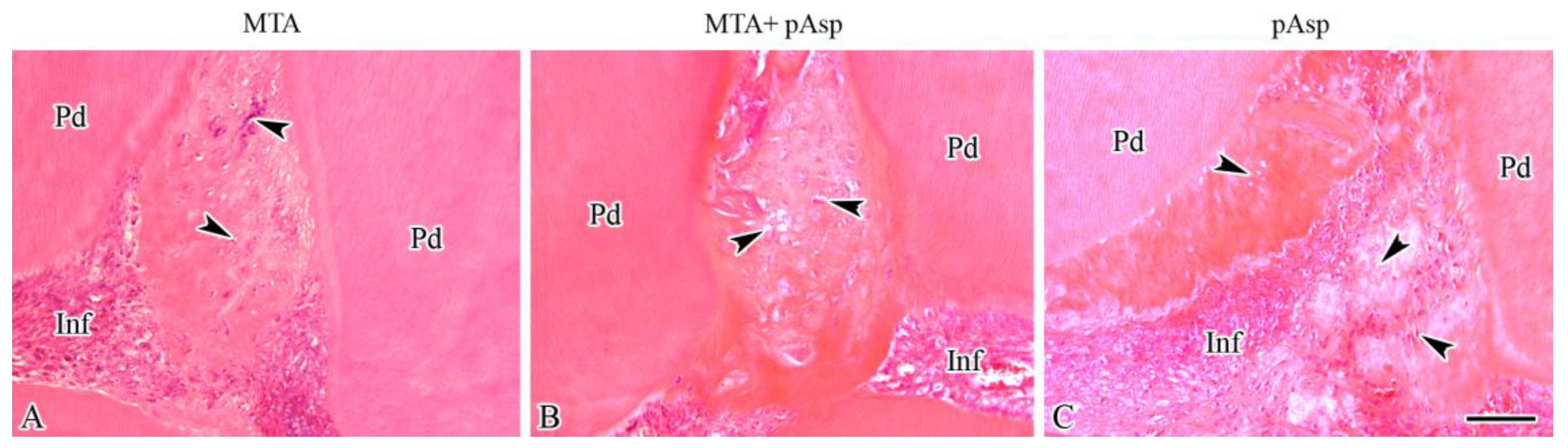
3.1. Histopathology
3.1.1. Seven Days
3.1.2. Twenty-One Days
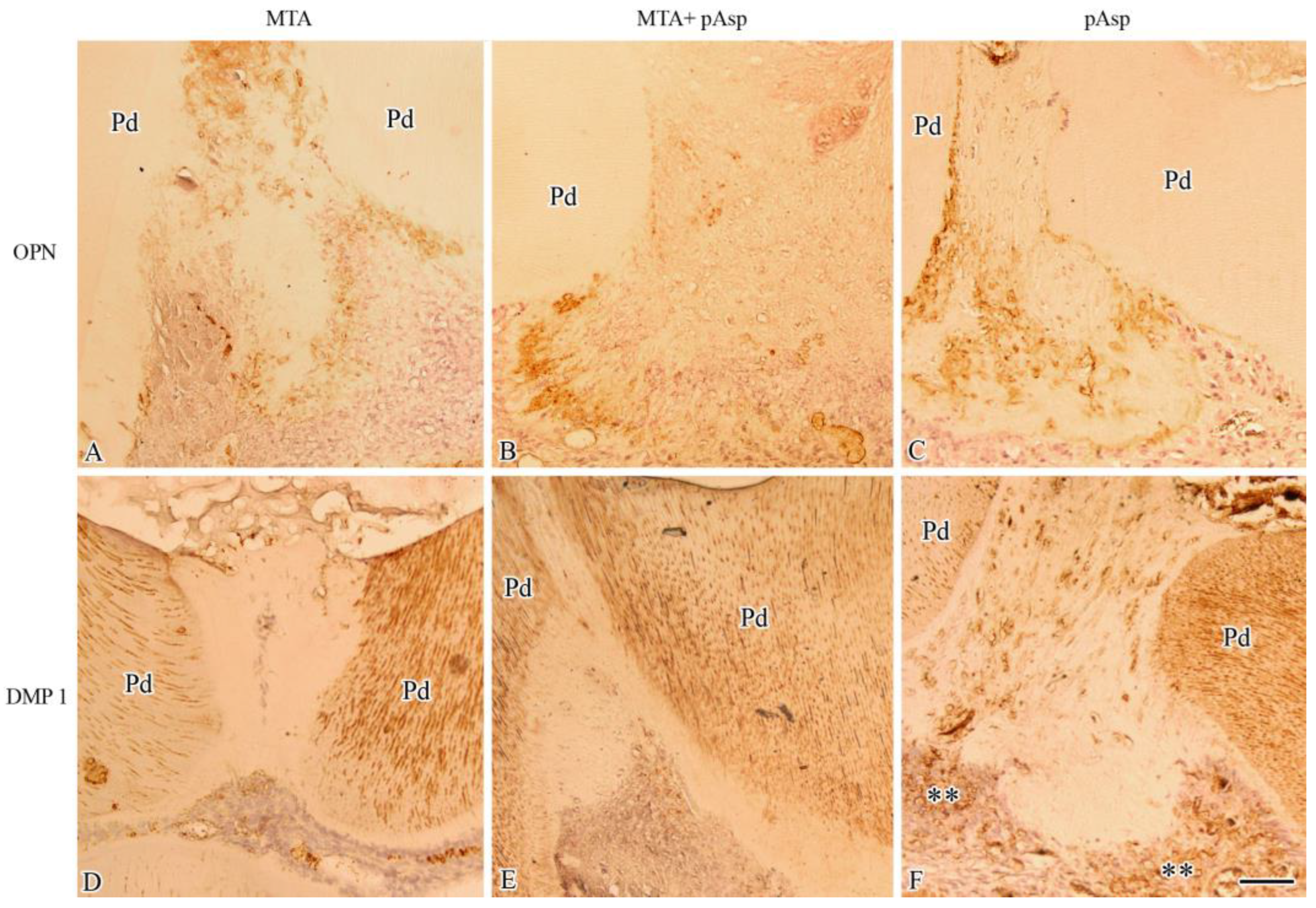
3.2. Immunohistochemistry
4. Discussion
5. Conclusions
Author Contributions
Funding
Institutional Review Board Statement
Data Availability Statement
Conflicts of Interest
References
- Paranjpe, A.; Zhang, H.; Johnson, J.D. Effects of mineral trioxide aggregate on human dental pulp cells after pulp-capping procedures. J. Endod. 2010, 36, 1042–1047. [Google Scholar] [CrossRef]
- Paula, A.B.; Laranjo, M.; Marto, C.M.; Paulo, S.; Abrantes, A.M.; Fernandes, B.; Casalta-Lopes, J.; Marques-Ferreira, M.; Botelho, M.F.; Carrilho, E. Evaluation of dentinogenesis inducer biomaterials: An in vivo study. J. Appl. Oral Sci. 2020, 28, e20190023. [Google Scholar] [CrossRef]
- Arana-Chavez, V.E.; Massa, L.F. Odontoblasts: The cells forming and maintaining dentine. Int. J. Biochem. Cell Biol. 2004, 36, 1367–1373. [Google Scholar] [CrossRef]
- Torabinejad, M.; Watson, T.F.; Pitt Ford, T.R. Sealing ability of a mineral trioxide aggregate when used as a root end filling material. J. Endod. 1993, 19, 591–595. [Google Scholar] [CrossRef]
- Mente, J.; Hufnagel, S.; Leo, M.; Michel, A.; Gehrig, H.; Panagidis, D.; Saure, D.; Pfefferle, T. Treatment outcome of mineral trioxide aggregate or calcium hydroxide direct pulp capping: Long-term results. J. Endod. 2014, 40, 1746–1751. [Google Scholar] [CrossRef]
- Olsson, H.; Petersson, K.; Rohlin, M. Formation of a hard tissue barrier after pulp cappings in humans. A systematic review. Int. Endod. J. 2006, 39, 429–442. [Google Scholar] [CrossRef]
- Schwendicke, F.; Brouwer, F.; Schwendicke, A.; Paris, S. Different materials for direct pulp capping: Systematic review and meta-analysis and trial sequential analysis. Clin. Oral Investig. 2016, 20, 1121–1132. [Google Scholar] [CrossRef]
- Aguilar, P.; Linsuwanont, P. Vital pulp therapy in vital permanent teeth with cariously exposed pulp: A systematic review. J. Endod. 2011, 37, 581–587. [Google Scholar] [CrossRef]
- Komabayashi, T.; Zhu, Q. Innovative endodontic therapy for anti-inflammatory direct pulp capping of permanent teeth with a mature apex. Oral. Surg. Oral. Med. Oral. Pathol. Oral. Radiol. Endod. 2010, 109, e75–e81. [Google Scholar] [CrossRef]
- Nowicka, A.; Wilk, G.; Lipski, M.; Kolecki, J.; Buczkowska-Radlinska, J. Tomographic Evaluation of Reparative Dentin Formation after Direct Pulp Capping with Ca(OH)2, MTA, Biodentine, and Dentin Bonding System in Human Teeth. J. Endod. 2015, 41, 1234–1240. [Google Scholar] [CrossRef]
- Li, Z.; Cao, L.; Fan, M.; Xu, Q. Direct Pulp Capping with Calcium Hydroxide or Mineral Trioxide Aggregate: A Meta-analysis. J. Endod. 2015, 41, 1412–1417. [Google Scholar] [CrossRef]
- Gandolfi, M.G.; Iezzi, G.; Piattelli, A.; Prati, C.; Scarano, A. Osteoinductive potential and bone-bonding ability of ProRoot MTA, MTA Plus and Biodentine in rabbit intramedullary model: Microchemical characterization and histological analysis. Dent. Mater. 2017, 33, e221–e238. [Google Scholar] [CrossRef]
- Barthel, C.R.; Rosenkranz, B.; Leuenberg, A.; Roulet, J.F. Pulp capping of carious exposures: Treatment outcome after 5 and 10 years: A retrospective study. J. Endod. 2000, 26, 525–528. [Google Scholar] [CrossRef]
- Brizuela, C.; Ormeno, A.; Cabrera, C.; Cabezas, R.; Silva, C.I.; Ramirez, V.; Mercade, M. Direct Pulp Capping with Calcium Hydroxide, Mineral Trioxide Aggregate, and Biodentine in Permanent Young Teeth with Caries: A Randomized Clinical Trial. J. Endod. 2017, 43, 1776–1780. [Google Scholar] [CrossRef]
- Dammaschke, T.; Leidinger, J.; Schafer, E. Long-term evaluation of direct pulp capping-treatment outcomes over an average period of 6.1 years. Clin. Oral. Investig. 2010, 14, 559–567. [Google Scholar] [CrossRef]
- Moussa, D.G.; Aparicio, C. Present and future of tissue engineering scaffolds for dentin-pulp complex regeneration. J. Tissue Eng. Regen. Med. 2019, 13, 58–75. [Google Scholar] [CrossRef]
- Sangwan, P.; Sangwan, A.; Duhan, J.; Rohilla, A. Tertiary dentinogenesis with calcium hydroxide: A review of proposed mechanisms. Int. Endod. J. 2013, 46, 3–19. [Google Scholar] [CrossRef]
- Zaugg, L.K.; Banu, A.; Walther, A.R.; Chandrasekaran, D.; Babb, R.C.; Salzlechner, C.; Hedegaard, M.A.B.; Gentleman, E.; Sharpe, P.T. Translation Approach for Dentine Regeneration Using GSK-3 Antagonists. J. Dent. Res. 2020, 99, 544–551. [Google Scholar] [CrossRef]
- Li, Z.; Hao, J.; Duan, X.; Wu, N.; Zhou, Z.; Yang, F.; Li, J.; Zhao, Z.; Huang, S. The Role of Semaphorin 3A in Bone Remodeling. Front. Cell Neurosci. 2017, 11, 40. [Google Scholar] [CrossRef]
- Yoshida, S.; Wada, N.; Hasegawa, D.; Miyaji, H.; Mitarai, H.; Tomokiyo, A.; Hamano, S.; Maeda, H. Semaphorin 3A Induces Odontoblastic Phenotype in Dental Pulp Stem Cells. J. Dent. Res. 2016, 95, 1282–1290. [Google Scholar] [CrossRef]
- Park, C.; Song, M.; Kim, S.Y.; Min, B.M. Vitronectin-Derived Peptide Promotes Reparative Dentin Formation. J. Dent. Res. 2022, 101, 1481–1489. [Google Scholar] [CrossRef]
- Komichi, S.; Takahashi, Y.; Okamoto, M.; Ali, M.; Watanabe, M.; Huang, H.; Nakai, T.; Cooper, P.; Hayashi, M. Protein S100-A7 Derived from Digested Dentin Is a Critical Molecule for Dentin Pulp Regeneration. Cells 2019, 8, 1002. [Google Scholar] [CrossRef]
- Watanabe, M.; Okamoto, M.; Komichi, S.; Huang, H.; Matsumoto, S.; Moriyama, K.; Ohshima, J.; Abe, S.; Morita, M.; Ali, M.; et al. Novel Functional Peptide for Next-Generation Vital Pulp Therapy. J. Dent. Res. 2022, 102, 322–330. [Google Scholar] [CrossRef]
- Thrivikraman, G.; Athirasala, A.; Gordon, R.; Zhang, L.; Bergan, R.; Keene, D.R.; Jones, J.M.; Xie, H.; Chen, Z.; Tao, J.; et al. Rapid fabrication of vascularized and innervated cell-laden bone models with biomimetic intrafibrillar collagen mineralization. Nat. Commun. 2019, 10, 3520. [Google Scholar] [CrossRef]
- Adelnia, H.; Tran, H.D.N.; Little, P.J.; Blakey, I.; Ta, H.T. Poly(aspartic acid) in Biomedical Applications: From Polymerization, Modification, Properties, Degradation, and Biocompatibility to Applications. ACS Biomater. Sci. Eng. 2021, 7, 2083–2105. [Google Scholar] [CrossRef]
- Lemke, T.; Edte, M.; Gebauer, D.; Peter, C. Three Reasons Why Aspartic Acid and Glutamic Acid Sequences Have a Surprisingly Different Influence on Mineralization. J. Phys. Chem. B 2021, 125, 10335–10343. [Google Scholar] [CrossRef]
- Giachelli, C.M.; Steitz, S. Osteopontin: A versatile regulator of inflammation and biomineralization. Matrix Biol. 2000, 19, 615–622. [Google Scholar] [CrossRef]
- Cai, K.; Yao, K.; Hou, X.; Wang, Y.; Hou, Y.; Yang, Z.; Li, X.; Xie, H. Improvement of the functions of osteoblasts seeded on modified poly(D,L-lactic acid) with poly(aspartic acid). J. Biomed. Mater. Res. 2002, 62, 283–291. [Google Scholar] [CrossRef]
- Gower, L.B. Biomimetic model systems for investigating the amorphous precursor pathway and its role in biomineralization. Chem. Rev. 2008, 108, 4551–4627. [Google Scholar] [CrossRef]
- Gower, L.B.; Odom, D.J. Deposition of calcium carbonate films by a polymer-induced liquid-precursor (PILP) process. J. Cryst. Growth 2000, 210, 719–734. [Google Scholar] [CrossRef]
- Thula, T.T.; Rodriguez, D.E.; Lee, M.H.; Pendi, L.; Podschun, J.; Gower, L.B. In vitro mineralization of dense collagen substrates: A biomimetic approach toward the development of bone-graft materials. Acta Biomater. 2011, 7, 3158–3169. [Google Scholar] [CrossRef]
- Olszta, M.J.; Cheng, X.; Jee, S.S.; Kumar, R.; Kim, Y.-Y.; Kaufman, M.J.; Douglas, E.P.; Gower, L.B. Bone structure and formation: A new perspective. Mater. Sci. Eng. R Rep. 2007, 58, 77–116. [Google Scholar] [CrossRef]
- Niu, L.N.; Jee, S.E.; Jiao, K.; Tonggu, L.; Li, M.; Wang, L.; Yang, Y.D.; Bian, J.H.; Breschi, L.; Jang, S.S.; et al. Collagen intrafibrillar mineralization as a result of the balance between osmotic equilibrium and electroneutrality. Nat. Mater. 2017, 16, 370–378. [Google Scholar] [CrossRef]
- Bacino, M.; Girn, V.; Nurrohman, H.; Saeki, K.; Marshall, S.J.; Gower, L.; Saeed, E.; Stewart, R.; Le, T.; Marshall, G.W.; et al. Integrating the PILP-mineralization process into a restorative dental treatment. Dent. Mater. 2019, 35, 53–63. [Google Scholar] [CrossRef]
- Nudelman, F.; Lausch, A.J.; Sommerdijk, N.A.; Sone, E.D. In vitro models of collagen biomineralization. J. Struct. Biol. 2013, 183, 258–269. [Google Scholar] [CrossRef]
- Dianat, O.; Mashhadiabbas, F.; Ahangari, Z.; Saedi, S.; Motamedian, S.R. Histologic comparison of direct pulp capping of rat molars with MTA and different concentrations of simvastatin gel. J. Oral. Sci. 2018, 60, 57–63. [Google Scholar] [CrossRef]
- Okamoto, M.; Takahashi, Y.; Komichi, S.; Ali, M.; Yoneda, N.; Ishimoto, T.; Nakano, T.; Hayashi, M. Novel evaluation method of dentin repair by direct pulp capping using high-resolution micro-computed tomography. Clin. Oral. Investig. 2018, 22, 2879–2887. [Google Scholar] [CrossRef]
- Yaemkleebbua, K.; Osathanon, T.; Nowwarote, N.; Limjeerajarus, C.N.; Sukarawan, W. Analysis of hard tissue regeneration and Wnt signalling in dental pulp tissues after direct pulp capping with different materials. Int. Endod. J. 2019, 52, 1605–1616. [Google Scholar] [CrossRef]
- Babaie, E.; Bacino, M.; White, J.; Nurrohman, H.; Marshall, G.W.; Saeki, K.; Habelitz, S. Polymer-Induced Liquid Precursor (PILP) remineralization of artificial and natural dentin carious lesions evaluated by nanoindentation and microcomputed tomography. J. Dent. 2021, 109, 103659. [Google Scholar] [CrossRef]
- Massa, L.F.; Arana-Chavez, V.E. Ultrastructural preservation of rat embryonic dental tissues after rapid fixation and dehydration under microwave irradiation. Eur. J. Oral. Sci. 2000, 108, 74–77. [Google Scholar] [CrossRef]
- Taylor, R.D. Modification of the Brown and Brenn gram stain for the differential staining of gram-positive and gram-negative bacteria in tissue sections. Am. J. Clin. Pathol. 1966, 46, 472–474. [Google Scholar] [CrossRef]
- Trongkij, P.; Sutimuntanakul, S.; Lapthanasupkul, P.; Chaimanakarn, C.; Wong, R.H.; Banomyong, D. Pulpal responses after direct pulp capping with two calcium-silicate cements in a rat model. Dent. Mater. J. 2019, 38, 584–590. [Google Scholar] [CrossRef]
- Reis, M.S.; Scarparo, R.K.; Signor, B.; Bolzan, J.T.; Steier, L.; Figueiredo, J.A.P. Pulp capping with mineral trioxide aggregate or Biodentine: A comparison of mineralized barrier formation and inflammatory and degenerative events. Braz. Oral. Res. 2021, 35, e118. [Google Scholar] [CrossRef]
- Bakland, L.K.; Andreasen, J.O. Will mineral trioxide aggregate replace calcium hydroxide in treating pulpal and periodontal healing complications subsequent to dental trauma? A review. Dent. Traumatol. 2012, 28, 25–32. [Google Scholar] [CrossRef]
- Andelin, W.E.; Shabahang, S.; Wright, K.; Torabinejad, M. Identification of hard tissue after experimental pulp capping using dentin sialoprotein (DSP) as a marker. J. Endod. 2003, 29, 646–650. [Google Scholar] [CrossRef]
- Dammaschke, T. Rat molar teeth as a study model for direct pulp capping research in dentistry. Lab. Anim. 2010, 44, 1–6. [Google Scholar] [CrossRef]
- Kuratate, M.; Yoshiba, K.; Shigetani, Y.; Yoshiba, N.; Ohshima, H.; Okiji, T. Immunohistochemical analysis of nestin, osteopontin, and proliferating cells in the reparative process of exposed dental pulp capped with mineral trioxide aggregate. J. Endod. 2008, 34, 970–974. [Google Scholar] [CrossRef]
- Shigetani, Y.; Yoshiba, K.; Kuratate, M.; Takei, E.; Yoshiba, N.; Yamanaka, Y.; Ohshima, H.; Okiji, T. Temporospatial localization of dentine matrix protein 1 following direct pulp capping with calcium hydroxide in rat molars. Int. Endod. J. 2015, 48, 573–581. [Google Scholar] [CrossRef]
- Sodek, J.; Ganss, B.; McKee, M.D. Osteopontin. Crit. Rev. Oral. Biol. Med. 2000, 11, 279–303. [Google Scholar] [CrossRef]
- Denhardt, D.T.; Guo, X. Osteopontin: A protein with diverse functions. FASEB J. 1993, 7, 1475–1482. [Google Scholar] [CrossRef]
- Calin, C.; Sajin, M.; Moldovan, V.T.; Coman, C.; Stratul, S.I.; Didilescu, A.C. Immunohistochemical expression of non-collagenous extracellular matrix molecules involved in tertiary dentinogenesis following direct pulp capping: A systematic review. Ann. Anat. 2021, 235, 151674. [Google Scholar] [CrossRef]
- Massa, L.F.; Ramachandran, A.; George, A.; Arana-Chavez, V.E. Developmental appearance of dentin matrix protein 1 during the early dentinogenesis in rat molars as identified by high-resolution immunocytochemistry. Histochem. Cell Biol. 2005, 124, 197–205. [Google Scholar] [CrossRef]
- Aguiar, M.C.; Arana-Chavez, V.E. Immunocytochemical detection of dentine matrix protein 1 in experimentally induced reactionary and reparative dentine in rat incisors. Arch. Oral. Biol. 2010, 55, 210–214. [Google Scholar] [CrossRef]
- Kim, Y.J.; Kim, W.J.; Bae, S.W.; Yang, S.M.; Park, S.Y.; Kim, S.M.; Jung, J.Y. Mineral trioxide aggregate-induced AMPK activation stimulates odontoblastic differentiation of human dental pulp cells. Int. Endod. J. 2021, 54, 753–767. [Google Scholar] [CrossRef]
- Quan, C.; Zhang, Z.; Liang, P.; Zheng, J.; Wang, J.; Hou, Y.; Tang, Q. Bioactive gel self-assembled from phosphorylate biomimetic peptide: A potential scaffold for enhanced osteogenesis. Int. J. Biol. Macromol. 2019, 121, 1054–1060. [Google Scholar] [CrossRef]
- Quan, B.D.; Wojtas, M.; Sone, E.D. Polyaminoacids in Biomimetic Collagen Mineralization: Roles of Isomerization and Disorder in Polyaspartic and Polyglutamic Acids. Biomacromolecules 2021, 22, 2996–3004. [Google Scholar] [CrossRef]


Disclaimer/Publisher’s Note: The statements, opinions and data contained in all publications are solely those of the individual author(s) and contributor(s) and not of MDPI and/or the editor(s). MDPI and/or the editor(s) disclaim responsibility for any injury to people or property resulting from any ideas, methods, instructions or products referred to in the content. |
© 2023 by the authors. Licensee MDPI, Basel, Switzerland. This article is an open access article distributed under the terms and conditions of the Creative Commons Attribution (CC BY) license (https://creativecommons.org/licenses/by/4.0/).
Share and Cite
dos Santos, F.F.V.; Habelitz, S.; Nascimento, F.D.; Arana-Chavez, V.E.; Braga, R.R. Poly(Aspartic Acid) Promotes Odontoblast-like Cell Differentiation in Rat Molars with Exposed Pulp. J. Funct. Biomater. 2023, 14, 537. https://doi.org/10.3390/jfb14110537
dos Santos FFV, Habelitz S, Nascimento FD, Arana-Chavez VE, Braga RR. Poly(Aspartic Acid) Promotes Odontoblast-like Cell Differentiation in Rat Molars with Exposed Pulp. Journal of Functional Biomaterials. 2023; 14(11):537. https://doi.org/10.3390/jfb14110537
Chicago/Turabian Styledos Santos, Fernanda Furuse Ventura, Stefan Habelitz, Fábio Dupart Nascimento, Victor Elias Arana-Chavez, and Roberto Ruggiero Braga. 2023. "Poly(Aspartic Acid) Promotes Odontoblast-like Cell Differentiation in Rat Molars with Exposed Pulp" Journal of Functional Biomaterials 14, no. 11: 537. https://doi.org/10.3390/jfb14110537