

Microbiological Properties and Cytotoxicity of PNVCL Hydrogels Containing Flavonoids as Intracanal Medication for Endodontic Therapy
 , , and
Abstract
:1. Introduction
2. Materials and Methods
2.1. Preparation of Flavonoids and Controls
2.2. Determination of Antimicrobial Activity
2.2.1. Microbial Strains and Growth Conditions
2.2.2. Determination of Minimum Inhibitory Concentration and Minimum Bactericidal Concentration
2.3. Cytotoxicity Assays
2.3.1. Cell Culture
2.3.2. Determination of Cell Viability
2.4. Synthesis and Characterization of Hydrogels
2.5. Evaluation of Antimicrobial Agents Release in PNVCL Hydrogels
2.6. Analysis of Antibiofilm Activity
2.6.1. Multispecies Biofilms Assays and Analysis by Scanning Electron Microscopy
2.6.2. Multi-Species Biofilm Assays on Dentin Tubules and Analysis by Confocal Laser Scanning Microscopy
2.7. Cytotoxicity Assays with Hydrogel Extracts
2.8. Statistical Analysis
3. Results
3.1. Antimicrobial Activity
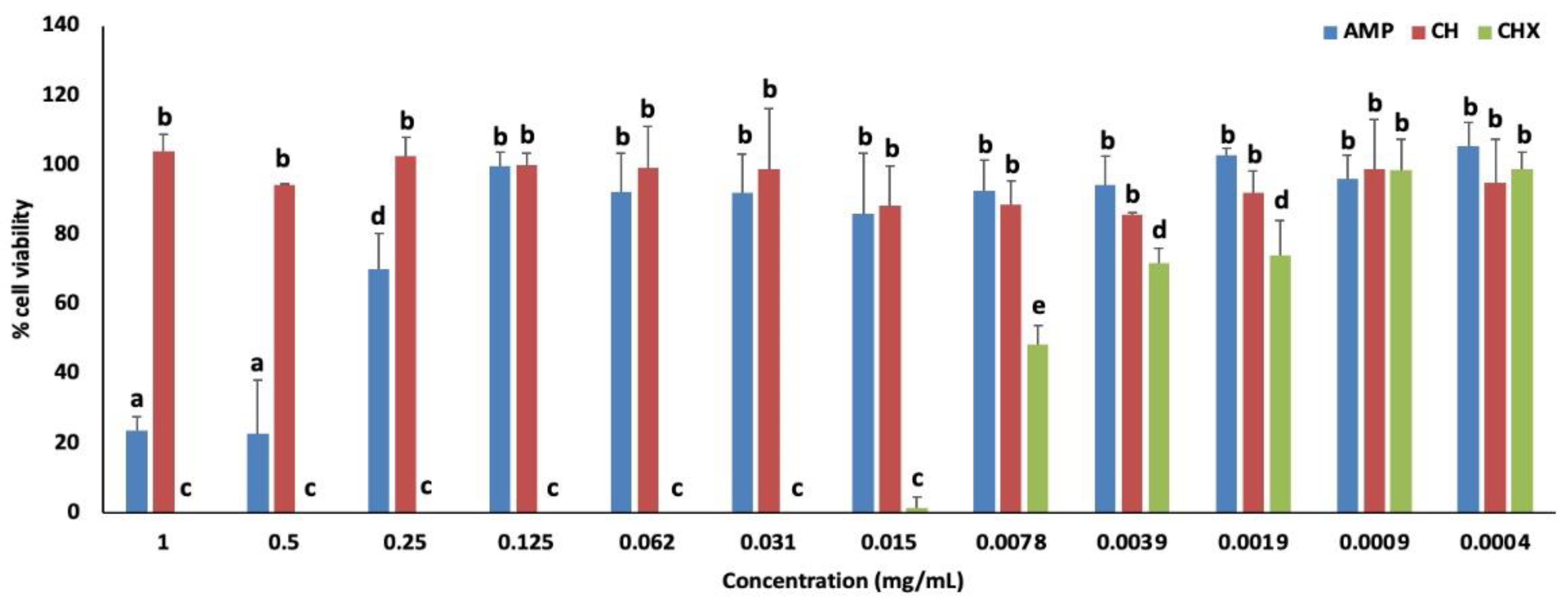
3.2. Cytotoxicity of AMP and Controls
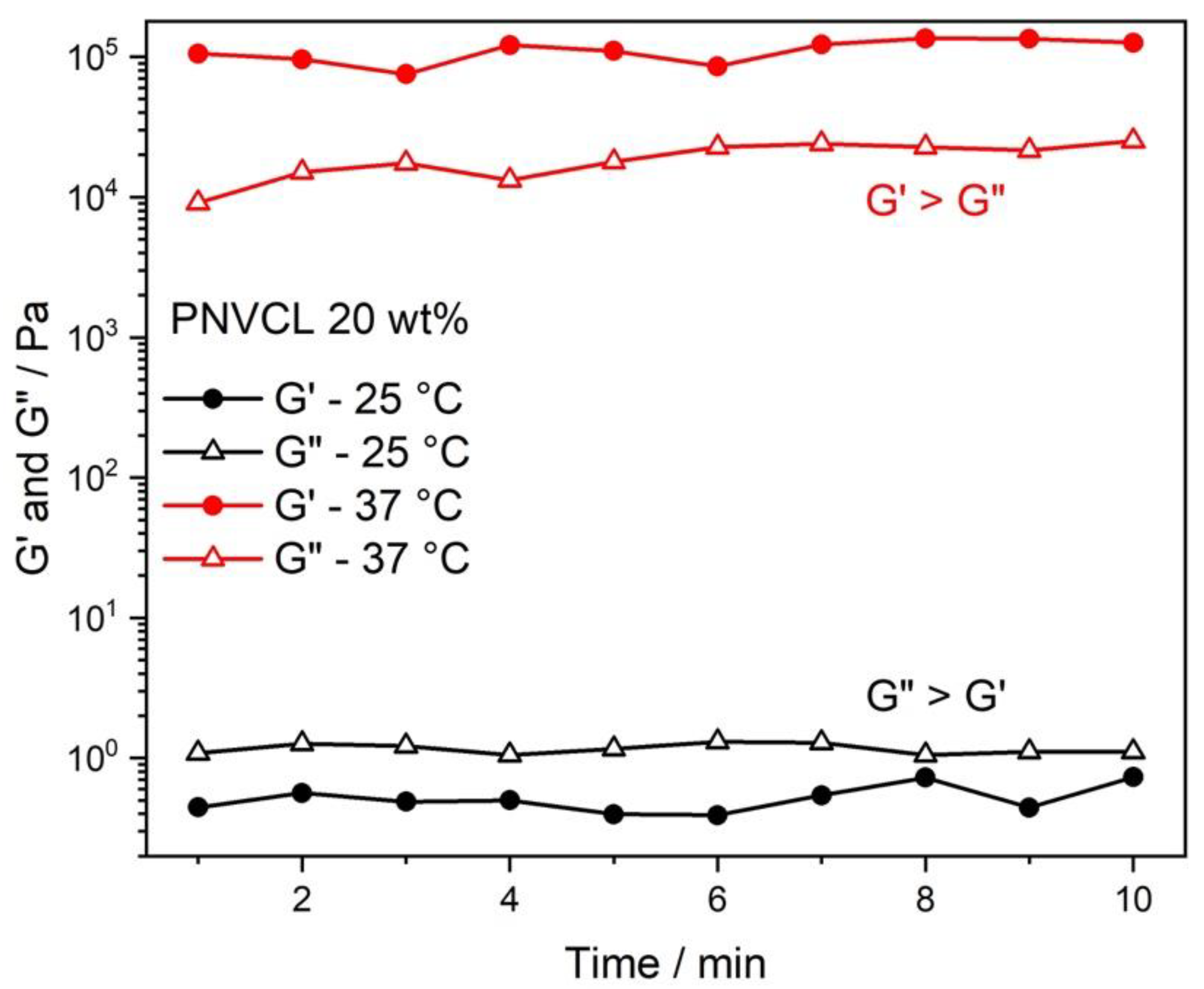
3.3. Rheological Analysis
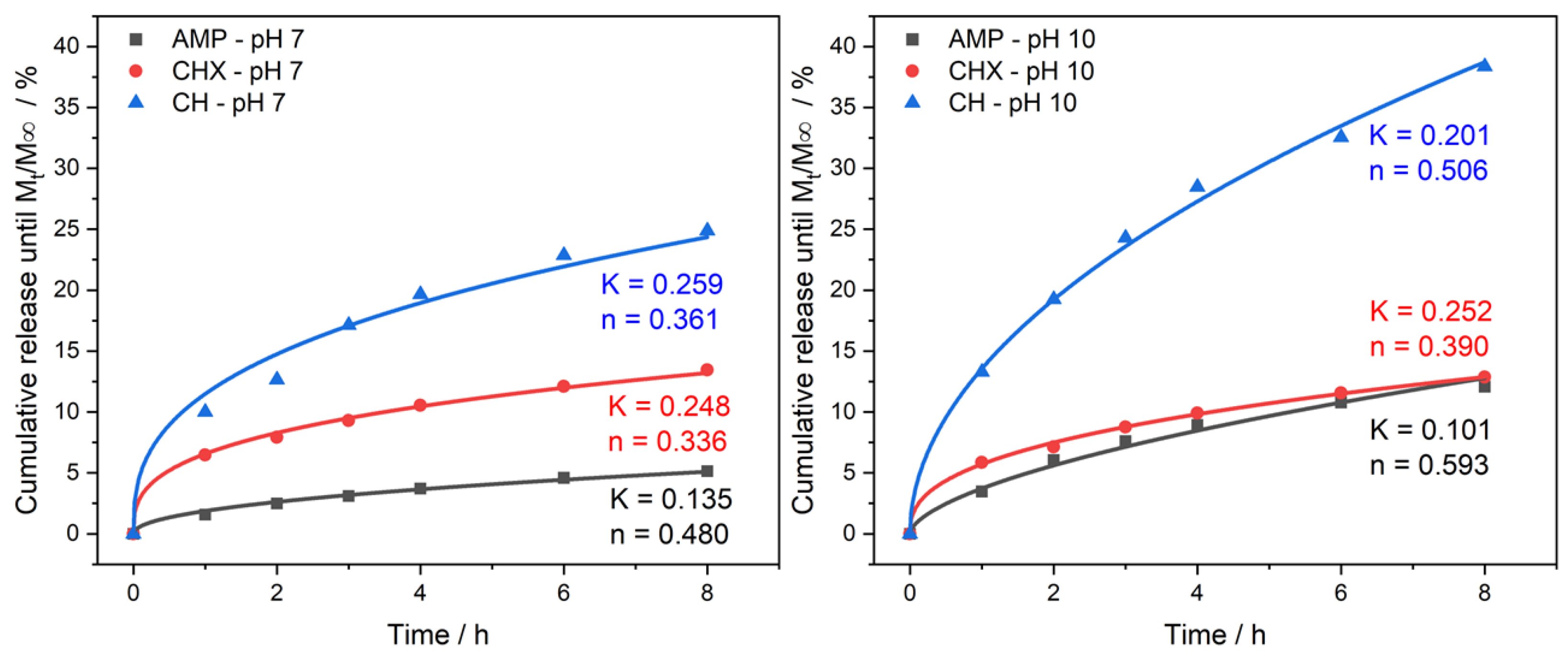
3.4. Release Assay
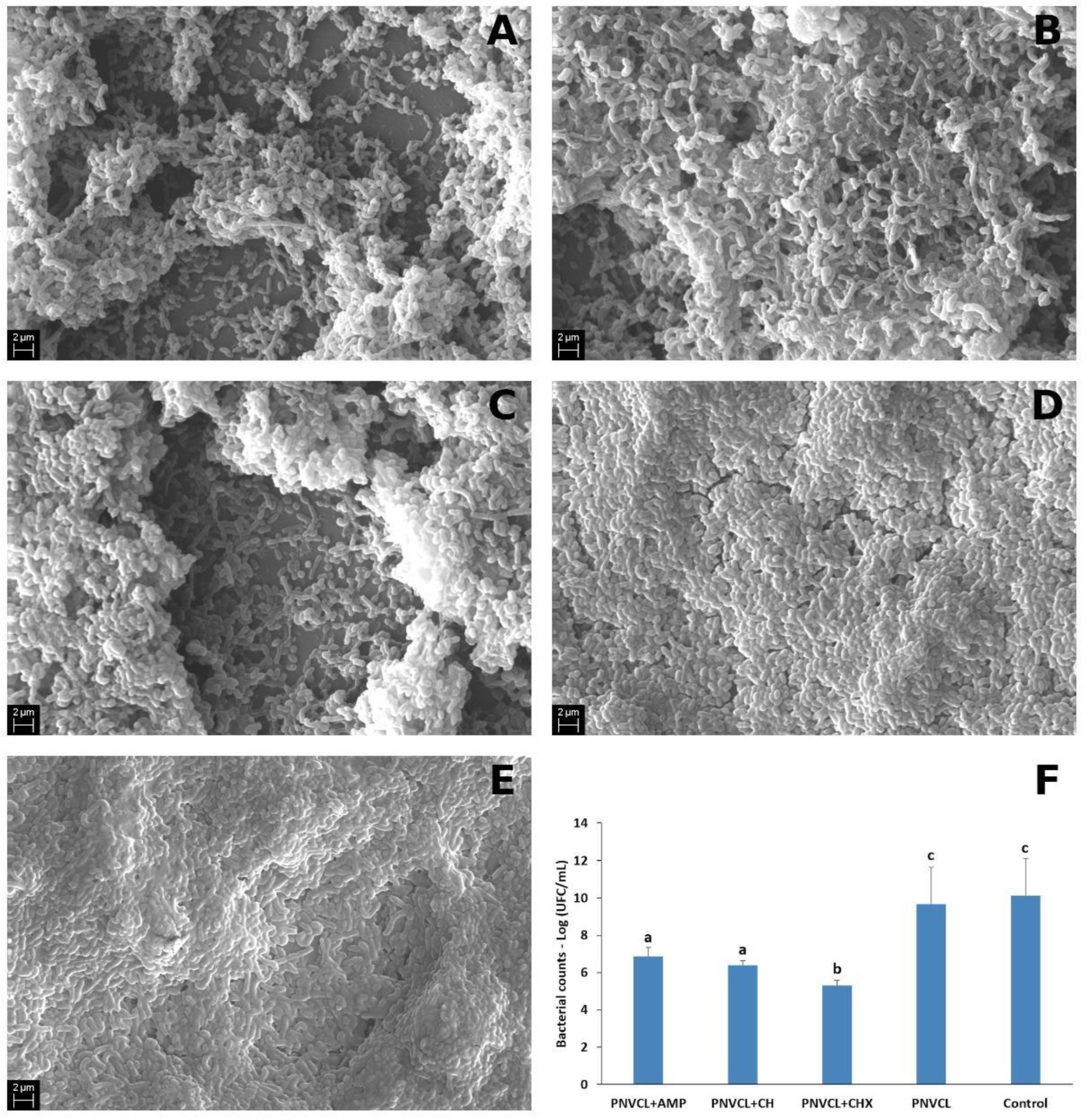
3.5. SEM Analysis
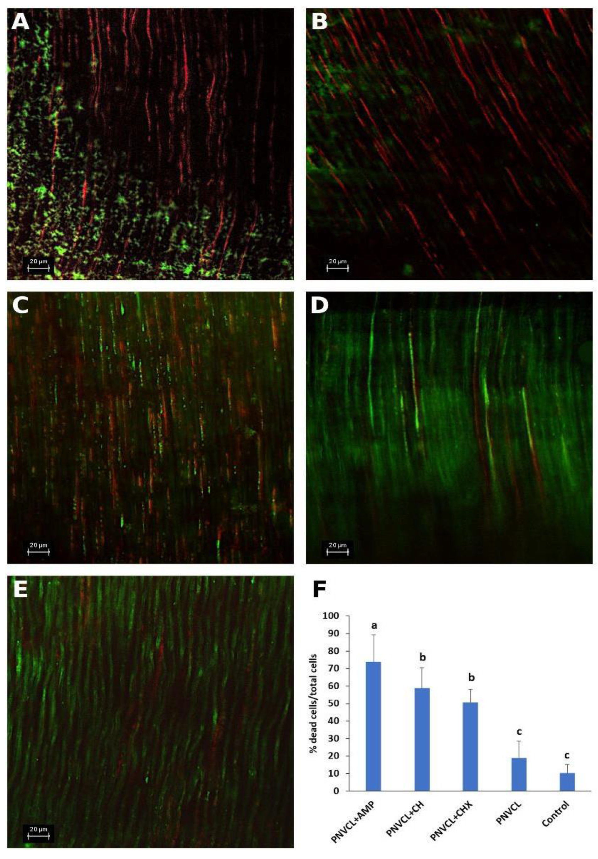
3.6. CLSM Analysis
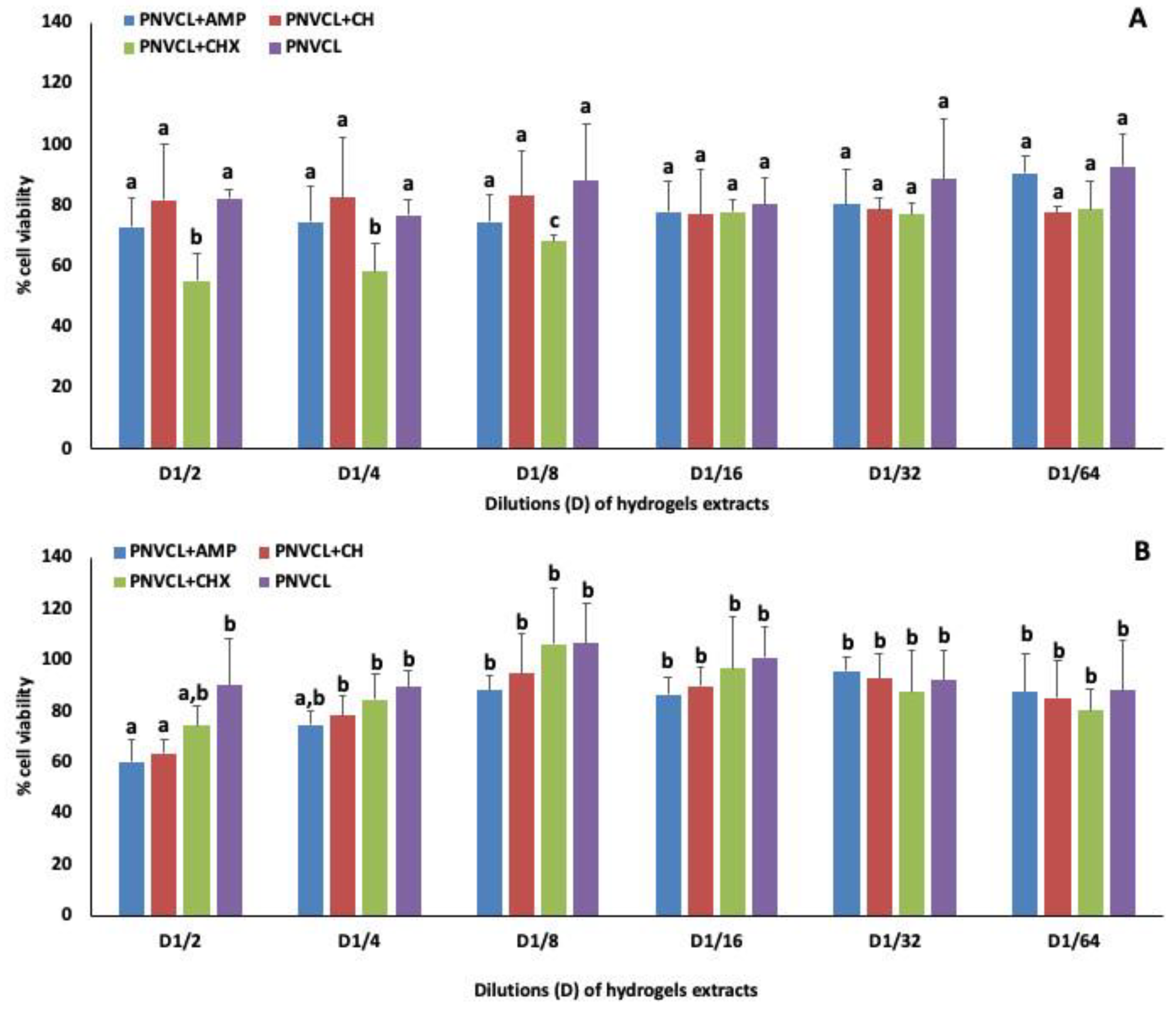
3.7. Cytotoxicity Tests with Hydrogels Extracts
4. Discussion
5. Conclusions
Supplementary Materials
Author Contributions
Funding
Data Availability Statement
Acknowledgments
Conflicts of Interest
References
- Siqueira, J.F., Jr.; Rôças, I.N. Present status and future directions: Microbiology of endodontic infections. Int. Endod. J. 2022, 3, 512–530. [Google Scholar] [CrossRef]
- Ricucci, D.; Siqueira, J.F. Biofilms and apical periodontitis: Study of prevalence and association with clinical and histopathologic findings. J. Endod. 2010, 36, 1277–1288. [Google Scholar] [CrossRef]
- de Paz, L.E.V.C. Fusobacterium nucleatum in endodontic flare-ups. Oral Surg. Oral Med. Oral Pathol. Oral Radiol. Endod. 2002, 93, 179–183. [Google Scholar]
- Ordinola-Zapata, R.; Noblett, W.C.; Perez-Ron, A.; Ye, Z.; Vera, J. Present status and future directions of intracanal medicaments. Int. Endod. J. 2022, 55 (Suppl. 3), 613–636. [Google Scholar] [CrossRef]
- Mohammadi, Z.; Dummer, P.M. Properties and applications of calcium hydroxide in endodontics and dental traumatology. Int. Endod. J. 2011, 44, 697–730. [Google Scholar] [CrossRef] [PubMed]
- Estrela, C.; Holland, R. Calcium hydroxide: Study based on scientific evidence. J. Appl. Oral Sci. 2003, 11, 269–282. [Google Scholar] [CrossRef] [Green Version]
- Vianna, M.E.; Horz, H.P.; Conrads, G.; Zaia, A.A.; Souza-Filho, F.J.; Gomes, B.P. Effect of root canal procedures on endotoxins and endodontic pathogens. Oral Microbiol. Immunol. 2007, 22, 411–418. [Google Scholar] [CrossRef] [PubMed]
- Rôças, I.N.; Siqueira, J.F., Jr. In vivo antimicrobial effects of endodontic treatment procedures as assessed by molecular microbiologic techniques. J. Endod. 2011, 37, 304–310. [Google Scholar] [CrossRef] [Green Version]
- Gomes, B.P.; Vianna, M.E.; Zaia, A.A.; Almeida, J.F.; Souza-Filho, F.J.; Ferraz, C.C. Chlorhexidine in endodontics. Braz. Dent. J. 2013, 24, 89–102. [Google Scholar] [CrossRef] [PubMed]
- Giannelli, M.; Chellini, F.; Margheri, M.; Tonelli, P.; Tani, A. Effect of chlorhexidine digluconate on different cell types: A molecular and ultrastructural investigation. Toxicol. Vitr. 2008, 22, 308–317. [Google Scholar] [CrossRef]
- Schmalz, G.; Widbiller, M.; Galler, K.M. Clinical Perspectives of Pulp Regeneration. J. Endod. 2020, 46, S161–S174. [Google Scholar] [CrossRef] [PubMed]
- Iglesias-Linares, A.; Yáñez-Vico, R.M.; Sánchez-Borrego, E.; Moreno-Fernández, A.M.; Solano-Reina, E.; Mendoza-Mendoza, A. Stem cells in current paediatric dentistry practice. Arch. Oral Biol. 2013, 58, 227–238. [Google Scholar] [CrossRef] [PubMed]
- Glynis, A.; Foschi, F.; Kefalou, I.; Koletsi, D.; Tzanetakis, G.N. Regenerative endodontic procedures for the treatment of necrotic mature teeth with apical periodontitis: A systematic review and meta-analysis of randomized controlled trials. J. Endond. 2021, 47, 873–882. [Google Scholar] [CrossRef] [PubMed]
- American Association of Endodontists (AAE). Clinical Considerations for a Regenerative Procedure. 2018. Available online: https://f3f142zs0k2w1kg84k5p9i1o-wpengine.netdna-ssl.com/specialty/wp-content/uploads/sites/2/2021/08/ClinicalConsiderationsApprovedByREC062921.pdf (accessed on 5 November 2022).
- Taneja, S.; Kumari, M.; Parkash, H. Nonsurgical healing of large periradicular lesions using a triple antibiotic paste: A case series. Contemp. Clin. Dent. 2010, 1, 31–35. [Google Scholar] [CrossRef]
- Trope, M. Treatment of the immature tooth with a non-vital pulp and apical periodontitis. Dent. Clin. N. Am. 2010, 54, 313–324. [Google Scholar] [CrossRef] [PubMed]
- Jucá, M.M.; Filho, F.M.S.C.; de Almeida, J.C.; Mesquita, D.D.S.; Barriga, J.R.M.; Dias, K.C.F.; Barbosa, T.M.; Vasconcelos, L.C.; Leal, L.K.A.M.; Ribeiro, J.E.; et al. Flavonoids: Biological activities and therapeutic potential. Nat. Prod. Res. 2020, 34, 692–705. [Google Scholar] [CrossRef] [PubMed]
- Carneiro, R.C.V.; Ye, L.; Baek, N.; Teixeira, G.H.A.; O’Keefe, S.F. Vine tea (Ampelopsis grossedentata): A review of chemical composition, functional properties, and potential food applications. J. Funct. Foods. 2021, 76, 104317. [Google Scholar] [CrossRef]
- Zhang, W.; Wang, S.; Yin, H.; Chen, E.; Xue, D.; Zheng, Q.; Gao, X.; Pan, Z. Dihydromyricetin enhances the osteogenic differentiation of human bone marrow mesenchymal stem cells in vitro partially via the activation of Wnt/β-catenin signaling pathway. Fund. Clin. Pharmacol. 2016, 30, 596–606. [Google Scholar] [CrossRef]
- Weng, L.; Zhang, H.; Li, X.; Zhan, H.; Chen, F.; Han, L.; Xu, Y.; Cao, X. Ampelopsin attenuates lipopolysaccharide-induced inflammatory response through the inhibition of the NF-κB and JAK2/STAT3 signaling pathways in microglia. Int. Immunopharmacol. 2017, 44, 1–8. [Google Scholar] [CrossRef]
- Yun, J.; Woo, E.R.; Lee, D.G. Effect of isoquercitrin on membrane dynamics and apoptosis-like death in Escherichia coli. Biochim. Bioph. Acta. Biomembr. 2018, 1860, 357–363. [Google Scholar] [CrossRef]
- Al-Shabib, N.A.; Husain, F.M.; Ahmad, I.; Khan, M.S.; Khan, R.A.; Khan, J.M. Rutin inhibits mono and multi-species biofilm formation by foodborne drug resistant Escherichia coli and Staphylococcus aureus. Food Control 2017, 79, 325–332. [Google Scholar] [CrossRef]
- Yong, D.O.C.; Saker, S.R.; Chellappan, D.K.; Madheswaran, T.; Panneerselvam, J.; Choudhury, H.; Pandey, M.; Chan, Y.L.; Collet, T.; Gupta, G.; et al. Molecular and immunological mechanisms underlying the various pharmacological properties of the potent bioflavonoid rutin. Endocr. Metab. Immune Disord. Drug Targets 2020, 20, 1590–1596. [Google Scholar] [CrossRef] [PubMed]
- Li, S.; Dong, S.; Xu, W.; Tu, S.; Yan, L.; Zhao, C.; Ding, J.; Chen, X. Antibacterial Hydrogels. Adv. Sci. 2018, 5, 1–17. [Google Scholar] [CrossRef] [Green Version]
- Yun, J.; Lee, H.; Ko, H.J.; Woo, E.R.; Lee, D.G. Fungicidal effect of isoquercitrin via inducing membrane disturbance. Biochim. Biophys. Acta (BBA)-Biomembr. 2015, 1848, 695–701. [Google Scholar] [CrossRef] [Green Version]
- Medeiros, S.F.; Lopes, M.V.; Rossi-Bergmann, B.; Ré, M.I.; Santos, A.M. Synthesis and characterization of poly(N-vinylcaprolactam)-based spray-dried microparticles exhibiting temperature and pH-sensitive properties for controlled release of ketoprofen. Drug Dev. Ind. Pharm. 2017, 43, 1519–1529. [Google Scholar] [CrossRef] [PubMed]
- Sala, R.L.; Kwon, M.Y.; Kim, M.; Gullbrand, S.E.; Henning, E.A.; Mauck, R.L.; Camargo, E.R.; Burdick, J.A. Thermosensitive poly(N-vinylcaprolactam) Injectable hydrogels for cartilage tissue engineering. Tissue Eng. Part A 2017, 23, 935–945. [Google Scholar] [CrossRef]
- Parameswaran-Thankam, A.; Parnell, C.M.; Watanabe, F.; Rangumagar, A.B.; Chhetri, B.P.; Szwedo, P.K.; Biris, A.S.; Ghosh, A. Guar-based injectable thermoresponsive hydrogel as a scaffold for bone cell growth and controlled drug delivery. ACS Omega 2018, 3, 15158–15167. [Google Scholar] [CrossRef]
- Caiaffa, K.S.; Massunari, L.; Danelon, M.; Abuna, G.F.; Bedran, T.B.L.; Santos-Filho, N.A.; Spolidorio, D.M.P.; Vizoto, N.L.; Cilli, E.M.; Duque, C. KR-12-a5 is a non-cytotoxic agent with potent antimicrobial effects against oral pathogens. Biofouling 2017, 33, 807–818. [Google Scholar] [CrossRef] [Green Version]
- Dos Santos, V.R.; Caiaffa, K.S.; Oliveira, W.C.; Pereira, J.A.; Abuna, G.F.; Polaquini, C.R.; Regasini, L.O.; Guiotti, A.M.; Duque, C. Cytotoxicity and effects of curcumin and cinnamaldehyde hybrids on biofilms of oral pathogens. Biofouling 2021, 37, 591–605. [Google Scholar] [CrossRef]
- CLSI—Clinical and Laboratory Standards Institute. Performance Standards for Antimicrobial Susceptibility Testing, 28th ed.; Clinical and Laboratory Standards Institute: Wayne, PA, USA, 2018. [Google Scholar]
- Duque, C.; Hussein, H.; Bortolatto, J.; Prakki, A.; Kishen, A. Effect of taxifolin and epigallocatechin-3-gallate on biomineralization potential of stem cells from dental apical papilla. Arch. Oral Biol. 2022, 138, 105413. [Google Scholar] [CrossRef]
- Borra, R.C.; Lotufo, M.A.; Gagioti, S.M.; de Barros, F.M.; Andrade, P.M. A simple method to measure cell viability in proliferation and cytotoxicity assays. Braz. Oral Res. 2009, 23, 255–262. [Google Scholar] [CrossRef]
- Jacob, V.P.; Paião, L.I.; da Silva, A.; Magario, M.; Kaneko, T.Y.; Martins, C.M.; Monteiro, D.R.; Mori, G.G. Antimicrobial action of NeoMTA Plus on mono- and dual-species biofilms of Enterococcus faecalis and Candida albicans: An in vitro study. Arch. Oral Biol. 2020, 120, 104925. [Google Scholar] [CrossRef]
- Ma, J.; Wang, Z.; Shen, Y.; Haapasalo, M.A. New noninvasive model to study the effectiveness of dentin disinfection by using confocal laser scanning microscopy. J. Endod. 2011, 37, 1380–1385. [Google Scholar] [CrossRef]
- Caiaffa, K.S.; Dos Santos, V.R.; Abuna, G.F.; Santos-Filho, N.A.; Cilli, E.M.; Sakai, V.T.; Cintra, L.T.A.; Duque, C. Cytocompatibility and synergy of EGCG and cationic peptides against bacteria related to endodontic infections, in planktonic and biofilm conditions. Prob. Antimicrob. Protein 2021, 13, 1808–1819. [Google Scholar] [CrossRef] [PubMed]
- Xiao, X.N.; Wang, F.; Yuan, Y.T.; Liu, J.; Liu, Y.Z.; Yi, X. Antibacterial activity and mode of action of dihydromyricetin from Ampelopsis grossedentata leaves against food-borne Bacteria. Molecules 2019, 24, 2831. [Google Scholar] [CrossRef] [Green Version]
- Wu, Y.P.; Bai, J.R.; Grosu, E.; Zhong, K.; Liu, L.J.; Tang, M.M.; Huang, Y.N.; Gao, H. Inhibitory effect of 2R,3R-dihydromyricetin on biofilm formation by Staphylococcus aureus. Foodborne Pathog. Dis. 2018, 15, 475–480. [Google Scholar] [CrossRef] [PubMed]
- Farhadi, F.; Khameneh, B.; Iranshahi, M.; Iranshahy, M. Antibacterial activity of flavonoids and their structure-activity relationship: An update review. Phytoth. Res. 2018, 33, 13–40. [Google Scholar] [CrossRef] [PubMed] [Green Version]
- Shevelev, A.B.; La Porta, N.; Isakova, E.P.; Martens, S.; Biryukova, Y.K.; Belous, A.S.; Sivokhin, D.A.; Trubnikova, E.V.; Zylkova, M.V.; Belyakova, A.V.; et al. In vivo antimicrobial and wound-healing activity of resveratrol, dihydroquercetin, and dihydromyricetin against Staphylococcus aureus, Pseudomonas aeruginosa, and Candida albicans. Pathogens 2020, 9, 296. [Google Scholar] [CrossRef] [PubMed]
- Liang, H.; He, K.; Li, T.; Cui, S.; Tang, M.; Kang, S.; Ma, W.; Song, L. Mechanism and antibacterial activity of vine tea extract and dihydromyricetin against Staphylococcus aureus. Sci. Rep. 2020, 10, 21416. [Google Scholar] [CrossRef]
- Gutiérrez-Venegas, G.; Gómez-Mora, J.A.; Meraz-Rodríguez, M.A.; Flores-Sánchez, M.A.; Ortiz-Miranda, L.F. Effect of flavonoids on antimicrobial activity of microorganisms present in dental plaque. Heliyon 2019, 5, 12. [Google Scholar] [CrossRef] [Green Version]
- Sklenarova, R.; Svrckova, M.; Hodek, P.; Ulrichova, J.; Frankova, J. Effect of the natural flavonoids myricetin and dihydromyricetin on the wound healing process in vitro. J. Appl. Biomed. 2021, 19, 149–158. [Google Scholar] [CrossRef] [PubMed]
- Ferreira, M.; Costa, D.; Sousa, Â. Flavonoids-based delivery systems towards cancer therapies. Bioengineering 2022, 9, 197. [Google Scholar] [CrossRef]
- Liu, L.; Bai, S.; Yang, H.; Li, S.; Quan, J.; Zhu, L.; Nie, H. Controlled release from thermo-sensitive PNVCL-co-MAA electrospun nanofibers: The effects of hydrophilicity/hydrophobicity of a drug. Mat. Sci. Eng. C Mat. Bio. Appl. 2016, 67, 581–589. [Google Scholar] [CrossRef] [PubMed]
- Solanki, S.S.; Sarkar, B.; Dhanwani, R.K. Microemulsion drug delivery system: For bioavailability enhancement of ampelopsin. ISRN Pharm. 2012, 108164. [Google Scholar] [CrossRef] [Green Version]
- Fallon, M.; Halligan, S.; Pezzoli, R.; Geever, L.; Higginbotham, C. Synthesis and characterisation of novel temperature and pH sensitive physically cross-linked poly (N-vinylcaprolactam-co-itaconic Acid) hydrogels for drug delivery. Gels 2019, 5, 41. [Google Scholar] [CrossRef] [PubMed] [Green Version]
- Dalcin, A.J.F.; Santos, C.G.; Gündel, S.S.; Roggia, I.; Raffin, R.P.; Ourique, A.F.; Santos, R.C.V.; Gomes, P. Anti-biofilm effect of dihydromyricetin-loaded nanocapsules on urinary catheter infected by Pseudomonas aeruginosa. Colloids Surf. B Biointerfaces 2017, 156, 282–291. [Google Scholar] [CrossRef]
- Hall, C.W.; Mah, T.F. Molecular mechanisms of biofilm-based antibiotic resistance and tolerance in pathogenic bacteria. FEMS Microbiol. Rev. 2017, 41, 276–301. [Google Scholar] [CrossRef] [Green Version]
- Oliveira, M.A.C.; Borges, A.C.; Brighenti, F.L.; Salvador, M.J.; Gontijo, A.V.L.; Koga-Ito, C.Y. Cymbopogon citratus essential oil: Effect on polymicrobial caries-related biofilm with low cytotoxicity. Braz. Oral Res. 2017, 31, 89. [Google Scholar] [CrossRef] [PubMed] [Green Version]
- O’Brien, T.J.; Figueroa, W.; Welch, M. Decreased efficacy of antimicrobial agents in a polymicrobial environment. ISME J. 2022, 16, 1694–1704. [Google Scholar] [CrossRef]
- Roy, R.; Tiwari, M.; Donelli, G.; Tiwari, V. Strategies for combating bacterial biofilms: A focus on anti-biofilm agents and their mechanisms of action. Virulence 2018, 9, 522–554. [Google Scholar] [CrossRef] [Green Version]
- Nabb, D.L.; Song, S.; Kluthe, K.E.; Daubert, T.A.; Luedtke, B.E.; Nuxoll, A.S. Polymicrobial interactions induce multidrug tolerance in Staphylococcus aureus through energy depletion. Front. Microbiol. 2019, 10, 2803. [Google Scholar] [CrossRef] [PubMed]
- Reddy, P.R.S.; Eswaramma, S.; Rao, K.K.; Lee, Y.I. Dual responsive pectin hydrogels and their silver nanocomposites: Swelling studies, controlled drug delivery and antimicrobial applications. Bull. Korean Chem. 2014, 35, 2391–2399. [Google Scholar] [CrossRef] [Green Version]
- Kumar, A.; Sharma, S.; Afgan, S.; Kumar, R.; Keshari, A.K.; Srivastava, R. Development of graft copolymer of carboxymethylcellulose and N-vinylcaprolactam towards strong antioxidant and antibacterial polymeric materials. Int. J. Biol. Macromol. 2018, 112, 780–787. [Google Scholar] [CrossRef] [PubMed]
- Vihola, H.; Laukkanen, A.; Valtola, L.; Tenhu, H.; Hirvonen, J. Cytotoxicity of thermosensitive polymers poly(N-isopropylacrylamide), poly(N-vinylcaprolactam) and amphiphilically modified poly(N-vinylcaprolactam). Biomaterials 2005, 26, 3055–3064. [Google Scholar] [CrossRef]
- Wang, Y.; Nie, J.; Chang, B.; Sun, Y.; Yang, W. Poly(vinylcaprolactam)-based biodegradable multiresponsive microgels for drug delivery. Biomacromolecules 2013, 14, 3034–3046. [Google Scholar] [CrossRef]
- Lessa, F.C.; Aranha, A.M.; Nogueira, I.; Giro, E.M.; Hebling, J.; Costa, C.A. Toxicity of chlorhexidine on odontoblast-like cells. J. Appl. Oral Sci. 2010, 18, 50–58. [Google Scholar] [CrossRef] [PubMed] [Green Version]
- Gonzalez-Urias, A.; Licea-Claverie, A.; Sañudo-Barajas, J.A.; González-Ayón, M.A. NVCL-Based Hydrogels and Composites for Biomedical Applications: Progress in the Last Ten Years. Int. J. Mol. Sci. 2022, 23, 4722. [Google Scholar] [CrossRef] [PubMed]


| E. faecalis | A. israelii | S. mutans | L. casei | F. nucleatum | |
|---|---|---|---|---|---|
| AMP | 1 (1) | 0.25 (1) | 0.25 (1) | 1 (1) | 0.125 (0.25) |
| ISO | 1 (>1) | 1 (>1) | 1 (>1) | 1 (>1) | 0.25 (1) |
| RUT | >1 (>1) | 1 (>1) | 1 (>1) | 1 (>1) | 0.5 (1) |
| CHX | 0.005 (0.005) | 0.002 (0.002) | 0.0002 (0.002) | 0.002 (0.004) | 0.004 (0.004) |
Publisher’s Note: MDPI stays neutral with regard to jurisdictional claims in published maps and institutional affiliations. |
© 2022 by the authors. Licensee MDPI, Basel, Switzerland. This article is an open access article distributed under the terms and conditions of the Creative Commons Attribution (CC BY) license (https://creativecommons.org/licenses/by/4.0/).
Share and Cite
Braga, G.P.d.A.; Caiaffa, K.S.; Pereira, J.A.; Santos, V.R.d.; Souza, A.C.A.; Ribeiro, L.d.S.; Camargo, E.R.; Prakki, A.; Duque, C. Microbiological Properties and Cytotoxicity of PNVCL Hydrogels Containing Flavonoids as Intracanal Medication for Endodontic Therapy. J. Funct. Biomater. 2022, 13, 305. https://doi.org/10.3390/jfb13040305
Braga GPdA, Caiaffa KS, Pereira JA, Santos VRd, Souza ACA, Ribeiro LdS, Camargo ER, Prakki A, Duque C. Microbiological Properties and Cytotoxicity of PNVCL Hydrogels Containing Flavonoids as Intracanal Medication for Endodontic Therapy. Journal of Functional Biomaterials. 2022; 13(4):305. https://doi.org/10.3390/jfb13040305
Chicago/Turabian StyleBraga, Gabriela Pacheco de Almeida, Karina Sampaio Caiaffa, Jesse Augusto Pereira, Vanessa Rodrigues dos Santos, Amanda Caselato Andolfatto Souza, Lucas da Silva Ribeiro, Emerson Rodrigues Camargo, Anuradha Prakki, and Cristiane Duque. 2022. "Microbiological Properties and Cytotoxicity of PNVCL Hydrogels Containing Flavonoids as Intracanal Medication for Endodontic Therapy" Journal of Functional Biomaterials 13, no. 4: 305. https://doi.org/10.3390/jfb13040305