Article

A Snapshot of the Taxonomic Composition and Metabolic Activity of the Microbial Community in an Arctic Harbour (Ny-Ålesund, Kongsfjorden, Svalbard)

1
Institute for Biological Resources and Marine Biotechnologies, Section of Messina, National Research Council (CNR-IRBIM), Spianata S. Rineri 86, 98122 Messina, Italy
2
Institute of Polar Sciences, Section of Messina, National Research Council (CNR-ISP), Spianata S. Raineri 86, 98122 Messina, Italy
3
PhD School of “Applied Biology and Experimental Medicine”, Faculty of Sciences, University of Messina, Viale F. Stagno D’Alcontres 5, 98166 Messina, Italy
*
Author to whom correspondence should be addressed.
J. Mar. Sci. Eng. 2023, 11(5), 1018; https://doi.org/10.3390/jmse11051018
Submission received: 9 March 2023 / Revised: 22 April 2023 / Accepted: 8 May 2023 / Published: 10 May 2023
(This article belongs to the Special Issue Microbes in Extreme Marine Environments)

Abstract

:
Within the Svalbard archipelago, Kongsfjorden is an important marine ecosystem that is recognised as one of the main representative Arctic glacial fjords. Prokaryotic organisms are key drivers of important ecological processes such as carbon fluxes, nutrient mineralisation, and energy transfer, as well as sentinels of environmental pollution, especially in sediments, that are a repository of contaminants. In some areas of the Arctic, the structure and metabolic activity of the microbial community in the organic matter turnover and globally in the functioning of the benthic domain are mostly still unknown. A snapshot of the main microbial parameters such as bacterial abundance (by microscopic and plate counts), structure (by 16S rRNA sequencing), and metabolic activity was provided in Ny-Ålesund harbour, contextually in seawater and sediment samples. Fluorogenic substrates were used to assess the microbial ability to utilise organic substrates such as proteins, polysaccharides, and organic phosphates through specific enzymatic assays (leucine aminopeptidase—LAP, beta-glucosidase—ß-GLU, and alkaline phosphatase—AP, respectively). The metabolic profiles of psychrophilic heterotrophic bacterial isolates were also screened using a qualitative assay. The phylogenetic analysis of the microbial community revealed that Proteobacteria prevailed among the observed taxonomic groups. Several of the observed sequences were assigned to clones found in harbours, microbial biofilms, antifouling paints, or oil-polluted facilities of cold environments, highlighting a signature of human pressure on the polar habitat of Ny-Ålesund harbour.

1. Introduction

Harbours are areas of economical relevance exposed to strong impacts from shipping, as well as urban and industrial waste, and consequently environmentally sensitive to anthropogenically derived pressure [1,2,3]. Intense maritime traffic, together with the low hydrodynamism of these sheltered bays, results in the organic matter enrichment of the waters, giving rise to anoxia and eutrophication events [4,5,6,7]. Traditionally, environmental characterisation of harbours regarded their chemical contamination-related features [1,4,5]. Heavy metals and polycyclic aromatic hydrocarbons are the main contaminants that impact harbour sites [8,9,10], especially in the benthic domain. The heavy weight oil fractions can have a long persistence in the environment due to their high resistance to degradation [6], making the successful restoration measures of these impacted areas difficult [11]. Moreover, quantification of contaminants’ concentration by itself cannot provide information on the effects on marine biota, resulting only in a partial assessment of the impacts of maritime activities on the benthic fauna [8,12].
In aquatic environments, prokaryotic organisms are key drivers of important ecological processes involved the organic matter biogeochemistry, such as carbon and nutrient mineralisation and consequent energy fluxes; they act also as sentinels of environmental change. Studies of microbial remineralisation offer information on the turnover of the pool of organic substrates, providing an integrated estimate of the polymers’ utilisation and of their bioavailability to the marine biota. In turn, the capability of microbes to react to environmental conditions is strictly related to their enzymatic profiles involved in several metabolic pathways [13].
Sediments are the domain most sensitive to environmental pollution, acting as a natural repository of organic contaminants (see [14] and the references therein). Nevertheless, to date, the changes in microbial community, especially regarding the benthic one, in response to environmental quality deterioration have been the subject of only a few studies [2,3,7,15]. In the assessment of chronically polluted areas, exploring microbial structure and metabolism is needed, not only due to their role as early warning sentinels of contamination [16,17,18] but also to apply appropriate remediation measures [6,19]. To recover marine polluted areas, such as harbours, microbial bioremediation measures using consortia of microorganisms able to degrade organic contaminants has been suggested as an effective and eco-friendly tool to remove biodegradable complex toxic substances and convert them into harmless end products, e.g., CO2 and H2O, via metabolic pathways [20].
In the last decade, several reports have documented in polar regions the occurrence of contamination related to increased human pressure [21,22,23]; nevertheless, knowledge of the variable dynamics of microbial communities in response to contamination is still lacking in Arctic harbour ecosystems. This main gap underlines the need of further research addressing how marine microorganisms living in these fragile environments react to stressors, with the goal of finding measures to prevent and/or reduce potentially detrimental impacts of contamination on the microbiota.
In marine sediments chronically exposed to oil contamination, microbial community composition and metabolism were reported to vary in relation with both temperature and chemical diversity [24]; furthermore, bacterial richness and catabolic diversification increased under chronic pollution at low temperature [24]. Moreover, climate warming, which is an ongoing issue in the Arctic region, as shown by glacier retreat, sea ice melting, and increases in suspended sediment inputs into coastal marine environments, might influence the dynamics and the trophic level interactions within the microbial community, with consequent implications on large-scale ecological processes [25,26].
Within the Svalbard archipelago (Norway), Kongsfjorden, located on the west coast of Spitsbergen, is considered a site representative of Arctic glacial fjords. Exposed to strong Atlantic and glacial runoff influence [27], it is a widely studied marine ecosystem [28]; here, many physical and biological features are being addressed by several countries which have established their research stations at Ny-Ålesund [29]. To date, an important number of studies have explored the structure and function of the bacterial community across the entire Kongsfjorden marine system, both at a pelagic and sedimentary level [25,30,31,32,33,34,35,36,37,38,39,40]. Data on microbial distribution and its role in this Arctic area are mostly fragmentary, due to the lack of comprehensive studies, as evidenced by recent literature reviews [29,40]. Comparatively more limited is the knowledge of the taxonomic composition and metabolic potential of the complex microbiota inhabiting the Ny-Ålesund harbour [41,42,43], where benthic and pelagic domains have never been investigated simultaneously. Originally, Ny-Ålesund was born as a coal mining community, which was decommissioned since the 1960s after a large accident; therefore, the soil of this site is affected by coal waste materials and heavy metals from mining operations [44]. In the Ny-Ålesund harbour sediments, the presence of cold-adapted oil-degrading bacteria was previously demonstrated [45]. As a contribution to fill this knowledge gap, during a field study performed in June 2015, the abundance, structure, and metabolic activity of the benthic microbial community hosted in this harbour were analysed; parallel measurements were performed in the waters of the same area. Psychrophilic heterotrophic bacteria were also isolated and identified.

2. Materials and Methods

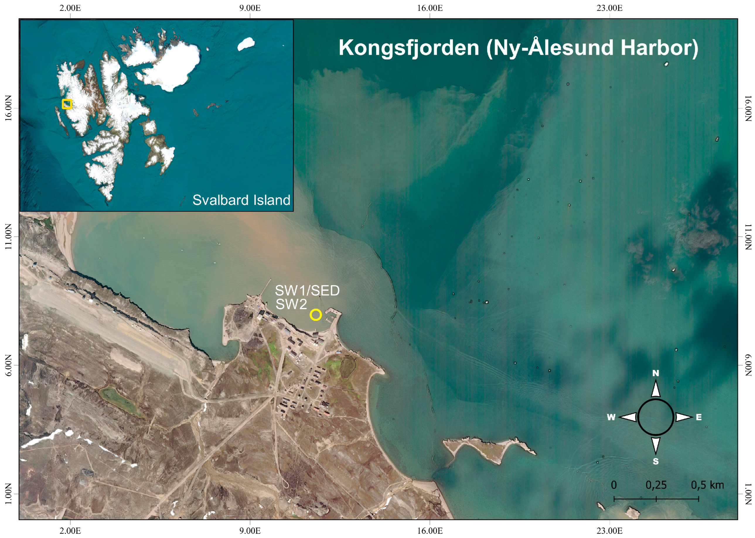
2.1. Sampling in the Study Area

Ny-Ålesund harbour is situated in Kongsfjorden within Spitsbergen, the largest island of the Svalbard archipelago (Arctic Ocean). Surface water samples were collected in June 2015 from two sites (further indicated as SW1 and SW2) in the Ny-Ålesund Harbour (78.928° N; 11.940° E; Figure 1), a popular shore break for cruises. Sampling was performed on board of the r/v Teisten with a 10 L Niskin bottle. The physico-chemical parameters of the sea water were acquired with a CTD SBE 911 probe (SeaBird Electronics, Bellevue, WA, USA). One surface sediment sample (indicated as SED1) was taken at a 4 m depth in the site corresponding to the SW1 area (Figure 1). The sampling was carried out using a 3 kg Van Veen Grab sampler. All samples, after collection, were transported and immediately treated in laboratory for microbiological and chemical analyses.

2.2. Measurement of the Hydrocarbon Concentration (GC-FID Analysis)

The total hydrocarbons and their derivates (TERHCs) in seawater (SW1 and SW2) and sediment (SED1) samples were extracted, and their composition was determined according to the EPA (Environmental Protection Agency) 3510 and 3550 methods, respectively. The extracts, reduced to 1 mL by a rotary evaporation (Rotavapor model R110; Büchi Labortechnik AG, Flawil, Switzerland), were analysed according to Genovese et al. [46] by high-resolution fast gas chromatography using a DANI Master GC Fast Gas Chromatograph System (DANI Instruments S.p.A., Milan, Italy). The concentrations of hydrocarbons were determined using the analytical protocols reported by Denaro et al. [47]. Generally, the detection limit of GC FID is approximately 20 pg of each component eluting from a capillary column.

2.3. Total Bacterial Count (DAPI)

For the quantification of the total bacterial abundance, seawater samples (1–2 mL) preserved in formaldehyde (2% final concentration) were stored at 4 °C until their treatment. After short-time (30 s) ultrasound exposure to an ultrasonic bath (Branson 1200 Ultrasonic Cleaner, Branson, Brookfield, CT, USA), they were filtered through Nuclepore black polycarbonate filters (0.2 µm pore size), which were incubated with 4,6-diamidino-2-phenylindole (DAPI; Sigma-Aldrich, Milan, Italy, 5 mg L−1 final concentration), following standard analytical protocols [48]. As reported by Cappello et al. [49], prior to the analytical protocol, a preliminary treatment with Tween 80 (final concentration, 1 mg L−1) for at least 15 min of the sediment samples was carried out. Bacteria were detached from the sediment through an ultrasonic treatment in a Branson 1200 Ultrasonic Cleaner bath (Branson, USA), using the protocol reported by Kuwae and Hosokawa [50]. The water-Tween 80 phase was collected after centrifugation of the samples at 8000× g for 8 min and stained with the above-indicated protocol. Observation of the obtained slides was performed using a Zeiss Axioplan 2 Imaging epifluorescence microscope (Zeiss; Carl Zeiss Inc., Thornwood, NY, USA); at least 30 microscopic fields were visualised, and the labelled cells were counted. Bacterial abundance was reported in terms of total number of cells mL−1.

2.4. Culturable Heterotrophic Bacterial Count (Marine Agar)

Culturable heterotrophic bacterial count in seawater samples was obtained through the “spread plate method”, through inoculation of 0.1 mL of sample on plates of Marine agar 2216 (Conda Pronadisa, Madrid, Spain), a medium specific for the cultivation of heterotrophic bacteria [51]. Marine agar was chosen in this study as the culture medium to better compare the heterotrophic bacterial counts with those determined in other Arctic environments [25,38].
One sediment aliquot was 10-times diluted into sterile physiological solution (in a weight/volume ratio); the obtained supernatant was further diluted (1:10) and treated as reported for seawater samples. Colonies grown after incubation for 20 days at +5 °C were counted, and the number of colony-forming units (CFU) obtained per ml or g of sample (for water or sediment, respectively) was reported, considering the dilutions performed for the sediment sample.

2.5. Hydrocarbon-Degrading Bacterial Count (MPN)

Hydrocarbon-degrading bacterial count was determined using a miniaturised most probable number (MPN) method, following the protocol of Brown and Braddock [52] with slight modifications [48]. The MPN index was calculated as indicated by the American Public Health Association [53].

2.6. 16S rDNA Clone Library

Extraction and PCR Amplification of Total DNA, and 16S rRNA Cloning and Sequencing

An amount of 1 L of seawater was treated by filtration through polycarbonate filtering membranes (Nuclepore, Costar, Pleasanton, CA, USA) with a 47 mm diameter and a 0.2 μm pore size. DNA extraction of total bacterial community of the seawater sample was performed using the MasterPure™ Complete DNA and RNA Purification Kit (Epicentre, Illumina San Diego, CA, USA). The total genomic DNA was eluted by washing with 35 μL of 1× TE (Tris and ethylenediaminetetraacetic acid) buffer; thereafter, it was measured with a Spectrophotomer ND-1000 (NanoDrop Technologies, Wilmington, DE, USA), and then nucleic acids were stored at −20 °C. From the total DNA, the 16S rRNA genes were amplified using the set of universal eubacterial primers 530F (5′-GTGCCAGCMGCCGCGG-3′) and 1492R (5′-TACGGYTACCTTGTTACGACT-3′) [54]. The PCR was performed in a 50 µL mixture (whole volume) containing 1 µM of each primer, 10 µM dNTPs (Gibco, Invitrogen, Carlsbad, CA, USA), 1× Q solution (Qiagen, Hilden, Germany), 1× Qiagen reaction buffer, 1 µL of DNA template (40–250 ng), and 2U of Taq Polymerase (Qiagen). The reaction included an initial denaturation step at 95 °C for 5 min, continued with an annealing step (1 min at 94 °C, 1 min at 50 °C, and 2 min at 72 °C, 30 cycles), and a final extension at 72 °C for 10 min. PCR products were run on agarose gel (1% w/vol) and treated with the Wizard® Genomic DNA Purification Kit (Promega, Madison, WI, USA); 16S rRNA gene fragments were cloned into the pGEM®-T Easy Vector (Promega) following the manufacturer’s instructions. Escherichia coli DH10β cells (Invitrogen, Waltham, MA, USA) were transformed using the obtained ligation products. From each clone library, a total of 100 white colonies were picked at random, and a PCR reaction was performed on the collected cells using the same conditions reported previously and the set of M13F and M13R primers. Gel electrophoresis was carried out to identify the amplified inserts according to their expected size (approx. 1200 bp). PCR products of the isolated strains were purified and sequenced (Sanger’s Method) by Macrogen Inc. (Amsterdam, the Netherlands) using only the reverse primer (1492R). The 16S rRNA gene sequences of the closest relatives were identified with the Basic Local Alignment Search Tool (BLAST), provided by the National Center for Biotechnology Information (NCBI, Bethesda, MA, USA).

2.7. Quantitative Determination of Enzymatic Activity Rates

The metabolic activities of the microbial assemblage on organic polymers (proteins, polysaccharides, and organic phosphates) were assessed through determination of the leucine aminopeptidase (LAP), beta-glucosidase (ß-GLU), and alkaline phosphatase (AP) activities. Enzymatic assays were performed through fluorogenic measurements [55]. L-leucine-4-methylcoumarinylamide hydrochloride, 4-methylumbelliferyl B-D-glucopyranoside, and 4-methylumbelliferyl phosphate (Merck Life Sciences, Milan, Italy) were the substrates specific for LAP, ß-GLU, and AP. Seawater samples were dispensed into three test tubes (10 mL each) plus one autoclaved sample (Blank tube), which were added increasing volumes (10 to 320 nmol L−1) of each substrate separately, according to the procedure already described in Caruso et al. [25]. The sediment was diluted 1:10 in sterile physiological solution, and the supernatant obtained after re-suspension was divided into triplicate sub-volumes that were incubated with each substrate in concentrations of 20 to 160 µM (stock solution 5 mM). After substrate hydrolysis by the specific enzyme, the released fluorescence was recorded using a Jenway fluorimeter (model 6280, Dunmow, UK); measurements were taken both at time zero (initial time) and after incubation at +5 ± 1 °C (LAP and AP, 2.5 h; ß-GLU, 4 h). Calibration was performed for LAP with increasing amounts (200 to 800 nM) of the standard 7-amino-4-methylcoumarin (MCA), while for ß-GLU and AP, the standard 4-methylumbelliferone (MUF) was used [56]. Results were expressed as the maximum velocity (Vmax) of substrate hydrolysis, in nanomoles of leucine, glucoside, and PO4 potentially released by LAP, ß-GLU and AP activities, respectively, per litre or per gram and per hour (nmol L−1 h−1 or nmol g−1 h−1, for water or sediment samples, respectively).

2.8. Isolation of Psychrophilic Heterotrophic Bacteria

To isolate strains of autochthonous heterotrophic bacteria, from the Marine Agar 2216 plates, a total number of 35 colonies (12 from each water sample and 11 from sediments) were selected on the basis of their different phenotypical traits (i.e., colony shape, size, presence of pigmentation) and subjected to purification by repeated streaking on the same Marine Agar medium until an axenic culture was obtained.

2.9. 16S rDNA Gene Phylogenetic Analyses

A 2 mL late-exponential phase cell culture of each bacterial isolate was used to extract the total genomic DNA with a MasterPure™ Complete DNA and RNA Purification Kit (Epicentre). The 16S rDNA genes were amplified by a PCR using the primers 27F (5′-AGAGTTTGATCMTGGCTCAG-3′) and 1492R (5′-TACGGYTACCTTGTTACGACTT-3′). The reaction was carried out under the conditions described previously. PCR products of both heterotrophic isolates and 16S rDNA seawater sample clone’s library were assayed for purity and sequenced by Macrogen (Amsterdam, the Netherlands). Checking of the sequences for possible chimeric origin was performed using the Pintail 1.1 software [57]. Alignment of the amplified 16S rRNA gene sequences and of the closest relative identified with BLAST [58] was initially made with the SILVA alignment tool [59], followed by manual alignment with ARB [60]. After these operations, using the neighbour-joining algorithm of ARB package and the distance analysis of the 16S rDNA genes, the phylogenetic trees were built. The sequences were deposited into the GenBank database under the accession numbers OQ753775-OQ753823.

2.10. Qualitative Study of Enzymatic Expression of Bacterial Isolates

Bacterial isolates were screened for their metabolic profiles in terms of presence/absence (+/−) of six enzymatic activities; this qualitative assay was performed with a miniaturised method using 96-well microtitre plates. For each bacterial strain, a suspension in sterile physiological saline was prepared, followed by the seeding of 75 μL of this suspension into each well and the addition of the same volume of each fluorogenic substrate (diluted to obtain a working solution of 20 μM). Stock solutions of the substrates in Cellosolve (5 mM final concentration) were prepared. The substrates used for the metabolic screening were leucine-amido-methyl coumarine (Leu-MCA), 4-methylumbelliferyl (MUF)-alpha-D-glucopyranoside, MUF-beta-D-glucopyranoside, and MUF-phosphate in order to assess whether the strains were positive for aminopeptidase (proteolytic enzyme), alpha- and beta-glucosidase (glycidic enzymes acting on maltose and cellobiose, respectively), and alkaline phosphatase (an enzyme able to decompose organic phosphoric compounds). The presence of lipolytic activity was assessed using MUF-heptanoate as the substrate; MUF-acetate was used as the substrate for esterase activity. After incubation at in situ temperature for 4 h, the fluorescence released by hydrolysis of the substrates was detected under a UV-Wood lamp, assigning a positive score to those wells that showed an intense and bright fluorescent signal [61].

3. Results

3.1. Environmental Characteristics

Temperature and salinity values recorded in the harbour were 2.79 °C and 32.88 psu, respectively; a low turbidity characterised the water column, as shown by a value of 4 m (by a Secchi disk) [45].

3.2. Analysis of Hydrocarbons (GC-FID Analysis)

The qualitative and quantitative analyses of hydrocarbons (GC-FID analysis) did not show the presence of these chemical compounds in the examined seawater and sediment samples.

3.3. Total Bacterial (DAPI), Heterotrophic Bacterial (CFU), and Hydrocarbon-Degrading Bacterial Counts (MPN)

Table 1 shows the total (DAPI), the cultivable heterotrophic (CFU), and the hydrocarbon-degrading bacterial counts as MPN values of the examined samples. Total microbial abundance evidenced similar values of ≈104 cell mL−1 in the seawater samples (SW1 and SW2), compared to values of ≈106 cell g−1 observed in the SED-1 sample. The abundance of heterotrophic bacteria able to grow on Marine Agar was in the order of 102 CFU mL−1 in the waters (4.9–6.1 × 10² CFU mL−1) and one order of magnitude higher in the sediment (2.1 ×10³ CFU g−1). Regarding the hydrocarbon-degrading bacteria, values of ≈102 MPN mL−1/g−1 were measured.

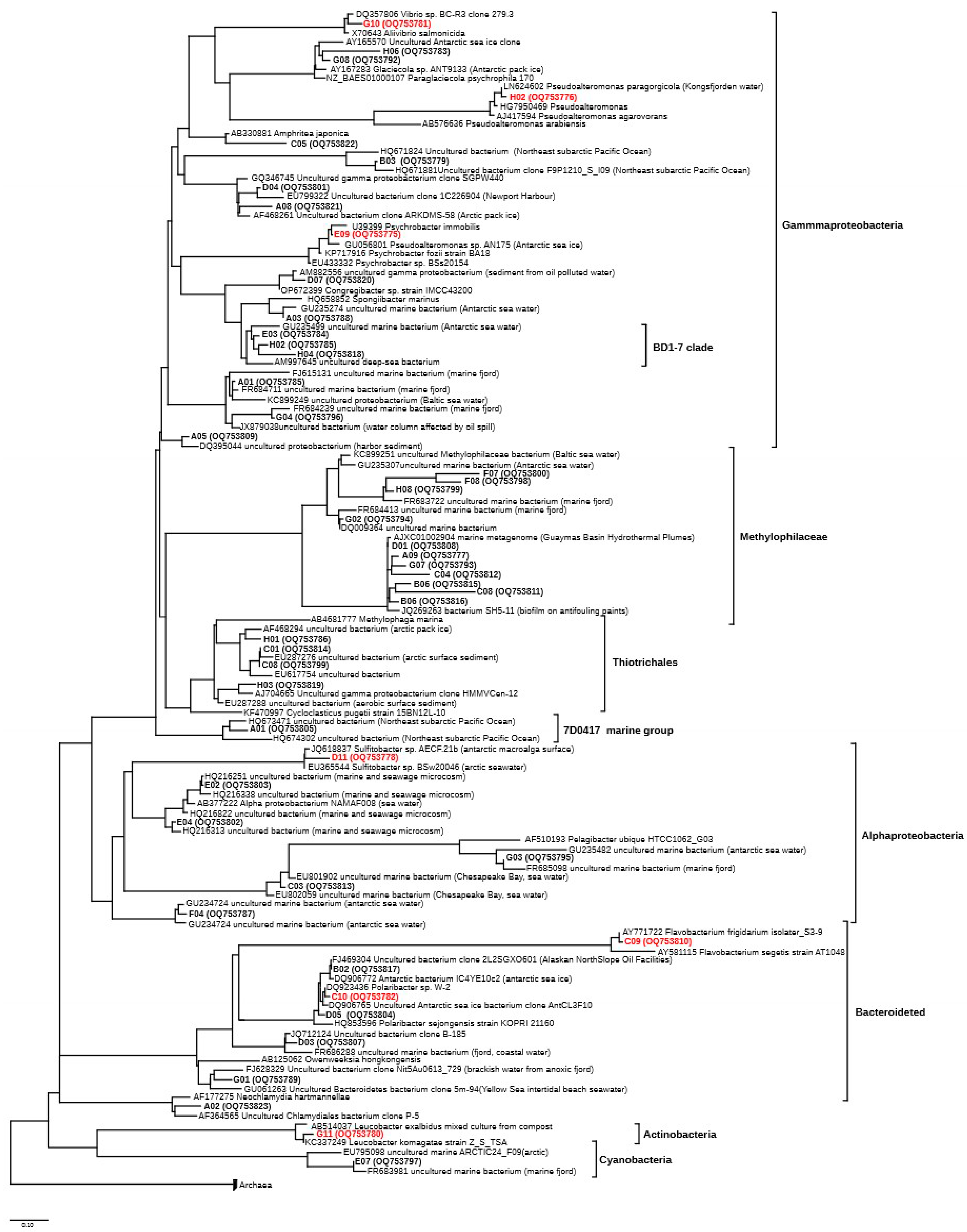
3.4. Structure of the Microbial Community (16S rRNA Clone Library)

Globally, 192 16S rRNA sequences were obtained from the SW1 and SW2 samples and analysed from the environmental clone library (Table S1 in Supplementary Materials). Statistical and taxonomic analysis did not reveal differences between the samples, and therefore the results obtained for the SW1 and SW2 were treated together, as a single sample. After clusterisation (at 97% of similarity), the sequences were assigned to 41 main taxa. The phylogenetic analysis of all the obtained 16S rDNA gene sequences revealed their distribution within four major taxonomic groups of prokaryotic organisms (Figure 2). The phylogenetic analysis showed that Proteobacteria were the most abundant group, with almost 66% of clones affiliated to Gammaproteobacteria, followed by 12% of sequences affiliated to the Bacteroidetes group and 9% of clones belonging to Alphaproteobacteria, with only one group related to Cyanobacteria (Figure 2).

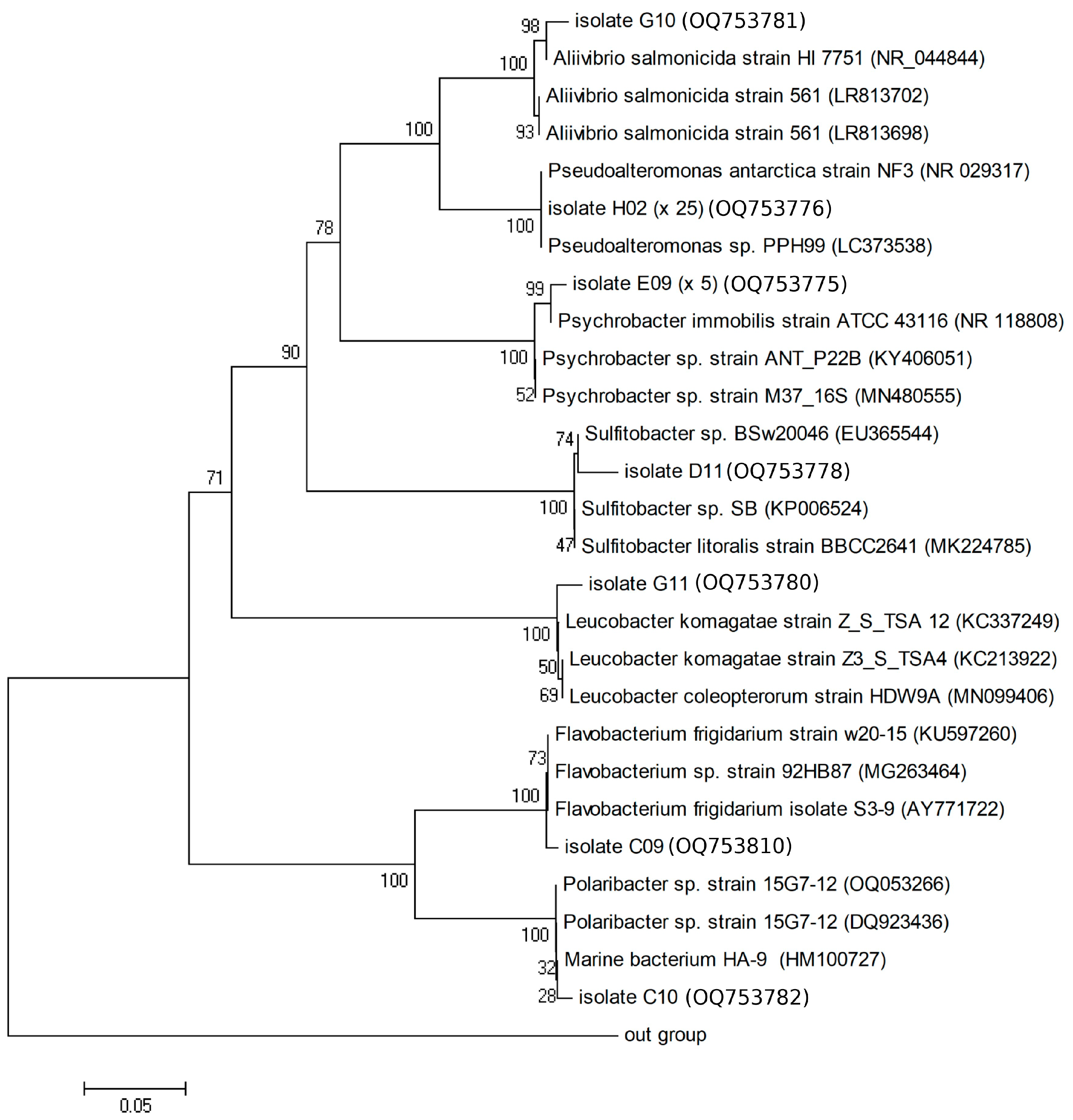
3.5. Taxonomic Identification of the Isolated Bacteria

Thirty-five bacterial strains were obtained by isolation on Marine Agar 2216 medium, and 16S rDNA gene amplicons of each isolate were subject to sequencing. Results of molecular identification are reported in Table 2, where the closest hits are indicated; the phylogenetic tree is shown in Figure 3. In detail, 25 sequences were related to Pseudoalteromonas antarctica strain NF3 (NR 029317, [62]) isolated from an Antarctic coastal environment; 5 sequences were closely related to the psychrophilic species Psychrobacter immobilis ATCC 43116 (NR 118808, [63]); and, finally, 1 sequence showed a high similarity to Aliivibrio salmonicida strain HI 7751 (NR 044844, [64]). Furthermore, one isolate, belonging to Alphaproteobacteria, had a phylogenetic affiliation to Sulfitobacter sp. BSw20046 identified in Arctic seawater [65] or on the Antarctic macroalga surface (JQ618837, [66]). An Actinobacteria-related sequence showed similarity with Leucobacter komagatae strain Z_S_TSA 12 (KC337249, [67]). Two isolates closely related to the Bacteroidetes group showed high similarity to the Flavobacterium frigidarium isolate S3-9 (AY771722, [68]) and to Polaribacter sp. W-2 (DQ923436, [69]).

3.6. Extracellular Enzymatic Activity Rates

The values of enzymatic activity measured in the water samples of the harbour showed for AP the highest hydrolytic rates, followed by LAP and GLU. AP values were below 3.8 nmol L−1 h−1, while GLU ranged around 1 nmol L−1 h−1. In the sediment of the harbour, AP values reached 5.2 nmol g−1 h−1, while comparatively lower LAP activity levels were found (Table 3).

3.7. Enzymatic Expression of Bacterial Isolates

Table 4 shows, in terms of mean percentage values obtained per each sample, the ability of bacterial isolates to decompose organic substrates through the secretion of specific enzymes. The enzymes most frequently produced by the bacterial isolates were lipase and protease, which were expressed on average by 41.7 and 33.3% of the total of water isolates, respectively. In the sediment, these enzymes were produced by 87.5 and 75% of the total of the isolates, respectively. Compared to benthic isolates, bacterial strains isolated from the water column exhibited a more diversified enzymatic spectrum; alkaline phosphatase was synthetised by over 20% of the total, while alpha- and beta-glucosidase as well as esterase were expressed by a lower percentage of bacteria (less than 10% of the total). Conversely, the bacterial strains isolated from the sediment exhibited a preferential utilisation of lipids and proteins, with the majority of the isolates able to decompose the substrates MUF-heptanoate and leucine-MCA, respectively; they were unable to utilise substrates such as maltose (MUF-alpha-glucosidase), cellobiose (MUF-beta-glucosidase), organic phosphates (MUF-phosphate), and acetate (MUF-acetate).

4. Discussion

Given the role of the Kongsfjorden as a harbinger of environmental changes occurring in the Arctic [40], the characterisation of microbial communities is relevant to understand the response of this biological component to environmental changes. The intrinsic peculiarity of a harbour environment makes this a confined environment where anthropogenic activities may cause the most evident effects on the marine biota. Both culture-dependent and -independent techniques were adopted in this study to provide a first characterisation of the microbial community occurring in the Ny-Ålesund harbour, both in pelagic and benthic domains. Data on bacterial abundance, taxonomic composition, and metabolic profiles reported in this study provide new insights on this particular polar habitat. Due to the low number of collected samples, low coverage of the 16S rRNA gene sequencing, and limited number of hydrolytic enzymes measured the obtained results should be treated with caution; as a consequence, the reported data cannot be considered to be representative of the complexity of the microbial community present in this site, preventing the inference of robust conclusions on microbial dynamics. Nevertheless, our research add important information on the abundance, taxonomic composition and metabolic activity of the microbial community.

4.1. Bacterial Abundance in the Ny-Ålesund Harbour

According to the type of the examined sample, total bacterial abundance values varied, with an increase in about two orders of magnitude recorded in the sediment compared to the water samples. Total bacterioplankton abundance in water was about 104 cells mL−1, a quite lower value compared to those reported in Arctic fjords (105–107 cells mL−1 [25,70]), but more similar to values of 1.46–4.26 × 10⁵ cells mL−1 reported by Kalinowska et al. [71] or those of 107–109 cells L−1 found in June–September by Sinha et al. [37]. Bacterial abundance values between 9.34 × 10⁶ and 1.98 × 10⁸ cells mL−1, increasing in during autumn and winter, were found in Kongfjorden sediments close to Ny-Ålesund [33]. Regarding the culturable fraction, average values of 1.1 × 10⁵ CFU g−1 were reported for viable heterotrophic bacteria in the sediments of the Brandalpynten site within Kongsfjorden [38]. The lower values recorded in our study (103 CFU g−1) could be explained by different properties (such as temperature, oxygen content, water mass and light patterns, and nutrient availability) of the overhanging water column or by changes occurring in the sedimentary burial of trophic inputs coming from coastal and riverine supplies. In addition, the presence of anaerobic methylotrophs could determine the low numbers of culturable bacteria, obtained under aerobic conditions. Hydrocarbon-degrading bacteria accounted, on average, for 20 and 10% of the culturable bacteria in seawater and sediment samples, respectively. This low percentage, however, could explained by the inability of obligate hydrocarbon-degrading marine bacteria to grow on the culture media commonly used for total heterotrophic bacteria.

4.2. Taxonomic Composition of the Microbial Community

Many of the sequences retrieved in this study were affiliated to isolates belonging to the group of Gammaproteobacteria. Despite the limited number of analysed sequences, which obviously cannot provide a complete picture of the microbial community of the study area, many of the analysed sequences were affiliated to clones identified in the polar regions of both the Artic and Antarctic [72,73], or the Baltic Sea [74] and Marine Fjord [75]. Interestingly, this tendency was maintained in all microbial groups detected in this clone libraries, regardless of taxonomic groups; this demonstrates that low temperatures seem to select entire microbial communities well defined and adapted to thrive in these conditions.
Similar results, with the predominance of Proteoteobacteria, Bacteroidetes, Firmicutes, and Actinobacteria, were reported in other Arctic sediments [38]; Alpha- and Gammaproteobacteria dominated in surface uncontaminated Kongsfjorden sediments [76]. Nevertheless, different signs of human settlement are highlighted by the presence of different sequences phylogenetically close to Gammaproteobacteria, already isolated in ports [77]. A set of other sequences, belonging to the Methylophilaceae group, were previously detected in biofilm covering an antifouling paint [78] and Bacteroidetes-related sequences detected in contaminated oil or oil processing facilities [79] (Figure 2). Compared to the molecular analysis, indicating that contamination occurred in the harbour, the quantitative determination of total hydrocarbon concentrations in our samples did not provide evidence for contaminated waters; a possible explanation for this controversial finding could be the detection limit of the GC-FID analysis.

4.3. Qualitative and Quantitative Characterisation of Microbial Metabolism

The rates of metabolic activity measured in terms of potential enzymatic activity showed the relative predominance of AP compared to LAP and beta-GLU. The enzymatic values examined in the harbour area fell in a range like that measured by Zappalà et al. [80] in the Svalbard region (min–max range AP: 1.33–10.81 nmol L−1h−1; LAP: 0.15–7.97 nmol L−1h−1 and beta-GLU: 0.09–5.19 nmol L−1h−1). This suggested that microbial metabolism was apparently unaffected by the anthropogenic impact in the harbour.
In natural ecosystems, enzyme activity assays provide useful indications on the microbial processes involved in the recycling of autochthonous and allochthonous organic matter, with consequent release of nutrients in water bodies [3,81]. The high enzyme activity levels measured in our study agreed with the summer season; indeed, temperature and organic matter availability are the main factors that stimulate the synthesis of enzymes, as observed also in temperate environments [3,13,15]. LAP was actively synthesised, suggesting the quick decomposition of proteins mediated by microorganisms. This biochemical trait is frequently observed in microbial communities inhabiting coastal marine environments [82], and a relationship between LAP values and eutrophication levels was observed in harbour environments [3,81]. GLU activity, another key enzyme in the carbon cycle in aquatic environments, was also relevant within the Ny Ålesund harbour, probably depending on the high amount of polysaccharidic substrates (i.e., high-carbohydrate-content organic polymers); a high metabolic versatility of the microbial community characterised the surface sediments of an Arctic fjord, where high-molecular-weight substrates were actively hydrolysed, even if a small fraction of the total community is generally found to express carbohydrate-hydrolysing enzymes [83]. In the waters of the harbour channel in Ustka, the estuarine part of the Stupia River (Poland), high levels of extracellular enzymes were found in the surface microlayer and subsurface water [81]. After lipase, phosphatase (133.3 nM MUF cm−3 h−1) and aminopeptidase (107.7 nM MCA cm−3 h−1) predominated within the enzyme spectra of the microbial community; higher values were detected in the surface microlayer, while no clear spatial distribution patterns were observed. Heterotrophic bacterial abundance, varying between 1.50 and 91.67 × 10⁶ CFU dm−3, was about six orders of magnitude higher than in our study (seawater: 102 CFU mL−1; sediment: 103 CFU g−1). Like what was observed in Ny Ålesund, in a touristic harbour of a temperate region (Rapallo harbour, along the Ligurian coast, Italy), enhanced enzyme activities (LAP and GLU) were detected in sediments (mean LAP and GLU: 125.3 ± 77.6 nmol g−1h−1 and 77.3 ± 3.1 nmol g−1 h−1, respectively) compared to seawater (mean LAP and GLU: 70.3 ± 28.28 nmol L−1h−1 and 10.1 ± 4.4 nmol L−1h−1 respectively) [3]. Proteolytic enzyme values were particularly higher in the benthic domain, reflecting the accumulation of organic matter due to the harbour activities.
Screening of bacterial isolates for the production of enzymes allowed us to identify the presence/absence of specific enzymes in each strain; however, the results of this qualitative assay cannot be compared with the quantitative determination of enzyme activity rates measured by the quantitative fluorimetric assay, which were referred to the whole microbial community. Moreover, variations in enzyme production are known to depend on the cell physiological state [84]. The enzymatic expression of bacterial isolates was affected by their source habitat; indeed, different spectra of enzymatic profiles were observed when comparing water to sediment-derived isolates. Bacteria isolated from the benthic compartment were not able to utilise maltose, cellobiose, organic phosphates, and acetate.
The enzymatic profiles obtained from the qualitative screening of enzymatic activities expressed by each bacterial isolate pointed out the wide expression of lipase and aminopeptidase activities, as suggested by the high percentages of utilisation of MUF-heptanoate and leucine-MCA. Lower percentages of strains were found to express alkaline phosphatase, beta-glucosidase, and esterase enzymes; these results pointed out that the bacterial community present in this Arctic harbour was able to preferentially metabolise lipids and proteins as organic substrates supporting their growth. Similar results were reported in a previous study performed close to a glacier in the same area [80]. The predominance of lipase among the enzymes synthesised by bacteria in the seawater of a harbour channel was attributed to anthropogenic pollution related to tourism and maritime activities as well as to lipidic components of marine organisms such as phytoplankton and zooplankton or detritus, accounting for a consistent fraction (3–55%) of the organic matter [81].

5. Conclusions

This study reports the first known description of the microbial characteristics of the Ny Ålesund harbour, a site where several activities related to research, maritime traffic, and tourism coexist. The obtained findings provide a baseline understanding of the composition and metabolic/functional potential of the microbial community in this polar marine ecosystem that could be monitored in future studies to assess the response of the microbial assemblage to increasing the anthropisation of the Svalbard region.
The phylogenetic analysis of the microbial community revealed that Proteobacteria were the most abundant group among the phyla retrieved in the studied sample. Many of the analysed sequences were affiliated to clones identified in cold areas, in ports, in biofilm-covering structures, in antifouling paints, and in oil-contaminated facilities, providing a signature of human pressure at the sampling site.

Supplementary Materials

The following supporting information can be downloaded at https://www.mdpi.com/article/10.3390/jmse11051018/s1, Table S1. List of the clones isolated with indication of their GenBank accession number, their closest hit with its accession number, and the percentage of identification.

Author Contributions

Conceptualisation, S.C., G.C., M.A.; methodology, all the authors; formal analysis, S.C., G.C., A.L., S.P.; investigation, M.A.; resources, M.A.; data curation, S.C.; writing—original draft preparation, all the authors; writing—review and editing, all the authors; supervision, S.C., F.S., G.C., M.A.; project administration, M.A.; funding acquisition, M.A. All authors have read and agreed to the published version of the manuscript.

Funding

This research was funded by the Italian Ministry of Education, University and Research (MIUR), project Arctic: present Climatic change and pAst extreme events (ARCA).

Institutional Review Board Statement

Not applicable.

Informed Consent Statement

Not applicable.

Data Availability Statement

Data are available upon request.

Acknowledgments

The authors would like to express their gratitude to the captain and crew of the r/v Teisten for the logistical support during the collection of water and sediment samples.

Conflicts of Interest

The authors declare no conflict of interest. The funders had no role in the design of the study; in the collection, analyses, or interpretation of data; in the writing of the manuscript; or in the decision to publish the results.

References

  1. Wong, Y.S.; Tam, N.F.Y.; Lau, P.S.; Xue, X.Z. The toxicity of marine sediments in Victoria Harbour, Hong Kong. Mar. Pollut. Bull. 1995, 31, 464–470. [Google Scholar] [CrossRef]
  2. Fabiano, M.; Marrale, D.; Misic, C. Bacteria and organic matter dynamics during a bioremediation treatment of organic-rich harbour sediments. Mar. Poll. Bull. 2003, 46, 1164–1173. [Google Scholar] [CrossRef] [PubMed]
  3. Misic, C.; Covazzi Harriague, A. Organic matter characterisation and turnover in the sediment and seawater of a tourist harbour. Mar. Environ. Res. 2009, 68, 227–235. [Google Scholar] [CrossRef] [PubMed]
  4. Danulat, E.; Muniz, P.; Garcia-Alonso, J.; Yannicelli, B. First assessment of the highly contaminated harbour of Montevideo, Uruguay. Mar. Poll. Bull. 2002, 44, 554–565. [Google Scholar] [CrossRef] [PubMed]
  5. Guerra-García, J.M.; García-Gómez, J.C. Oxygen levels versus chemical pollutants: Do they have similar influence on macrofaunal assemblages? A case study in a harbour with two opposing entrances. Environ. Pollut. 2005, 135, 281–291. [Google Scholar] [CrossRef]
  6. Todorova, N.H.; Mironova, R.S.; Karamfilov, V.K. Comparative molecular analysis of bacterial communities inhabiting pristine and polluted with polycyclic aromatic hydrocarbons Black Sea coastal sediments. Mar. Pollut. Bull. 2014, 83, 231–240. [Google Scholar] [CrossRef]
  7. Caruso, G.; Giacobbe, M.G.; Azzaro, F.; Decembrini, F.; Leonardi, M.; Miserocchi, S.; Cao, X.; Song, C.; Zhou, Y. All-In-One: Microbial Response to Natural and Anthropogenic Forcings in a Coastal Mediterranean Ecosystem, the Syracuse Bay (Ionian Sea, Italy). J. Mar. Sci. Eng. 2022, 10, 19. [Google Scholar] [CrossRef]
  8. Blanchard, A.L.; Feder, H.M.; Shaw, D.G. Variations in benthic fauna underneath an effluent mixing zone at a marine oil terminal in Port Valdez, Alaska. Mar. Pollut. Bull. 2003, 46, 1583–1589. [Google Scholar] [CrossRef]
  9. Je, J.G.; Belan, T.; Levings, C.; Koo, B.J. Changes in benthic communities along a presumed pollution gradient in Vancouver Harbour. Mar. Environ. Res. 2003, 57, 121–135. [Google Scholar] [CrossRef]
  10. Caplat, C.; Texier, H.; Barillier, D.; Lelievre, C. Heavy metals mobility in harbour contaminated sediments: The case of Port-en-Bessin. Mar. Pollut. Bull. 2005, 50, 504–511. [Google Scholar] [CrossRef]
  11. Camphuysen, C.J. IFAW Chronic Oil Pollution in Europe. Status Report 2006; Royal Netherlands Institute for Sea Research: Texel, The Netherlands, 2007; pp. 1–88. [Google Scholar]
  12. Mali, M.; Dell’Anna, M.M.; Mastrorilli, P.; Damiani, L.; Ungaro, N.; Marinski, J.; Korsachka, M. Integrated Assessment of the Quality of Harbor Sediments: Case Study Based on a Comparative Analysis of Sediments Quality of Two Industrial Ports: Bourgas (BG) and Bari (IT). In Sustainable Development of Sea-Corridors and Coastal Waters; Stylios, C., Floqi, T., Marinski, J., Damiani, L., Eds.; Springer International Publishing: Cham, Switzerland, 2015; Chapter 1; pp. 1–20. [Google Scholar]
  13. Zaccone, R.; Caruso, G. Microbial enzymes in the Mediterranean Sea: Relationship with climate changes. AIMS Microbiol. 2019, 5, 251–271. [Google Scholar] [CrossRef] [PubMed]
  14. Wessels Perelo, L. Review: In situ and bioremediation of organic pollutants in aquatic sediments. J. Haz. Mat. 2010, 177, 81–89. [Google Scholar] [CrossRef] [PubMed]
  15. Misic, C.; Castellano, M.; Covazzi Harriague, A. Organic matter features, degradation and remineralisation at two coastal sites in the Ligurian Sea (NW Mediterranean) differently influenced by anthropogenic forcing. Mar. Environ. Res. 2011, 72, 67–74. [Google Scholar] [CrossRef] [PubMed]
  16. Ford, T.E. Response of marine microbial communities to anthropogenic stress. J. Aquat. Ecosys. Stress Recov. 2000, 7, 75–89. [Google Scholar] [CrossRef]
  17. Ford, T.; Jay, J.; Patel, A.; Kile, M.; Prommasith, P.; Galloway, T.; Sanger, R.; Smith, K.; Depledge, M. Use of Ecotoxicological Tools to Evaluate the Health of New Bedford Harbor Sediments: A Microbial Biomarker Approach. Environ. Health Perspect. 2005, 113, 186–191. [Google Scholar] [CrossRef]
  18. Caruso, G.; Azzaro, M.; Caroppo, C.; Decembrini, F.; Monticelli, L.S.; Leonardi, M.; Maimone, G.; Zaccone, R.; La Ferla, R. Microbial community and its potential as descriptor of environmental status. ICES J. Mar. Sci. 2016, 73, 2174–2177. [Google Scholar] [CrossRef]
  19. Guibert, L.M.; Loviso, C.L.; Borglin, S.; Jansson, J.K.; Dionisi, H.; Lozada, M. Diverse Bacterial Groups Contribute to the Alkane Degradation Potential of Chronically Polluted Subantarctic Coastal Sediments. Microb. Ecol. 2016, 71, 100–112. [Google Scholar] [CrossRef]
  20. Dash, H.R.; Mangwani, N.; Chakraborty, J.; Kumari, S.; Das, S. Marine bacteria: Potential candidates for enhanced bioremediation. Appl. Microbiol. Biotechnol. 2013, 97, 561–571. [Google Scholar] [CrossRef]
  21. Lo Giudice, A.; Caruso, G.; Rizzo, C.; Papale, M.; Azzaro, M. Bacterial communities versus anthropogenic disturbances in the Antarctic coastal marine environment. Environ. Sustain. 2019, 2, 297–310. [Google Scholar] [CrossRef]
  22. Varotsos, C.A.; Krapivin, V.F. Pollution of Arctic Waters Has Reached a Critical Point: An Innovative Approach to This Problem. Water Air Soil Pollut. 2018, 229, 343. [Google Scholar] [CrossRef]
  23. Pouch, A.; Zaborska, A.; Mazurkiewicz, M.; Winogradow, A.; Pazdro, K. PCBs, HCB and PAHs in the seawater of Arctic fjords—Distribution, sources and risk assessment. Mar. Pollut. Bull. 2021, 164, 11980. [Google Scholar] [CrossRef] [PubMed]
  24. Bargiela, R.; Mapelli, F.; Rojo, D.; Chouaia, B.; Tornés, J.; Borin, S.; Richter, M.; Del Pozo, M.V.; Cappello, S.; Gertler, C.; et al. Bacterial population and biodegradation potential in chronically crude oil-contaminated marine sediments are strongly linked to temperature. Sci. Rep. 2015, 5, 11651. [Google Scholar] [CrossRef] [PubMed]
  25. Caruso, G.; Madonia, A.; Bonamano, S.; Miserocchi, S.; Giglio, F.; Maimone, G.; Azzaro, F.; Decembrini, F.; La Ferla, R.; Piermattei, V.; et al. Microbial abundance and enzyme activity patterns: Response to changing environmental characteristics along a transect in Kongsfjorden (Svalbard Islands). J. Mar. Sci. Eng. 2020, 8, 824. [Google Scholar] [CrossRef]
  26. Papale, M.; Caruso, G.; Maimone, G.; La Ferla, R.; Lo Giudice, A.; Rappazzo, A.C.; Cosenza, A.; Azzaro, F.; Ferretti, R.; Paranhos, R.; et al. Microbial Community Abundance and Metabolism Close to the Ice-Water Interface of the Blomstrandbreen Glacier (Kongsfjorden, Svalbard): A Sampling Survey Using an Unmanned Autonomous Vehicle. Water 2023, 15, 556. [Google Scholar] [CrossRef]
  27. Cottier, F.; Tverberg, V.; Inall, M.; Svendsen, H.; Nilsen, F.; Griffiths, C. Water mass modification in an Arctic fjord through cross-shelf exchange: The seasonal hydrography of Kongsfjorden, Svalbard. J. Geophys. Res. Ocean. 2005, 110, C12005. [Google Scholar] [CrossRef]
  28. Hop, H.; Pearson, T.; Nøst Hegseth, E.; Kovacs, K.M.; Wiencke, C.; Kwasniewski, S.; Eiane, K.; Mehlum, F.; Gulliksen, B.; Wlodarska-Kowalczuk, M.; et al. The marine ecosystem of Kongsfjorden, Svalbard. Polar Res. 2002, 21, 167–208. [Google Scholar] [CrossRef]
  29. Pedersen, Å.Ø.; Convey, P.; Newsham, K.K.; Mosbacher, J.B.; Fuglei, E.; Ravolainen, V.; Hansen, B.B.; Jensen, T.C.; Augusti, A.; Biersma, E.M.; et al. Five decades of terrestrial and freshwater research at Ny-Ålesund, Svalbard. Polar Res. 2022, 41, 1–39. [Google Scholar] [CrossRef]
  30. Piquet, A.M.-T.; Scheepens, J.F.; Bolhuis, H.; Wiencke, C.; Buma, A.G.J. Variability of protistan and bacterial communities in two Arctic fjords (Spitsbergen). Polar Biol. 2010, 33, 1521–1536. [Google Scholar] [CrossRef]
  31. Iversen, K.R.; Seuthe, L. Seasonal microbial processes in a high-latitude fjord (Kongsfjorden, Svalbard): I. Heterotrophic bacteria, picoplankton and nanoflagellates. Polar Biol. 2011, 34, 731–749. [Google Scholar] [CrossRef]
  32. Prasad, S.; Manasa, P.; Buddhi, S.; Tirunagari, P.; Begum, Z.; Rajan, S.; Shivaji, S. Diversity and Bioprospective Potential (Cold-Active Enzymes) of Cultivable Marine Bacteria from the Subarctic Glacial Fjord, Kongsfjorden. Curr. Microbiol. 2014, 68, 233–238. [Google Scholar] [CrossRef]
  33. Bourgeois, S.; Kerhervé, P.; Calleja, M.L.; Many, G.; Morata, N. Glacier inputs influence organic matter composition and prokaryotic distribution in a high Arctic fjord (Kongsfjorden, Svalbard). J. Mar. Syst. 2016, 164, 112–127. [Google Scholar] [CrossRef]
  34. Jain, A.; Krishnan, K.P. Differences in free-living and particle-associated bacterial communities and their spatial variation in Kongsfjorden, Arctic. J. Basic Microbiol. 2017, 57, 827–838. [Google Scholar] [CrossRef] [PubMed]
  35. Salam, S.; Lekshmi, S.; Silvester, R.; Krishnan, K.P.; Saramma, A.V.; Hatha, A.A.M. Effect of environmental factors on growth and enzyme production of cold adapted bacteria from water and sediment of Kongsfjord, Ny-Alesund, Arctic. J. Environ. Biol. 2017, 38, 579–585. [Google Scholar] [CrossRef]
  36. Sinha, R.K.; Krishnan, K.P.; Hatha, A.A.M. Diversity of retrievable heterotrophic bacteria in Kongsfjorden, an Arctic fjord. Braz. J. Microbiol. 2017, 48, 51–61. [Google Scholar] [CrossRef]
  37. Sinha, R.K.; Krishnan, K.P.; Kerkar, S.; David, T.D. Spatio-Temporal Monitoring and Ecological Significance of Retrievable Pelagic Heterotrophic Bacteria in Kongsfjorden, an Arctic Fjord. Indian J. Microbiol. 2017, 57, 116–120. [Google Scholar] [CrossRef]
  38. Conte, A.; Papale, M.; Amalfitano, S.; Mikkonen, A.; Rizzo, C.; De Domenico, E.; Michaud, L.; Lo Giudice, A. Bacterial community structure along the subtidal sandy sediment belt of a high Arctic fjord (Kongsfjorden, Svalbard Islands). Sci. Total Environ. 2018, 619–620, 203–211. [Google Scholar] [CrossRef]
  39. Fang, X.M.; Zhang, T.; Li, J.; Wang, N.-F.; Wang, Z.; Yu, L.-Y. Bacterial community pattern along the sediment seafloor of the Arctic fjorden (Kongsfjorden, Svalbard). Antonie Van Leeuwenhoek 2019, 112, 1121–1136. [Google Scholar] [CrossRef]
  40. Bischof, K.; Convey, P.; Duarte, P.; Gattuso, J.-P.; Granberg, M.E.; Hop, H.; Hoppe, C.; Jiménez, C.; Lisitsyn, L.; Martinez, B.; et al. Kongsfjorden as harbinger of the future Arctic: Knowns, unknowns and research priorities. In The Ecosystem of Kongsfjorden, Svalbard; Hop, H., Wiencke, C., Eds.; Springer Nature: Cham, Switzerland, 2019; pp. 537–562. [Google Scholar]
  41. Srinivas, T.N.R.; Rao, S.; Reddy, P.V.V.; Pratibha, M.S.; Sailaja, B.; Kavya, B.; Kishore, K.H.; Begum, Z.; Singh, S.M.; Shivaji, S. Bacterial diversity and bioprospecting for cold-active lipases, amylases and proteases, from culturable bacteria of Kongsfjorden and Ny-Alesund, Svalbard, Arctic. Curr. Microbiol. 2009, 59, 537–547. [Google Scholar] [CrossRef]
  42. Thomas, F.A.; Mohan, M.; Krishnan, K.P. Bacterial diversity and their metabolic profiles in the sedimentary environments of Ny-Ålesund, Arctic. Antonie Van Leeuwenhoek 2021, 114, 1339–1360. [Google Scholar] [CrossRef]
  43. Venkatachalam, S.; Gopinath, A.; Krishnan, K.P. Fjords of the western and northern regions of Svalbard harbour distinct bacterioplankton community structures. World J. Microbiol. Biotechnol. 2023, 39, 57. [Google Scholar] [CrossRef]
  44. Dowdall, M.; Vicat, K.; Frearson, I.; Gerland, S.; Lind, B.; Shaw, G. Assessment of the radiological impacts of historical coal mining operations on the environment of Ny-Ålesund, Svalbard. J. Environ. Radioact. 2004, 71, 101–114. [Google Scholar] [CrossRef] [PubMed]
  45. Crisafi, F.; Giuliano, L.; Yakimov, M.M.; Az.zaro, M.; Denaro, R. Isolation and degradation potential of a cold-adapted oil/PAH degrading marine bacterial consortium from Kongsfjorden (Arctic region). Rend. Fis. Acc. Lincei 2016, 27 (Suppl. S1), S261–S270. [Google Scholar] [CrossRef]
  46. Genovese, M.; Crisafi, F.; Denaro, R.; Cappello, S.; Russo, D.; Calogero, R.; Santisi, S.; Catalfamo, M.; Modica, A.; Smedile, F.; et al. Effective bioremediation strategy for rapid in situ cleanup of anoxic marine sediments in mesocosm oil spill simulation. Front. Microbiol. 2014, 5, 162. [Google Scholar] [CrossRef]
  47. Denaro, R.; D’Auria, G.; Di Marco, G.; Genovese, M.; Troussellier, M.; Yakimov, M.M.; Giuliano, L. Assessing terminal restriction fragment length polymorphism suitability for the description of bacterial community structure and dynamics in hydrocarbon-polluted marine environments. Environ. Microbiol. 2005, 7, 78–87. [Google Scholar] [CrossRef] [PubMed]
  48. Cappello, S.; Caruso, G.; Zampino, D.; Monticelli, L.S.; Maimone, G.; Denaro, R.; Tripodo, B.; Troussellier, M.; Yakimov, M.M.; Giuliano, L. Microbial community dynamics during assays of harbor oil spill bioremediation: A microscale simulation study. J. Appl. Microbiol. 2007, 102, 184–194. [Google Scholar] [CrossRef] [PubMed]
  49. Cappello, S.; Calogero, R.; Santisi, S.; Genovese, M.; Denaro, R.; Genovese, L.; Giuliano, L.; Mancini, G.; Yakimov, M.M. Bioremediation of oil polluted marine sediments: A bio-engineering treatment. Int. Microbiol. 2015, 18, 127–134. [Google Scholar] [CrossRef] [PubMed]
  50. Kuwae, T.; Hosokawa, Y. Determination of abundance and biovolume of bacteria in sediments by dual staining with 4′,6-diamidino-2-phenylindole and acridine orange: Relationship to dispersion treatment and sediment characteristics. Appl. Environ. Microbiol. 1999, 65, 3407–3412. [Google Scholar] [CrossRef]
  51. Cappello, S.; Caruso, G.; Bergami, E.; Macrì, A.; Venuti, V.; Majolino, D.; Corsi, I. New insights into the structure and function of the prokaryotic communities colonizing plastic debris collected in King George Island (Antarctica): Preliminary observations from two plastic fragments. J. Hazard. Mater. 2021, 414, 125586. [Google Scholar] [CrossRef]
  52. Brown, E.J.; Braddock, J.F. Sheen screen, a miniaturized most-probable-number method for enumeration of oil-degrading microorganisms. Appl. Environ. Microbiol. 1990, 56, 3895–3896. [Google Scholar] [CrossRef]
  53. American Public Health Association (APHA). Standard Methods for the Examination of Water and Waste Water, 18th ed.; APHA: Washington, DC, USA, 1992; pp. 1–1100. [Google Scholar]
  54. Lane, D.J. 16S/23S rRNA sequencing. In Nucleic Acid Techniques in Bacterial Systematics; Stackerbrandt, E., Goodfellow, M., Eds.; Wiley: New York, NY, USA, 1991; pp. 115–175. [Google Scholar]
  55. Hoppe, H.-G. Use of fluorogenic model substrate for extracellular enzyme activity (EEA) measurement of bacteria. In Handbook of Methods in Aquatic Microbial Ecology; Kemp, P.F., Sherr, B.F., Sherr, E.B., Cole, J.J., Eds.; Lewis Publishers: Boca Raton, FL, USA, 1993; pp. 423–431. [Google Scholar]
  56. La Ferla, R.; Azzaro, M.; Caruso, G.; Monticelli, L.S.; Maimone, G.; Zaccone, R.; Packard, T.T. Prokaryotic abundance and heterotrophic metabolism in the deep Mediterranean Sea. Adv. Oceanogr. Limnol. 2010, 1, 143–166. [Google Scholar] [CrossRef]
  57. Ashelford, K.E.; Chuzhanova, N.A.; Fry, J.C.; Jones, A.J.; Weightman, A.J. At least 1 in 20 16S rRNA sequence records currently held in public repositories is estimated to contain substantial anomalies. Appl. Environ. Microbiol. 2005, 71, 7724–7736. [Google Scholar] [CrossRef] [PubMed]
  58. Altschul, S.F.; Gish, W.; Miller, W.; Myers, E.W.; Lipman, D.J. Basic local alignment search tool. J. Mol. Biol. 1990, 215, 403–410. [Google Scholar] [CrossRef]
  59. Pruesse, E.; Quast, C.; Knittel, K.; Fuchs, B.M.; Ludwig, W.; Peplies, J.; Glöckner, F.O. SILVA: A comprehensive online resource for quality checked and aligned ribosomal RNA sequence data compatible with ARB. Nucleic Acids Res. 2007, 35, 7188–7196. [Google Scholar] [CrossRef] [PubMed]
  60. Ludwig, W.; Strunk, O.; Westram, R.; Richter, L.; Meier, H.; Yadhukumar Buchner, A.; Lai, T.; Steppi, S.; Jobb, G.; Förster, W.; et al. ARB: A software environment for sequence data. Nucleic Acids Res. 2004, 32, 1363–1371. [Google Scholar] [CrossRef]
  61. Caruso, G.; Dell’Acqua, O.; Caruso, R.; Azzaro, M. Phenotypic characterization of bacterial isolates from marine waters and plastisphere communities of the Ross Sea (Antarctica). J. Clin. Microbiol. Biochem. Technol. 2022, 8, 1–9. [Google Scholar] [CrossRef]
  62. Bozal, N.; Tudela, E.; Rossello-Mora, R.; Lalucat, J.; Guinea, J. Pseudoalteromonas antarctica sp. nov., isolated from an Antarctic coastal environment. Int. J. Syst. Bacteriol. 1997, 47, 345–351. [Google Scholar] [CrossRef] [PubMed]
  63. Juni, E.; Heym, G.A. Psychrobacter immobilis gen. nov., sp. nov.: Genospecies composed of gram-negative, aerobic, oxidase-positive Coccobacilli. Int. J. Syst. Bacteriol. 1986, 36, 388–391. [Google Scholar] [CrossRef]
  64. Egidius, E.; Wiik, R.; Andersen, K.; Hoff, K.A.; Hjeltnes, B. Vibrio salmonicida sp. nov., a new fish pathogen. Int. J. Syst. Bacteriol. 1986, 36, 518–520. [Google Scholar] [CrossRef]
  65. Ji, Q.; Yu, Y.; Chen, B. Bacteria Isolated from Arctic Seawater; direct submission to GenBank database 23 December 2007; Polar Research Institute of China, Biology Department: Shangai, China, 2007; Unpublished data. [Google Scholar]
  66. Barreto, C.C.; Lima, D.V.; de Oliveira, E.C.; Absher, T.M.; Pellizari, V.H. Species-Specific Microbiota Associated with Antarctic Macroalgae; direct submission to GenBank database 3 February 2012; Universidade de Sao Paulo, Instituto de Ciencias Biomedicas: Sao Paulo, Brazil, 2012; Unpublished data. [Google Scholar]
  67. Skrodenyte-Arbaciauskiene, V.; Pliuraite, V.; Virbickas, T.; Bartasiute, A. Intestinal Microbial Community of Roach (Rutilus rutilus) and Its Changes under the Impact of River Pollution; direct submission to GenBank database 10 December 2012; Institute of Ecology, Nature Research Centre: Vilnius, Lithuania, 2012; Unpublished data. [Google Scholar]
  68. Lee, H.K.; Sung, K.C.; Cho, H.H.; Lee, Y.K. Diversity of Arctic Bacteria; direct submission to GenBank database 5 October 2004; Korea Polar Research Institute (KOPRI), Microbiology Lab.: Ansan, Korea, 2004; Unpublished data. [Google Scholar]
  69. Lin, X.; Yi, D.; Wang, C. Investigation of Oceanic Microbial Diversity in Qingdao Fushan Bay; direct submission to GenBank datahase 28 August 2006; First Institute of Oceanography (SOA), Key Lab of Marine Bioactive Substances: Qingdao, China, 2006; Unpublished data.
  70. Guo, C.Y.; He, J.F.; Zhang, F.; Cai, M.H.; Eang, G.Z. Spatial heterogeneity of a microbial community in Kongsfjorden, Svalbard during late summer 2006 and its relationship to biotic and abiotic factors. Adv. Polar Sci. 2011, 22, 55–66. [Google Scholar] [CrossRef]
  71. Kalinowska, A.; Ameryk, A.; Jankowska, K. Microbiological Survey in Two Arctic Fjords: Total Bacterial Number and Biomass Comparison of Hornsund and Kongsfjorden. In Impact of Climate Changes on Marine Environments; Zielinski, T., Weslawski, M., Kulinski, K., Eds.; GeoPlanet: Earth and Planetary Sciences; Springer: Cham, Switzerland, 2015. [Google Scholar]
  72. Vishnupriya, S.; Jabir, T.; Krishnan, K.P.; Hatha, A.A.M. Bacterial community structure and functional profiling of high Arctic fjord sediments. World J. Microbiol. Biotechnol. 2021, 37, 133. [Google Scholar] [CrossRef]
  73. Brinkmeyer, R.; Knittel, K.; Jurgens, J.; Weyland, H.; Amann, R.; Helmke, E. Diversity and structure of bacterial communities in Arctic versus Antarctic pack ice. Appl. Environ. Microbiol. 2003, 69, 6610–6619. [Google Scholar] [CrossRef] [PubMed]
  74. Allers, E.; Wright, J.J.; Konwar, K.M.; Howes, C.G.; Beneze, E.; Hallam, S.J.; Sullivan, M.B. Diversity and population structure of Marine Group A bacteria in the Northeast subarctic Pacific Ocean. ISME J. 2013, 7, 256–268. [Google Scholar] [CrossRef] [PubMed]
  75. von Scheibner, M.; Dorge, P.; Biermann, A.; Sommer, U.; Hoppe, H.G.; Jurgens, K. Impact of warming on phyto-bacterioplankton coupling and bacterial community composition in experimental mesocosms. Environ. Microbiol. 2014, 16, 718–733. [Google Scholar] [CrossRef] [PubMed]
  76. Zaikova, E.; Walsh, D.A.; Stilwell, C.P.; Mohn, W.W.; Tortell, P.D.; Hallam, S.J. Microbial community dynamics in a seasonally anoxic fjord: Saanich Inlet, British Columbia. Environ. Microbiol. 2010, 12, 172–191. [Google Scholar] [CrossRef] [PubMed]
  77. Paissé, S.; Coulon, F.; Goni-Urriza, M.; Peperzak, L.; McGenity, T.J.; Duran, R. Structure of bacterial communities along a hydrocarbon contamination gradient in a coastal sediment. FEMS Microbiol. Ecol. 2008, 66, 295–305. [Google Scholar] [CrossRef]
  78. Guo, F.; School of Life Sciences, Xiamen University, Xiamen, China; Ke, C.-H.; State Key Laboratory of Marine Environmental Science, Xiamen University, Xiamen, China; Huang, Z.-B.; State Key Laboratory of Marine Environmental Science, Xiamen University, Xiamen, China. Bacteria on the Surface of Antifouling Paint Immersed in the Estuary Zone of Jiulong River, Xiamen University, Xiamen, China. Personal communication, 2012. [Google Scholar]
  79. Duncan, K.E.; Gieg, L.M.; Parisi, V.A.; Tanner, R.S.; Tringe, S.G.; Bristow, J.; Suflita, J.M. Biocorrosive Thermophilic Microbial Communities in Alaskan North Slope Oil Facilities. Environ. Sci. Technol. 2009, 43, 7977–7984. [Google Scholar] [CrossRef]
  80. Zappalà, G.; Bruzzone, G.; Caruso, G.; Azzaro, M. Development of an automatic sampler for extreme polar environments: First in situ application in Svalbard Islands. Rend. Fis. Acc. Lincei 2016, 27, 251–259. [Google Scholar] [CrossRef]
  81. Perlinski, P.; Mudryk, Z.J.; Antonowicz, J. Enzymatic activity in the surface microlayer and subsurface water in the harbour channel. Estuar. Coast. Shelf Sci. 2017, 196, 150–158. [Google Scholar] [CrossRef]
  82. Obayashi, Y.; Suzuki, S. Proteolytic enzymes in coastal surface seawater: Significant activity of endopeptidases and exopeptidases. Limnol. Oceanogr. 2008, 50, 722–726. [Google Scholar] [CrossRef]
  83. Arnosti, C. Microbial Extracellular Enzymes and the Marine Carbon Cycle. Ann. Rev. Mar. Sci. 2011, 3, 401–425. [Google Scholar] [CrossRef]
  84. Hughes, S.G. Variations in Enzyme Production as a Function of the Physiological State of Bacteria. Master’s Thesis, University of Edinburgh, Edinburgh, UK, 1971; pp. 1–143. [Google Scholar]
Figure 1. Location of the sampling sites at Ny Ålesund.
Jmse 11 01018 g001
Figure 2. Rooted phylogenetic tree clustered by neighbour-joining of maximum likelihood values showing the affiliation of partial bacterial 16S rRNA gene sequences to closest-related sequences from members of different bacterial clusters. Isolates obtained in the present study are indicated in red bold type. Percentages of 1000 bootstrap resampling that supported the branching orders in each analysis are shown above or near the relevant nodes (only values P50% are shown). The number within the parenthesis indicates the number of times that gene has been detected in the analysis. A sequence of an uncultured archaeon clone Arch_AE_G08 (FJ968062) was used as an out-group.
Jmse 11 01018 g002
Figure 3. Rooted phylogenetic tree clustered by neighbour-joining of the maximum likelihood values showing the affiliation of bacterial isolates to the closest related sequences. Percentages of 1000 bootstrap resampling that supported the branching orders in each analysis are shown above or near the relevant nodes (only values P50% are shown). A sequence of an uncultured archaeon clone Arch_AE_G08 (FJ968062) was used as an out-group.
Jmse 11 01018 g003
Table 1. Mean ± standard error (from n = 3 replicates) of total bacterial abundance obtained by microscopic counts after DAPI staining (cell mL−1/g−1), heterotrophic bacterial counts obtained by culture on Marine Agar medium (CFU mL−1/g−1), and hydrocarbon-degrading bacteria estimated by most probable number (MPN mL−1/g−1) in the examined samples.
SW1SW2SED1
DAPI (CFU mL−1/g−1)1.60 × 104 ± 1.52 × 1032.50 × 104 ± 3.00 × 1031.12 × 106 ± 1.1 × 10⁵
Culturable (CFU mL−1/g−1)4.90 × 102 ± 4.6 × 1016.10 × 102 ± 5.8 × 1012.1 × 103 ± 1.80 × 10²
Hydrocarbon-degrading bacteria (MPN mL−1/g−1)1.00 × 102 ± 8.00 × 1011.30 × 102 ± 1.9 × 1012.10 × 102 ± 0.25 × 101
Table 2. Outputs of the sequencing of the 16S rDNA gene amplicons of each bacterial isolate. Reported are the length of the fragment (in base pairs) and the closest hits, with an indication of the GenBank accession numbers and percentage of identification.
GenBank Accession NumberIsolate CodeLength (bp)Closest HitAccession No.ID, %
OQ753781G10780Aliivibrio salmonicida strain HI 7751NR_04484499.3
OQ753776H02840Pseudoalteromonas antarctica strain NF3NR_02931798.7
OQ753775E09568Psychrobacter immobilis strain ATCC 43116NR_11880899.1
OQ753778D11630Sulfitobacter sp. BSw20046EU36554498.1
OQ753810C09590Flavobacterium frigidarium isolate S3-9AY77172298.9
OQ753782C10880Polaribacter sp. W-2DQ92343698.1
OQ753780G11680Leucobacter komagatae strain Z_S_TSA 12KC33724998.2
Table 3. Mean ± standard error (from n = 3 replicates) of enzymatic activity rates (alkaline phosphatase, AP; beta-glucosidase, GLU; and leucine aminopeptidase, LAP) measured in the seawater (SW1 and SW2) and sediment (SED1) samples.
LAPGLUAP
SW1 (nmol L−1 h−1)1.40 ± 0.131.1 ± 0.102.7 ± 0.3
SW2 (nmol L−1 h−1)1.50 ± 0.141.30 ± 0.143.8 ± 0.41
SED (nmol g−1 h−1)2.90 ± 3.03.50 ± 0.325.2 ± 0.55
Table 4. Mean percentage of utilisation of organic polymers by the bacterial isolates in the examined seawater and sediment isolates.
SW 1SW2SED1
No. of isolates12128
Alpha-glucosidase0.0 ± 0.09.2 ± 0.80.0 ± 0.0
Beta-glucosidase18.2 ± 1.618.1 ± 2.00.0 ± 0.0
Protease33.1 ± 3.533.4 ± 3.476.0 ± 8.0
Phosphatase33.0 ± 2.833.2 ± 2.90.0 ± 0.0
Lipase24.2 ± 2.258.1 ± 0.686.1 ± 0.9
Esterase0.0 ± 0.09.0 ± 0.90.0 ± 0.0
Disclaimer/Publisher’s Note: The statements, opinions and data contained in all publications are solely those of the individual author(s) and contributor(s) and not of MDPI and/or the editor(s). MDPI and/or the editor(s) disclaim responsibility for any injury to people or property resulting from any ideas, methods, instructions or products referred to in the content.

