Review

MicroRNA-Mediated Responses: Adaptations to Marine Extreme Environments

Ecosustainable Marine Biotechnology Department, Stazione Zoologica Anton Dohrn, Via Acton 55, 80133 Naples, Italy
*
Author to whom correspondence should be addressed.
J. Mar. Sci. Eng. 2023, 11(2), 361; https://doi.org/10.3390/jmse11020361
Submission received: 9 January 2023 / Revised: 25 January 2023 / Accepted: 27 January 2023 / Published: 5 February 2023

Abstract

:
Extreme environments are characterized by peculiar conditions, such as hypoxia/anoxia, freezing/heat temperatures, and desiccation. With climate change, more and more habitats are facing extreme conditions and living communities are finding ways to adapt in order to survive. In this study, we show several species which have been shown to adapt to marine extreme conditions also via miRNA-mediated responses. miRNAs are a class of small non-coding RNAs that mediate gene regulation via interactions with transcripts. Their action can directly or indirectly regulate pathways that can result in a response to a specific condition. Furthermore, the study of these miRNA-mediated responses could help in the biotechnological field for their application in the development of environmental biomarkers of stress conditions, or in the genetic engineering of algal species for the production of high-value compounds.

1. Introduction

In an anthropocentric point of view, “extreme” is the furthest living condition with respect to our tolerance range. Therefore, we define “extreme” as an environment in which physical (temperature, pressure, radiation, etc.), geochemical (desiccation, salinity, redox potential, etc.), and biological (predation levels, population density levels, low nutrition, etc.) factors are at the limits of a normal range, taking into consideration typical model organisms like Escherichia coli or humans [1,2]. Examples of extreme environments are hydrothermal vents, deep sea, hot springs, anoxic environments, etc. In addition, climate changes play a key role in the creation of constantly changing habitats [3] that are challenging the inhabitant species, influencing their distribution and biodiversity, as well as abiotic conditions such as temperature, pH, salinity, oxygen levels, and many others [4,5].
These extreme environments are full of life, ruled by organisms called extremophiles. Firstly, it was thought they were only micro-organisms [6], but soon after, all domains of life were discovered to live under these conditions. For example, these organisms are referred to as thermophiles when living at high temperatures; psychrophiles when living under frozen ice; piezophiles when living under high pressures; or acidophiles when living in highly acidic habitats [7]. Historically, there was little knowledge about these organisms due to the inaccessibility of their habitats, but, nowadays, with the advent of new sampling technologies (e.g., Remotely Operated Vehicles, ROVs, for deep sea samplings) much more information is available. New research methodologies (e.g., metagenomics, high-throughput analyses, etc.) have been developed as well, in order to obtain higher- quality data starting from low-quantity sample materials. The study of extremophiles is very important in many scientific fields because these environments have persistent evolutive pressures that make living organisms continuously adapt and develop novel strategies to survive. For instance, extreme temperatures lead to the denaturation of biomolecules on one side, and the formation of ice crystals in the cell on the other. In order to overcome these conditions, organisms have evolved different strategies such as the production of thermostable enzymes, such as saccharifying amylolytic enzymes (active at ~90 °C) in Archaea [8], or the accumulation of glycerol as an antifreeze in Osmerus mordax [9]. Another limiting factor is pressure that challenges organisms by forcing volumes to change, compressing lipids, and resulting in the destabilization of the cell membranes [10]. Piezophiles overcome it via changing the structure of their biomolecules [11]. Additionally, one of the most extreme conditions is hypoxia, i.e., the absence or very low concentrations of oxygen. This feature characterizes many environments, from the deep sea to high-altitude regions, but it can also interest temporarily some habitats, such as lakes during eutrophication events or intertidal ponds during desiccation. Furthermore, when hypoxia is related to aquatic environments, it refers to the availability of dissolved oxygen, which is influenced by depth, pressure, temperature, and salinity of water [12]. Generally, organisms have adapted to hypoxia/anoxia conditions by changing their metabolic rates and requirements.
In this review, we aim to summarize how organisms have adapted to extreme environmental conditions in the ocean, via gene expression regulation, and, in particular, through RNA-mediated gene silencing by the use of fine-tuned modulators called microRNAs (miRNAs).

1.1. Organism Metabolic Adaptations

One of the main strategies to adapt to extreme conditions is to severely reduce metabolic rates, which is called hypometabolism [13]. Entering in a hypometabolic state means going through the following different criteria: (1) global metabolic rate depression; (2) strategies to handle endogenous pollution by the accumulation of end products; (3) synchronization of metabolic responses by all cells by signaling; (4) reorganization of ATP expenditure at the cellular level; (5) modulation of gene expression; and (6) stabilization of macromolecules [14]. The first point is achieved by organisms via a strong net repression of the metabolic rate, that can be nearly 100% in cryptobiotic systems. This mechanism can be facilitated by several ways: a decrease in body temperature to near ambient [15], reducing physiological activities (movement, predation, feeding, etc.), and a specific inhibition of metabolic pathways. Furthermore, in order to limit internal pollution (point 2), especially in anoxic environments, organisms have developed different strategies: preference for producing easily excretable compounds, minimizing acidosis from anaerobic products, and use of end products for useful purposes (such as urea accumulation during estivation in desiccation resistance) [16,17]. In order to achieve a global modulation of the metabolism, signal transduction is a key step. For instance, in anoxia-tolerant species, this mechanism is led by the products of ATP degradation (such as AMP and IMP + NH + 4) acting as neurotransmitters [18].
Additionally, another strategy to respond to stressful conditions is to modulate specific metabolic processes. The main regulated pathways are glycolysis, tricarboxylic acid cycle (TCA cycle), and mitochondrial respiration. Glycolysis can occur both in anaerobic or aerobic conditions; it differs in the end products of the process, being lactate in an anaerobic process and pyruvate in an aerobic one [19]. It is one of the major pathways for ATP production, and it is also important for its intermediates that are used in the synthesis of amino acids and fat [20]. Subsequently, pyruvate is the initial substrate for the TCA cycle, which will be converted into acetyl-CoA and finally into malate, producing reducing power [21]. In this pathway, there is the production of the great majority of intermediates which are used in many other processes (such as amino acid production, inflammatory response, and regulation of immune response [22]). Thus, at the end of the pathway for the production of ATP, there is the mitochondrial respiration, or mitochondrial electron transport process. All the reducing power produced in the previous metabolic pathways is used in the mitochondria for the synthesis of ATP. The potential energy generated is used to power ATP synthase via letting protons cross the mitochondrial membrane through the complex channel [21]. When an environmental condition becomes stressful, organisms can down-regulate these pathways in order to reduce the metabolic rate and the expense of energetic molecules, and to accumulate intermediates in order to use them in other mechanisms.
Furthermore, another strategy is to accumulate antioxidants during oxidative stress. Oxidative stress is produced under extreme conditions such as desiccation, high salinity levels, low not-freezing temperatures, high irradiances, and the presence of pathogens [23]. In addition, metabolic processes, especially oxygenic photosynthesis, can lead to the production of by-products called reactive oxygen species (ROS). These are a major threat to living organisms because they are a category of free radicals that include superoxide, hydroxyl radical, and singlet oxygen, which are highly reactive and can compromise cellular components [24]. Nonetheless, ROS have been reconsidered as signaling molecules for oxidative stress, that lead to the development of acclimatory responses [25]. One way by which organisms respond to the accumulation of ROS is the enhancement of the production of antioxidant molecules, and subsequently their accumulation in the cells. Commonly, an antioxidant is a molecule that significantly delays or suppresses the oxidation of a certain substrate [26]. For instance, the cyanobacteria Spirulina platensis enhances the production of lipophilic antioxidants (carotenoids and α-tocopherol) and hydrophilic ones (glutathione and ascorbic acid) under increasing H2O2 levels [27]. Since it has been observed that different conditions lead to the generation of ROS, it has been shown that stress-induced changes in ROS/REDOX homeostasis also enhance the production of flavonoids [28,29], which are ROS scavengers that inhibit and reduce ROS concentration, once formed [30,31]. Other important and most studied antioxidants are carotenoids, that are produced in plants, algae, and microbes [32,33]. They are a class of compounds that structurally derives from tetraterpene lycopene. Usually, they are synthesized as primary compounds for photosynthesis, as accessory pigments, and also as secondary storage metabolites [34], but in the presence of oxidative stress, they act both as ROS scavengers and radical quenchers [35]. Among all the carotenoids, there are several of industrial relevance, such as β-carotene, echinenone, lutein, or zeaxanthin [36].
In addition, an overproduction of lipids by the organisms during oxidative stress [37] has been observed. Interestingly, lipids are one of the main targets of ROS, and are oxidized to the maximum possible extent or to form lipid peroxides via lipid peroxidation [38]. However, the lipid action in the oxidative stress response still remains unclear.

1.2. Cellular Adaptations

Generally, the cell cycle is comprehensive of signaling and regulatory pathways that are called cell cycle checkpoints [39]. These checkpoints control the successful execution of events before going through the next cellular phase. Regulation factors can temporarily block the cell cycle in these points due to the presence of cellular damage, or of environmental/exogenous stress, or due to lack of essential growth factors, nutrients, or hormones [40]. If the damage or stress is not solved, there is the activation of pathways for programmed cell death, such as apoptosis.
In order to achieve cell cycle arrest and/or cell death, there is the activation of different signaling molecules. When stress conditions (such as hypoxia, oxidative stress, or DNA damage) are present, there can be the activation of p53 complex via phosphorylation and acetylation, and subsequently the activation of target genes, that express for cell-cycle arrest, DNA repair, apoptosis, or autophagy [41]. Another important signaling factor is a protein kinase called mechanistic target of rapamycin (mTOR), that is part of the catalytic subunit of two protein complexes called mTOR Complex 1 (mTORC1) and 2 (mTORC2). The first is known to control global metabolic pathways; moreover, according to exogenous conditions, it can balance anabolism and catabolism by repressing catabolic processes. Mainly, mTORC1 responds to environmental stress, such as hypoxia, DNA damage, and low ATP or nutrient levels [42]. On the other hand, mTORC2 promotes cell proliferation, growth, and survival by phosphorylating and activating the protein kinase B (Akt), a key effector of insulin/PI3K signaling [43]. In the end, if the cell fails to overcome the damage or stress, it goes through programmed cell death.

2. RNA-Mediated Gene Silencing: A Way to Adapt to the Environment

2.1. Gene Silencing: The Role of Non-Coding RNAs

Gene silencing is a regulatory mechanism that allows an organism, or a cell, to knockdown the expression of a certain gene [44]. Among all the organisms, there are different types of gene silencing, e.g., histone modification, transposon silencing, RNA interference (RNAi), etc., and they are grouped into transcriptional and post-transcriptional regulation, depending on if they interact with DNA or mRNA [45].
RNAi is an ancient mechanism conserved in all organisms, which was primordially evolved as a defense against exogenous nucleic acids [46]. It uses non-coding RNAs that bind nucleic acids and recall protein complexes in order to suppress the transcript. It may have originated during the period known as “RNA World”, where all the biological and genetic functions of archaic organisms were sustained by RNAs [47]. Its discovery occurred in the 1993, when in Caenorhabditis elegans was found the gene lin-4 to code for a small non-coding RNA (now known as microRNA), that knockdowns the gene lin-14 by interacting with its mRNA via RNA–RNA interaction [48]. Generally, the production of miRNAs begins with a pri-miRNA that undergoes different maturation processes, depending on the organism under analysis, obtaining a molecule that is long ~21 nt in plants and ~22–24 nt in animals [49]. All in all, miRNAs are emerging as an important tool to increase the knowledge of regulatory pathways, but also in ecological analysis to understand the adaptative responses of organisms to changing environments.

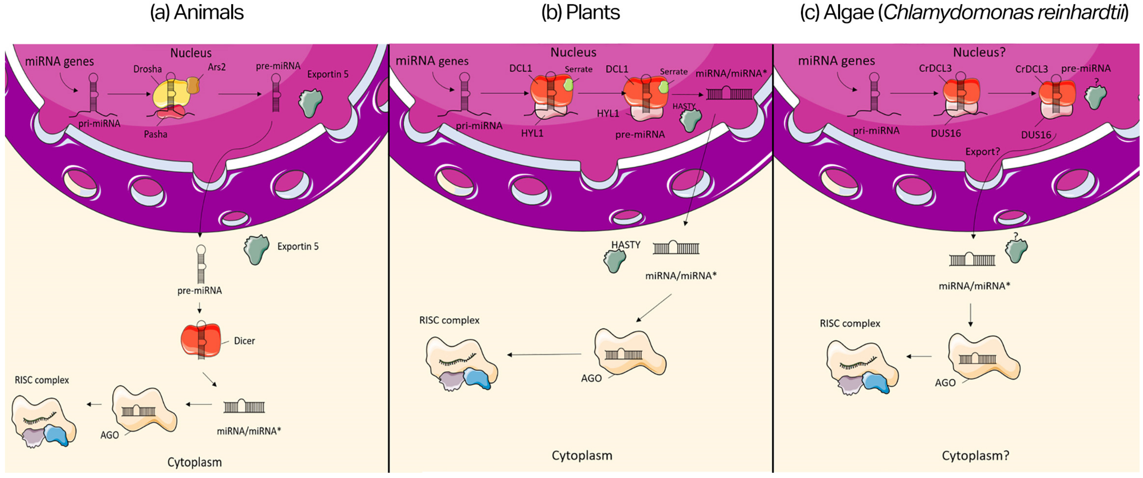
2.2. miRNA Biogenesis

miRNA biogenesis is a process that involves the action of several enzymes that process small precursors into mature molecules (Figure 1) [50]. miRNAs are endogenous molecules that are expressed by both intronic or exonic genes or can be organized in clusters. They are synthesized by RNA polymerase II in the form of a hairpin-shaped precursor, called pri-miRNA. From this point of their biogenesis, different processes and enzymes are known for animals, plants, and algae.
The pri-miRNA of animals (Figure 1a) is processed in the nucleus by a RNAse III enzyme called Drosha, with the help of two proteins Pasha and Ars2 [51], forming another precursor of ~70 nt called pre-miRNA. The complex of Drosha–Pasha cuts asymmetrically the pri-miRNA generating a 3′ protruding end, that facilitates the recognition by an exportin (Exp5) and by another RNAse III enzyme called Dicer [52]. At this point, the pre-miRNA molecule is exported into the cytoplasm by the Exp5 and here it is processed by Dicer, that cleaves the hairpin and generates a ~22 nt miRNA/miRNA* (where “*” stands for the miRNA strand that will be later released and degraded) duplex with 2 nt 3′ overhangs [49]. In the end, one of the two strands is loaded onto an Argonaute (AGO) protein, which will constitute the catalytic compartment of the RNA-induced silencing complex (RISC), and the other strand is later degraded [53].
On the other hand, in plants (Figure 1b), both the pri-miRNA and the pre-miRNA are processed by a Dicer homolog called Dicer-like 1 (DCL1), assisted by the Hyponastic Leaves 1 (HYL1/DRB1) and Serrate (SE), forming the miRNA/miRNA* duplex inside of the nucleus [54]. It is later exported into the cytoplasm by a transporter called HASTY [55]. Finally, one of the two strands is loaded onto an AGO protein, forming the RISC complex.
Regarding algae, there is less knowledge about the mechanisms of biogenesis. Taking into consideration the studies in Chlamydomonas reinhardtii (Figure 1c), for which much information is available compared to other microalgae, both the pri-miRNA and the pre-miRNA molecules are processed by Dicer-like 3 (CrDCL3) protein, associated with DUS16, as a component of the microprocessor complex, forming the miRNA/miRNA* duplex [56]. It is not known whether these processes occur both in the nucleus or there is the exportation of one of the two precursors into the cytoplasm. In the end, one of the two strands is loaded onto an AGO protein, forming the RISC complex.

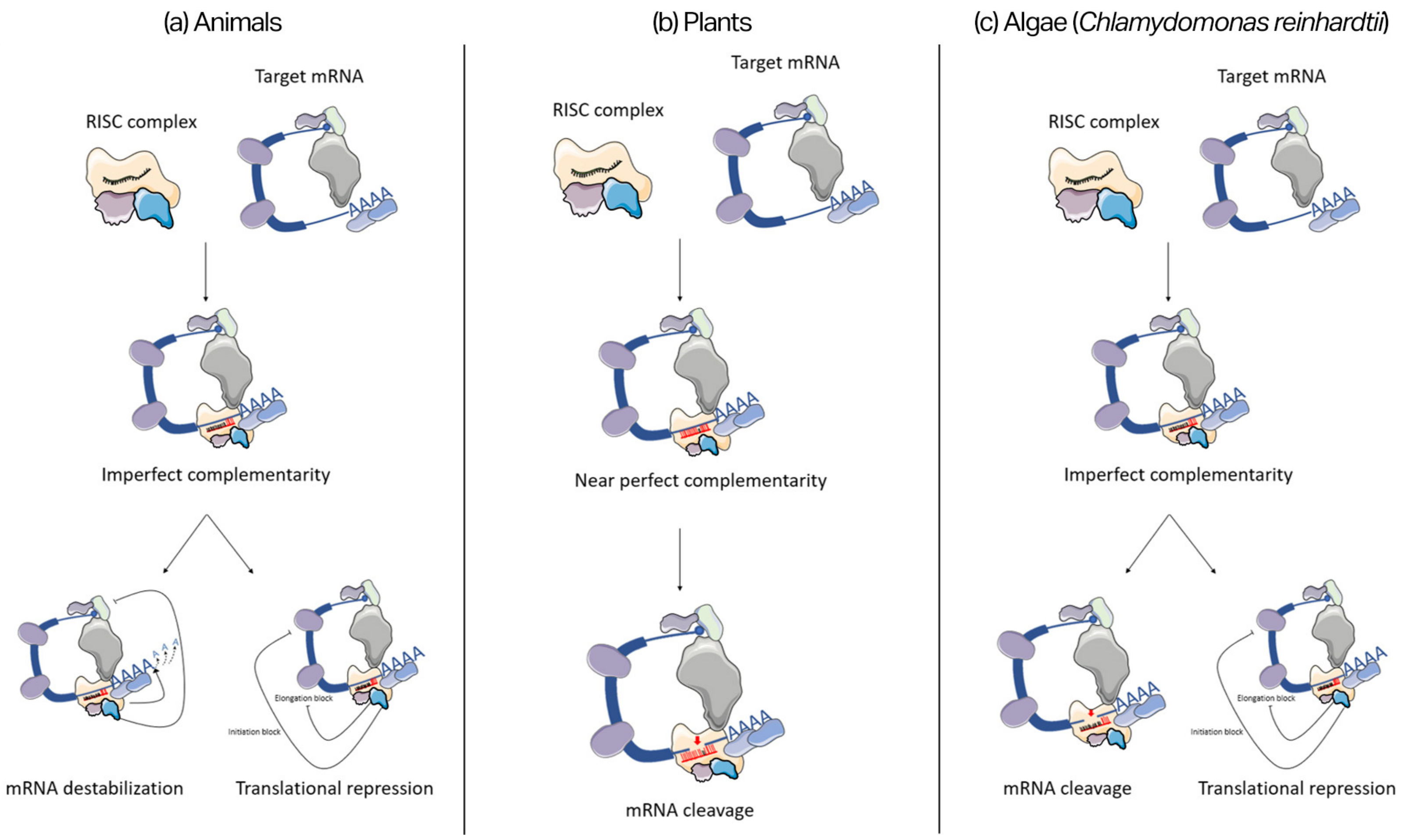
2.3. miRNA Mode of Action

Once the RISC complex is formed, it is active and can modulate gene expression via the binding of a mRNA target. The core of this complex is made of an AGO protein and a single strand of miRNA. The Argonaute family is an evolutionarily very conserved group of proteins, that share two common structural features: a PAZ (Piwi-Argonaute-Zwille) domain and a PIWI (P-element Induced WImpy) domain [57]. The first contains the binding site for the miRNAs and the second showed extensive homology to RNase H [58]. The RISC complex is able to recognize a target by pairing its miRNA to a region of the mRNA, usually at the 3′ UTR. The specificity of miRNAs is not so strict; in fact, it has been reported that miRNAs can recognize hundreds of different targets, overlapping their functions [59]. The mode of action of miRNAs differs among the organisms. Here, we report the main processes in animals, plants, and algae (Figure 2).
In animals (Figure 2a), miRNAs can bind their mRNA targets via imperfect complementarity, where only the nucleotide 2–7 of the miRNA (called “seed” region) have to perfectly pair with the target binding site. Generally, mRNAs are folded into a circular molecule by several interacting proteins. The process begins with the binding of a complex of eukaryotic translation initiation factor (eIF) to the 5′-terminal cap structure of the mRNA. The eIF4G interacts with poly(A)-binding protein (PABP), that binds the poly(A) tail, and the circularization of the molecule begins [60]. When the RISC complex recognizes and pairs with the binding region on the mRNA target, two kinds of repression can occur: destabilization of the molecule or translational repression. When the RISC complex is formed by an AGO1 protein, it can interact with the P-body protein GW182 [61]. This interaction is needed to recruit the carbon catabolite repression–negative on TATA-less (CCR4-NOT) deadenylating complex, that starts to degrade the poly(A) tail of the mRNA [62]. Subsequently, there is the decapping of the target, resulting in the destabilization of the molecule, and later it will be degraded. The translational repression event can occur at two different steps of the process: preventing translation initiation or preventing translation elongation. The first is made by the interaction of the AGO2-Dicer-TRBP (TAR-RNA Binding Protein) with the 60S ribosomal subunit protein and eIF6 [63]. It has been shown that eIF6 is important in the synthesis process of the 60S subunit in the nucleus, and it also carries this subunit to the cytoplasm [64]. On the other hand, the elongation is decelerated and later repressed via the interaction, observed by the co-sedimentation of the miRNA-AGO with the polysomes (ribosomes already bound to the mRNA) [65], although, in both translational repression mechanisms, there is not much information, so it is still unclear how exactly the processes occur.
Alternatively, in plants (Figure 2b), miRNAs show a near-perfect complementarity with their targets. When the RISC complex binds the mRNA, the AGO2 protein is able to cut the molecule, that will be later degraded. The PIWI domain of AGO proteins has a tertiary structure belonging to the RNase H family of enzymes, making them able to cleave RNA [66]. Even though this feature is conserved among the AGO proteins family, only the AGO2 is able to show a slicing mechanism.
On the other hand, the C. reinhardtii (Figure 2c) microalga’s miRNAs behave more like the ones of animals. They show an imperfect complementarity with their targets, binding only a seed region of the miRNA. When the RISC complex is bound to the mRNA, it can repress it via both translational repression and mRNA cleavage [49,67].

2.4. miRNA Conservation

The miRNA regulatory mechanism is a very ancient defense against exogenous molecules, and it is well conserved among the species [46]. Generally, during evolution, not only have organisms conserved the protein machinery that participate in miRNA processes, but also many miRNA sequences are shared across organisms, such as the fact that more than 30% of miRNAs are conserved in all bilaterian animals [68]. Moreover, it has been observed that the knockout of essential miRNAs and their associated protein machinery is not tolerated by animals, highlighting the importance of miRNA-mediated gene silencing for the organism survival. For instance, the loss of Dicer in Danio rerio results in lethal abnormalities in gastrulation and brain development in larvae [69]; and the lack of miRNA-specific Argonautes alg-1 and alg-2 causes the arrest in the morphogenetic elongation during the embryo stages [70]. Furthermore, miRNAs across organisms tend to conserve mostly the seed region, that is the site of specificity, because parallel to the evolution of miRNAs, there has been the evolution of target genes [71]. However, many essential miRNAs are conserved mostly in their entirety. For instance, mir-9a, a central modulator in the embryonic neural development [72], is conserved identically in Drosophila, mouse, and human [73]; and let-7, regulator of the late larval development [74], has not accumulated mutations between humans and worms [75].

3. miRNA-Mediated Adaptations to Marine Extreme Conditions

3.1. Organisms Experiencing Extreme Conditions during Their Lifetime

In order to adapt to different environmental conditions, living organisms can modulate their gene expression through differential expression of miRNAs. In this paragraph, we are going to present the adaptation of marine organisms via miRNA-mediated silencing to different extreme conditions, such as: hypoxia/anoxia, freezing temperatures, high salinity, and high light intensity. In particular, we report examples regarding six species for which information in this perspective are available: Dosidicus gigas, Hemiscyllium ocellatum, Apostichopus japonicus, Littorina littorea, Trematomus bernacchii, and Dunaliella salina (Table 1).
Dosidicus gigas, the so-called jumbo squid, is a marine species that during night is present at the surface of the oceans in order to feed. During the day, in order to avoid predators, they descend into the deep sea, dealing with hypoxia, high pressure, and cold water [76]. During hypoxia, the entering of the animal into a hypometabolic state has been observed, and a change in the expression of the miRNAs in the brain, mantle muscle, and heart [77] (Table 1). For instance, there is an up-regulation of miR-133 in the brain of the organism, that could have a neuroprotective action by inhibiting programmed neuronal death, targeting death-associated protein kinase 2 (DAPK2) [78]. This is a serine/threonine kinase that directly inhibits mTORC1 by phosphorylation, inducing programmed cell death, such as autophagy [79]. Additionally, miR-33 is up-regulated in brain and down-regulated in mantle muscle during hypoxia. It is generally involved in various metabolic pathways, such as the inhibition of cholesterol transporters ATP binding cassette subfamily A member 1 and subfamily G member 1 (ABCA1 and ABCG1) [80], and the repression of the genes Crot, Cpt1a, Handhb, Ampkα, and Irs2, that are known to code key enzymes for fatty acid metabolism [81]. More specifically, miR-33, with the cooperation of sterol regulatory element-binding protein (SREBP), inhibits two key enzymes of the gluconeogenesis: phosphoenolpyruvate carboxykinase (PCK1) and glucose-6-phosphatase (G6PC) [82]. The reason why it is overexpressed in the squid brain could be related to the attempt of achieving a metabolic rate suppression. Furthermore, in the mantle muscle, miR-100-5p is up-regulated. This miRNA has an important role in reducing oxidative stress by targeting NADPH oxidase 4 (NOX4) mRNA, in order to decrease the release of H2O2 [83]. In the heart, there is an overexpression of miR-29-3p, that is a modulator of the cardiac hypertrophy, acting on the synthetic pathways of proteins involved in cardiac fibrosis, potentially inhibiting target mRNAs such as: fibrillin 1 (FBN1), collagen type I, alpha 1 and 2 (COL1A1 and COL1A2), and collagen type III, alpha 1 (COL3A1) [84,85].
Another species that adapted to hypoxic environments is Hemiscyllium ocellatum, known also as epaulette shark. This species has adapted not only to live in cycling hypoxic environments, but also to temporary anoxia (~2 h at 19 °C) [86]. In order to survive, it seems that it is able to enter into a hypometabolic state, based on the switching to anaerobic ATP production in the shark brain [87]. Interestingly, the differential expression of miRNAs has been observed in response to anoxia, with an up-regulation of specific miRNAs [88] (Table 1). As such, there is an overexpression of miR-92, which is a modulator of the hypoxia-inducible factor (HIF) pathway [89]. More specifically, HIF-1α (one of the two factors composing the heterodimer HIF1) can lead to hypoxia-induced apoptosis through two processes: it can bind and stabilize p53, that can mediate for apoptosis, or it can induce the expression of the gene for BCL2/adenovirus interacting protein 3 (BNIP3), a pro-apoptotic protein [90]. Thus, the action of miR-92 results in an anti-apoptotic and anti-proliferative response. Additionally, miR-146b is up-regulated during anoxia, which prevents damages against ischemia, and it also has anti-apoptotic activity [91]. This miRNA attenuates the activation of the nuclear factor-κB (NF-κB) signaling pathway, by directly targeting multiple elements, including the Toll-like receptor 4 (TLR4) and the key adaptor/signaling proteins myeloid differentiation primary response (MyD88), interleukin-1 receptor-associated kinase 1 (IRAK-1), and tumor necrosis factor receptor-associated factor 6 (TRAF6) [92,93]. NF-κB is known to regulate different pathways, including inflammation responses.
On the other hand, another species that is interested in seasonal hypoxia is the sea cucumber Apostichopus japonicus, that lives in the estuaries. The regulation of 26 differentially expressed miRNAs has been observed in the respiratory tree (an organ for oxygen extraction) of the species under hypoxia stress [94] (Table 1). For instance, there are up-regulated miR-31 and miR-153 that act antagonistically on the HIF pathway, the first by promoting it via the inhibition of HIF suppressors, while the second represses the HIF-1α translation by targeting its mRNA [95]. In addition, miR-184 and miR-375 are up-regulated during hypoxia, generally in order to reduce autophagy [87]. Nonetheless, they are also linked to high oxidative stress levels, and miR-375 is associated also with increased apoptosis [96,97].
Furthermore, an interesting example is Littorina littorea, a sea snail that inhabits the intertidal zone, in which there is the formation of continuously changing microenvironments. Thus, the differential expression of miRNAs has been observed during freezing temperatures (−6.7 °C to −7.5 °C), and anoxia in the foot muscle and hepatopancreas of the species [98] (Table 1). Interestingly, the levels of Dicer protein increased in the foot and in the hepatopancreas during freezing, suggesting an increased production of mature miRNAs during stress conditions. For example, there is an up-regulation of miR-210 in anoxic hepatopancreas, which may act as a repressor of mitochondrial respiration, and may be associated with the metabolic shift that the species undergoes during freezing and anoxia [99]. Indeed, this miRNA is able to regulate several targets, such as the repression of: the iron-sulfur cluster scaffold proteins (ISCU1 and ISCU2), which are involved in the TCA cycle and mitochondrial respiration; the NADH dehydrogenase (ubiquinone) 1 alpha subcomplex 4 (NDUFA4); the succinate dehydrogenase complex subunit D (SDHD); and the glycerol-3-phosphate dehydrogenase 1-like (GPD1-L) [100]. In addition, there is the up-regulation of miR-29b in freezing hepatopancreas and foot muscle, but also a down-regulation of it in anoxic hepatopancreas. This miRNA is known to act in the Akt/PI3K pathway via inhibiting the phosphatidylinositol-3 kinase (PI3K) directly targeting its subunit p58α [101], thus it could negatively regulate the pathway via the indirect modulation of mTOR, negatively affecting the protein synthesis [102,103]. However, miR-29b can also directly act on p53 [104], which promotes a shift to anaerobic metabolism under oxidative stress [105]. That may be the reason why miR-29b is down-regulated in anoxic hepatopancreas.
In addition, Trematomus bernacchii is an Antarctic fish species that lives in cold freezing waters (−18 °C). Generally, under heat stress (+4 °C), this fish modulates its immune and inflammatory response via miRNAs-mediated gene silencing [106] (Table 1). In particular, tbe-miR-146a during heat stress regulates two pathways: the toll-like receptor (TLR) and the FoxO signaling pathway. The first is negatively modulated via the inhibition of the genes TRAF6 and IRAK1, that express the production of pro-inflammatory cytokines [107]. Furthermore, the FoxO signaling pathway is up-regulated indirectly by the action of miR-146a during acute heat stress. This miRNA decreases the translation of forkhead transcription factor O subfamily member 3a (FOXO3a) inhibitors, promoting apoptotic signaling [106]. Additionally, the miR-21 family (tbe-miR-21, tbe-miR-21a, and tbe-miR-21b) is differentially expressed under heat stress. The targets of these miRNAs are the genes FOXO3a and FOXO3b, repressing the FoxO signaling pathway and resulting in an anti-apoptotic response [108], in contrast to the one showed by miR-146a. Moreover, miR-21 targets also the phospholipase C (PLC) and phosphatidylinositol 3-kinase (PIK3c) involved in the PIK3-AKT signaling pathway, that modulate positively cell proliferation and growth [109,110].
Finally, Dunaliella salina is a halophilic microalga, that can live in extreme environments characterized by high salt concentrations [111]. In response to specific stresses, such as light intensity and salinity, it can stock a high quantity of β-carotene [112]. In correspondence to the above-mentioned stresses, the differential expression of miRNAs has been observed [113] (Table 1). Among all these miRNAs, novel-m0533-3p is the one that may be involved in the accumulation of β-carotene. It can inhibit malate dehydrogenase, which plays a central role in the TCA cycle [114]. This repression may decrease the participation of acetyl-CoA in the TCA cycle, promoting its involvement in the synthesis of geranylgeranyl pyrophosphate (GGPP), which is a precursor of carotenoids [115].
Table 1. Differential expressed (DE) miRNAs of marine species under different extreme conditions and relative adaptations.
SpeciesConditionDE miRNAsAdaptationsReferences
Dosidicus gigasHypoxia and freezingmiR-1175;
miR-133, miR-33; miR-67; miR-29; miR-2a; miR-100; miR-12; miR-1985; miR-2001; miR-2722; miR-190; miR-34
Hypometabolic state; anti-apoptotic responses; reduction of oxidative stress; modulation of cardiac hypertrophy[77]
Hemiscyllium
ocellatum
Hypoxia and
anoxia
miR-92; miR181a; miR-146b;
miR-140;
miR-20a; miR-17; miR-138; miR-143
Metabolic rate depression; anti-ischemic responses; modulation of HIF; anti-apoptotic responses[88]
Apostichopus
japonicus
HypoxiaAja-miR-2008,;
Aja-miR-10-5p; Aja-miR-184; Aja-miR-71b; Aja-miR-125-5p; novel-miR-1;
Aja-let-7a-5p; Aja-miR-375-3p; Aja-miR-2013-3p; novel-miR-2;
Aja-miR-2835; Aja-miR-1;
Aja-miR-71-5p; Aja-miR-200-3p; Aja-miR-2011-3p; Aja-miR-2478a; Aja-miR-31-5p; Aja-miR-7977; Aja-miR-71a; Aja-miR-29b-3p; Aja-miR-2478b; Aja-miR-2008-5p; Aja-miR-1a-3p; novel-miR-3;
Aja-miR-153-3p; Aja-miR-153
Negatively
regulate HIF pathway;
reduction of
cellular
autophagy;
induction of cell cycle arrest
[94]
Littorina littoreaAnoxia and freezingmiR-1a; miR-210; miR-34a; miR-133a; miR-125b; miR-29b; miR-2aHypometabolic state; anti-
apoptotic
responses;
reduction of
protein synthesis; activation of
oxidative stress response
pathways
[98]
Trematomus
bernacchi
Heat stresstbe-miR-22a; tbe-let-7; tbe-miR-21; tbe-let-7a; tbe-miR26a; tbe-miR30a; tbe-miR-146a; tbe-miR-203b; tbe-miR-200a; tbe-miR-725Regulation of: FoxO signaling cascade, TLR pathway, PI3KT-AKT signaling pathway.
Anti-apoptotic responses
[106]
Dunaliella salinaHigh salinity and light intensitymiR-482; miR-162; miR-3630; miR-166;
miR-858;
novel-m0038-5p; novel-m0783-5p; novel-m1007-3p; novel-m0533-3p
Accumulation of antioxidants (β-carotene)[113]

3.2. Identification of miRNAs through Stress-Response Laboratory Experiments

In order to find and annotate novel miRNAs, organisms of interest are put under different stressful conditions and their miRNome (the complete set of miRNAs in an organism) is analyzed. Here, we show some examples reporting these kinds of experiments on marine species, such as: Branchiostoma belcheri, Strongylocentrotus purpuratus, Mytilus galloprovincialis, and Isochrysis galbana.
Firstly, the amphioxus Branchiostoma belcheri has been analyzed under chemical stress from the xenobiotic polycyclic aromatic hydrocarbon benzo(a)pyrene (BaP), which is a severe environmental carcinogen, mainly produced and released in the seawater by human activities [116]. Thus, the expression of 11 already known and 47 novel miRNAs has been detected, that were differentially expressed in the treated amphioxus with 0.1 mg/L of BaP [117]. Moreover, it has been found that a total of 16 miRNAs regulated different key xenobiotics and toxicant biodegradation-related signaling pathways. For example, bbe-miR-182b-5p is up-regulated and positively modulate the hypoxia-inducible factor 1α (HIF-1α). In addition, bbe-miR-281-3p was also up-regulated and interact with ornithine decarboxylase antizyme (ODA) [118]; this may promote BaP metabolism activation by ornithine decarboxylase, as already seen in experiments with rat lungs [119].
An interesting study has identified the miRNA expression during stress by zinc (Zn) in sea urchins [120], and more recently, in Strongylocentrotus purpuratus under abiotic stress from pH acidification (from pH 8.01 of the control to pH 7.88 of the stressed condition); a total of 682 conserved miRNAs and 17 new ones have been found [121]. Among these, spu-miR-92a, spu-miR-92c, and spu-miR-92e were down-regulated in the larvae exposed to acidified water. This family of miRNAs may activate the Wnt/β-catenin signaling pathways, that has a key role in the sea urchin skeletogenesis [122] and endomesoderm formation in S. purpuratus [123]. In addition, this miR-92 family and spu-miR-2002-3p are predicted to target the carbonic anhydrase transcript variant X1. The enzyme carbonic anhydrase catalyzes the reaction from CO2 to HCO3 [124], but also this enzyme has an important role in blocking the spicule formation in sea urchins [125]. Additionally, spu-miR-133 is up-regulated in the larvae in acidified water. This miRNA has been predicted to target the breast carcinoma amplified sequence (BCAS2). The BCAS2 regulates the β-catenin pre-RNA splicing [126], and so indirectly also the Wnt/β-catenin signaling pathways.
Additionally, another organism that has been put under chemical stress is the mussel Mytilus galloprovincialis, exposed to 5 µg/L and 50 µg/L of cadmium (Cd). Under these conditions, 107 known miRNAs have been validated and 32 novel ones have been identified [127], of which 66 known and 19 novel miRNAs were differentially expressed and up-regulated during the Cd treatment. For instance, miR-745a is up-regulated and may target the apoptosis-resistant E3 gene, which is a key player in the regulation of apoptosis [128]. Another up-regulated miRNA is miR-2a-3p-6, which has been predicted to target the calponin-like protein (Cap) gene, that repress the actomyosin ATPase activity in mussels [129]. The expression profile of these miRNAs suggests that Cd affects cell responses such as apoptosis, the stabilization of the cytoskeleton, and energy metabolism in mussels [127].
Finally, the microalga Isochrysis galbana has been put under abiotic stress, exposed to high temperatures (from 20 °C in the control to 35 °C in the stressed condition), in which nine conserved miRNAs and 149 novel ones have been identified [130]. Under heat stimuli, the miRNAs novel_152 and novel_190 were down-regulated, corresponding to an over-expression of their target genes histidine kinase and superoxide-generating NADPH oxidase, respectively. Related to this, the glutathione S-transferase (GST) gene also, a ROS scavenger related gene [130], was down-regulated by the expression of the miRNA novel_33. This regulation may carry out the accumulation of ROS, and the subsequent activation of downstream pathways through heat stress transcription factors (HSFs), altering the redox state of the cell [130,131]. Furthermore, the miRNA novel_64 was down-regulated, corresponding to the over-expression of its target gene E3 ubiquitin-protein ligase. The E3 ubiquitin-ligase with the help of heat-shock proteins (HSPs) might mediate the degradation of misfolded protein due to heat stress [132], implying that novel_64 could be involved in the protein ubiquitination, and thus in the thermotolerance of this microalga.

4. Discussion

In this review, we have summarized available information on how marine organisms can adapt to extreme environmental conditions by modifying gene expression through miRNA-mediated silencing. For instance, many species that faces hypoxia/anoxia can enter in a hypometabolic state via the expression of specific miRNAs that can slow the metabolic rates and activate pathways to protect cells from programmed death, or algal species facing high irradiance can activate pathways for the accumulation of antioxidants (e.g., carotenoids) via miRNA expression. We report available studies on miRNA identification and expression in different marine organisms living in extreme conditions. In addition, we also summarized how laboratory experiments mimicking extreme conditions, such as in terms of temperature, acidification, or pollutant exposure, can allow the identification of miRNA variations. However, our review shows that this field is still in its infancy and there is still a lack of knowledge on the differential expression of miRNAs, especially for marine plants and algae. Reaching a better comprehension of gene regulatory networks can help to shed light on marine organism adaptations to different habitats and enhance biotechnological applications. As such, it can help to enhance the production of high-value bioproducts, for instance, by engineering microalgae by the overexpression or inhibition of miRNAs. These techniques are already in use in the medical field, such as in the treatment of human diseases [133,134,135], and additional strategies are under evaluation in disease animal models [136,137,138]. On the other hand, in the case of the microalga Chlamydomonas reinhardtii, artificial miRNAs (amiRNAs) have been used that targeted Chlamydomonas phosphoenolpyruvate carboxylase isoform 1 and 2 (CrPEPC1 and CrPEPC2) genes to inhibit phosphoenolpyruvate carboxylase activity, obtaining an enhancement in fatty acid production [139]. A similar study was performed on the microalga Phaeodactylum tricornutum, where the endogenous phytoene synthase (PSY) gene has been targeted, with the use of amiRNAs, in order to reduce carotenoid levels [140].
Furthermore, miRNAs can be exploited as biomarkers of generic or specific environmental stresses. For instance, miR-166 is a very conserved miRNA in land plants, proposed as a biomarker for biotic and abiotic stresses in major crop plants, due to its regulatory activity during drought, salinity, temperature, and during biotic stress management [141]. Additionally, other miRNAs are found in plants to respond to abiotic stressors that can work as useful environmental biomarkers [142]. For marine species, there is still a lack of knowledge in the application of miRNAs as environmental biomarkers, although some efforts have been made to identify miRNAs deregulated in different stress conditions in marine organisms. Examples of these efforts are the works conducted on bivalve species. Bivalve mollusks are ubiquitous and abundant marine organisms, important for biological monitoring because of their abilities to adapt to different environments. Bivalve miRNAs have been reported to rapidly respond and to adjust the adaptation and physiological functions of bivalves during environmental stressors such as pollution, salinity and temperature changes, and desiccation [127,143]. Considering that nowadays climate change is one of the major environmental issues, the identification of miRNAs involved in adaptation mechanisms, the evaluation of their expression, and the identification of which related pathways are activated or switched off could be extremely beneficial to the monitoring and prediction strategies, as well as for the study of their possible biotechnological applications.

Author Contributions

Conceptualization, G.D.F. and S.C.; writing—original draft preparation, G.D.F.; writing—review and editing, G.D.F., C.L. and S.C. All authors have read and agreed to the published version of the manuscript.

Funding

G.D.F. was supported by a Stazione Zoologica Anton Dohrn Ph.D. fellowship via the Open University.

Institutional Review Board Statement

Not applicable.

Informed Consent Statement

Not applicable.

Data Availability Statement

Not applicable.

Acknowledgments

The authors thank the Servier Medical Art (SMART) website (https://smart.servier.com; accessed on 30 January 2023) by Servier for the elements in Figure 1 and Figure 2. In addition, thanks to the Flaticon website (https://www.flaticon.com; accessed on 30 January 2023) for the images used in the Graphical Abstract, made by the Freepik and Flaticon staff. Furthermore, thanks are also due to Sebastiano Urciuoli for the graphical support.

Conflicts of Interest

The authors declare no conflict of interest.

References

  1. Lindgren, A.R.; Buckley, B.A.; Eppley, S.M.; Reysenbach, A.-L.; Stedman, K.M.; Wagner, J.T. Life on the Edge—The Biology of Organisms Inhabiting Extreme Environments: An Introduction to the Symposium. Integr. Comp. Biol. 2016, 56, 493–499. [Google Scholar] [CrossRef] [PubMed]
  2. Rothschild, L.J.; Mancinelli, R.L. Life in Extreme Environments. Nature 2001, 409, 1092–1101. [Google Scholar] [CrossRef] [PubMed]
  3. Bijma, J.; Pörtner, H.-O.; Yesson, C.; Rogers, A.D. Climate Change and the Oceans—What Does the Future Hold? Mar. Pollut. Bull. 2013, 74, 495–505. [Google Scholar] [CrossRef]
  4. Schmidtko, S.; Stramma, L.; Visbeck, M. Decline in Global Oceanic Oxygen Content during the Past Five Decades. Nature 2017, 542, 335–339. [Google Scholar] [CrossRef] [PubMed]
  5. Camp, E.F.; Schoepf, V.; Mumby, P.J.; Hardtke, L.A.; Rodolfo-Metalpa, R.; Smith, D.J.; Suggett, D.J. The Future of Coral Reefs Subject to Rapid Climate Change: Lessons from Natural Extreme Environments. Front. Mar. Sci. 2018, 5, 4. [Google Scholar] [CrossRef]
  6. Macelroy, R.D. Some Comments on the Evolution of Extremophiles. Biosystems 1974, 6, 74–75. [Google Scholar] [CrossRef]
  7. Morita, R.Y. Extremes of Biodiversity. BioScience 1999, 49, 245–248. [Google Scholar] [CrossRef]
  8. Lévêque, E.; Janeček, Š.; Haye, B.; Belarbi, A. Thermophilic Archaeal Amylolytic Enzymes. Enzyme Microb. Technol. 2000, 26, 3–14. [Google Scholar] [CrossRef]
  9. Driedzic, W.R.; Clow, K.A.; Short, C.E.; Ewart, K.V. Glycerol Production in Rainbow Smelt (Osmerus mordax) May Be Triggered by Low Temperature Alone and Is Associated with the Activation of Glycerol-3-Phosphate Dehydrogenase and Glycerol-3-Phosphatase. J. Exp. Biol. 2006, 209, 1016–1023. [Google Scholar] [CrossRef]
  10. Seckbach, J. Enigmatic Microorganisms and Life in Extreme Environments; Springer: Dordrecht, The Netherlands, 2013; ISBN 978-94-011-4838-2. [Google Scholar]
  11. Chilukuri, L.N.; Bartlett, D.H. Isolation and Characterization of the Gene Encoding Single-Stranded-DNA-Binding Protein (SSB) from Four Marine Shewanella Strains That Differ in Their Temperature and Pressure Optima for Growth. Microbiology 1997, 143, 1163–1174. [Google Scholar] [CrossRef] [Green Version]
  12. Wetzel, R.G. Limnology: Lake and River Ecosystems, 3rd ed.; Academic Press: San Diego, CA, USA, 2001; ISBN 978-0-12-744760-5. [Google Scholar]
  13. Storey, K.B.; Storey, J.M. Biochemical Adaptation to Extreme Environments. In Integrative Physiology in the Proteomics and Post-Genomics Age; Walz, W., Ed.; Humana Press: Totowa, NJ, USA, 2005; pp. 169–200. ISBN 978-1-58829-315-2. [Google Scholar]
  14. Storey, K.B.; Storey, J.M. Tribute to P. L. Lutz: Putting Life on ‘pause’—Molecular Regulation of Hypometabolism. J. Exp. Biol. 2007, 210, 1700–1714. [Google Scholar] [CrossRef]
  15. Heldmaier, G.; Ortmann, S.; Elvert, R. Natural Hypometabolism during Hibernation and Daily Torpor in Mammals. Resp. Physiol. Neurobiol. 2004, 141, 317–329. [Google Scholar] [CrossRef] [PubMed]
  16. Hochachka, P.W.; Somero, G.N. Biochemical Adaptation: Mechanism and Process in Physiological Evolution; Oxford University Press: New York, NY, USA, 2002; ISBN 978-0-19-511702-8. [Google Scholar]
  17. Storey, K.B. Life in the Slow Lane: Molecular Mechanisms of Estivation. Comp. Biochem. Physiol. Part A Mol. Integr. Physiol. 2002, 133, 733–754. [Google Scholar] [CrossRef] [PubMed]
  18. Lutz, P.L.; Nilsson, G.E. Vertebrate Brains at the Pilot Light. Resp. Physiol. Neurobiol. 2004, 141, 285–296. [Google Scholar] [CrossRef] [PubMed]
  19. Chaudhry, R.; Varacallo, M. Biochemistry, Glycolysis. In StatPearls; StatPearls Publishing: Treasure Island, FL, USA, 2022. [Google Scholar]
  20. Akram, M. Mini-Review on Glycolysis and Cancer. J. Cancer Educ. 2013, 28, 454–457. [Google Scholar] [CrossRef] [PubMed]
  21. Fernie, A.R.; Carrari, F.; Sweetlove, L.J. Respiratory Metabolism: Glycolysis, the TCA Cycle and Mitochondrial Electron Transport. Curr. Opin. Plant Biol. 2004, 7, 254–261. [Google Scholar] [CrossRef]
  22. Choi, I.; Son, H.; Baek, J.-H. Tricarboxylic Acid (TCA) Cycle Intermediates: Regulators of Immune Responses. Life 2021, 11, 69. [Google Scholar] [CrossRef]
  23. Sharma, P.; Jha, A.B.; Dubey, R.S.; Pessarakli, M. Reactive Oxygen Species, Oxidative Damage, and Antioxidative Defense Mechanism in Plants under Stressful Conditions. J. Bot. 2012, 2012, 217037. [Google Scholar] [CrossRef]
  24. Zhang, J.; Wang, X.; Vikash, V.; Ye, Q.; Wu, D.; Liu, Y.; Dong, W. ROS and ROS-Mediated Cellular Signaling. Oxid. Med. Cell Longev. 2016, 2016, 4350965. [Google Scholar] [CrossRef]
  25. Foyer, C.H.; Noctor, G. Redox Regulation in Photosynthetic Organisms: Signaling, Acclimation, and Practical Implications. Antioxid. Redox Signal 2009, 11, 861–905. [Google Scholar] [CrossRef] [Green Version]
  26. Halliwell, B. How to Characterize an Antioxidant: An Update. Biochem. Soc. Symp. 1995, 61, 73–101. [Google Scholar] [CrossRef] [PubMed]
  27. Abd El-Baky, H.H.; El Baz, F.K.; El-Baroty, G.S. Enhancement of Antioxidant Production in Spirulina platensis under Oxidative Stress. Acta Physiol. Plant. 2009, 31, 623–631. [Google Scholar] [CrossRef]
  28. Akhtar, T.A.; Lees, H.A.; Lampi, M.A.; Enstone, D.; Brain, R.A.; Greenberg, B.M. Photosynthetic Redox Imbalance Influences Flavonoid Biosynthesis in Lemna gibba. Plant Cell Environ. 2010, 33, 1205–1219. [Google Scholar] [CrossRef]
  29. Agati, G.; Tattini, M. Multiple Functional Roles of Flavonoids in Photoprotection. New Phytol. 2010, 186, 786–793. [Google Scholar] [CrossRef] [PubMed]
  30. Agati, G.; Matteini, P.; Goti, A.; Tattini, M. Chloroplast-located Flavonoids Can Scavenge Singlet Oxygen. New Phytol. 2007, 174, 77–89. [Google Scholar] [CrossRef] [PubMed]
  31. Gould, K.S.; McKelvie, J.; Markham, K.R. Do Anthocyanins Function as Antioxidants in Leaves? Imaging of H2O2 in Red and Green Leaves after Mechanical Injury: H2O2 Scavenging by Anthocyanins. Plant Cell Environ. 2002, 25, 1261–1269. [Google Scholar] [CrossRef]
  32. Maoka, T. Carotenoids as Natural Functional Pigments. J. Nat. Med. 2020, 74, 1–16. [Google Scholar] [CrossRef]
  33. Remias, D.; Karsten, U.; Lütz, C.; Leya, T. Physiological and Morphological Processes in the Alpine Snow Alga Chloromonas nivalis (Chlorophyceae) during Cyst Formation. Protoplasma 2010, 243, 73–86. [Google Scholar] [CrossRef]
  34. Doppler, P.; Kornpointner, C.; Halbwirth, H.; Remias, D.; Spadiut, O. Tetraedron Minimum, First Reported Member of Hydrodictyaceae to Accumulate Secondary Carotenoids. Life 2021, 11, 107. [Google Scholar] [CrossRef]
  35. Han, R.-M.; Zhang, J.-P.; Skibsted, L.H. Reaction Dynamics of Flavonoids and Carotenoids as Antioxidants. Molecules 2012, 17, 2140–2160. [Google Scholar] [CrossRef] [Green Version]
  36. Molino, A.; Iovine, A.; Casella, P.; Mehariya, S.; Chianese, S.; Cerbone, A.; Rimauro, J.; Musmarra, D. Microalgae Characterization for Consolidated and New Application in Human Food, Animal Feed and Nutraceuticals. Int. J. Environ. Res. Public Health 2018, 15, 2436. [Google Scholar] [CrossRef]
  37. Yilancioglu, K.; Cokol, M.; Pastirmaci, I.; Erman, B.; Cetiner, S. Oxidative Stress Is a Mediator for Increased Lipid Accumulation in a Newly Isolated Dunaliella salina Strain. PLoS ONE 2014, 9, e91957. [Google Scholar] [CrossRef] [PubMed]
  38. Cai, Z. Lipid Peroxidation. In Encyclopedia of Toxicology; Elsevier: Amsterdam, The Netherlands, 2005; pp. 730–734. ISBN 978-0-12-369400-3. [Google Scholar]
  39. Hartwell, L.H.; Weinert, T.A. Checkpoints: Controls That Ensure the Order of Cell Cycle Events. Science 1989, 246, 629–634. [Google Scholar] [CrossRef] [PubMed]
  40. Pietenpol, J.A.; Stewart, Z.A. Cell Cycle Checkpoint Signaling. Toxicology 2002, 181–182, 475–481. [Google Scholar] [CrossRef]
  41. Joerger, A.C.; Fersht, A.R. The P53 Pathway: Origins, Inactivation in Cancer, and Emerging Therapeutic Approaches. Annu. Rev. Biochem. 2016, 85, 375–404. [Google Scholar] [CrossRef] [PubMed]
  42. Saxton, R.A.; Sabatini, D.M. MTOR Signaling in Growth, Metabolism, and Disease. Cell 2017, 168, 960–976. [Google Scholar] [CrossRef]
  43. Sarbassov, D.D.; Guertin, D.A.; Ali, S.M.; Sabatini, D.M. Phosphorylation and Regulation of Akt/PKB by the Rictor-MTOR Complex. Science 2005, 307, 1098–1101. [Google Scholar] [CrossRef]
  44. Mocellin, S.; Provenzano, M.M. RNA Interference: Learning Gene Knock-down from Cell Physiology. J. Transl. Med. 2004, 2, 39. [Google Scholar] [CrossRef]
  45. El-Sappah, A.H.; Yan, K.; Huang, Q.; Islam, M.M.; Li, Q.; Wang, Y.; Khan, M.S.; Zhao, X.; Mir, R.R.; Li, J.; et al. Comprehensive Mechanism of Gene Silencing and Its Role in Plant Growth and Development. Front. Plant Sci. 2021, 12, 705249. [Google Scholar] [CrossRef]
  46. Shabalina, S.; Koonin, E. Origins and Evolution of Eukaryotic RNA Interference. Trends Ecol. Evol. 2008, 23, 578–587. [Google Scholar] [CrossRef] [Green Version]
  47. Higgs, P.G.; Lehman, N. The RNA World: Molecular Cooperation at the Origins of Life. Nat. Rev. Genet. 2015, 16, 7–17. [Google Scholar] [CrossRef] [PubMed]
  48. Wightman, B.; Ha, I.; Ruvkun, G. Posttranscriptional Regulation of the Heterochronic Gene Lin-14 by Lin-4 Mediates Temporal Pattern Formation in C. elegans. Cell 1993, 75, 855–862. [Google Scholar] [CrossRef] [PubMed]
  49. Lou, S.; Sun, T.; Li, H.; Hu, Z. Mechanisms of MicroRNA-Mediated Gene Regulation in Unicellular Model Alga Chlamydomonas reinhardtii. Biotechnol. Biofuels 2018, 11, 244. [Google Scholar] [CrossRef] [PubMed]
  50. Bonin, C.A.; van Wijnen, A.J.; Lewallen, E.A. MicroRNA Applications in Marine Biology. Curr. Mol. Biol. Rep. 2019, 5, 167–175. [Google Scholar] [CrossRef]
  51. Wilson, M.D.; Wang, D.; Wagner, R.; Breyssens, H.; Gertsenstein, M.; Lobe, C.; Lu, X.; Nagy, A.; Burke, R.D.; Koop, B.F.; et al. ARS2 Is a Conserved Eukaryotic Gene Essential for Early Mammalian Development. Mol. Cell Biol. 2008, 28, 1503–1514. [Google Scholar] [CrossRef]
  52. Han, J.; Lee, Y.; Yeom, K.-H.; Kim, Y.-K.; Jin, H.; Kim, V.N. The Drosha-DGCR8 Complex in Primary MicroRNA Processing. Genes Dev. 2004, 18, 3016–3027. [Google Scholar] [CrossRef]
  53. Du, T.; Zamore, P.D. Beginning to Understand MicroRNA Function. Cell Res. 2007, 17, 661–663. [Google Scholar] [CrossRef]
  54. Park, M.Y.; Wu, G.; Gonzalez-Sulser, A.; Vaucheret, H.; Poethig, R.S. Nuclear Processing and Export of MicroRNAs in Arabidopsis. Proc. Natl. Acad. Sci. USA 2005, 102, 3691–3696. [Google Scholar] [CrossRef]
  55. Bollman, K.M.; Aukerman, M.J.; Park, M.-Y.; Hunter, C.; Berardini, T.Z.; Poethig, R.S. HASTY, the Arabidopsis ortholog of Exportin 5/MSN5, Regulates Phase Change and Morphogenesis. Development 2003, 130, 1493–1504. [Google Scholar] [CrossRef]
  56. Yamasaki, T.; Onishi, M.; Kim, E.-J.; Cerutti, H.; Ohama, T. RNA-Binding Protein DUS16 Plays an Essential Role in Primary MiRNA Processing in the Unicellular Alga Chlamydomonas reinhardtii. Proc. Natl. Acad. Sci. USA 2016, 113, 10720–10725. [Google Scholar] [CrossRef] [Green Version]
  57. Höck, J.; Meister, G. The Argonaute Protein Family. Genome Biol. 2008, 9, 210. [Google Scholar] [CrossRef] [PubMed]
  58. Rashid, U.J.; Paterok, D.; Koglin, A.; Gohlke, H.; Piehler, J.; Chen, J.C.-H. Structure of Aquifex Aeolicus Argonaute Highlights Conformational Flexibility of the PAZ Domain as a Potential Regulator of RNA-Induced Silencing Complex Function. J. Biol. Chem. 2007, 282, 13824–13832. [Google Scholar] [CrossRef] [PubMed]
  59. Brennecke, J.; Stark, A.; Russell, R.B.; Cohen, S.M. Principles of MicroRNA–Target Recognition. PLoS Biol. 2005, 3, e85. [Google Scholar] [CrossRef] [PubMed]
  60. Gebauer, F.; Hentze, M.W. Molecular Mechanisms of Translational Control. Nat. Rev. Mol. Cell Biol. 2004, 5, 827–835. [Google Scholar] [CrossRef]
  61. Till, S.; Lejeune, E.; Thermann, R.; Bortfeld, M.; Hothorn, M.; Enderle, D.; Heinrich, C.; Hentze, M.W.; Ladurner, A.G. A Conserved Motif in Argonaute-Interacting Proteins Mediates Functional Interactions through the Argonaute PIWI Domain. Nat. Struct. Mol. Biol. 2007, 14, 897–903. [Google Scholar] [CrossRef]
  62. Filipowicz, W.; Bhattacharyya, S.N.; Sonenberg, N. Mechanisms of Post-Transcriptional Regulation by MicroRNAs: Are the Answers in Sight? Nat. Rev. Genet. 2008, 9, 102–114. [Google Scholar] [CrossRef]
  63. Chendrimada, T.P.; Finn, K.J.; Ji, X.; Baillat, D.; Gregory, R.I.; Liebhaber, S.A.; Pasquinelli, A.E.; Shiekhattar, R. MicroRNA Silencing through RISC Recruitment of EIF6. Nature 2007, 447, 823–828. [Google Scholar] [CrossRef]
  64. Basu, U.; Si, K.; Warner, J.R.; Maitra, U. The Saccharomyces cerevisiae TIF6 Gene Encoding Translation Initiation Factor 6 Is Required for 60S Ribosomal Subunit Biogenesis. Mol. Cell. Biol. 2001, 21, 1453–1462. [Google Scholar] [CrossRef]
  65. Kim, J.; Krichevsky, A.; Grad, Y.; Hayes, G.D.; Kosik, K.S.; Church, G.M.; Ruvkun, G. Identification of Many MicroRNAs That Copurify with Polyribosomes in Mammalian Neurons. Proc. Natl. Acad. Sci. USA 2004, 101, 360–365. [Google Scholar] [CrossRef]
  66. Park, J.H.; Shin, C. MicroRNA-Directed Cleavage of Targets: Mechanism and Experimental Approaches. BMB Rep. 2014, 47, 417–423. [Google Scholar] [CrossRef] [Green Version]
  67. Chung, B.Y.-W.; Deery, M.J.; Groen, A.J.; Howard, J.; Baulcombe, D.C. Endogenous MiRNA in the Green Alga Chlamydomonas Regulates Gene Expression through CDS-Targeting. Nat. Plants 2017, 3, 787–794. [Google Scholar] [CrossRef] [PubMed]
  68. Dexheimer, P.J.; Cochella, L. MicroRNAs: From Mechanism to Organism. Front. Cell Dev. Biol. 2020, 8, 409. [Google Scholar] [CrossRef]
  69. Giraldez, A.J.; Cinalli, R.M.; Glasner, M.E.; Enright, A.J.; Thomson, J.M.; Baskerville, S.; Hammond, S.M.; Bartel, D.P.; Schier, A.F. MicroRNAs Regulate Brain Morphogenesis in Zebrafish. Science 2005, 308, 833–838. [Google Scholar] [CrossRef] [PubMed]
  70. Vasquez-Rifo, A.; Jannot, G.; Armisen, J.; Labouesse, M.; Bukhari, S.I.A.; Rondeau, E.L.; Miska, E.A.; Simard, M.J. Developmental Characterization of the MicroRNA-Specific, C. elegans Argonautes Alg-1 and Alg-2. PLoS ONE 2012, 7, e33750. [Google Scholar] [CrossRef] [PubMed]
  71. Nozawa, M.; Fujimi, M.; Iwamoto, C.; Onizuka, K.; Fukuda, N.; Ikeo, K.; Gojobori, T. Evolutionary Transitions of MicroRNA-Target Pairs. Genome Biol. Evol. 2016, 8, 1621–1633. [Google Scholar] [CrossRef]
  72. Coolen, M.; Katz, S.; Bally-Cuif, L. MiR-9: A Versatile Regulator of Neurogenesis. Front. Cell Neurosci. 2013, 7, 220. [Google Scholar] [CrossRef] [PubMed]
  73. Li, Y.; Wang, F.; Lee, J.-A.; Gao, F.-B. MicroRNA-9a Ensures the Precise Specification of Sensory Organ Precursors in Drosophila. Genes Dev. 2006, 20, 2793–2805. [Google Scholar] [CrossRef]
  74. Bagga, S.; Bracht, J.; Hunter, S.; Massirer, K.; Holtz, J.; Eachus, R.; Pasquinelli, A.E. Regulation by Let-7 and Lin-4 MiRNAs Results in Target MRNA Degradation. Cell 2005, 122, 553–563. [Google Scholar] [CrossRef]
  75. Pasquinelli, A.E.; Reinhart, B.J.; Slack, F.; Martindale, M.Q.; Kuroda, M.I.; Maller, B.; Hayward, D.C.; Ball, E.E.; Degnan, B.; Müller, P.; et al. Conservation of the Sequence and Temporal Expression of Let-7 Heterochronic Regulatory RNA. Nature 2000, 408, 86–89. [Google Scholar] [CrossRef] [PubMed]
  76. Gilly, W.F.; Zeidberg, L.D.; Booth, J.A.T.; Stewart, J.S.; Marshall, G.; Abernathy, K.; Bell, L.E. Locomotion and Behavior of Humboldt Squid, Dosidicus gigas, in Relation to Natural Hypoxia in the Gulf of California, Mexico. J. Exp. Biol. 2012, 215, 3175–3190. [Google Scholar] [CrossRef] [Green Version]
  77. Hadj-Moussa, H.; Logan, S.M.; Seibel, B.A.; Storey, K.B. Potential Role for MicroRNA in Regulating Hypoxia-Induced Metabolic Suppression in Jumbo Squids. Biochim. Biophys. Acta Gene Regul. Mech. 2018, 1861, 586–593. [Google Scholar] [CrossRef] [PubMed]
  78. Li, S.; Xiao, F.-Y.; Shan, P.-R.; Su, L.; Chen, D.-L.; Ding, J.-Y.; Wang, Z.-Q. Overexpression of MicroRNA-133a Inhibits Ischemia-Reperfusion-Induced Cardiomyocyte Apoptosis by Targeting DAPK2. J. Hum. Genet. 2015, 60, 709–716. [Google Scholar] [CrossRef] [PubMed]
  79. Ber, Y.; Shiloh, R.; Gilad, Y.; Degani, N.; Bialik, S.; Kimchi, A. DAPK2 Is a Novel Regulator of MTORC1 Activity and Autophagy. Cell Death Differ. 2015, 22, 465–475. [Google Scholar] [CrossRef] [PubMed]
  80. Näär, A.M. MiR-33: A Metabolic Conundrum. Trends Endocrinol. Metab. 2018, 29, 667–668. [Google Scholar] [CrossRef]
  81. Dávalos, A.; Goedeke, L.; Smibert, P.; Ramírez, C.M.; Warrier, N.P.; Andreo, U.; Cirera-Salinas, D.; Rayner, K.; Suresh, U.; Pastor-Pareja, J.C.; et al. MiR-33a/b Contribute to the Regulation of Fatty Acid Metabolism and Insulin Signaling. Proc. Natl. Acad. Sci. USA 2011, 108, 9232–9237. [Google Scholar] [CrossRef]
  82. Ramírez, C.M.; Goedeke, L.; Rotllan, N.; Yoon, J.-H.; Cirera-Salinas, D.; Mattison, J.A.; Suárez, Y.; de Cabo, R.; Gorospe, M.; Fernández-Hernando, C. MicroRNA 33 Regulates Glucose Metabolism. Mol. Cell Biol. 2013, 33, 2891–2902. [Google Scholar] [CrossRef]
  83. Kriegel, A.J.; Baker, M.A.; Liu, Y.; Liu, P.; Cowley, A.W.; Liang, M. Endogenous MicroRNAs in Human Microvascular Endothelial Cells Regulate MRNAs Encoded by Hypertension-Related Genes. Hypertension 2015, 66, 793–799. [Google Scholar] [CrossRef]
  84. van Rooij, E.; Sutherland, L.B.; Thatcher, J.E.; DiMaio, J.M.; Naseem, R.H.; Marshall, W.S.; Hill, J.A.; Olson, E.N. Dysregulation of MicroRNAs after Myocardial Infarction Reveals a Role of MiR-29 in Cardiac Fibrosis. Proc. Natl. Acad. Sci. USA 2008, 105, 13027–13032. [Google Scholar] [CrossRef]
  85. Dong, D.; Yang, B. Role of MicroRNAs in Cardiac Hypertrophy, Myocardial Fibrosis and Heart Failure. Acta Pharm. Sin. 2011, 1, 1–7. [Google Scholar] [CrossRef]
  86. Dowd, W.W.; Renshaw, G.M.C.; Cech, J.J.; Kültz, D. Compensatory Proteome Adjustments Imply Tissue-Specific Structural and Metabolic Reorganization Following Episodic Hypoxia or Anoxia in the Epaulette Shark (Hemiscyllium ocellatum). Physiol. Genomics 2010, 42, 93–114. [Google Scholar] [CrossRef]
  87. Hadj-Moussa, H.; Storey, K.B. The OxymiR Response to Oxygen Limitation: A Comparative MicroRNA Perspective. J. Exp. Biol. 2020, 223, jeb204594. [Google Scholar] [CrossRef] [PubMed]
  88. Riggs, C.L.; Summers, A.; Warren, D.E.; Nilsson, G.E.; Lefevre, S.; Dowd, W.W.; Milton, S.; Podrabsky, J.E. Small Non-Coding RNA Expression and Vertebrate Anoxia Tolerance. Front. Genet. 2018, 9, 230. [Google Scholar] [CrossRef]
  89. Valera, V.A.; Walter, B.A.; Linehan, W.M.; Merino, M.J. Regulatory Effects of MicroRNA-92 (MiR-92) on VHL Gene Expression and the Hypoxic Activation of MiR-210 in Clear Cell Renal Cell Carcinoma. J. Cancer 2011, 2, 515–526. [Google Scholar] [CrossRef] [PubMed]
  90. Greijer, A.E. The Role of Hypoxia Inducible Factor 1 (HIF-1) in Hypoxia Induced Apoptosis. J. Clin. Pathol. 2004, 57, 1009–1014. [Google Scholar] [CrossRef] [PubMed]
  91. Di, Y.-F.; Li, D.-C.; Shen, Y.-Q.; Wang, C.-L.; Zhang, D.-Y.; Shang, A.-Q.; Hu, T. MiR-146b Protects Cardiomyocytes Injury in Myocardial Ischemia/Reperfusion by Targeting Smad4. Am. J. Transl. Res. 2017, 9, 656–663. [Google Scholar] [PubMed]
  92. Li, J.-W.; He, S.-Y.; Feng, Z.-Z.; Zhao, L.; Jia, W.-K.; Liu, P.; Zhu, Y.; Jian, Z.; Xiao, Y.-B. MicroRNA-146b Inhibition Augments Hypoxia-Induced Cardiomyocyte Apoptosis. Mol. Med. Rep. 2015, 12, 6903–6910. [Google Scholar] [CrossRef]
  93. Curtale, G.; Mirolo, M.; Renzi, T.A.; Rossato, M.; Bazzoni, F.; Locati, M. Negative Regulation of Toll-like Receptor 4 Signaling by IL-10–Dependent MicroRNA-146b. Proc. Natl. Acad. Sci. USA 2013, 110, 11499–11504. [Google Scholar] [CrossRef]
  94. Huo, D.; Sun, L.; Li, X.; Ru, X.; Liu, S.; Zhang, L.; Xing, L.; Yang, H. Differential Expression of MiRNAs in the Respiratory Tree of the Sea Cucumber Apostichopus japonicus Under Hypoxia Stress. G3 Genes Genomes Genet. 2017, 7, 3681–3692. [Google Scholar] [CrossRef]
  95. Liu, C.-J.; Tsai, M.-M.; Hung, P.-S.; Kao, S.-Y.; Liu, T.-Y.; Wu, K.-J.; Chiou, S.-H.; Lin, S.-C.; Chang, K.-W. MiR-31 Ablates Expression of the HIF Regulatory Factor FIH to Activate the HIF Pathway in Head and Neck Carcinoma. Cancer Res. 2010, 70, 1635–1644. [Google Scholar] [CrossRef]
  96. Chang, Y.; Yan, W.; He, X.; Zhang, L.; Li, C.; Huang, H.; Nace, G.; Geller, D.A.; Lin, J.; Tsung, A. MiR-375 Inhibits Autophagy and Reduces Viability of Hepatocellular Carcinoma Cells Under Hypoxic Conditions. Gastroenterology 2012, 143, 177–187.e8. [Google Scholar] [CrossRef]
  97. Liu, X.; Fu, B.; Chen, D.; Hong, Q.; Cui, J.; Li, J.; Bai, X.; Chen, X. MiR-184 and MiR-150 Promote Renal Glomerular Mesangial Cell Aging by Targeting Rab1a and Rab31. Exp. Cell Res. 2015, 336, 192–203. [Google Scholar] [CrossRef] [PubMed]
  98. Biggar, K.K.; Kornfeld, S.F.; Maistrovski, Y.; Storey, K.B. MicroRNA Regulation in Extreme Environments: Differential Expression of MicroRNAs in the Intertidal Snail Littorina Littorea During Extended Periods of Freezing and Anoxia. Genom. Proteom. Bioinform. 2012, 10, 302–309. [Google Scholar] [CrossRef] [PubMed]
  99. Churchill, T.A.; Storey, K.B. Metabolic Responses to Freezing and Anoxia by the Periwinkle Littorina littorea. J. Therm. Biol. 1996, 21, 57–63. [Google Scholar] [CrossRef]
  100. Devlin, C.; Greco, S.; Martelli, F.; Ivan, M. MiR-210: More than a Silent Player in Hypoxia. IUBMB Life 2011, 63, 94–100. [Google Scholar] [CrossRef]
  101. Li, N.; Cui, J.; Duan, X.; Chen, H.; Fan, F. Suppression of Type I Collagen Expression by MiR-29b via PI3K, Akt, and Sp1 Pathway in Human Tenon’s Fibroblasts. Investig. Ophthalmol. Vis. Sci. 2012, 53, 1670. [Google Scholar] [CrossRef]
  102. Faridi, J.; Fawcett, J.; Wang, L.; Roth, R.A. Akt Promotes Increased Mammalian Cell Size by Stimulating Protein Synthesis and Inhibiting Protein Degradation. Am. J. Physiol. Endocrinol. Metab. 2003, 285, E964–E972. [Google Scholar] [CrossRef]
  103. Ruggero, D.; Sonenberg, N. The Akt of Translational Control. Oncogene 2005, 24, 7426–7434. [Google Scholar] [CrossRef] [PubMed]
  104. He, L.; He, X.; Lim, L.P.; de Stanchina, E.; Xuan, Z.; Liang, Y.; Xue, W.; Zender, L.; Magnus, J.; Ridzon, D.; et al. A MicroRNA Component of the P53 Tumour Suppressor Network. Nature 2007, 447, 1130–1134. [Google Scholar] [CrossRef] [PubMed]
  105. Yeung, S.J.; Pan, J.; Lee, M.-H. Roles of P53, Myc and HIF-1 in Regulating Glycolysis—The Seventh Hallmark of Cancer. Cell. Mol. Life Sci. 2008, 65, 3981–3999. [Google Scholar] [CrossRef]
  106. Vasadia, D.J.; Zippay, M.L.; Place, S.P. Characterization of Thermally Sensitive MiRNAs Reveals a Central Role of the FoxO Signaling Pathway in Regulating the Cellular Stress Response of an Extreme Stenotherm, Trematomus bernacchii. Mar. Genom. 2019, 48, 100698. [Google Scholar] [CrossRef]
  107. Bi, Y.; Liu, G.; Yang, R. MicroRNAs: Novel Regulators During the Immune Response. J. Cell Physiol. 2009, 218, 467–472. [Google Scholar] [CrossRef] [PubMed]
  108. Pfeffer, S.R.; Yang, C.H.; Pfeffer, L.M. The Role of MiR-21 in Cancer: The Role of MiR-21 In Cancer. Drug Dev. Res. 2015, 76, 270–277. [Google Scholar] [CrossRef] [PubMed]
  109. Zhang, X.; Tang, N.; Hadden, T.J.; Rishi, A.K. Akt, FoxO and Regulation of Apoptosis. Biochim. Biophys. Acta Mol. Cell Res. 2011, 1813, 1978–1986. [Google Scholar] [CrossRef] [PubMed]
  110. Berridge, M.J. Calcium Signalling and Cell Proliferation. Bioessays 1995, 17, 491–500. [Google Scholar] [CrossRef]
  111. Wei, S.; Bian, Y.; Zhao, Q.; Chen, S.; Mao, J.; Song, C.; Cheng, K.; Xiao, Z.; Zhang, C.; Ma, W.; et al. Salinity-Induced Palmella Formation Mechanism in Halotolerant Algae Dunaliella salina Revealed by Quantitative Proteomics and Phosphoproteomics. Front. Plant Sci. 2017, 8, 810. [Google Scholar] [CrossRef]
  112. Jeon, H.; Jeong, J.; Baek, K.; McKie-Krisberg, Z.; Polle, J.E.W.; Jin, E. Identification of the Carbonic Anhydrases from the Unicellular Green Alga Dunaliella salina Strain CCAP19/18. Algal Res. 2016, 19, 12–20. [Google Scholar] [CrossRef]
  113. Lou, S.; Zhu, X.; Zeng, Z.; Wang, H.; Jia, B.; Li, H.; Hu, Z. Identification of MicroRNAs Response to High Light and Salinity That Involved in Beta-Carotene Accumulation in Microalga Dunaliella salina. Algal Res. 2020, 48, 101925. [Google Scholar] [CrossRef]
  114. Takahashi-Íñiguez, T.; Aburto-Rodríguez, N.; Vilchis-González, A.L.; Flores, M.E. Function, Kinetic Properties, Crystallization, and Regulation of Microbial Malate Dehydrogenase. J. Zhejiang Univ. Sci. B 2016, 17, 247–261. [Google Scholar] [CrossRef]
  115. Goodwin, T.W. Biosynthesis of Carotenoids. In The Biochemistry of the Carotenoids; Springer: Dordrecht, The Netherlands, 1980; pp. 33–76. ISBN 978-94-009-5862-3. [Google Scholar]
  116. Xiao, R.; Zhou, H.; Chen, C.-M.; Cheng, H.; Li, H.; Xie, J.; Zhao, H.; Han, Q.; Diao, X. Transcriptional Responses of Acropora Hyacinthus Embryo under the Benzo(a)Pyrene Stress by Deep Sequencing. Chemosphere 2018, 206, 387–397. [Google Scholar] [CrossRef]
  117. Zhang, Q.-L.; Dong, Z.-X.; Xiong, Y.; Li, H.-W.; Guo, J.; Wang, F.; Deng, X.-Y.; Chen, J.-Y.; Lin, L.-B. Genome-Wide Transcriptional Response of MicroRNAs to the Benzo(a)Pyrene Stress in Amphioxus Branchiostoma belcheri. Chemosphere 2019, 218, 205–210. [Google Scholar] [CrossRef]
  118. Xiong, H.; Qian, J.; He, T.; Li, F. Independent Transcription of MiR-281 in the Intron of ODA in Drosophila melanogaster. Biochem. Biophys. Res. Commun. 2009, 378, 883–889. [Google Scholar] [CrossRef] [PubMed]
  119. Raunio, H.; Vähäkangas, K.; Saarni, H.; Pelkonen, O. Effects of Cigarette Smoke on Rat Lung and Liver Ornithine Decarboxylase and Aryl Hydrocarbon Hydroxylase Activities and Lung Benzo(a)Pyrene Metabolism. Acta Pharmacol. Toxicol. 1983, 52, 168–174. [Google Scholar] [CrossRef] [PubMed]
  120. Wei, Z.; Liu, X.; Zhang, H.; Mi, X. Identification of Micro-RNAs Responding to ZnSO4 Stress in Gonads. Fresenius Environ. Bull. 2015, 24, 4446–4451. [Google Scholar]
  121. Pan, Y.; Zhao, Z.; Zhou, Z. Identification of MiRNAs in Sea Urchin Strongylocentrotus purpuratus Larvae Response to PH Stress. Aquac. Res. 2021, 52, 4735–4744. [Google Scholar] [CrossRef]
  122. Wei, Z.; Range, R.; Angerer, R.; Angerer, L. Axial Patterning Interactions in the Sea Urchin Embryo: Suppression of Nodal by Wnt1 Signaling. Development 2012, 139, 1662–1669. [Google Scholar] [CrossRef]
  123. Wikramanayake, A.H.; Huang, L.; Klein, W.H. Beta-Catenin Is Essential for Patterning the Maternally Specified Animal-Vegetal Axis in the Sea Urchin Embryo. Proc. Natl. Acad. Sci. USA 1998, 95, 9343–9348. [Google Scholar] [CrossRef]
  124. Weis, V.M.; Smith, G.J.; Muscatine, L. A “CO2 Supply” Mechanism in Zooxanthellate Cnidarians: Role of Carbonic Anhydrase. Mar. Biol. 1989, 100, 195–202. [Google Scholar] [CrossRef]
  125. Mitsunaga, K.; Akasaka, K.; Shimada, H.; Fujino, Y.; Yasumasu, I.; Numanoi, H. Carbonic Anhydrase Activity in Developing Sea Urchin Embryos with Special Reference to Calcification of Spicules. Cell Differ. 1986, 18, 257–262. [Google Scholar] [CrossRef]
  126. Huang, C.-W.; Chen, Y.-W.; Lin, Y.-R.; Chen, P.-H.; Chou, M.-H.; Lee, L.-J.; Wang, P.-Y.; Wu, J.-T.; Tsao, Y.-P.; Chen, S.-L. Conditional Knockout of Breast Carcinoma Amplified Sequence 2 (BCAS2) in Mouse Forebrain Causes Dendritic Malformation via β-Catenin. Sci. Rep. 2016, 6, 34927. [Google Scholar] [CrossRef]
  127. Yu, D.; Peng, Z.; Wu, H.; Zhang, X.; Ji, C.; Peng, X. Stress Responses in Expressions of MicroRNAs in Mussel Mytilus galloprovincialis Exposed to Cadmium. Ecotoxicol. Environ. Saf. 2021, 212, 111927. [Google Scholar] [CrossRef]
  128. Chromik, J.; Safferthal, C.; Serve, H.; Fulda, S. Smac Mimetic Primes Apoptosis-Resistant Acute Myeloid Leukaemia Cells for Cytarabine-Induced Cell Death by Triggering Necroptosis. Cancer Lett. 2014, 344, 101–109. [Google Scholar] [CrossRef] [PubMed]
  129. Sirenko, V.V.; Dobrzhanskaya, A.V.; Shelud’ko, N.S.; Borovikov, Y.S. Calponin-like Protein from Mussel Smooth Muscle Is a Competitive Inhibitor of Actomyosin ATPase. Biochemistry 2016, 81, 28–33. [Google Scholar] [CrossRef] [PubMed]
  130. Cao, J.-Y.; Xu, S.-M.; Wang, Y.-Y.; Long, X.-D.; Ma, S.-N.; Zhou, C.-X.; Xu, J.-L.; Yan, X.-J. Integrated Analyses of MiRNome and Transcriptome Reveal the Critical Role of MiRNAs Toward Heat Stress Response in Isochrysis galbana. Mar. Biotechnol. 2022, 24, 753–762. [Google Scholar] [CrossRef] [PubMed]
  131. Mittler, R.; Finka, A.; Goloubinoff, P. How Do Plants Feel the Heat? Trends Biochem. Sci. 2012, 37, 118–125. [Google Scholar] [CrossRef] [PubMed]
  132. Wang, S.; Lv, X.; Zhang, J.; Chen, D.; Chen, S.; Fan, G.; Ma, C.; Wang, Y. Roles of E3 Ubiquitin Ligases in Plant Responses to Abiotic Stresses. Int. J. Mol. Sci. 2022, 23, 2308. [Google Scholar] [CrossRef]
  133. Diener, C.; Keller, A.; Meese, E. Emerging Concepts of MiRNA Therapeutics: From Cells to Clinic. Trends Genet. 2022, 38, 613–626. [Google Scholar] [CrossRef]
  134. Liang, L.; He, X. A Narrative Review of MicroRNA Therapeutics: Understanding the Future of MicroRNA Research. Precis. Cancer Med. 2021, 4, 33. [Google Scholar] [CrossRef]
  135. Hanna, J.; Hossain, G.S.; Kocerha, J. The Potential for MicroRNA Therapeutics and Clinical Research. Front. Genet. 2019, 10, 478. [Google Scholar] [CrossRef]
  136. John, A.A.; Xie, J.; Yang, Y.-S.; Kim, J.-M.; Lin, C.; Ma, H.; Gao, G.; Shim, J.-H. AAV-Mediated Delivery of Osteoblast/Osteoclast-Regulating MiRNAs for Osteoporosis Therapy. Mol. Ther. Nucleic Acids 2022, 29, 296–311. [Google Scholar] [CrossRef]
  137. Carrella, S.; Di Guida, M.; Brillante, S.; Piccolo, D.; Ciampi, L.; Guadagnino, I.; Garcia Piqueras, J.; Pizzo, M.; Marrocco, E.; Molinari, M.; et al. miR-181a/b Downregulation: A Mutation-independent Therapeutic Approach for Inherited Retinal Diseases. EMBO Mol. Med. 2022, 14, e15941. [Google Scholar] [CrossRef]
  138. Wang, C.; Li, Y.; Yi, Y.; Liu, G.; Guo, R.; Wang, L.; Lan, T.; Wang, W.; Chen, X.; Chen, S.; et al. Hippocampal MicroRNA-26a-3p Deficit Contributes to Neuroinflammation and Behavioral Disorders via P38 MAPK Signaling Pathway in Rats. J. Neuroinflamm. 2022, 19, 283. [Google Scholar] [CrossRef] [PubMed]
  139. Wang, C.; Chen, X.; Li, H.; Wang, J.; Hu, Z. Artificial MiRNA Inhibition of Phosphoenolpyruvate Carboxylase Increases Fatty Acid Production in a Green Microalga Chlamydomonas reinhardtii. Biotechnol. Biofuels 2017, 10, 91. [Google Scholar] [CrossRef] [PubMed]
  140. Kaur, S.; Spillane, C. Reduction in Carotenoid Levels in the Marine Diatom Phaeodactylum tricornutum by Artificial MicroRNAs Targeted Against the Endogenous Phytoene synthase Gene. Mar. Biotechnol. 2015, 17, 1–7. [Google Scholar] [CrossRef] [PubMed]
  141. Yadav, A.; Kumar, S.; Verma, R.; Lata, C.; Sanyal, I.; Rai, S.P. MicroRNA 166: An Evolutionarily Conserved Stress Biomarker in Land Plants Targeting HD-ZIP Family. Physiol. Mol. Biol. Plants 2021, 27, 2471–2485. [Google Scholar] [CrossRef]
  142. Singh, R.; Parihar, P.; Singh, S.; Singh, M.P.V.V.B.; Singh, V.P.; Prasad, S.M. Micro RNAs and Nitric Oxide Cross Talk in Stress Tolerance in Plants. Plant Growth Regul. 2017, 83, 199–205. [Google Scholar] [CrossRef]
  143. Abo-Al-Ela, H.G.; Faggio, C. MicroRNA-Mediated Stress Response in Bivalve Species. Ecotoxicol. Environ. Saf. 2021, 208, 111442. [Google Scholar] [CrossRef]
Figure 1. Schematic representation of miRNA biogenesis in animals, plants, and algae. For algae, the example of Chlamydomonas reinhardtii has been taken into consideration, for which greater information is available on miRNA processes with respect to other algae. Elements that have the same color exert similar actions in the process, whereas the “?” symbol indicates unknown processes or factors, and the “*” symbol shows the miRNA strand that will be not maintained on the AGO protein, but later released and degraded. (a) In animals, miRNA genes code for miRNA precursor (pri-miRNA) that is processed in the nucleus by an RNAse III enzyme Drosha, and by the co-factors Pasha and Ars2. The processing of pri-miRNA forms another precursor (pre-miRNA) that is exported by Exportin 5 in the cytoplasm, where it is again processed by another RNAse III enzyme Dicer. The Dicer activity forms a duplex of miRNA/miRNA* that is loaded onto an Argonaute (AGO) protein. (b) In plants, the pri-miRNA is processed by the RNAse III enzyme Dicer-like 1 (DCL1), a homologue of Dicer, with the help of HYL1 and SERRATE. DCL1 processes both the pri-miRNA and pre-miRNA precursors forming a duplex of miRNA/miRNA* in the nucleus, which is exported in the cytoplasm by the HASTY protein. Here, it is loaded onto an AGO protein. (c) In Chlamydomonas reinhardtii, both the pri-miRNA and pre-miRNA are processed by the RNAse III enzyme CrDCL3, with the help of DUS16. This forms the duplex of miRNA/miRNA* that is loaded onto an AGO protein. In all species, animals, plants, and algae, the AGO protein shows affinity for only one of the two strands of the duplex miRNA/miRNA*, that will be retained and will constitute the core component of the RISC complex.
Jmse 11 00361 g001
Figure 2. Schematic view of miRNA mode of action in animals, plants, and algae. For algae, the example of Chlamydomonas reinhardtii has been taken into consideration for which greater information is available on miRNA functions with respect to other algae. Regions of miRNAs colored in red are the ones that shows the perfect pairing between miRNA and mRNA, whereas the ones colored in black are those not paired. (a) In animals, the RISC complex is able to bind the target mRNA by an imperfect complementarity of the miRNA with its target site, pairing only the so-called “seed” region. The modes of target suppression are: the destabilization of the transcript by deadenylation of the poly(A) tail and decapping, or the translational repression by blocking translational initiation or elongation. (b) In plants, there is an almost-perfect complementarity between the miRNA and the target site. In addition, the mRNA cleavage is carried out by the splicing activity of the AGO protein present in the RISC complex. (c) In Chlamydomonas reinhardtii, there is an imperfect complementarity of the miRNA and its target site, pairing only the so-called “seed” region. The mRNA suppression can be obtained via mRNA cleavage or translational repression by blocking translational initiation or elongation.
Jmse 11 00361 g002
Disclaimer/Publisher’s Note: The statements, opinions and data contained in all publications are solely those of the individual author(s) and contributor(s) and not of MDPI and/or the editor(s). MDPI and/or the editor(s) disclaim responsibility for any injury to people or property resulting from any ideas, methods, instructions or products referred to in the content.

