Article

Biodiversity of UV-Resistant Bacteria in Antarctic Aquatic Environments

1
Department of Ecosustainable Marine Biotechnology, Stazione Zoologica Anton Dohrn, Via Ammiraglio Ferdinando Acton 55, 80133 Napoli, Italy
2
Department of Research Infrastructure for Marine Biological Resources, Stazione Zoologica Anton Dohrn, Villa Comunale, 80121 Napoli, Italy
3
Istituto di Chimica Biomolecolare (ICB), National Research Council (CNR), Via Campi Flegrei 34, 80078 Pozzuoli (Napoli), Italy
4
Department of Biology, University of Naples “Federico II”, Via Cinthia, 80126 Napoli, Italy
5
Institute of Biosciences and BioResources (IBBR), National Research Council (CNR), Via Pietro Castellino 111, 80131 Napoli, Italy
*
Author to whom correspondence should be addressed.
J. Mar. Sci. Eng. 2023, 11(5), 968; https://doi.org/10.3390/jmse11050968
Submission received: 23 March 2023 / Revised: 27 April 2023 / Accepted: 28 April 2023 / Published: 1 May 2023

Abstract

:
Antarctica is an untapped reservoir of bacterial communities, which are able to adapt to a huge variety of strategies to cope with extreme conditions and, therefore, are capable of producing potentially valuable compounds for biotechnological applications. In this study, 31 UV-resistant bacteria collected from different Antarctic aquatic environments (surface sea waters/ice and shallow lake sediments) were isolated by UV-C assay and subsequently identified. A phylogenetic analysis based on 16S rRNA gene sequence similarities showed that the isolates were affiliated with Proteobacteria, Actinobacteria and Firmicutes phyla, and they were clustered into 15 bacterial genera, 5 of which were Gram negative (Brevundimonas, Qipengyuania, Sphingorhabdus, Sphingobium, and Psychrobacter) and 10 of which were Gram positive (Staphylococcus, Bacillus, Mesobacillus, Kocuria, Gordonia, Rhodococcus, Micrococcus, Arthrobacter, Agrococcus, and Salinibacterium). Strains belonging to Proteobacteria and Actinobacteria phyla were the most abundant species in all environments. The genus Psychrobacter was dominant in all collection sites, whereas bacteria belonging to Actinobacteria appeared to be the most diverse and rich in terms of species among the investigated sites. Many of these isolates (20 of 31 isolates) were pigmented. Bacterial pigments, which are generally carotenoid-type compounds, are often involved in the protection of cells against the negative effects of UV radiation. For this reason, these pigments may help bacteria to successfully tolerate Antarctic extreme conditions of low temperature and harmful levels of UV radiation.

1. Introduction

Although solar radiation represents one of the most important abiotic factors that allows life on Earth, excessive ultraviolet radiation (UV-R) of certain wavelengths can also threaten living organisms by causing damage to their molecular machinery. In fact, UV-mediated cellular stress produces an increase in reactive oxygen species (ROS), which damage DNA, lipids, and proteins [1,2]. Such deleterious processes can change aquatic ecosystems, thereby influencing biodiversity, ecosystem stability, trophic interactions, and global biogeochemical cycles [3].
Through evolution, aquatic microorganisms have developed unique metabolic, physiological, and adaptive strategies to survive in diverse and hostile environments. For this reason, they are a huge source of unique chemical and biochemical diversity, and their metabolites are useful for counteracting environmental pressures [4]. Among others, aquatic microorganisms display important mechanisms to counteract UV damage, including avoidance mechanisms, the synthesis of UV-absorbing substances, the enzymatic and non-enzymatic quenching of ROS, and the activation of DNA repairing pathways [5].
Generally, local UV incidence is influenced by the total ozone column, cloudiness, ground reflectivity (i.e., albedo), and local aerosols, but, in Antarctica, surface UV is mostly driven by ozone and albedo effects [6]. In this context, the extreme and remote Antarctic environment offers a unique opportunity to isolate and study UV-resistant microorganisms. In fact, the Antarctic Ocean is characterized by a low attenuation of UV-R, especially during episodes of ozone holes, where surface incidence can increase by 35% [7]. In recent decades, over much of Antarctica, the “ozone hole” has grown in size (up to 27 million km2 in 2006, which is almost double the area of the Antarctic continent) and duration (from August to early December). Although it recently seemed to be slightly on the decline (15–18 million km2 in November 2017 [3]), the 2021 Antarctic ozone hole (24 million km2 in November 2021) has ranked 13th largest since 1979, which is probably due to colder-than-average 2021 stratospheric conditions in the Southern hemisphere (https://www.noaa.gov/news/antarctic-ozone-hole-is-13th-largest-on-record-and-expected-to-persist-into-november, accessed on 16 March 2023).
Therefore, for Antarctic aquatic microorganisms living in one of most inhospitable scenarios on Earth, with extreme temperatures, UV-R, and ice, photoprotective defence mechanisms are fundamental to mitigate the effects of solar radiation of rays B (UV-B) [3]. In fact, UV-resistant Antarctic aquatic bacteria use a non-enzymatic antioxidant defence system, such as the synthesis of pigments, mostly carotenoids, to provide photo-oxidative protection to the cells [8,9,10,11,12,13]. Behind non-enzymatic antioxidants, other quenching mechanisms used by microorganisms for minimizing UV damage included an efficient enzymatic system to cope with ROS, represented by antioxidants, such as superoxide dismutase (SOD), catalase (CAT), glutathione peroxidase, and other enzymes that can neutralize the effects of radicals [14]. In some cases, the presence of more than one copy of these genes ensures an enhanced antioxidant capacity in the cold environment [14]. Since ROS are formed at a higher abundance as a result of increased oxygen solubility at low temperatures [15], the antioxidant enzyme system also plays an important role in the adaptation to low temperatures in marine cold-adapted microorganisms [3]. Moreover, Antarctic aquatic microorganisms have developed repair mechanisms to correct DNA damage induced by UV-B rays, such as cyclobutane pyrimidine dimers (CPD), photolyases, and 6,4-photolyases [16,17,18,19,20].
However, to date, there is still little information available on photoprotection products from Antarctic organisms, due to the limited accessibility to this extreme ecosystem. The current study aims to expand the still scarce knowledge on the biodiversity of UV-resistant bacterial communities inhabiting Antarctica. Several bacterial genera collected from different Antarctic aquatic sites have here been identified for their UV resistance and phylogenetically identified by molecular approaches.

2. Materials and Methods
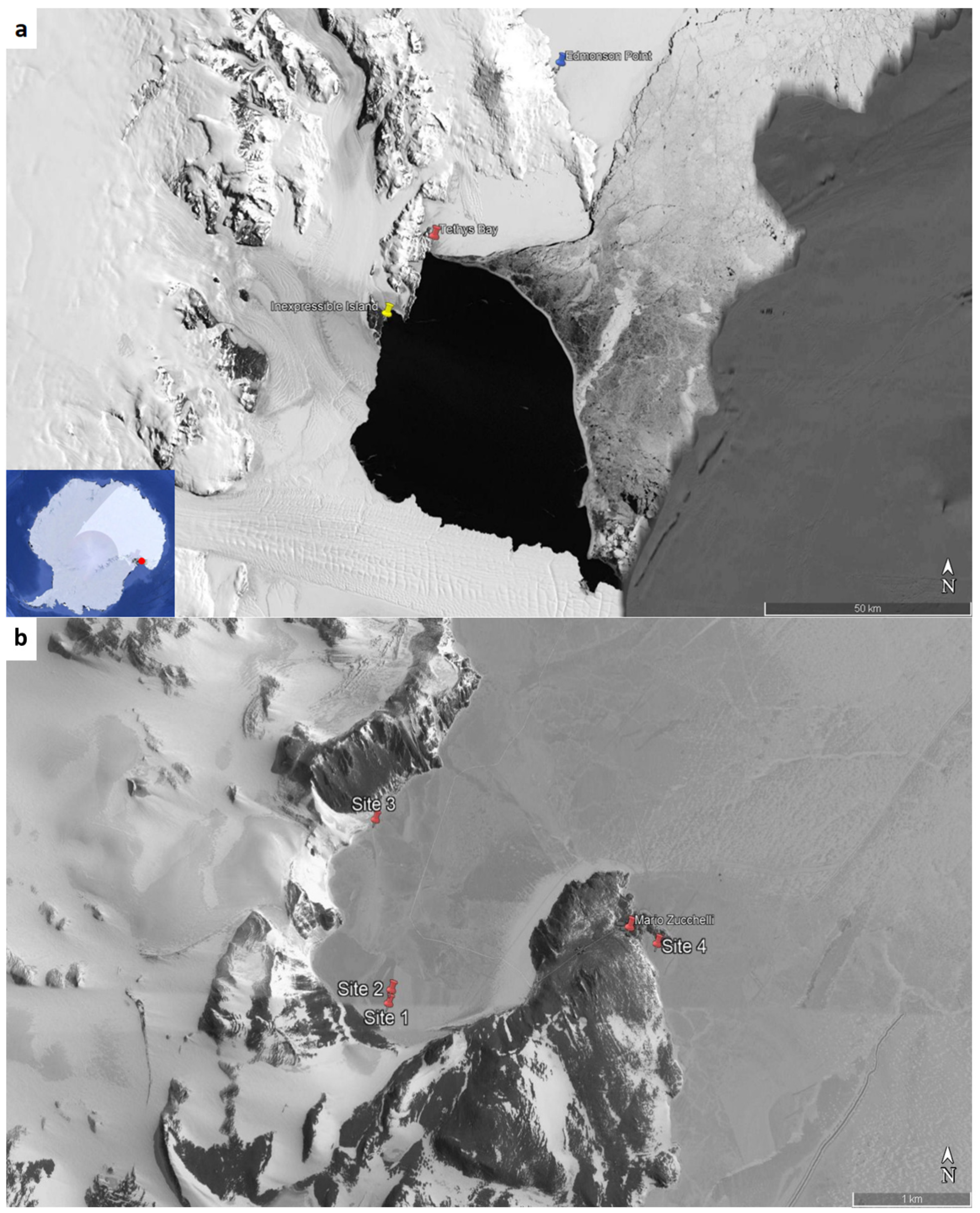
2.1. Sampling

Marine surface water, ice, and sediment samples from different coastal areas in Antarctica, listed in Table 1, were collected during the Italian XXIX (January 2014), XXXIII (October–November 2017) and XXXIV (November–December 2018) expeditions at the Mario Zucchelli Station, including different sites in Tethys Bay and Road Bay near the Italian station and 2 sites, Edmonson Point and Inexpressible Island, reached by helicopter.
All samples were collected in duplicate into sterile 50 mL conical tubes and stored at +4 °C and −20 °C until further use. In some cases (where possible for logistical constraints), 5–10 L of surface water were also collected, filtered (0.45 μm) by a peristaltic pump, and stored in 20% glycerol at −80 °C.

2.2. Media

Marine Broth (MB) and R2A media were those of Conda Pronadisa (Madrid, Spain). The Luria–Bertani (LB) growth medium (per litre) contained bacto-tryptone 10.0 g (VWR, Leuven, Belgium), yeast extract 5.0 g (Applichem, Darmstadt, Germany), and sodium chloride 10.0 g (Applichem, Darmstadt, Germany). The TGY growth medium (per litre) contained bacto-tryptone 3.0 g (VWR, Leuven, Belgium), yeast extract 3.0 g (Applichem, Darmstadt, Germany), glucose 3.0 g (Applichem, Darmstadt, Germany). For solid media, 15 g of bacteriological agar (VWR, Leuven, Belgium) was added to 1 L of liquid medium.

2.3. Isolation and Selection of UV-Resistant Bacteria by UV-C Assay

The isolation of UV-resistant bacteria was carried out as previous described by [21]. Briefly, 100 μL of each marine water/melted ice sample or 100 μL aliquots stored in 20% glycerol were incubated into 0.9 mL of MB and R2A at 15 °C, 180 rpm, for 1 week. After that, bacterial cells were serially diluted ten-fold until 10−6 in MB and R2A. Moreover, 1 g of each sediment sample was suspended in 9 mL of sterile sea water, vigorously stirred by vortexing and incubated at 15 °C, 180 rpm, overnight. The supernatant was serially diluted tenfold until 10−6 in sterile sea water.
Aliquots (100 µL) of each sample prepared as described above were spread in duplicate on Marine Agar (MA) and R2A agar plates. One plate for each sample was exposed to UV-C irradiation for 45 s (corresponding to 0.135 J/cm2) inside a Bio-Link crosslinker and, subsequently, incubated at 15 °C for 1 week (or more, if necessary). After that, only the UV-resistant colonies were visible on plates. On the contrary, one plate for each sample was incubated directly at 15 °C without UV exposure and used as negative control.
A UV-C assay was performed as previously described by [21] to characterize the UV resistance of Antarctic bacteria. Identified bacterial isolates were grown in MB or R2A broth at 15 °C with constant shaking until an OD600 ~0.3 was reached [22]. Then, 100 μL of each culture were plated in MA or R2A agar (in triplicates). Each plate was exposed to UV-C irradiation for 0, 1, 2, and 3 min (corresponding to 0, 0.180, 0.360, and 0.540 J/cm2, respectively) inside a Bio-Link crosslinker and then incubated at 15 °C for 1 week. Some strains, which were very resistant (showing more colonies on plates after 3 min of UV-C irradiation), were also exposed to UV-C for 4, 5, and 6 min (corresponding to 0.720, 0.900, and 1.08 J/cm2, respectively) and incubated at 15 °C for 1 week [21]. The same protocol was used for Escherichia coli MG1655 (UV-C sensitive) and Deinococcus radiodurans (UV-C resistant), as negative and positive controls, respectively. They were grown at 37 °C in LB (E. coli) or at 30 °C in TGY (D. radiodurans); colonies were visible on plates after only 2–4 days [21].

2.4. PCR Amplification of 16S rRNA Gene of UV-Resistant Bacteria

Selected isolates (from the plates exposed to UV), resistant to UV-C irradiation for 45 s (0.135 J/cm2) (Section 2.2), were identified by 16S rRNA sequencing. UV-resistant Antarctic bacterial colonies, grown overnight at 15 °C on MA or R2A agar plates, were resuspended in 50 μL of sterile distilled water, frozen for 60 min, then heated to 95 °C for 10 min, and cooled on ice for 5 min. PCR amplification of 16S rDNA gene was carried out using universal primers 27F (5′-AGAGTTTGATCCTGGCTCAG-3′) and 1492R (5′-GGTTACCTTGTTACGACTT-3′) designed from the conserved bacterial sequences at the 5′ and 3′ ends of the 16S rRNA gene of Escherichia coli rDNA, thus allowing the amplification of nearly the entire gene [23]. The reaction mix consisted of 1 μL of PCR colony as template, 1.25 μL of each primer, 5 μL of Q5® High-Fidelity Reaction Buffer (New England Biolabs), 0.5 μL of dNTP, 0.25 μL of Q5® High-Fidelity DNA Polymerase (New England Biolabs) and 15.75 μL of molecular grade water. The amplification steps included initial denaturation at 98 °C for 3 min, 35 cycles at 98 °C for 10 s, 60 °C for 30 s, 72 °C for 40 s, and a final extension at 72 °C for 10 min.
The PCR products were analysed on 1% agarose gel and checked by sequencing of both strands by a commercial sequencing service (Eurofins Genomics, Ebersberg, Germany). Other universal bacterial primers were used to sequence the initial (518R: 5′-GTATTACCGCGGCTGCTGG-3′) and the final (967F: 5′-CAACGCGAAGAACCTTACC-3′) parts of the 16S rRNA gene [24].

2.5. Identification and Phylogenetic Analysis

The sequences of 16S rRNA obtained from the bacterial isolates were analysed with the BioEdit program and compared with sequences deposited in the NCBI GenBank database by using the BLASTn tool. Furthermore, evolutionary analyses were conducted using Molecular Evolutionary Genetics Analysis (MEGA) version 6 [25] with Clustal W sequence alignments [26]. The evolutionary history was inferred by using the Maximum Likelihood method based on the Jukes–Cantor model [27].
Each 16S rRNA gene sequence was deposited in the NCBI GenBank database under the accession numbers shown in Table 2 and in the phylogenetic trees.

3. Results ad Discussion

3.1. Sampling

Sampling was performed in several marine coastal sites in Victoria Land (Figure 1a,b), a region in eastern Antarctica, which fronts the western side of the Ross Sea (Southern Ocean) and is the most extensive continental shelf ecosystem South of the Antarctic Polar Front.
This area is characterized by the rigid Antarctic climate, with the average monthly air temperature ranging between −25.9 °C (August) and −0.1 °C (January). Furthermore, there is complete darkness during winter (from May to August) and 24 h of light during the summer (from October to February), with December and January showing the maximum monthly average radiation, corresponding to 320 and 300 Wm−2, respectively [28].
Sea water/ice sampling was carried out in different sites of Tethys Bay (Figure 1a,b, red sites 1, 2, and 3) and Road Bay (Figure 1b, red site 4), in the surroundings of the Mario Zucchelli Station, the Italian seasonal research station, located at Terra Nova Bay, Ross Sea. The bay is a coastal marine area encompassing 29.4 km2, which is very important for scientific investigations in Antarctica. It is generally free of ice during the summer due to the western katabatic winds that blow from the plateau towards the sea.
Sea water/ice sampling was also performed in the Inexpressible Island area (Figure 1a, yellow site). Site 5 is far from the others (around 30 km) whereas sites 6, 7, and 8 are about 200 m from each other. It is almost completely free from glaciers and culminates at 390 m.
Sea water/ice sampling was also performed in the Edmonson Point area (Figure 1a, blue site), located approximately 50 km NE of the Mario Zucchelli Station, in Wood Bay, Ross Sea. Site 10 is far at around 20 km from site 11, whereas site 11 is about 5 km from sites 9 and 12. These latter sites are about 500 m from each other. Edmonson Point is covered by hills, knolls, and moraines of volcanic material, and it is divided by small valleys with streams, ponds, and lakes [28]. In general, the soil is dark in colour, which favours the melting of snow in spring; in summer, there are some shallow lakes free of ice cover, while deeper lakes are permanently frozen. Generally, Antarctic lakes are dominated by microbial organisms, including bacteria, protozoa, and phytoplankton [29,30], of which little information is available. Interestingly, they include freshwater and saline systems that have experienced little or no anthropogenic impact and, therefore, harbour pristine biotopes. In this area, in addition to sea water sampling, sediments from a shallow lake, which was free of ice, were sampled as well (site 12).

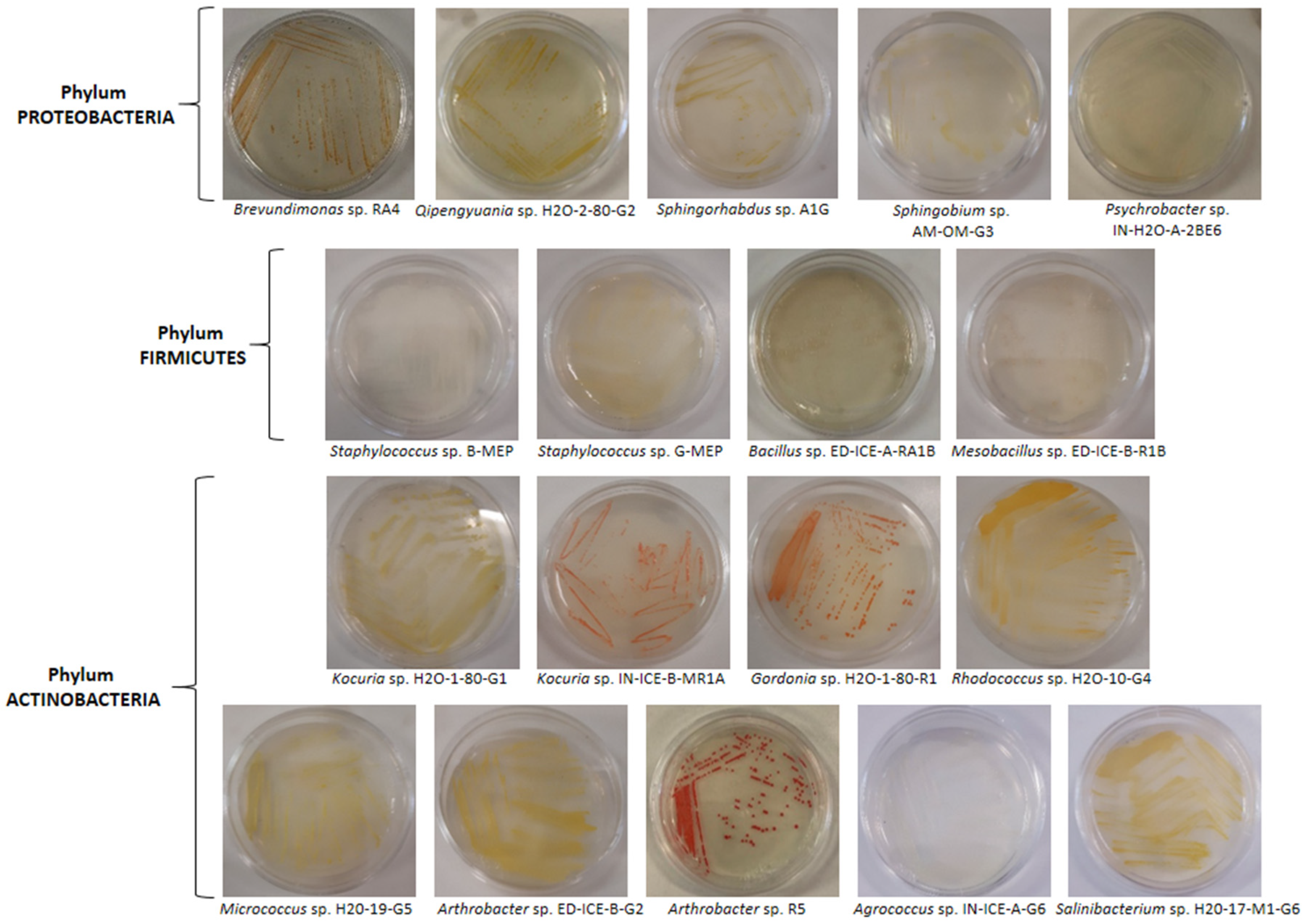
3.2. Isolation and Identification of UV-Resistant Bacteria by UV-C Assay

In order to obtain cultivable UV-resistant bacteria, each sea water/melted ice sample or sediment sample collected from different sites in Antarctica was prepared as described previously (Section 2.2). Several colonies were present on all MA plates subjected to UV-C irradiation, whereas very few bacterial isolates were present on the R2A agar plates. On the contrary, bacteria, incubated directly at 15 °C for 1 week and not exposed to UV-C rays, grew to confluence on the plates (no UV-C control plate).
The tolerance to UV-R of all the isolates obtained was studied by exposing them to UV-C radiation for different times (from 1 to 3 min corresponding to 0.180, 0.360, and 0.540 J/cm2, respectively), as shown in Table 2. The four most UV-C resistant isolates derived from surface sediment samples, which showed more colonies on plates after 3 min of UV-C irradiation, were exposed to UV-C for longer times (from 4 to 6 min) and compared with the positive control D. radiodurans (UV-C resistant) [21]. As shown in Table 2, these strains still displayed colonies after up to 6 min of exposure to UV-C.
Table 2. UV-resistant bacterial strains isolated from Antarctic marine and lake environments.
StrainPigmentationSample Source-
Collection Site
Identification by
16S rRNA
UV-C
Resistance (min)
Accession Number (AN)Next Relative by GenBank Alignment (AN, Organism)Sequence Homology (%)
Phylum PROTEOBACTERIA
Class Alphaproteobacteria (Gram negative)
RA4Reddish orangeLake sediments—Edmonson Point (site 12)Brevundimonas sp.6OQ540805EF093132.1, Brevundimonas sp. VTT E-05291499.93
ED-ICE-A-RA1Reddish orangeSea ice—Edmonson Point (site 10)Brevundimonas sp.3OQ540806EF093132.1, Brevundimonas sp. VTT E-05291499.79
IN-H2O-B-RA3Reddish orangeSea water—Inexpressible Island (site 8)Brevundimonas sp.3OQ540807AJ244710.1, Brevundimonas sp. V4.BP.05100
H2O-2-80-G2Brilliant yellowSea water/ice—Tethys Bay (site 1)Qipengyuania sp.3OQ540808MZ749497.1, Qipengyuania aerophila GH25100
G5GYellowLake sediments—Edmonson Point (site 12)Sphingorhabdus sp.6OQ540809MN255826.1, Sphingorhabdus soli
D-2Q-5-6
100
A1GYellowLake sediments—Edmonson Point (site 12)Sphingorhabdus sp.6OQ540810MN255826.1, Sphingorhabdus soli
D-2Q-5-6
100
AM-0m-G3 1Pale yellowSea ice—Tethys Bay (site 3)Sphingobium sp.3OQ540811CP016456.1, Sphingobium sp. RAC03100
Class Gammaproteobacteria (Gram negative)
IN-H2O-A-2BE6CreamySea water—Inexpressible Island (site 7)Psychrobacter sp.2OQ540812EU196303.1, Psychrobacter sp. NP5299.64
IN-ICE-A-A1BBeigeSea ice—Inexpressible Island (site 7)Psychrobacter sp.2OQ540813CP012533.1, Psychrobacter sp. P11G599.87
H2O-1-80-BE1BeigeSea water—Edmonson Point (site 9)Psychrobacter sp.3OQ540814ON209525.1, Psychrobacter okhotskensis PCRB17b99.86
IN-ICE-A-A1GCreamySea ice—Inexpressible Island (site 7)Psychrobacter sp.1OQ540815FR750957.1, Psychrobacter nivimaris CMS161 99.66
Gly6-2-BE4BeigeSea water—Tethys Bay (site 2)Psychrobacter sp.3OQ540816KU579272.1, Psychrobacter nivimaris OUCMDZ421999.93
Gly9-1-BE7BeigeSea water—Road Bay (site 4)Psychrobacter sp.1OQ540817CP106752.1, Psychrobacter sp. SC65A.3100
Phylum FIRMICUTES
Class Bacilli (Gram positive)
B-MEPWhiteSea water—Edmonson Point (site 9)Staphylococcus sp.2OQ540818CP054831.1, Staphylococcus saprophyticus UTI-04599.93
G-MEPPale YellowSea water—Edmonson Point (site 9)Staphylococcus sp.2OQ540819CP054831.1, Staphylococcus saprophyticus UTI-04599.93
H2O-2-80-R3WWhiteSea water/ice—Tethys Bay (site 1)Staphylococcus sp.1OQ540820MN826459.1, Staphylococcus saprophyticus TA1100
ED-ICE-B-R1BTranslucent whiteSea ice—Edmonson Point (site 11)Mesobacillus sp.1OQ540821KY202702.1, Mesobacillus subterraneus A999.86
ED-ICE-A-RA1BTranslucent whiteSea ice—Edmonson Point (site 10)Bacillus sp.3OQ540822MT332156.1, Bacillus cereus DBA1.1100
Phylum ACTINOBACTERIA
Class Actinobacteria (Gram positive)
H2O-1-80-G1Brilliant yellowSea water—Edmonson Point (site 9)Kocuria sp.3OQ540823AM237350.1, Kocuria carniphila OS-32.d199.93
IN-H2O-A-BE6RRed coralSea water—Inexpressible Island (site 7)Kocuria sp.1OQ540824AM418390.1, Kocuria sp. 29Y1zhy99.93
IN-ICE-B-MR1ARed coralSea ice—Inexpressible Island (site 8)Kocuria sp.1OQ540825CP035103.1, Kocuria rosea ATCC 18699.93
H2O-1-80-R1Brilliant orangeSea water—Edmonson Point (site 9)Gordonia sp.1OQ540826CP049836.1, Gordonia terrae RL-JC0299.86
H2O-2-80-R4Brilliant orangeSea water/ice—Tethys Bay (site1)Gordonia sp.1OQ540827CP049836.1, Gordonia terrae RL-JC02100
H2O-10-G4Yellowish orangeSea water—Tethys Bay (site 2)Rhodococcus sp.1OQ540828JX428873.1, Rhodococcus sp. ZS33399.80
H2O-19-G5Brilliant yellowSea water—Inexpressible Island (site 6)Micrococcus sp.3OQ540829CP097650.1, Micrococcus yunnanensis TT999.86
ED-ICE-B-R1GBrilliant yellowSea ice—Edmonson Point (site 11)Micrococcus sp.2OQ540830CP043842.1, Micrococcus luteus NCCP 1683199.79
ED-ICE-B-G2Brilliant yellowSea ice-Edmonson Point (site 11)Arthrobacter sp.2OQ540831KR085775.1, Arthrobacter flavus IHBB 9551100
ED-ICE-B-R1Brilliant redSea ice—Edmonson Point (site 11)Arthrobacter sp.1OQ540832JX949321.2, Arthrobacter cheniae99.73
R5Brilliant redLake sediments—Edmonson Point (site 12)Arthrobacter sp.6OQ540833KU921543.1, Arthrobacter agilis IHBB 997999.66
IN-ICE-A-G6 1CreamySea ice—Inexpressible Island (site 7)Agrococcus sp.2OQ540834MW580037.1, Agrococcus sp. AHE_PA_07299.72
H2O-17-M1-G6Brilliant yellowSea ice—Inexpressible Island (site 5)Salinibacterium sp.2OQ540835MK140978.1, Salinibacterium sp. s4a41-1099.86
1 These strains were isolated and grown in R2A medium.
All bacterial UV-C-resistant strains were identified by 16S rRNA sequencing, and their sequences were compared with the closest relatives of isolates deposited in the NCBI GenBank database using the BLASTn tool to determine their taxonomical relationships (Table 2). Comparative sequence analysis indicated that the UV-C resistant Antarctic bacteria were closely related to known bacteria with a 16S rRNA sequence homology >99%.
The bacterial strains belonged to Proteobacteria (class Alpha and Gammaproteobacteria), Actinobacteria (class Actinobacteria), and Firmicutes (class Bacilli) phyla (Table 2). The results revealed that the 31 isolates were representative of 15 bacterial genera: 5 of which were Gram negative (Brevundimonas, Qipengyuania, Sphingorhabdus, Sphingobium, and Psychrobacter) and 10 of which were Gram positive (Staphylococcus, Bacillus, Mesobacillus, Kocuria, Gordonia, Rhodococcus, Micrococcus, Arthrobacter, Agrococcus, and Salinibacterium).
Strains affiliated with Proteobacteria and Actinobacteria phyla were detected in all sites and were the most abundant species in all of the different environments included in this study, as has already been reported in other Antarctic environments [31,32,33]. Affiliates to the genera Psychrobacter (among the Gammaproteobacteria) appeared to be predominant in all investigated Antarctic sites. Conversely, isolates belonging to Actinobacteria appeared to be the most diverse and rich in species among all the collected sites. Interestingly, Alpha and Gammaproteobacteria species, which are more traditionally associated with the marine environment, were also isolated from the lake sediment (Edmonson Point, Sites 12). Considering the proximity of the lake to the sea, it is not excluded that there was a contribution of bacteria from the marine environment to the Antarctic Lake through atmospheric deposition (aerosols) and/or seabird droppings [30,34,35].
Interestingly, 20 of 31 isolates showed pigmentation exhibiting various colours with different shades ranging from brilliant red to brilliant orange, to reddish or yellowish orange, and from pale yellow to brilliant yellow and yellowish. The other 11 strains were not pigmented, showing from cream to translucent white colonies (Figure 2, Table 2). The colonies of Arthrobacter spp. exhibited different colors, including brilliant yellow (ED-ICE-B-G2) and brilliant red (ED-ICE-B-R1 and R5) with colonies of Kocuria spp. that were brilliant yellow (H2O-1-80-G1) and red coral (IN-H2O-A-BE6R and IN-ICE-B-MR1A), as has already been reported for both strains [36,37].
Bacterial pigmentation is determined by the expression of pigments that, in many cases, are represented by carotenoid-type compounds, which are often involved in the protective role against the negative effects of UV-R [3,12]. As chemical quenchers of singlet oxygen, they function as potent scavengers of ROS [38]. In addition to being essential constituents of photosynthetic organisms (e.g., plants, algae, and cyanobacteria) [39], in heterotrophic bacteria, carotenoids provide protection against exposure to solar UV and freeze–thaw cycles [40]. In the Antarctic environment, they may contribute to membrane stability by maintaining proton permeability and augmenting oxidative stress resistance [41,42]. The presence of carotenoids has been already reported in the literature for most genera studied in this work (see below Section 3.3).
Bacterial pigments are promising and sustainable bioactive compounds, which may be used in cosmetics, food, textiles, printing, and pharmaceutical products [43,44,45]. They can be produced from bacterial sources easily and cheaply, compared to plant sources, obtaining a huge biomass by culturing them in controlled conditions in bioreactors [44,46]. For their wide range of applications in different industries, bacterial pigments are getting more attention in the global pigments market (https://www.grandviewresearch.com/industry-analysis/dyes-and-pigments-market, accessed on 16 March 2023) and in the global carotenoids market (https://www.researchandmarkets.com/reports/5682374/carotenoids-market-global-industry-trends, accessed on 16 March 2023). Among bacteria, marine species are a valuable source of these pigments with potential cosmeceutical and nutraceutical applications [46,47,48,49].

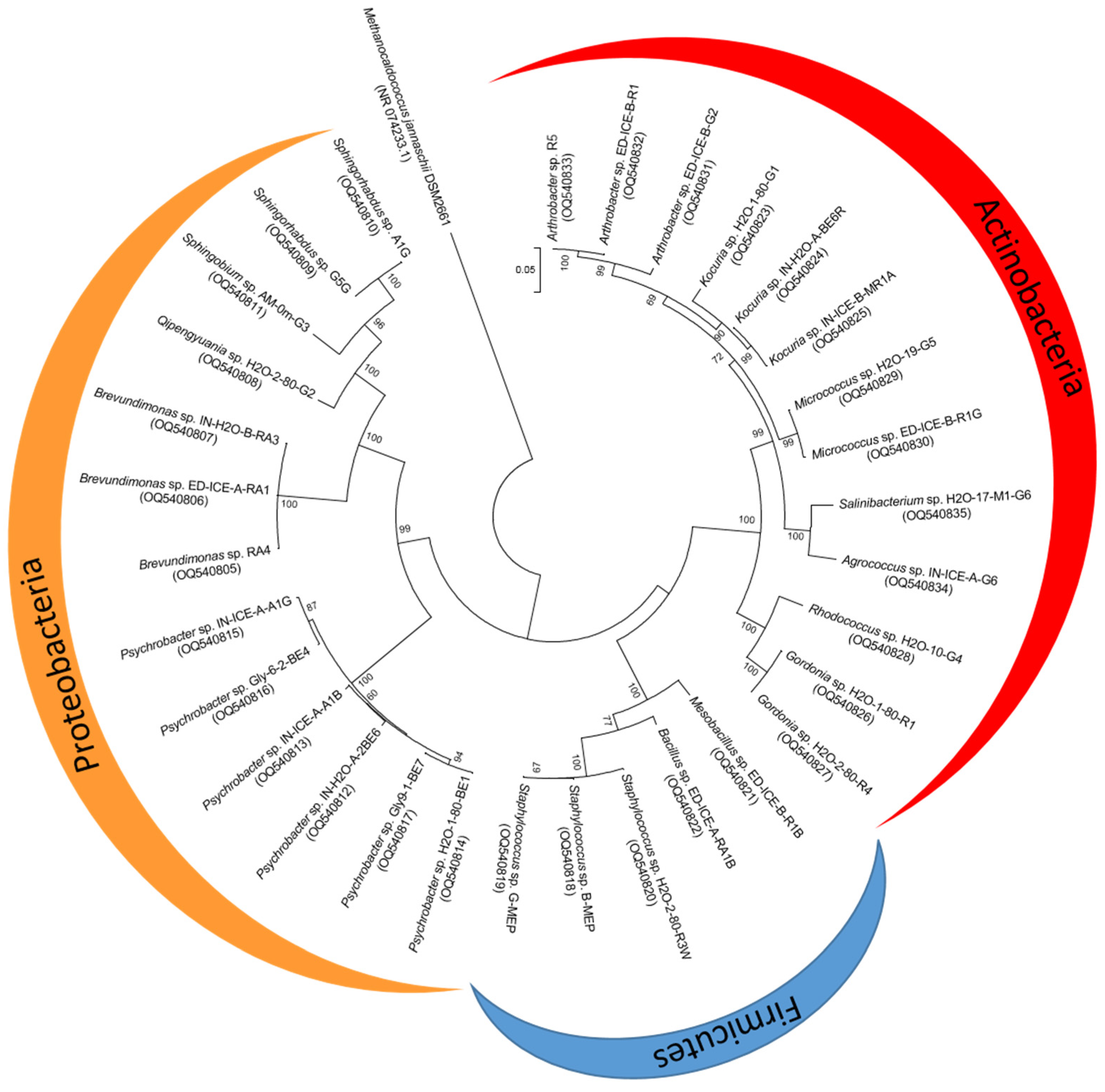
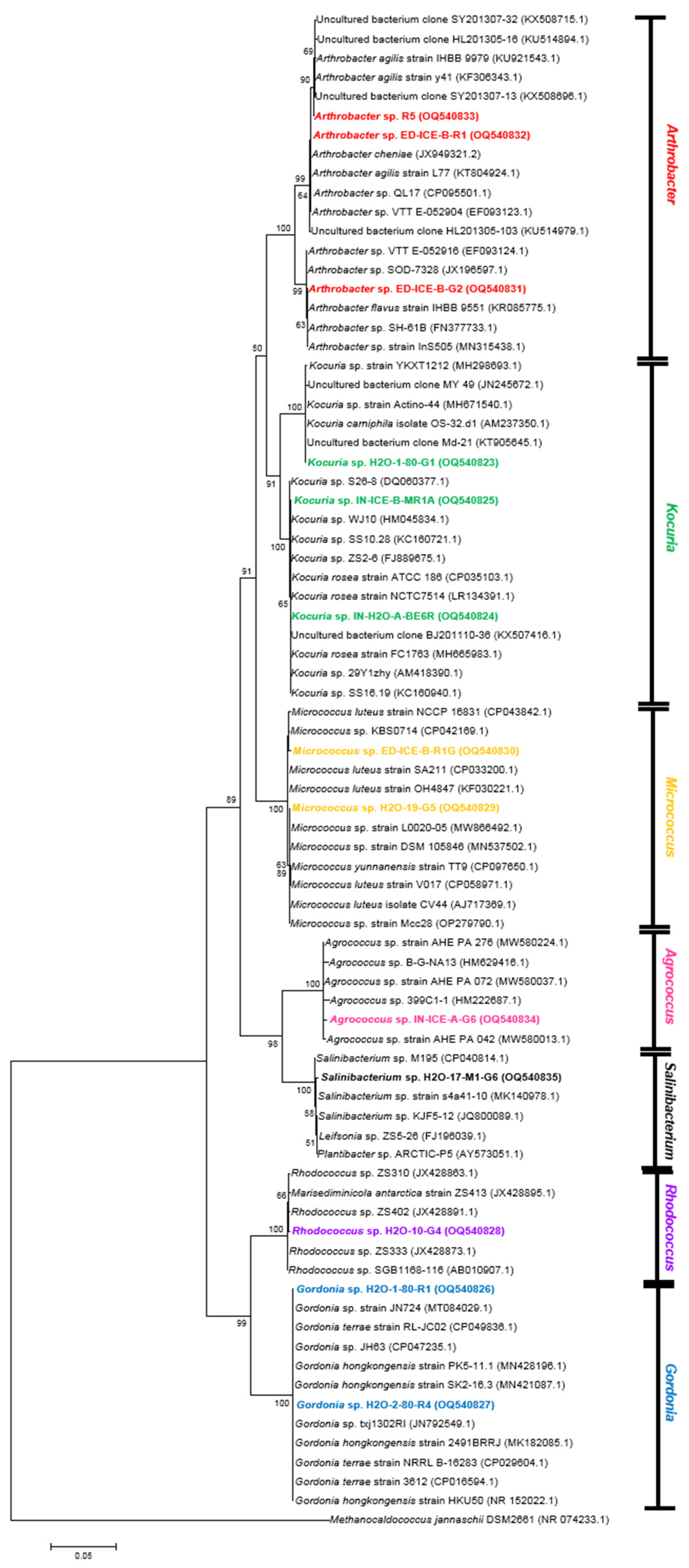
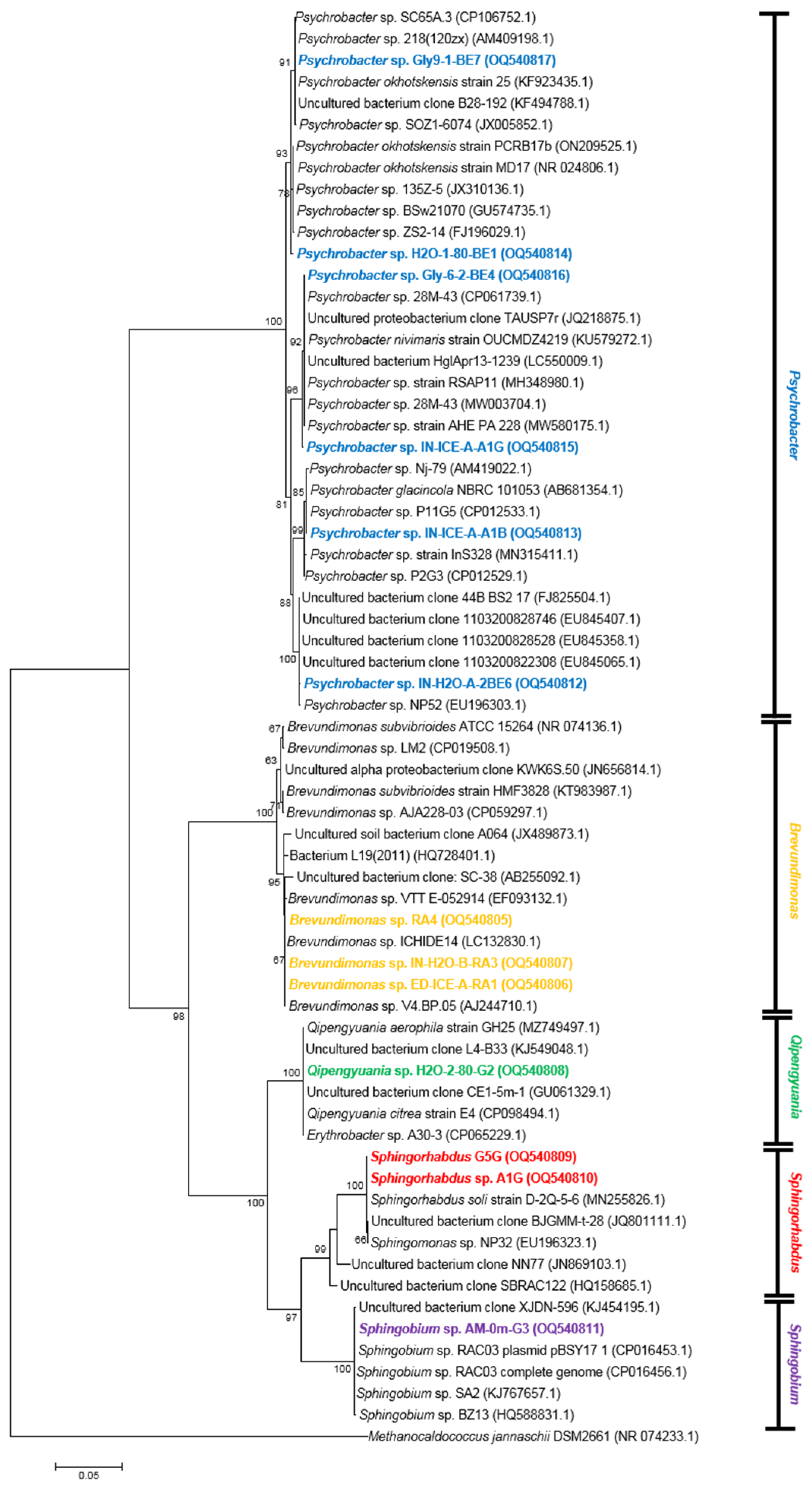
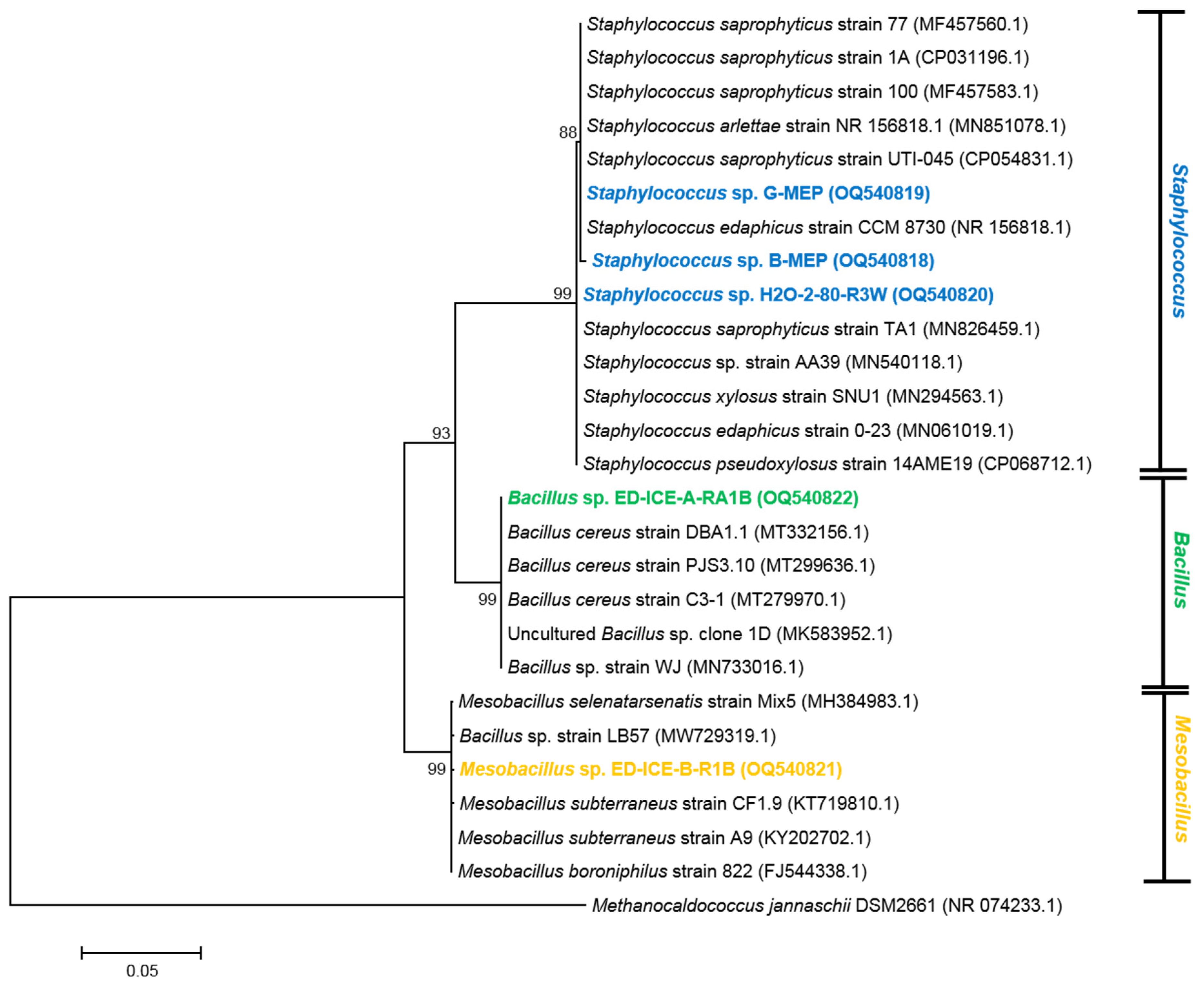
3.3. Diversity of UV-Resistant Antarctic Aquatic Bacteria and Phylogenetic Analysis

A phylogenetic tree made by using the 16S rRNA gene sequences of Antarctic marine and lake bacterial strains was constructed by using the Maximum Likelihood method based on the Jukes–Cantor model (bootstrap 500), Figure 3.
The analysis of the phylogenetic tree revealed the presence of two distinct phylogenetic clusters, one split into Gram-negative Proteobacteria phylum and the other including Gram-positive Actinobacteria and Firmicutes phyla. In the cluster of Gram positive bacteria, the Firmicutes phylum was split into a separate group of Staphylococcus, Bacillus, and Mesobacillus.
To further investigate the evolutionary relatedness between each strain and other members of the same genus, representative sequences (5 for each strain with the highest percentage of identity sequence) were downloaded from the NCBI to build phylogenetic trees (Figure 4, Figure 5 and Figure 6).

3.3.1. Actinobacteria

Phylogenetic studies of the bacteria belonging to the Phylum Actinobacteria revealed that the tree was split in two lineages. The first one divided in many subgroups comprising Arthrobacter, Kocuria, Micrococcus, Agrococcus, and Salinibacterium spp., whereas the second clade was divided in two subgroups represented by Rhodococcus and Gordonia spp. (Figure 4).
Arthrobacter, Micrococcus, and Kocuria species are aerobic Gram-positive bacteria of the family Micrococcaceae, and they all show interesting characteristics of UV-R resistance. Phylogenetic studies, supported by BLAST analyses of 16S rRNA gene sequences, indicated that the strains R5, ED-ICE-B-R1, and ED-ICE-B-G2 grouped together in the Arthrobacter clade, which was subdivided into two subgroups. One group comprised the red-pigmented R5 and ED-ICE-B-R1, which shared 99.53% of 16S rRNA gene sequence identity and were closely related to the red-pigmented Arthrobacter agilis strain IHBB 9979 [50] and Arthrobacter cheniae strain [51], respectively. The other group comprised the yellow-pigmented ED-ICE-B-G2, which was closely related to the yellow Antarctic Arthrobacter flavus strain IHBB 9551 [52] and shared 100% sequence identity with it (Figure 4).
Members of the genus Arthrobacter were among the most frequently isolated bacteria in extreme cold environments ranging from the Antarctic to Arctic and Himalaya, and they are well known for their metabolic versatility and wide prevalence in stressful environments [53,54,55,56]. This genus is a good carotenoid producer. For example, different carotenoids with high antioxidant activity and good stability under exposure to UV light were isolated from the Antarctic bacteria Arthrobacter agilis 50cyt and Arthrobacter psychrochitiniphilus 366 [57]. Other Antarctic isolates of Arthrobacter agilis produce the red carotenoid bacterioruberin and a C50 hydrocarbon known as tetraanhydrobacterioruberin [41]. The rare C50 carotenoid bacterioruberin in Arthrobacter agilis DSM 20550T and Arthrobacter bussei DSM 109896T has been reported to modulate membrane fluidity, thereby increasing cell resistance to freeze–thaw stress [58]. Arthrobacter sp. P40 strains, isolated from Fildes Peninsula in King George Island, produced C50 carotenoids, such as decaprenoxanthin and its glucosylated derivatives, as well as lycopene used as synthesis precursors of C50 carotenoids [59]. Other strains, Arthrobacter sp. QL17 and Arthrobacter sp. G20, produced carotenoids with possible applications in the food and pharmaceutical industries [60,61].
Arthrobacter genomes isolated from Antarctic soils contained several genes that conferred protection from free radical damage and from ROS. Among them, there were up to two copies of the SOD gene and up to three copies of the CAT gene, as well as several copies of the peroxiredoxin gene and thioredoxin genes [54]. Other Antarctic Arthrobacter strains showed the presence of genes encoding proteins involved in the DNA repair system in response to DNA damage caused by UV-R [62].
In the phylogenetic tree (Figure 4), Kocuria species, IN-ICE-B-MR1A, IN-H2O-A-BE6R, and H2O-1-80-G1, grouped together within the genus Kocuria and shared a high sequence identity of 16S rRNA genes between them (from 96.3–99.9%) (Figure 4). As occurred for Arthobacter, they were subdivided into two groups—one group consisting of the yellow-pigmented H2O-1-80-G1, which is closely related to the yellow Kocuria carniphila [63], and a second group comprising the two red coral IN-ICE-B-MR1A and IN-H2O-A-BE6R, which are closely related to the pink Kocuria rosea (CP035103.1), which is capable of tolerating extreme conditions [64]. The genus Kocuria, which was generated by the taxonomic dissection of the genus Micrococcus [65], included species resistant to gamma-irradiation [66] and UV-C radiation [64] with the ability to produce relatively large quantities of CAT [67]. Kocuria sp. 301 showed high resistance against UV-R, comparable to the remarkably radiotolerant D. radiotolerans, free radicals and desiccation [68]. Studies conducted on the carotenoids extracted from the Kocuria marina DAGII strain showed a higher antioxidant activity compared to β−carotene, thus suggesting several potential applications [69].
In this study, the yellow H2O-19-G5 and ED-ICE-B-R1G isolates, which shared 99.6% of 16S rRNA gene sequence identity, were grouped within the cluster of the Micrococcus genus (Figure 4). The Micrococcus sp. ED-ICE-B-R1G was closely related to Micrococcus luteus NCCP 16831 and Micrococcus sp. KBS0714, and the latter is resistant to environmental stressors [70]. Strains belonging to the genus Micrococcus are commonly found in temperate soil, water, mammalian skin, Antarctic ice, and desert soil, which is probably due to the ability of these bacteria to form biofilms or enter dormant stages following conditions such as desiccation and starvation [70,71,72].
Agrococcus and Salinibacterium species belong to the same family Microbacteriaceae. In Figure 4, they were grouped together and subdivided in two subgroups. Agrococcus sp. IN-ICE-A-G6, isolated in R2A medium, clustered tightly with species of marine Agrococcus isolated from the North Sea (AHE_PA_276, AHE_PA_072, AHE_PA_042). The yellow Salinibacterium sp. H2O-17-M1-G6 grouped closely to Salinibacterium sp. KJF5-12, which were isolated from the subarctic glacial Fjord Kongsfjorden [73], and Leifsonia sp. ZS5-26 isolated from Antarctic sea ice.
The genus Salinibacterium has mainly been isolated from sea water samples from the East Sea, from frozen soil from a Chinese glacier, and from Antarctic sediments or green snow [74,75,76,77]. Members of the bacterial genus Agrococcus are globally distributed and found across diverse environments, including polar ones [78]. Both genera are potentially able to produce carotenoids. Salinibacterium strains isolated from the Fildes Peninsula in King George Island presented a pigment profile composed of the carotenoid C.p. 450 free form and its glucosylated derivatives [59]. The Agrococcus pavilionensis strain RW1, which was isolated from microbialite collected in Pavilion Lake, British Columbia, was screened for genes involved in carotenoid production, and they had the genetic potential to produce lycopene, β-carotene, canthaxanthin, echinenone, and zeaxanthin or astaxanthin [79]. In this study, the isolated strain of Agrococcus sp. IN-ICE-A-G6 showed no pigmentation with creamy colonies isolated on R2A agar (Figure 2).
The 16S rRNA gene sequence analysis indicated that the yellowish orange strain H2O-10-G4 may be affiliated within the genus Rhodococcus, which belongs to the family Nocardiaceae, particularly with Rhodococcus sp. ZS333, which was isolated from Antarctic sandy intertidal sediments, as its closest relative (99.80% of 16S rRNA gene sequence identity) (Figure 4). However, the strain H2O-10-G4 shared 99.7% of 16S rRNA gene sequence identity also with the Marisediminicola antarctica strain ZS413T, which belongs to the family Microbacteriaceae and was isolated from the coastal area off the Chinese Antarctic Zhongshan Station [80]. To date, only two strains have been reported belonging to this genus: Marisediminicola antarctica strain ZS413T and Marisediminicola senii sp. nov., which was isolated from Queen Maud Land, Antarctica [81]. Comparison with 16S rRNA gene sequences (100 sequences showing the highest sequence identity with H2O-10-G4 strain) deposited in the NCBI GenBank database showed that the Marisediminicola antarctica strain ZS413T was the only species belonging to this genus, whereas all other strains with high sequence identities (from 98.00 to 99.80%) belonged to the genus Rhodococcus sp. Species belonging to the Rhodococcus genus are reported in widespread environments, including deep-sea and sea level sediments, alpine soils, and Arctic and Antarctic regions. They are able to survive under several stressful conditions, including UV irradiation [82]. The Antarctic marine bacterium Rhodococcus sp. NJ-530, which was isolated from floating ice, showed several UV adaptive characteristics, including significant repair activities of DNA damage induced by UV rays of the purified DNA photolyase [16]. The red-orange strain Rhodococcus sp. B7740, which was isolated from deep-sea water in the Arctic Ocean, is a promising source of natural carotenoids and isoprenoid quinones, which is interesting for their application in the food industry [83].
In Figure 4, the orange Gordonia species, H2O-1-80-R1, and H2O-2-80-R4, with a 99.7% of 16S rRNA gene sequence identity between them, were closely related to different strains of the Gordonia genus, Gordonia terrae and Gordonia hongkongensis, from the family Gordoniaceae. A strain of Gordonia terrae showing antimicrobial activity was already isolated from Antarctic volcanic soils in Deception Island [33]. Species belonging to the genus Gordonia are ecologically adaptable and produce a large variety and significant quantities of carotenoids that are recently gaining attention for their biotechnological potential [84]. An important example is provided by Gordonia jacobaea MV-1, which is capable of producing great amounts of carotenoids [85], including ketocarotenoid all-trans-canthaxanthin (4,4′-diketo-β-carotene) and all-trans-astaxanthin (3,3′-dihydroxy-4,4′-diketo-β-carotene), which are currently produced by Hoffmann-La Roche, Ltd. and used as food additives in feeding.

3.3.2. Proteobacteria

Proteobacteria constituted one of the most dominant phyla in Antarctica [31,32,33] and in this study comprised 5 Gram-negative genera Brevundimonas, Qipengyuania, Sphingorhabdus, Sphingobium, and Psychrobacter. The phylogenetic tree of bacteria affiliated with the Phylum Proteobacteria was split into two distinct phylogenetic lineages (Figure 5) that, in turn, were divided in other groups: two groups for the Psychrobacter genus and another two groups divided in other subgroups that comprised Brevundimonas, Qipengyuania, Sphingobium, and Sphingorhabdus.
In the current study, Psychrobacter was the most abundant genus and comprised 19% of the identified sequences. Psychrobacter spp. IN-H2O-A-2BE6, IN-ICE-A-A1B, H2O-1-80-BE1, IN-ICE-A-A1G, Gly6-2-BE4, and Gly9-1-BE7 were isolated from different sites, shared from 97.6 to 99.7% of 16S rRNA gene sequence identity, and were not pigmented. They grouped within the Psychrobacter genus, of the family Moraxellaceae, and were split into three groups. This genus is known to be widely distributed in different habitats, including fish, food, clinical specimens, Antarctic soils, and sea water [86]. Psychrobacter is a good producer of antioxidants involved in oxidative stress defence in Antarctic and Arctic environments [87,88,89].
The 16S rRNA gene sequence analysis indicated that Brevundimonas spp. RA4, ED-ICE-A-RA1, and IN-H2O-B-RA3 shared more than 99.70% of sequence identity between them, and they were grouped together within the clade of the genus Brevundimonas, of the family Caulobacteraceae, which were close to species found from different sources (Figure 5). One of these, Brevundimonas sp. V4.BP.05, was identified as Brevundimonas mediterranea sp. nov., which was isolated from the Mediterranean Sea [90]. Although the majority of Antarctic Brevundimonas strains reported in the literature displayed no pigmentation [35,91,92,93], in this study, all three isolates, RA4, ED-ICE-A-RA1, and IN-H2O-B-RA3, showed a reddish orange pigmentation, as has already been reported in other environments [94,95]. Some orange Brevundimonas strains isolated from aquatic environments in Japan were producers of astaxanthin and adonixanthin [94]. Red 2,2′-dihydroxy-astaxanthin was the major carotenoid produced by B. scallop and has strong antioxidative activity and potential industrial use [96].
In Figure 5, the yellow species H2O-2-80-G2 aligned closely with Erythrobacter and Qipengyuania species in sharing 100% of 16S rRNA gene sequence identity with them. Both genera, Erythrobacter and Qipengyuania, belong to the family Erythrobacteraceae of the order Sphingomonadales within the class Alphaproteobacteria. Recently, the taxonomy of the family Erythrobacteraceae has been revised, thereby leading to the formation of many novel genera within the family. Some species of the genus Erythrobacter were proposed to be reclassified into the genus Qipengyuania. The genus Qipengyuania currently comprises 13 species with validly published names, thus showing a high degree of genetic diversity, metabolic versatility, and environmental adaptation [97,98,99,100], (https://lpsn.dsmz.de/genus/qipengyuania, accessed on 24 January 2023). For this reason, the species H2O-2-80-G2 was assigned to the genus Qipengyuania. Members of the genus Qipengyuania are heterotrophic bacteria generally that are found worldwide in diverse marine environments with great possible applications in several fields (e.g., carotenoid producer) [100,101].
In Figure 5, the three yellow Sphingorhabdus spp. A1G, G5G, and Sphingobium sp. AM-0m-G3 belong to the same family Sphingomonadaceae. The 16S rRNA gene analysis revealed that Sphingorhabdus spp. A1G and G5G shared 100% of 16S rRNA gene sequence identity with the Sphingorhabdus soli strain D-2Q-5-6, whereas 99.93% of 16S rRNA gene sequence identity was shared with species affiliated with the genus of Sphingomonas sp. NP32. Sphingomonas strains are highly UVC-resistant bacteria, which is probably due to their ability to produce carotenoids [102], as well as CPD photolyases and a 6,4-photolyase to repair DNA damage [18]. Members of Sphingorhabdus have been isolated from various habitats, including freshwater, sea water, and polar environments [103,104], and analysis of the genome sequencing of some strains revealed the presence of a terpenoid gene cluster with high similarity encoding the carotenoid astaxanthin [105].
Sphingobium sp. AM-0m-G3, which was isolated in R2A medium, shared a 100% sequence identity with Sphingobium sp. RAC03 (Figure 5). Sphingobium is one of the four genera commonly called Sphingomonas, together with Sphingomonas, Novosphingobium, and Sphingopyxis. Strains belonging to the genus Sphingobium have been found in different environments, including in Antarctica, where they have been characterized for their ability to degrade aromatic and aliphatic hydrocarbons [106,107]. Analysis of the carotenoids extracted from the yellow-pigmented carbazole-degrading bacterium Sphingobium yanoikuyae XLDN2-5 showed the production of zeaxanthin, which is likely involved in the modulation of membrane fluidity and protection against oxidative stress [108].

3.3.3. Firmicutes

The phylogenetic tree of bacteria affiliated with the Phylum Firmicutes was split into two distinct phylogenetic clusters (Figure 6).
The bigger clade was formed by Staphylococcus and some strains of Bacillus genera, which were split in a separate branch. The more recent clade was formed by other Bacillus genera of the family Bacillales. The genus Bacillus is a heterogeneous group of species exhibiting polyphyletic branching with other new genera of the family Bacillaceae that were recently proposed [109]. In fact, Mesobacillus sp. ED-ICE-B-R1B shared 99.86% of its sequence identity with other sequences of the group, with some of these belonging to Mesobacillus or Bacillus genera. Mesobacillus sp. ED-ICE-B-R1B and Bacillus sp. ED-ICE-A-RA1B shared 93.35% of 16S rRNA gene sequence identity between them. Bacillus sp. ED-ICE-A-RA1B was phylogenetically close to Bacillus cereus, which has been found in the waters of Lake Vostok, Antarctica [93]. Staphylococcus spp. B-MEP, G-MEP, and H2O-2-80-R3W shared 99.15% of 16S rRNA gene sequence identity and were closely related to the genus Staphylococcus, of the family Staphylococcaceae. Members of the genus Staphylococcus are widespread in nature, and their ability to act as pathogens is partly due to their ability to alleviate oxidative and nitrosative stress through the development of different protection, detoxification, and repair mechanisms, including the production of carotenoids or detoxifying enzymes [110]. Staphylococcus saprophyticus, which is the closest phylogenetic relative of the Staphylococcus strains isolated in this study, is an opportunistic pathogen that is able to infect humans, lower mammals, and birds, and it can cause human urinary tract infections, wound infections, and septicaemia [111,112]. The Antarctic Staphylococcus edaphicus sp. nov., which is closely related to Staphylococcus sp. H2O-2-80-R3W, presented characteristics that are essential to adaptation to extreme environments [113].
Mesobacillus sp. ED-ICE-B-R1B and Bacillus sp. ED-ICE-A-RA1B exhibited no pigmentation. Only a few studies have reported the isolation of pigmented Bacillus isolates from marine sources [114]. However, marine Bacillus species showed good antioxidant activity by producing a wide range of enzymes and metabolites [115,116,117]. In the case of Staphylococcus species, only Staphylococcus sp. G-MEP exhibited pale yellow pigmentation, even though it shared 100% of its 16S rRNA sequence identity with the white B-MEP (Figure 2). This yellow pigment in G-MEP may be due to the formation of a carotenoid intermediate pigment found in the same Staphylococcus species [118,119].

4. Conclusions

The extreme conditions of the Antarctic aquatic environment have selected a high biodiversity within the bacterial communities, which are able to tolerate and survive to the constant exposure to UV-R. Our results highlight the importance of the Antarctic environment as a rich and novel source of pigmented UV-resistant Gram-positive and Gram-negative bacteria. In total, 42% of the UV-resistant strains isolated from the surface sea waters/ice and shallow lake sediments were assigned to the Phylum Proteobacteria that was represented by 5 genera (Brevundimonas, Psychrobacter, Qipengyuania, Sphingorhabdus, and Sphingobium); the other 42% was affiliated with Actinobacteria with 7 genera (Kocuria, Gordonia, Rhodococcus, Micrococcus, Arthrobacter, Agrococcus, and Salinibacterium), and the remaining 16% was assigned to the Phylum of Firmicutes represented by 3 genera, i.e., Staphylococcus, Mesobacillus, and Bacillus. Most of the Antarctic strains showed a close phylogenetic relationship to bacteria obtained from diverse Antarctic and non-Antarctic ecosystems, as well as marine and non-marine environments. However, many of them shared the harsh conditions in which they inhabited, which suggests that these species possessed adaptive features that allowed them to survive in these hostile environments.
It is widely reported that the bacterial genera selected in this work have been previously characterized as good producers of molecules that are able to minimize UV damage, including antioxidant molecules and enzymes. Many of them are represented by pigments, in particular carotenoids, that function as protection against UV-R and ROS, as well as modulate the membrane fluidity in the cold.
Antarctica is a huge reservoir of pigmented bacterial biodiversity but a still poorly explored resource for pigment discovery, production, and applications. There could be promising candidates for novel chemical structures and for cell factories of bio-pigments. Although the literature studies on Antarctic pigmented bacteria are increasing rapidly, currently, there are only few examples of Antarctic pigments that have been used in biotechnological applications, such as the carotenoids isolated from UV-resistant Antarctic bacteria used to develop green solar cells, called photosensitizers, in dye-sensitized solar cells [22]. Therefore, further studies are needed to investigate the potential of pigmented Antarctic bacteria and related molecular and enzymatic machineries to be used in bio-technological discovery pipelines and pharmaceutical applications.

Author Contributions

Conceptualization, D.C. and D.G.; collection of samples in the framework of Italian National Antarctic Research Program (PNRA) expeditions, D.C. and G.Z.; methodology, D.C., C.L., G.N., M.C. and D.G.; formal analysis, data curation, D.C., C.L., M.C. and D.G.; writing—original draft preparation, D.C., C.L., G.N., M.C. and D.G.; writing—review and editing, D.C., C.L., G.Z., G.N., A.F., A.I., M.C., C.V. and D.G.; funding acquisition, C.L., G.N., A.F., A.I., M.C. and D.G. All authors have read and agreed to the published version of the manuscript.

Funding

This research was supported by National Programme of Antarctic Research (PNRA), Ministry of Universiy and Research (MUR), PNRA16_00043 Cosmeceuticals And Nutraceuticals From Antarctic biological REsources (CAN FARE). It was carried out in the framework of the SCAR Programme “Antarctic Thresholds–Ecosystem Resilience and Adaptation” (AnT-ERA).

Institutional Review Board Statement

Not applicable.

Informed Consent Statement

Not applicable.

Data Availability Statement

The datasets generated for this study can be found in the GenBank nucleotide repository, https://www.ncbi.nlm.nih.gov/nucleotide/, using either the strain names or accession numbers highlighted in Table 2 and Figure 3.

Acknowledgments

D.G. and C.V. wish to thank Chiara Nobile and Flavia Lo Passo for their technical support and assistance.

Conflicts of Interest

The authors declare no conflict of interest.

References

  1. Santos, A.L.; Gomes, N.C.; Henriques, I.; Almeida, A.; Correia, A.; Cunha, Â. Contribution of reactive oxygen species to UV-B-induced damage in bacteria. J. Photochem. Photobiol. B Biol. 2012, 117, 40–46. [Google Scholar] [CrossRef] [PubMed]
  2. Zhao, X.; Drlica, K. Reactive oxygen species and the bacterial response to lethal stress. Curr. Opin. Microbiol. 2014, 21, 1–6. [Google Scholar] [CrossRef] [PubMed]
  3. Núñez-Pons, L.; Avila, C.; Romano, G.; Verde, C.; Giordano, D. UV-protective compounds in marine organisms from the Southern Ocean. Mar. Drugs 2018, 16, 336. [Google Scholar] [CrossRef] [PubMed]
  4. Blunt, J.W.; Carroll, A.R.; Copp, B.R.; Davis, R.A.; Keyzers, R.A.; Prinsep, M.R. Marine natural products. Nat. Prod. Rep. 2018, 35, 8–53. [Google Scholar] [CrossRef]
  5. Häder, D.-P.; Williamson, C.E.; Wängberg, S.-Å.; Rautio, M.; Rose, K.C.; Gao, K.; Helbling, E.W.; Sinha, R.P.; Worrest, R. Effects of UV radiation on aquatic ecosystems and interactions with other environmental factors. Photochem. Photobiol. Sci. 2015, 14, 108–126. [Google Scholar] [CrossRef]
  6. Cordero, R.R.; Damiani, A.; Ferrer, J.; Jorquera, J.; Tobar, M.; Labbe, F.; Carrasco, J.; Laroze, D. UV irradiance and albedo at Union Glacier Camp (Antarctica): A case study. PLoS ONE 2014, 9, e90705. [Google Scholar] [CrossRef]
  7. Tedetti, M.; Sempéré, R. Penetration of ultraviolet radiation in the marine environment. A review. Photochem. Photobiol. 2006, 82, 389–397. [Google Scholar] [CrossRef]
  8. Correa-Llantén, D.N.; Amenábar, M.J.; Blamey, J.M. Antioxidant capacity of novel pigments from an Antarctic bacterium. J. Microbiol. 2012, 50, 374–379. [Google Scholar] [CrossRef]
  9. Martín-Cerezo, M.L.; García-López, E.; Cid, C. Isolation and identification of a red pigment from the antarctic bacterium Shewanella; frigidimarina. Protein Pept. Lett. 2015, 22, 1076–1082. [Google Scholar] [CrossRef]
  10. Liao, L.; Su, S.; Zhao, B.; Fan, C.; Zhang, J.; Li, H.; Chen, B. Biosynthetic potential of a novel Antarctic Actinobacterium Marisediminicola antarctica ZS314T revealed by genomic data mining and pigment characterization. Mar. Drugs 2019, 17, 388. [Google Scholar] [CrossRef]
  11. Silva, T.R.; Canela-Garayoa, R.; Eras, J.; Rodrigues, M.V.; Dos Santos, F.N.; Eberlin, M.N.; Neri-Numa, I.A.; Pastore, G.M.; Tavares, R.S.; Debonsi, H.M. Pigments in an iridescent bacterium, Cellulophaga fucicola, isolated from Antarctica. Antonie Van Leeuwenhoek 2019, 112, 479–490. [Google Scholar] [CrossRef] [PubMed]
  12. Núñez-Pons, L.; Shilling, A.; Verde, C.; Baker, B.J.; Giordano, D. Marine terpenoids from polar latitudes and their potential applications in biotechnology. Mar. Drugs 2020, 18, 401. [Google Scholar] [CrossRef] [PubMed]
  13. Silva, T.R.e.; Silva, L.C.F.; de Queiroz, A.C.; Alexandre Moreira, M.S.; de Carvalho Fraga, C.A.; de Menezes, G.C.A.; Rosa, L.H.; Bicas, J.; de Oliveira, V.M.; Duarte, A.W.F. Pigments from Antarctic bacteria and their biotechnological applications. Crit. Rev. Biotechnol. 2021, 41, 809–826. [Google Scholar] [CrossRef] [PubMed]
  14. Giordano, D.; Coppola, D.; Russo, R.; Tinajero-Trejo, M.; di Prisco, G.; Lauro, F.; Ascenzi, P.; Verde, C. The globins of cold-adapted Pseudoalteromonas haloplanktis TAC125: From the structure to the physiological functions. Adv. Microb. Physiol. 2013, 63, 329–389. [Google Scholar] [PubMed]
  15. Chattopadhyay, M.; Raghu, G.; Sharma, Y.; Biju, A.; Rajasekharan, M.; Shivaji, S. Increase in oxidative stress at low temperature in an Antarctic bacterium. Curr. Microbiol. 2011, 62, 544–546. [Google Scholar] [CrossRef] [PubMed]
  16. He, Y.; Qu, C.; Zhang, L.; Miao, J. DNA photolyase from Antarctic marine bacterium Rhodococcus sp. NJ-530 can repair DNA damage caused by ultraviolet. 3 Biotech 2021, 11, 102. [Google Scholar] [CrossRef]
  17. Marizcurrena, J.J.; Martínez-López, W.; Ma, H.; Lamparter, T.; Castro-Sowinski, S. A highly efficient and cost-effective recombinant production of a bacterial photolyase from the Antarctic isolate Hymenobacter sp. UV11. Extremophiles 2019, 23, 49–57. [Google Scholar] [CrossRef]
  18. Marizcurrena, J.J.; Morel, M.A.; Braña, V.; Morales, D.; Martinez-López, W.; Castro-Sowinski, S. Searching for novel photolyases in UVC-resistant Antarctic bacteria. Extremophiles 2017, 21, 409–418. [Google Scholar] [CrossRef]
  19. Marizcurrena, J.J.; Herrera, L.M.; Costábile, A.; Morales, D.; Villadóniga, C.; Eizmendi, A.; Davyt, D.; Castro-Sowinski, S. Validating biochemical features at the genome level in the Antarctic bacterium Hymenobacter sp. strain UV11. FEMS Microbiol. Lett. 2019, 366, fnz177. [Google Scholar] [CrossRef]
  20. Marizcurrena, J.J.; Acosta, S.; Canclini, L.; Hernández, P.; Vallés, D.; Lamparter, T.; Castro-Sowinski, S. A natural occurring bifunctional CPD/(6-4)-photolyase from the Antarctic bacterium Sphingomonas sp. UV9. Appl. Microbiol. Biotechnol. 2020, 104, 7037–7050. [Google Scholar] [CrossRef]
  21. Coppola, D.; Verde, C.; Giordano, D. Isolation of UV-Resistant Marine Bacteria by UV-C Assays. In Marine Genomics; Springer: New York, NY, USA, 2022; Volume 2498, pp. 293–305. [Google Scholar]
  22. Órdenes-Aenishanslins, N.; Anziani-Ostuni, G.; Vargas-Reyes, M.; Alarcón, J.; Tello, A.; Pérez-Donoso, J. Pigments from UV-resistant Antarctic bacteria as photosensitizers in dye sensitized solar cells. J. Photochem. Photobiol. B Biol. 2016, 162, 707–714. [Google Scholar] [CrossRef] [PubMed]
  23. Weisburg, W.G.; Barns, S.M.; Pelletier, D.A.; Lane, D.J. 16S ribosomal DNA amplification for phylogenetic study. J. Bacteriol. 1991, 173, 697–703. [Google Scholar] [CrossRef] [PubMed]
  24. Youssef, N.H.; Couger, M.B.; McCully, A.L.; Criado, A.E.; Elshahed, M.S. Assessing the global phylum level diversity within the bacterial domain: A review. J. Adv. Res. 2015, 6, 269–282. [Google Scholar] [CrossRef] [PubMed]
  25. Tamura, K.; Stecher, G.; Peterson, D.; Filipski, A.; Kumar, S. MEGA6: Molecular evolutionary genetics analysis version 6.0. Mol. Biol. Evol. 2013, 30, 2725–2729. [Google Scholar] [CrossRef] [PubMed]
  26. Thompson, J.D.; Higgins, D.G.; Gibson, T.J. CLUSTAL W: Improving the sensitivity of progressive multiple sequence alignment through sequence weighting, position-specific gap penalties and weight matrix choice. Nucleic Acids Res. 1994, 22, 4673–4680. [Google Scholar] [CrossRef] [PubMed]
  27. Jukes, T.H.; Cantor, C.R. Evolution of protein molecules. Mamm. Protein Metab. 1969, 3, 21–132. [Google Scholar]
  28. Giovannetti, R.; Alibabaei, L.; Zannotti, M.; Ferraro, S.; Petetta, L. HPLC-DAD-ESI/MS identification of light harvesting and light screening pigments in the lake sediments at edmonson point. Sci. World J. 2013, 2013, 741906. [Google Scholar] [CrossRef]
  29. Laybourn-Parry, J. Survival mechanisms in Antarctic lakes. Philos. Trans. R. Soc. Lond. Ser. B Biol. Sci. 2002, 357, 863–869. [Google Scholar] [CrossRef]
  30. Michaud, L.; Caruso, C.; Mangano, S.; Interdonato, F.; Bruni, V.; Lo Giudice, A. Predominance of Flavobacterium, Pseudomonas, and Polaromonas within the prokaryotic community of freshwater shallow lakes in the northern Victoria Land, East Antarctica. FEMS Microbiol. Ecol. 2012, 82, 391–404. [Google Scholar] [CrossRef]
  31. Archer, S.D.; McDonald, I.R.; Herbold, C.W.; Lee, C.K.; Cary, C.S. Benthic microbial communities of coastal terrestrial and ice shelf Antarctic meltwater ponds. Front. Microbiol. 2015, 6, 485. [Google Scholar] [CrossRef]
  32. Antony, R.; Sanyal, A.; Kapse, N.; Dhakephalkar, P.K.; Thamban, M.; Nair, S. Microbial communities associated with Antarctic snow pack and their biogeochemical implications. Microbiol. Res. 2016, 192, 192–202. [Google Scholar] [CrossRef] [PubMed]
  33. Núñez-Montero, K.; Barrientos, L. Advances in Antarctic research for antimicrobial discovery: A comprehensive narrative review of bacteria from Antarctic environments as potential sources of novel antibiotic compounds against human pathogens and microorganisms of industrial importance. Antibiotics 2018, 7, 90. [Google Scholar] [CrossRef] [PubMed]
  34. Camacho, A. Planktonic microbial assemblages and the potential effects of metazooplankton predation on the food web of lakes from the maritime Antarctica and sub-Antarctic islands. Rev. Environ. Sci. Bio/Technol. 2006, 5, 167–185. [Google Scholar] [CrossRef]
  35. Baker, J.M.; Vander Schaaf, N.A.; Cunningham, A.M.; Hang, A.C.; Reeves, C.L.; Huffman, E.R.; Riester, C.J.; Madigan, M.T.; Sattley, W.M. Chemoorganotrophic bacteria from Lake Fryxell, Antarctica, including Pseudomonas strain LFY10, a cold-adapted, halotolerant bacterium useful in teaching labs. Front. Microbiol. 2019, 10, 156. [Google Scholar] [CrossRef]
  36. Kim, S.B.; Nedashkovskaya, O.I.; Mikhailov, V.V.; Han, S.K.; Kim, K.-O.; Rhee, M.-S.; Bae, K.S. Kocuria marina sp. nov. a novel actinobacterium isolated from marine sediment. Int. J. Syst. Evol. Microbiol. 2004, 54, 1617–1620. [Google Scholar] [CrossRef] [PubMed]
  37. Sutthiwong, N.; Fouillaud, M.; Valla, A.; Caro, Y.; Dufossé, L. Bacteria belonging to the extremely versatile genus Arthrobacter as novel source of natural pigments with extended hue range. Food Res. Int. 2014, 65, 156–162. [Google Scholar] [CrossRef]
  38. Fiedor, J.; Burda, K. Potential role of carotenoids as antioxidants in human health and disease. Nutrients 2014, 6, 466–488. [Google Scholar] [CrossRef]
  39. Maoka, T. Carotenoids as natural functional pigments. J. Nat. Med. 2020, 74, 1–16. [Google Scholar] [CrossRef]
  40. Dieser, M.; Greenwood, M.; Foreman, C.M. Carotenoid pigmentation in Antarctic heterotrophic bacteria as a strategy to withstand environmental stresses. Arct. Antarct. Alp. Res. 2010, 42, 396–405. [Google Scholar] [CrossRef]
  41. Fong, N.; Burgess, M.; Barrow, K.; Glenn, D. Carotenoid accumulation in the psychrotrophic bacterium Arthrobacter agilis in response to thermal and salt stress. Appl. Microbiol. Biotechnol. 2001, 56, 750–756. [Google Scholar] [CrossRef]
  42. Reis-Mansur, M.C.P.; Cardoso-Rurr, J.S.; Silva, J.V.; de Souza, G.R.; Cardoso, V.d.S.; Mansoldo, F.R.P.; Pinheiro, Y.; Schultz, J.; Lopez Balottin, L.B.; da Silva, A.J.R. Carotenoids from UV-resistant Antarctic Microbacterium sp. LEMMJ01. Sci. Rep. 2019, 9, 9554. [Google Scholar] [CrossRef] [PubMed]
  43. Ram, S.; Mitra, M.; Shah, F.; Tirkey, S.R.; Mishra, S. Bacteria as an alternate biofactory for carotenoid production: A review of its applications, opportunities and challenges. J. Funct. Foods 2020, 67, 103867. [Google Scholar] [CrossRef]
  44. Venil, C.K.; Dufossé, L.; Renuka Devi, P. Bacterial pigments: Sustainable compounds with market potential for pharma and food industry. Front. Sustain. Food Syst. 2020, 4, 100. [Google Scholar] [CrossRef]
  45. Lopes, F.C.; Ligabue-Braun, R. Agro-industrial residues: Eco-friendly and inexpensive substrates for microbial pigments production. Front. Sustain. Food Syst. 2021, 5, 589414. [Google Scholar] [CrossRef]
  46. Velmurugan, P.; Venil, C.K.; Veera Ravi, A.; Dufossé, L. Marine bacteria is the cell factory to produce bioactive pigments: A prospective pigment source in the ocean. Front. Sustain. Food Syst. 2020, 4, 589655. [Google Scholar] [CrossRef]
  47. Soliev, A.B.; Hosokawa, K.; Enomoto, K. Bioactive pigments from marine bacteria: Applications and physiological roles. Evid.-Based Complement. Altern. Med. Ecam 2011, 2011, 670349. [Google Scholar] [CrossRef]
  48. Galasso, C.; Corinaldesi, C.; Sansone, C. Carotenoids from marine organisms: Biological functions and industrial applications. Antioxidants 2017, 6, 96. [Google Scholar] [CrossRef]
  49. Nawaz, A.; Chaudhary, R.; Shah, Z.; Dufossé, L.; Fouillaud, M.; Mukhtar, H.; ul Haq, I. An overview on industrial and medical applications of bio-pigments synthesized by marine bacteria. Microorganisms 2020, 9, 11. [Google Scholar] [CrossRef]
  50. Koch, C.; Schumann, P.; Stackebrandt, E. Reclassification of Micrococcus agilis (Ali-Cohen 1889) to the genus Arthrobacter as Arthrobacter agilis comb. nov. and emendation of the genus Arthrobacter. Int. J. Syst. Evol. Microbiol. 1995, 45, 837–839. [Google Scholar] [CrossRef]
  51. Yang, L.-L.; Liu, H.-C.; Liu, Q.; Xin, Y.-H. Arthrobacter cheniae and Arthrobacter frigidicola sp. nov. isolated from a glacier. Int. J. Syst. Evol. Microbiol. 2021, 71, 005177. [Google Scholar] [CrossRef]
  52. Reddy, G.; Aggarwal, R.; Matsumoto, G.; Shivaji, S. Arthrobacter flavus sp. nov. a psychrophilic bacterium isolated from a pond in McMurdo Dry Valley, Antarctica. Int. J. Syst. Evol. Microbiol. 2000, 50, 1553–1561. [Google Scholar] [CrossRef] [PubMed]
  53. Wietz, M.; Månsson, M.; Bowman, J.S.; Blom, N.; Ng, Y.; Gram, L. Wide distribution of closely related, antibiotic-producing Arthrobacter strains throughout the Arctic Ocean. Appl. Environ. Microbiol. 2012, 78, 2039–2042. [Google Scholar] [CrossRef] [PubMed]
  54. Dsouza, M.; Taylor, M.W.; Turner, S.J.; Aislabie, J. Genomic and phenotypic insights into the ecology of Arthrobacter from Antarctic soils. BMC Genom. 2015, 16, 36. [Google Scholar] [CrossRef] [PubMed]
  55. Yadav, A.N.; Sachan, S.G.; Verma, P.; Saxena, A.K. Prospecting cold deserts of north western Himalayas for microbial diversity and plant growth promoting attributes. J. Biosci. Bioeng. 2015, 119, 683–693. [Google Scholar] [CrossRef] [PubMed]
  56. Mukhia, S.; Khatri, A.; Acharya, V.; Kumar, R. Comparative genomics and molecular adaptational analysis of Arthrobacter from Sikkim Himalaya provided insights into its survivability under multiple high-altitude stress. Genomics 2021, 113, 151–158. [Google Scholar] [CrossRef] [PubMed]
  57. Silva, T.R.; Tavares, R.S.; Canela-Garayoa, R.; Eras, J.; Rodrigues, M.V.; Neri-Numa, I.A.; Pastore, G.M.; Rosa, L.H.; Schultz, J.A.; Debonsi, H.M. Chemical characterization and biotechnological applicability of pigments isolated from Antarctic bacteria. Mar. Biotechnol. 2019, 21, 416–429. [Google Scholar] [CrossRef] [PubMed]
  58. Flegler, A.; Lipski, A. The C50 carotenoid bacterioruberin regulates membrane fluidity in pink-pigmented Arthrobacter species. Arch. Microbiol. 2022, 204, 70. [Google Scholar] [CrossRef] [PubMed]
  59. Vila, E.; Hornero-Méndez, D.; Azziz, G.; Lareo, C.; Saravia, V. Carotenoids from heterotrophic bacteria isolated from fildes peninsula, king george island, antarctica. Biotechnol. Rep. 2019, 21, e00306. [Google Scholar] [CrossRef]
  60. Yu, X.; Jiang, K.; Zhang, W.; Dong, S.; Wu, Y.; Zhang, G.; Wu, S.; Chen, T.; Liu, G. Purification, Identification, and Properties of a Novel Carotenoid Produced by Arthrobacter sp. QL17 Isolated from Mount Qomolangma. Antioxidants 2022, 11, 1493. [Google Scholar] [CrossRef]
  61. Afra, S.; Makhdoumi, A.; Matin, M.; Feizy, J. A novel red pigment from marine Arthrobacter sp. G20 with specific anticancer activity. J. Appl. Microbiol. 2017, 123, 1228–1236. [Google Scholar] [CrossRef]
  62. Romaniuk, K.; Golec, P.; Dziewit, L. Insight into the diversity and possible role of plasmids in the adaptation of psychrotolerant and metalotolerant Arthrobacter spp. to extreme Antarctic environments. Front. Microbiol. 2018, 9, 3144. [Google Scholar] [CrossRef]
  63. Tvrzova, L.; Schumann, P.; Sedláček, I.; Páčová, Z.; Spröer, C.; Verbarg, S.; Kroppenstedt, R.M. Reclassification of strain CCM 132, previously classified as Kocuria varians, as Kocuria carniphila sp. nov. Int. J. Syst. Evol. Microbiol. 2005, 55, 139–142. [Google Scholar] [CrossRef] [PubMed]
  64. Gholami, M.; Etemadifar, Z.; Bouzari, M. Isolation a new strain of Kocuria rosea capable of tolerating extreme conditions. J. Environ. Radioact. 2015, 144, 113–119. [Google Scholar] [CrossRef] [PubMed]
  65. Lee, J.Y.; Kim, S.H.; Jeong, H.S.; Oh, S.H.; Kim, H.R.; Kim, Y.H.; Lee, J.N.; Kook, J.-K.; Kho, W.-G.; Bae, I.K. Two cases of peritonitis caused by Kocuria marina in patients undergoing continuous ambulatory peritoneal dialysis. J. Clin. Microbiol. 2009, 47, 3376–3378. [Google Scholar] [CrossRef] [PubMed]
  66. Shirsalimian, M.S.; Sepahy, A.A.; Amoozegar, M.A.; Kalantar, S.M.; Dabbagh, R. Isolation of a mesophilic and halotolerant strain of Kocuriapolaris from Gandom Beryan area in the Lut Desert of Iran, moderately resistant to gamma radiation and desiccation. Biosci. Biotechnol. Res. Asia 2016, 13, 2343–2350. [Google Scholar] [CrossRef]
  67. Godini, E.; Asgarani, E.; Fooladi, J.; Ghashghaei, T. Optimization of catalase production by Kocuria sp. ASB107 using response surface methodology and molasses as the carbon source. J. Appl. Biotechnol. Rep. 2017, 4, 519–525. [Google Scholar]
  68. Timkina, E.; Drábová, L.; Palyzová, A.; Řezanka, T.; Maťátková, O.; Kolouchová, I. Kocuria Strains from Unique Radon Spring Water from Jachymov Spa. Fermentation 2022, 8, 35. [Google Scholar] [CrossRef]
  69. Samanta, A.K.; Chaudhuri, S.; Dutta, D. Antioxidant efficacy of carotenoid extract from bacterial strain Kocuria marina DAGII. Mater. Today Proc. 2016, 3, 3427–3433. [Google Scholar] [CrossRef]
  70. Kuo, V.; Shoemaker, W.; Muscarella, M.; Lennon, J. Whole-genome sequence of the soil bacterium Micrococcus sp. KBS0714. Genome Announc. 2017, 5, e00697-17. [Google Scholar] [CrossRef]
  71. Liu, H.; Xu, Y.; Ma, Y.; Zhou, P. Characterization of Micrococcus antarcticus sp. nov. a psychrophilic bacterium from Antarctica. Int. J. Syst. Evol. Microbiol. 2000, 50, 715–719. [Google Scholar] [CrossRef]
  72. Lennon, J.T.; Aanderud, Z.T.; Lehmkuhl, B.; Schoolmaster, D.R., Jr. Mapping the niche space of soil microorganisms using taxonomy and traits. Ecology 2012, 93, 1867–1879. [Google Scholar] [CrossRef] [PubMed]
  73. Prasad, S.; Manasa, P.; Buddhi, S.; Tirunagari, P.; Begum, Z.; Rajan, S.; Shivaji, S. Diversity and bioprospective potential (cold-active enzymes) of cultivable marine bacteria from the subarctic glacial fjord, Kongsfjorden. Curr. Microbiol. 2014, 68, 233–238. [Google Scholar] [CrossRef] [PubMed]
  74. Han, S.K.; Nedashkovskaya, O.I.; Mikhailov, V.V.; Kim, S.B.; Bae, K.S. Salinibacterium amurskyense gen. nov. sp. nov. a novel genus of the family Microbacteriaceae from the marine environment. Int. J. Syst. Evol. Microbiol. 2003, 53, 2061–2066. [Google Scholar] [CrossRef]
  75. Zhang, D.-C.; Liu, H.-C.; Xin, Y.-H.; Yu, Y.; Zhou, P.-J.; Zhou, Y.-G. Salinibacterium xinjiangense sp. nov. a psychrophilic bacterium isolated from the China No. 1 glacier. Int. J. Syst. Evol. Microbiol. 2008, 58, 2739–2742. [Google Scholar] [CrossRef] [PubMed]
  76. Shin, S.C.; Kim, S.J.; Ahn, D.H.; Lee, J.K.; Lee, H.; Lee, J.; Hong, S.G.; Lee, Y.M.; Park, H. Genome sequence of a Salinibacterium sp. isolated from Antarctic soil. J Bacteriol. 2012, 194, 2404. [Google Scholar] [CrossRef]
  77. Smirnova, M.; Miamin, U.; Kohler, A.; Valentovich, L.; Akhremchuk, A.; Sidarenka, A.; Dolgikh, A.; Shapaval, V. Isolation and characterization of fast-growing green snow bacteria from coastal East Antarctica. MicrobiologyOpen 2021, 10, e1152. [Google Scholar] [CrossRef] [PubMed]
  78. Millán-Aguiñaga, N.; Soldatou, S.; Brozio, S.; Munnoch, J.T.; Howe, J.; Hoskisson, P.A.; Duncan, K.R. Awakening ancient polar Actinobacteria: Diversity, evolution and specialized metabolite potential. Microbiology 2019, 165, 1169–1180. [Google Scholar] [CrossRef] [PubMed]
  79. White III, R.A.; Gavelis, G.; Soles, S.A.; Gosselin, E.; Slater, G.F.; Lim, D.S.; Leander, B.; Suttle, C.A. The Complete genome and physiological analysis of the microbialite-dwelling Agrococcus pavilionensis sp. nov; reveals genetic promiscuity and predicted adaptations to environmental stress. Front. Microbiol. 2018, 9, 2180. [Google Scholar] [CrossRef]
  80. Li, H.-R.; Yu, Y.; Luo, W.; Zeng, Y.-X. Marisediminicola antarctica gen. nov. sp. nov. an actinobacterium isolated from the Antarctic. Int. J. Syst. Evol. Microbiol. 2010, 60, 2535–2539. [Google Scholar] [CrossRef]
  81. Jani, K.; Kajale, S.; Shetye, M.; Palkar, S.; Sharma, A. Marisediminicola senii sp. nov. isolated from Queen Maud Land, Antarctica. Int. J. Syst. Evol. Microbiol. 2021, 71, 004641. [Google Scholar] [CrossRef]
  82. Donini, E.; Firrincieli, A.; Cappelletti, M. Systems biology and metabolic engineering of Rhodococcus for bioconversion and biosynthesis processes. Folia Microbiol. 2021, 66, 701–713. [Google Scholar] [CrossRef] [PubMed]
  83. Chen, Y.; Xie, B.; Yang, J.; Chen, J.; Sun, Z. Identification of microbial carotenoids and isoprenoid quinones from Rhodococcus sp. B7740 and its stability in the presence of iron in model gastric conditions. Food Chem. 2018, 240, 204–211. [Google Scholar] [CrossRef]
  84. Drzyzga, O. The strengths and weaknesses of Gordonia: A review of an emerging genus with increasing biotechnological potential. Crit. Rev. Microbiol. 2012, 38, 300–316. [Google Scholar] [CrossRef] [PubMed]
  85. de Miguel, T.; Sieiro, C.; Poza, M.; Villa, T.G. Isolation and taxonomic study of a new canthaxanthin-containing bacterium, Gordonia jacobaea MV-1 sp. nov. Int. Microbiol. 2000, 3, 107–111. [Google Scholar] [PubMed]
  86. Yoon, J.-H.; Yeo, S.-H.; Oh, T.-K.; Park, Y.-H. Psychrobacter alimentarius sp. nov., isolated from squid jeotgal, a traditional Korean fermented seafood. Int. J. Syst. Evol. Microbiol. 2005, 55, 171–176. [Google Scholar] [CrossRef] [PubMed]
  87. Wang, Y.; Hou, Y.; Wang, Q. Cloning, expression, characterization, and antioxidant protection of glutaredoxin3 from psychrophilic bacterium Psychrobacter sp. ANT206. Front. Microbiol. 2021, 12, 633362. [Google Scholar] [CrossRef] [PubMed]
  88. Dziewit, L.; Cegielski, A.; Romaniuk, K.; Uhrynowski, W.; Szych, A.; Niesiobedzki, P.; Zmuda-Baranowska, M.J.; Zdanowski, M.K.; Bartosik, D. Plasmid diversity in arctic strains of Psychrobacter spp. Extremophiles 2013, 17, 433–444. [Google Scholar] [CrossRef]
  89. Ciok, A.; Dziewit, L. Exploring the genome of Arctic Psychrobacter sp. DAB_AL32B and construction of novel Psychrobacter-specific cloning vectors of an increased carrying capacity. Arch. Microbiol. 2019, 201, 559–569. [Google Scholar] [CrossRef]
  90. Fritz, I.; Strömpl, C.; Nikitin, D.I.; Lysenko, A.; Abraham, W.-R. Brevundimonas mediterranea sp. nov., a non-stalked species from the Mediterranean Sea. Int. J. Syst. Evol. Microbiol. 2005, 55, 479–486. [Google Scholar] [CrossRef]
  91. Tahon, G.; Willems, A. Isolation and characterization of aerobic anoxygenic phototrophs from exposed soils from the Sør Rondane Mountains, East Antarctica. Syst. Appl. Microbiol. 2017, 40, 357–369. [Google Scholar] [CrossRef]
  92. Antony, R.; Krishnan, K.; Laluraj, C.; Thamban, M.; Dhakephalkar, P.; Engineer, A.S.; Shivaji, S. Diversity and physiology of culturable bacteria associated with a coastal Antarctic ice core. Microbiol. Res. 2012, 167, 372–380. [Google Scholar] [CrossRef] [PubMed]
  93. Epova, E.Y.; Shevelev, A.B.; Akbayev, R.M.; Biryukova, Y.K.; Zylkova, M.V.; Bogdanova, E.S.; Guseva, M.A.; Tynio, Y.Y.; Egorov, V.V. Heterotrophic Microbiota from the Oligotrophic Waters of Lake Vostok, Antarctica. Int. J. Environ. Res. Public Health 2022, 19, 4025. [Google Scholar] [CrossRef] [PubMed]
  94. Asker, D.; Awad, T.S.; Beppu, T.; Ueda, K. Screening and profiling of natural ketocarotenoids from environmental aquatic bacterial isolates. Food Chem. 2018, 253, 247–254. [Google Scholar] [CrossRef] [PubMed]
  95. Qu, J.-H.; Fu, Y.-H.; Li, X.-D.; Li, H.-F.; Tian, H.-L. Brevundimonas lutea sp. nov., isolated from lake sediment. Int. J. Syst. Evol. Microbiol. 2019, 69, 1417–1422. [Google Scholar] [CrossRef] [PubMed]
  96. Liu, H.; Zhang, C.; Zhang, X.; Tan, K.; Zhang, H.; Cheng, D.; Ye, T.; Li, S.; Ma, H.; Zheng, H. A novel carotenoids-producing marine bacterium from noble scallop Chlamys nobilis and antioxidant activities of its carotenoid compositions. Food Chem. 2020, 320, 126629. [Google Scholar] [CrossRef] [PubMed]
  97. Xu, L.; Sun, C.; Fang, C.; Oren, A.; Xu, X.-W. Genomic-based taxonomic classification of the family Erythrobacteraceae. Int. J. Syst. Evol. Microbiol. 2020, 70, 4470. [Google Scholar] [CrossRef]
  98. Lee, S.D.; Kim, I.S. Aurantiacibacter rhizosphaerae sp. nov., isolated from a rhizosphere mudflat of a halophyte and proposal to reclassify Erythrobacter suaedae Lee et al. 2019. and Erythrobacter flavus Yoon et al. 2003 as Aurantiacibacter suaedae comb. nov. and Qipengyuania flava comb. nov., respectively. Int. J. Syst. Evol. Microbiol. 2020, 70, 6257–6265. [Google Scholar]
  99. Liu, Y.; Pei, T.; Deng, M.-R.; Zhu, H. Qipengyuania soli sp. nov., Isolated from Mangrove Soil. Curr. Microbiol. 2021, 78, 2806–2814. [Google Scholar] [CrossRef]
  100. Liu, Y.; Pei, T.; Du, J.; Yao, Q.; Deng, M.-R.; Zhu, H. Comparative Genomics Reveals Genetic Diversity and Metabolic Potentials of the Genus Qipengyuania and Suggests Fifteen Novel Species. Microbiol. Spectr. 2022, 10, e01264-21. [Google Scholar] [CrossRef]
  101. Tareen, S.; Risdian, C.; Müsken, M.; Wink, J. Qipengyuania pacifica sp. nov., a Novel Carotenoid-Producing Marine Bacterium of the Family Erythrobacteraceae, Isolated from Sponge (Demospongiae), and Antimicrobial Potential of Its Crude Extract. Diversity 2022, 14, 295. [Google Scholar] [CrossRef]
  102. Kikukawa, H.; Okaya, T.; Maoka, T.; Miyazaki, M.; Murofushi, K.; Kato, T.; Hirono-Hara, Y.; Katsumata, M.; Miyahara, S.; Hara, K.Y. Carotenoid nostoxanthin production by Sphingomonas sp. SG73 isolated from deep sea sediment. Mar. Drugs 2021, 19, 274. [Google Scholar] [CrossRef] [PubMed]
  103. Li, A.-Z.; Han, X.-B.; Zhang, M.-X.; Zhou, Y.; Chen, M.; Yao, Q.; Zhu, H.-H. Culture-dependent and-independent analyses reveal the diversity, structure, and assembly mechanism of benthic bacterial community in the Ross Sea, Antarctica. Front. Microbiol. 2019, 10, 2523. [Google Scholar] [CrossRef] [PubMed]
  104. Marois, C.; Girard, C.; Klanten, Y.; Vincent, W.F.; Culley, A.I.; Antoniades, D. Local habitat filtering shapes microbial community structure in four closely spaced lakes in the High Arctic. Front. Microbiol. 2022, 13, 779505. [Google Scholar] [CrossRef] [PubMed]
  105. Silva, S.G.; Lago-Lestón, A.; Costa, R.; Keller-Costa, T. Draft genome sequence of Sphingorhabdus sp. strain EL138, a metabolically versatile Alphaproteobacterium isolated from the gorgonian coral Eunicella labiata. Genome Announc. 2018, 6, e00142-18. [Google Scholar] [CrossRef]
  106. Baraniecki, C.; Aislabie, J.; Foght, J.M. Characterization of Sphingomonas sp. Ant 17, an aromatic hydrocarbon-degrading bacterium isolated from Antarctic soil. Microb. Ecol. 2002, 43, 44–54. [Google Scholar] [CrossRef]
  107. Gran-Scheuch, A.; Fuentes, E.; Bravo, D.M.; Jiménez, J.C.; Pérez-Donoso, J.M. Isolation and Characterization of Phenanthrene Degrading Bacteria from Diesel Fuel-Contaminated Antarctic Soils. Front. Microbiol. 2017, 8, 1634. [Google Scholar] [CrossRef]
  108. Liu, X.; Gai, Z.; Tao, F.; Tang, H.; Xu, P. Carotenoids play a positive role in the degradation of heterocycles by Sphingobium yanoikuyae. PLoS ONE 2012, 7, e39522. [Google Scholar] [CrossRef]
  109. Patel, S.; Gupta, R.S. A phylogenomic and comparative genomic framework for resolving the polyphyly of the genus Bacillus: Proposal for six new genera of Bacillus species, Peribacillus gen. nov., Cytobacillus gen. nov., Mesobacillus gen. nov., Neobacillus gen. nov., Metabacillus gen. nov. and Alkalihalobacillus gen. nov. Int. J. Syst. Evol. Microbiol. 2020, 70, 406–438. [Google Scholar]
  110. Gaupp, R.; Ledala, N.; Somerville, G.A. Staphylococcal response to oxidative stress. Front. Cell. Infect. Microbiol. 2012, 2, 33. [Google Scholar] [CrossRef]
  111. Schleifer, K.H.; Kloos, W.E. Isolation and characterization of Staphylococci from human skin I. Amended descriptions of Staphylococcus epidermidis and Staphylococcus saprophyticus and descriptions of three new species: Staphylococcus cohnii, Staphylococcus haemolyticus, and Staphylococcus xylosus. Int. J. Syst. Evol. Microbiol. 1975, 25, 50–61. [Google Scholar]
  112. Becker, K.; Von Eiff, C. Staphylococcus, Micrococcus, and other catalase-positive cocci. In Manual of Clinical Microbiology, 10th ed.; ASM Press: Washington, DC, USA, 2011; pp. 308–330. [Google Scholar]
  113. Pantůček, R.; Sedláček, I.; Indráková, A.; Vrbovská, V.; Mašlaňová, I.; Kovařovic, V.; Švec, P.; Králová, S.; Krištofová, L.; Kekláková, J. Staphylococcus edaphicus sp. nov., isolated in Antarctica, harbors the mecC gene and genomic islands with a suspected role in adaptation to extreme environments. Appl. Environ. Microbiol. 2018, 84, e01746-17. [Google Scholar] [CrossRef] [PubMed]
  114. Du, H.; Jiao, N.; Hu, Y.; Zeng, Y. Diversity and distribution of pigmented heterotrophic bacteria in marine environments. FEMS Microbiol. Ecol. 2006, 57, 92–105. [Google Scholar] [CrossRef] [PubMed]
  115. Chiquito-Contreras, R.G.; Murillo-Amador, B.; Carmona-Hernandez, S.; Chiquito-Contreras, C.J.; Hernandez-Montiel, L.G. Effect of marine bacteria and ulvan on the activity of antioxidant defense enzymes and the bio-protection of papaya fruit against Colletotrichum gloeosporioides. Antioxidants 2019, 8, 580. [Google Scholar] [CrossRef] [PubMed]
  116. Khalid, N.; Asgher, M.; Hussain, F.; Iqbal, J. Exopolysaccharides production from marine Bacillus strains and their antioxidant and bio-flocculant capacities. Arch. Microbiol. 2022, 204, 250. [Google Scholar] [CrossRef] [PubMed]
  117. Abdel-Wahab, B.A.; Abd El-Kareem, H.F.; Alzamami, A.; Fahmy, C.A.; Elesawy, B.H.; Mostafa Mahmoud, M.; Ghareeb, A.; El Askary, A.; Abo Nahas, H.H.; Attallah, N.G.M.; et al. Novel Exopolysaccharide from Marine Bacillus subtilis with Broad Potential Biological Activities: Insights into Antioxidant, Anti-Inflammatory, Cytotoxicity, and Anti-Alzheimer Activity. Metabolites 2022, 12, 715. [Google Scholar] [CrossRef] [PubMed]
  118. Liu, W.; Boudry, P.; Bohn, C.; Bouloc, P. Staphylococcus aureus pigmentation is not controlled by Hfq. BMC Res. Notes 2020, 13, 63. [Google Scholar] [CrossRef]
  119. Otani, Y.; Maoka, T.; Kawai-Noma, S.; Saito, K.; Umeno, D. A novel carotenoid biosynthetic route via oxidosqualene. Biochem. Biophys. Res. Commun. 2022, 599, 75–80. [Google Scholar] [CrossRef]
Figure 1. Map of Victoria Land, Ross Sea, Antarctica. (a) Collection coastal areas in Tethys Bay (red site), Inexpressible Island (yellow site), and Edmonson Point (blue site), scale bar = 50 km. (b) Collection sites in Tethys Bay: scale bar = 1 km. In each location, collection sites were numbered as indicated in Table 1.
Jmse 11 00968 g001
Figure 2. Representative Antarctic UV-resistant bacteria belonging to different genera and showing different pigmentation.
Jmse 11 00968 g002
Figure 3. 16S rRNA genes phylogenetic tree created using the Maximum Likelihood method based on the Jukes–Cantor model of Antarctic UV-resistant bacteria isolated from marine and lake environments. The percentage of trees in which the associated taxa clustered together in the bootstrap test (500 replicates) is shown next to the branches (values below 50% are not shown). The tree is drawn to scale, with branch lengths measured in the number of substitutions per site. The analysis involved 1257 positions in the final dataset. All positions containing gaps and missing data were eliminated. The tree was outgrouped with the 16S rRNA gene sequence of Methanocaldococcus jannaschii DSM2661 (NR_074233.1). All accession numbers are in parentheses.
Jmse 11 00968 g003
Figure 4. 16S rRNA genes phylogenetic tree created using the Maximum Likelihood method based on the Jukes–Cantor model of Antarctic UV-resistant bacteria isolated from marine and lake environments affiliated with Actinobacteria. The percentage of trees in which the associated taxa clustered together in the bootstrap test (500 replicates) is shown next to the branches (values below 50% are not shown). The tree is drawn to scale, with branch lengths measured in the number of substitutions per site. The analysis involved 1319 positions in the final dataset. All positions containing gaps and missing data were eliminated. The tree was outgrouped with the 16S rRNA gene sequence of Methanocaldococcus jannaschii DSM2661 (NR_074233.1). All accession numbers are in parentheses.
Jmse 11 00968 g004
Figure 5. 16S rRNA genes phylogenetic tree created using the Maximum Likelihood method based on the Jukes–Cantor model of Antarctic UV-resistant bacteria isolated from marine and lake environments affiliated with Proteobacteria. The percentage of trees in which the associated taxa clustered together in the bootstrap test (500 replicates) is shown next to the branches (values below 50% are not shown). The tree is drawn to scale, with branch lengths measured in the number of substitutions per site. The analysis involved 1274 positions in the final dataset. All positions containing gaps and missing data were eliminated. The tree was outgrouped with the 16S rRNA gene sequence of Methanocaldococcus jannaschii DSM2661 (NR_074233.1). All accession numbers are in parentheses.
Jmse 11 00968 g005
Figure 6. 16S rRNA genes phylogenetic tree created using the Maximum Likelihood method based on the Jukes–Cantor model of Antarctic UV-resistant bacteria isolated from marine and lake environments affiliated with Firmicutes. The percentage of trees in which the associated taxa clustered together in the bootstrap test (500 replicates) is shown next to the branches (values below 50% are not shown). The tree is drawn to scale, with branch lengths measured in the number of substitutions per site. The analysis involved 1321 positions in the final dataset. All positions containing gaps and missing data were eliminated. The tree was outgrouped with the 16S rRNA gene sequence of Methanocaldococcus jannaschii DSM2661 (NR_074233.1). All accession numbers are in parentheses.
Jmse 11 00968 g006
Table 1. Collection sites, samples, expeditions, and geographic coordinates are reported.
Collection SiteLocationSamplesExpeditionCoordinates
Site 1Tethys BaySurface sea water/iceXXXIIIS 74°42′03.8″
E 164°02′32.5″
Site 2Tethys BaySurface sea water 1XXXIIIS 74°42′00.2″
E 164°02′34.8″
Site 3 Tethys BaySurface sea water/iceXXXIVS 74°41′13.98″
E 164° 2′11.76″
Site 4Road BaySurface sea water 1XXXIIIS 74°41′47.2″
E 164°07′16.1″
Site 5Inexpressible IslandSurface sea iceXXXIIIS 74°53′46.7″
E 164°44′26.3″
Site 6Inexpressible IslandSurface sea waterXXXIIIS 74°53′47.1″
E 163°44′27.2″
Site 7Inexpressible IslandSurface sea water/iceXXXIVS 74°53′45.60″
E 63°44′31.68″
Site 8Inexpressible IslandSurface sea water/iceXXXIVS 74°53′47.94″
E 63°44′37.80″
Site 9Edmonson PointSurface sea waterXXXIIIS 74°19′57.4″
E 165°08′52.1″
Site 10Edmonson PointSurface sea water/iceXXXIVS 74°19′24″
E 166°07′12″
Site 11Edmonson PointSurface sea water/iceXXXIVS 74°19′44″
E 165°25′40″
Site 12Edmonson PointShallow lake sedimentsXXIXS 74°20′11.12″
E 65°07′53.02″
1 In these collection sites, 5–10 L of surface water were collected, filtered (0.45 μm) by a peristaltic pump, and stored in 20% glycerol.
Disclaimer/Publisher’s Note: The statements, opinions and data contained in all publications are solely those of the individual author(s) and contributor(s) and not of MDPI and/or the editor(s). MDPI and/or the editor(s) disclaim responsibility for any injury to people or property resulting from any ideas, methods, instructions or products referred to in the content.

